Note: Descriptions are shown in the official language in which they were submitted.
2126952
-1-
BACKGROUND
The present invention relates to nucleic acid
hybridization assays which are useful as a means of locating
specific nucleic acid sequences. Examples of nucleic acid
sequences are deoxyribonucleic acid (DNA) and ribonucleic acid
(RNA) sequences. The molecular subunits of both DNA and RNA
are called nucleotides which are linked together to form long
polynucleotide chains. Each nucleotide subunit is made of a
sugar moiety, a phosphate moiety and a base moiety. It is the
sequential ordering of the base moieties [adenine (A),
cytosine (C), guanine (G), thymine (T), uracil (U)] that
contains DNA or RNA's genetic information. The ordering of
these base moieties in a polynucleotide chain and the tendency
of the bases to attract and bond with other specific base
moieties, is exploited by this invention to locate, detect and
isolate specific DNA or RNA sequences.
DNA normally contains two polynucleotide strands
twisted about one another lengthwise in a helical manner
resembling a ladder where the sides are made of identical
sugar (deoxyribose) and phosphate molecules while the rungs
are made up of bases extending out from each strand, held
together by weak attractive forces. In DNA, the base thymine
on one strand always pairs with the base adenine on the
opposing strand, and the base guanine always pairs with the
base cytosine. This is called complementary base pairing.
RNA is also a polynucleotide strand. However, the
sugar moiety is ribose (versus deoxyribose in DNA) and the
bases are adenine, guanine, cytosine and uracil. In RNA, the
base uracil on one strand always pairs with the base adenine
on the opposing strand, and the base guanine always pairs with
the base cytosine. Although RNA can pair with either a
complementary strand of RNA or DNA, it is normally single
stranded so does not form a helical structure.
CA 02126952 2004-08-04
-2-
The present invention is founded, in part, upon the
technique that single stranded nucleic acid sequences can be
combined, or hybridized, under appropriate conditions with
complementary single stranded nucleic acid sequences to form
double stranded molecules. This technique was developed as
-a means for detecting and/or and isolating particular nucleic
acid sequences of interest. It has increased in popularity
during recent years in its application for detecting the
presence of the DNA or RNA within such pathogens as viruses,
bacteria, or other microorganisms and therefore the presence
of these pathogens themselves. The technique can also be used
for other purposes such as to screen bacteria for antibiotic
resistance, to aid in the diagnosis of genetic disorders (for
example in sickle cell anaemia and thalassaemia), and to
detect cancerous cells. Several applications have been
developed for the microbiological analysis of clinical, food,
environmental, and forensic samples. A general review of the
technique and its present and future significance is provided
in Biotechnology (August 1983), pp. 471-478.
The following definitions are provided to facilitate
an understanding of the present invention. The term `probe'
refers to a nucleic acid sequence of which there are at least
three types: the primary probe, the amplification probe, and
the labelling probe. The primary probe contains at least one
nucleic acid sequence that is complementary (or will base
pair) to some portion of a nucleic acid sequence on the target
DNA or RNA molecule of interest. The amplification probe
contains sequences that are complimentary to some sequences
on the primary probe, and contains a region that is typically
of at least one type of repeating sequence unit. The
labelling probe contains sequences complementary to one of the
repeating sequence units, in addition to a chemical label.
Labels are detectable chemical groups, either radioactive
molecules or non-radioactive molecules and can include:
2126952
-3-
radioactive isotopes; enzymatically active groups such as
horse radish peroxidase; fluorescent agents; chemiluminescent
agents; precipitating agents; and/or dyes. The term `signal'
is used loosely to indicate the detectable characteristic of
a detectable chemical group, which can include: a change in
the light adsorption characteristics of a reaction solution
resulting from enzymatic action of an enzyme attached to a
labelling probe acting on a substrate; the color or change in
color of a dye; fluorescence; phosphorescence; radioactivity;
or any other indicia that will be evident to one skilled in
the art.
The amplification probe is so named because it is
used to cause many detectable chemical labels to become
attached to one probe-target complex, such that the resulting
signal is amplified in direct proportion to the number of
labelled probes that hybridize to the amplification probe.
If the amplification probe were to contain only one sequence
unit that comprises sequences compatible to the labelling
probe, only one labelling probe would become attached to the
probe-target complex, and the signal would not be amplified.
However, the amplification probe disclosed in the present
invention contains typically five or more sequence units that
are compatible to the labelling probe, such that five
labelling probes will attach to one probe-target complex,
resulting in five times the amount of detectable chemical
label signalling the presence of one probe-target complex;
thus, the indication that one probe-target complex was formed
will be amplified five times. Moreover, if the amplification
probe contains twenty sequence units that are compatible to
the labelling probe, twenty labelling probes will attach to
one probe-target complex, resulting in twenty times the amount
of detectable chemical label signalling the presence of one
probe-target complex; the indication that one probe-target
complex was formed will thereby be amplified twenty times.
The degree of amplification is optional and can be manipulated
2126952
-4-
by the design and construction of the amplification probe as
described herein.
One objective of a nucleic acid hybridization assay
is to detect the presence of a specific nucleic acid sequence
(the target sequence) in a given sample by contacting the
sample with a complementary nucleic acid sequence (the probe)
under hybridising conditions and observing the formation or
absence of any probe-target complexes. The probe-target
complex can be detected directly by a label attached to the
probe. The complex can also be detected indirectly through
such techniques as the hybridization of another nucleic acid
sequence conjugated to a label or by the binding of an
antibody labelled with a detectable chemical group.
One detection strategy currently employed in the art
is exemplified by PCT Application 84/03520 and EPA 124221
which use an enzyme labelled nucleic acid sequence to detect
the probe-target complex by hybridization to complementary
sequences on the tail of the probe. For example, the Enzo
Biochem "Bio-Bridge" system uses a biotin molecule conjugated
to a poly(A) tail (a nucleic acid sequence comprised solely
of adenine nucleotides) as the detection system following
hybridization of a DNA probe possessing a poly(T)' tail (a
nucleic acid sequence comprised solely of thymine nucleotides)
to the target DNA sequence.
In order to employ such a technique as an assay, one
must be able to detect the presence or absence of probe-target
complexes with a high degree of sensitivity. The sensitivity
of a nucleic acid hybridization assay is determined primarily
by the detection limit of the label to demonstrate the
formation of the probe-target complex against back-ground
noise and/or false-positives. Different strategies have been
employed to improve the sensitivity of nucleic acid
hybridization assays, which can be classified into four broad
categories: 1) separation of the probe-target complex; 2)
CA 02126952 2004-08-04
-5-
target amplification; 3) probe amplification; 4) multiple
labelling, or combinations thereof.
Some nucleic acid hybridization assays involve
immobilization of the target sequence on a solid support
followed by washing away the remainder of the reaction
mixture. This first category involves techniques that attempt
to either immobilize the target sequence before adding a label
probe or use an immobilized labelled probe to capture the
target nucleotide sequence. Alternatively, techniques have
been developed that immobilize the probe-target complex after
its formation. For example, EPA Publication No. 0225807
discloses a nucleic acid hybridization assay in which the
probe-target complex is removed from solution by hybridization
with a complementary solid-supported capture probe. The solid
phase complex is then detected by subsequent hybridization to
a labelled probe. Generally, procedures attempting to
immobilize the probe-target complex at this stage using a
nucleic acid sequence suffer from the fact that proteins and
other materials in the heterogeneous sample may have a higher
tendency to interfere with the immobilization of the nucleic
acids. Furthermore, the sensitivity is low as the label to
target ratio is 1:1.
A second category of strategies involves increasing
the sensitivity of a nucleic acid hybridization assay through
target amplification. An example of target amplification
entails assaying for ribosomal RNA (rRNA) of a microorganism
rather than chromosomal DNA. Since rRNA is present in any
given cell at 104 times higher concentration than DNA, the
number of possible probe-target complexes increases, thereby
increasing the probability of detecting the target organism.
Alternatively, the polymerase chain reaction (PORT") described
in United States patents 4,683,105 and 4,683,202 has been used
to amplify target nucleic acid sequences. The advantages and
limitations of this technique has been reviewed by Gyllensten
(Biotechniques 7, 700-706, 1989).
CA 02126952 2004-08-04
-6-
For example, this transcription-based
amplification system can produce a 2-5 million-fold
amplification of a RNA target after 4 cycles (Lizardi et al.,
Biotechnology 6, 1197-1202, 1988). However, this technique
suffers from such problems as excessive noise, false
positives, requires considerable technical expertise, and
relatively expensive instruments and reagents (Walcott et al.
Food Protein 54:387-401, 1991).
A third category of strategies for increasing the
sensitivity of a nucleic acid hybridization assay entails
probe amplification, by employing a combination of primary
probes. Examples of this method are disclosed in United
States patents 4,731,325 and 4,868,105 wherein techniques
describe the use of more than one probe that binds to the
target nucleic acid sequence. A further example is found in
U.S. Patent 4,868,105, where the labelled secondary probes
hybridizes to the multiple primary probes bound to the target
nucleic acid sequence.
Finally, some have attempted to employ multiple
probes in a cascading or sandwich fashion as a strategy for
amplifying the signal. These methods fall under the fourth
category of signal amplification because they result in the
attachment of multiple labelling groups to the primary probe-
target complex. It is within this category that the present
invention could be said to reside.
An early attempt to develop strategies within this
fourth category is exemplified by PCT Application WO 90/13667
which describes an amplified solution-phase sandwich nucleic
acid hybridization assay for the hepatitis B virus nucleic
acid sequence in which the analyte is hybridized in solution
with sets of amplifier probe polynucleotides and capture probe
polynucleotides each have a first segment that is
complementary to the target nucleic acid; furthermore, the
amplifier probe has a second segment that is complementary to
a unit of a polynucleotide multimer whereas the capture probe
2126952
-7-
has a second segment that is complimentary to a polynucleotide
bound to a solid phase. The resulting product is reacted with
the polynucleotide'bound to a solid phase and then with the
multimer. The multimer probe is a chemically cross-linked
single stranded oligodeoxyribonucleotide star-structured
complex with arms possessing sequences complementary to the
primary probe. Detection occurs when the bound materials are
reacted with a labelled probe complementary to the
polynucleotide units of the multimer.
In spite of the limited use of this strategy for
detecting hepatitis B virus, the difficulties one would face
devising the assay reagents are manifold, especially for
general use. Constructing the chemical cross-linking in the
secondary probe involves high levels of technical expertise
as well as careful chemical modification of these
polynucleotides in order to bring about the desired cross-
linking. Overloading the star-like structures of the
secondary probe can lead to steric hindrance between the star-
like structures as well as between the anchoring arms attached
to the solid phase. Furthermore, two sets of primary probes,
both requiring the sequences complementary to the target
nucleic acid, demand synthesis of probes designated to only
one given target which is the case for a consensus hepatitis
B virus double-stranded region sequence based on a
multiplicity of hepatitis B viral subtypes. With these
methods the cost of preparing probe reagents becomes
significantly elevated eventually reflecting in the overall
cost of the assay or the diagnostic kit. In short, this
strategy is technically complex and the probe reagents are not
applicable to a large variety of targets. Therefore, there
remains a general need for a simpler system of signal
amplification.
In yet a further method, Canadian Patent Application
2,039,517 provides a method for amplifying a signal wherein
the amplification is obtained through use of a bridging
2126952
-8-
nucleic acid sequence which can hybridize to the primary probe
and to a developer nucleic acid sequence. This method entails
hybridizing the primary probe to the target sequence, followed
by exposure to the bridging sequence, followed by exposure to
a first developer molecule, and finally followed by a second
developer molecule to form a developer chain. One of the
developer molecules is labelled, and the labelling can be
detected in the developer chain. Again, the major limitation
of this strategy is its complexity and there still remains a
need for a simple system that allows for an increase in
sensitivity.
A further method for this category is found in
United States Patent 4,716,106 where the primary probe
sequence is first cloned in the filamentous phage M13 DNA.
The single stranded form of the M13 DNA carrying the target-
complementary sequence is isolated and then used as a primary
probe. The DNA strand complementary to that carrying the
probe is also separately isolated, labelled at multiple sites
along its length and then used as the detector probe. Even
though this assay involves the use of multiple labelling, this
strategy necessarily involves cloning of the probe sequences
in the M13 phage. Given the present molecular biology
methodology, cloning of M13 is a difficult and cumbersome
process, even for someone skilled in the art. The cloning
must be performed every time anew in order to prepare the
probe reagents directed towards a given target nucleic acid
sequence. This increases the time, effort, and cost involved.
Therefore, there remains a need for a simple and sensitive
assay system for detection of specific nucleic acid sequences.
Due to the complexity and involvement of each of the
strategies described above, these techniques are used to a
limited extent by laboratories. Therefore, a need continues
to exist for a simplified, rapid, and adaptable hybridization
assay wherein a primary probe hybridizes to a target and to
2126952
-9-
an amplifier polynucleotide strand that allows for attachment
of multiple copies of a labelling molecule.
The present invention serves to overcome the
limitations of the assays currently available in the art.
Those ordinarily skilled in the art will appreciate that a
preferred embodiment of the present invention provides a
method for amplifying a signal during the detection of target
nucleic acid sequences comprising:
a) hybridizing a polynucleotide probe (the
primary probe) to the target nucleic acid
sequence wherein the primary probe has a means
for binding to a amplification nucleic acid
sequence (the amplification probe), the
amplifying molecule being capable of
hybridizing to at least one labelling nucleic
acid sequence, conjugated to a chemical label
(the labelling probe);
b) immobilizing the probe-target complex;
c) exposing the immobilized probe-target complex
to an amplification probe (a polynucleotide
sequence containing a region of multiple
repeating sequence units and a region of
sequences complementary to sequences in a
region of the primary probe) under conditions
that allow the amplification probe to
hybridize to the probe-target complex;
d) exposing the hybridized amplification probe to
a labelling probe (a nucleic acid sequence
conjugated to a chemical label and containing
a region of sequences complementary to a
repeating sequence unit on the amplification
probe) under conditions that allow many
labelling probes to hybridize to the
amplification probe;
2126952
-10-
e) detecting the labelling probes in the
resulting complex.
There are many advantages over the prior art
attained by this invention. First and foremost, the present
invention greatly facilitates label detection in nucleic acid
hybridization assays by providing a plurality of repeating
units in the amplification probe whereby it may receive up to
20 molecules (or more) of a selected labelling probe for every
probe-target complex. This results in significantly increased
sensitivity, and enhanced utility of hybridization assays.
A further advantage resides in the design of the
probe used in the cascade of reactions. These probes could
be easily and economically synthesized with the currently
available methods and instruments. The rate of hybridization
reaction is much higher in this invention due to the use of
many shorter probes than when longer probes are used.
Moreover, use of single stranded probes also avoids the
problem of self-annealing of the probes during the
hybridization reaction. Since the label is not directly
linked to the primary probe, the hybridization properties of
primary probe are not altered, which contributes to the
ability of probe reagents to be applicable in a large number
of target detection assays. Therefore, the preparation of
such reagents is achieved by simple methods, reducing the
overall cost of the assay.
Yet a further advantage is seen in the fact that
since the primary probe could always be synthesized with a
poly(A) sequence, the other components used in the assay such
as the amplification probe and the labelling probe could be
used universally in any nucleic acid hybridization assay.
This translates into a large economy of efforts, costs, and
uniformity of reaction conditions. Further, this allows the
possibility for quickly developing assays for any given
target; the only new requirement for each project would be the
construction of the specific portion of the primary probe.
2126952
-11-
A further advantage of the present invention is to
provide hybridization assays having less background noise,
greater sensitivity, higher signal/noise ratios, as well as
greater speed than has been achievable with previously known
methods. Traditionally in nucleic acid hybridization assays,
the primary probe is labelled with biotin or other markers
along its entire length. When required to expedite the
hybridization reaction using polynucleotides, such extensive
modification of the probe might lead to a reduced efficiency
of its hybridization with the target molecule and therefore
the probe-target complexes might not be formed rapidly or
stably. Since the primary probes of these traditional assays
are end labelled, each of the probe-target complexes would
carry only one marker and therefore the sensitivity of the
assay would be limited to the detection of one marker per
probe-target complex. In contrast, one embodiment of the
present invention would amplify the signal by adding twenty
or more marker molecules per each probe-target complex. The
sensitivity of the assay would therefore be amplified to the
extent proportional to the number of marker molecules attached
to the amplification probe.
In a preferred embodiment of the present invention,
a method is provided for amplifying a signal during the
detection of target nucleic acid sequences in a test sample
containing cellular material comprising the steps of
hybridizing a polynucleotide probe (the primary probe) to the
target nucleic acid sequence wherein the primary probe has a
means for binding to a amplification nucleic acid sequence
(the amplification probe), the amplifying molecule being
capable of hybridizing to at least one labelling nucleic acid
sequence, conjugated to a chemical label (the labelling
probe); immobilizing the probe-target complex; exposing the
immobilized probe-target complex to an amplification probe (a
polynucleotide sequence containing a region of multiple
repeating sequence units and a region of sequences
2126952
-12-
complementary to sequences in a region of the primary probe)
under conditions that allow the amplification probe to
hybridize to the probe-target complex; exposing the hybridized
amplification probe to a labelling probe (a nucleic acid
sequence conjugated to a chemical label and containing a
region of sequences complementary to a repeating sequence unit
on the amplification probe) under conditions that allow many
labelling probes to hybridize to the amplification probe;
detecting the labelling probes in the resulting complex.
In a further preferred embodiment, this invention
involves an amplification probe adapted to permit enhanced
detectable labelling of a selected nucleic acid target, such
probe comprising at least two regions of nucleic acid
sequences: a first region including a sequence complementary
to a sequence on a selected primary probe which also contains
a sequence complementary to a sequence of said selected
nucleic acid target, and a second region including a plurality
of discretely labelable sequence units.
Yet another preferred embodiment of this invention
involves a method for enhancing detectable labelling of probe-
target complexes in nucleic acid hybridization assays
incorporating an amplification probe adapted to permit
enhanced detectable labelling of a selected nucleic acid
target, such probe comprising at least two regions of nucleic
acid sequences: a first region including a sequence
complementary to a sequence on a selected primary probe which
also contains a sequence complimentary to a sequence of said
selected nucleic acid target, and a second region including
a plurality of discretely labelable sequence units.
In a further preferred embodiment, this invention
involves a method for detecting specific nucleic acid
sequences comprising: a) hybridizing a first sequence of a
primary polynucleotide probe to a selectable target nucleic
acid sequence wherein the primary probe has a means for
binding to an amplification probe comprising a nucleic acid
2126952
-13-
sequence adapted to permit enhanced detectable labelling, the
amplification probe being capable of hybridizing to at least
one labelling probe comprising a nucleic acid sequence
conjugated to a chemical label; b) immobilizing the target-
probe complex; c) exposing the immobilized target-probe
complex to said amplification probe, such probe comprising at
least two regions of nucleic acid sequences: a first region
including a sequence complementary to a sequence on a selected
primary probe which also contains a sequence complementary to
a sequence of said selected nucleic acid target, and a second
region including a plurality of discretely labelable sequence
units, under conditions that allows the amplification probe
to hybridize to the target-probe complex; d) exposing the
hybridized amplification probe to a labelling probe covalently
attached to a detectable chemical label, such probe comprising
sequences complementary to sequences on the amplification
probe, under conditions that allows many labelling probes to
hybridize to the amplification probe; e) observing the
presence or absence of the detectable chemical label,
covalently attached to said labelling probe, in association
with the sample as indicating the presence or absence of the
target sequence.
.In yet another embodiment, this invention involves
a reagent for detecting a particular polynucleotide sequence
in a test sample, comprising: (i) a primary nucleic acid probe
comprising at least one single stranded base sequence that is
substantially complementary to the sequence to be detected;
(ii) an antibody reagent capable of binding to hybrids formed
between any of the particular polynucleotide sequences to be
detected in the sample and the primary probe, but incapable
of binding substantially to single stranded nucleic acids;
(iii) an amplification probe adapted to permit enhanced
detectable labelling of a selected nucleic acid target, such
probe comprising at least two regions of nucleic acid
sequences: a first region including a sequence complementary
2126952
-14-
to a sequence of said selected nucleic acid target, and a
second region including a plurality of discreetly labelable
sequence units; (iv) a labelling probe covalently attached to
a detectable chemical label, such probe comprising sequences
complementary to sequences on the amplification probe.
Another embodiment of this invention involves a
diagnostic kit for detecting a particular polynucleotide
sequence within a sample comprising: (i) a primary nucleic
acid probe comprising at least one single stranded base
sequence that is substantially complementary to the sequence
to be detected; (ii) an antibody reagent capable of binding
to hybrids formed between any of the particular polynucleotide
sequences to be detected in the sample and the primary probe,
but incapable of binding substantially to single stranded
nucleic acids; (iii) an amplification probe adapted to permit
enhanced detectable labelling of a selected nucleic acid
target, such probe comprising at least two regions of nucleic
acid sequences: a first region including a sequence
complementary to a sequence on a selected primary probe which
also contains a sequence complementary to a sequence of said
selected nucleic acid target, and a second region including
a plurality of discretely labelable sequence units; (iv) a
labelling probe covalently attached to a detectable chemical
label, such probe comprising sequences complementary to
sequences on the amplification probe.
In another embodiment, cells to be assayed are lysed
with a lysis buffer solution and the nucleic acid sequences
are denatured. The primary probe is contacted with the
denatured nucleic acids in solution phase. Any probe-target
complexes formed are removed from other material by
immunocapture of the complexes. This is achieved by
transferring the hybridization reaction mixture to microtiter
plate wells that have been previously coated with monoclonal
anti-probe-target complex. After washing, the immobilized
probe-target complex is detected by contacting it with the
2126952
-15-
amplification probe, then the labelling probe. The presence
of an enzyme attached to the immobilized probe-target complex
is determined by contacting the enzyme with its substrate and
measuring the resulting reaction. Such an embodiment can
involve the detection of Listeriarnonocytogenes in cheese, the
detection of Escherichia coli in meat, or the detection of
Salmonella typhi in human blood.
FIGURES
Certain embodiments of the invention may be seen
from the Figures.
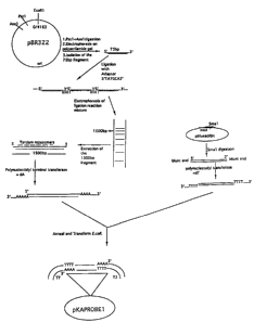
Figure 1 is a schematic representation of plasmid
preparation for reproducing the amplification probe.
Figure 2 is a schematic representation of the
processes for preparing the amplification probe.
Figure 3 depicts an embodiment wherein multiple
probes are used in a sequential fashion in an assay for
detecting a target nucleic acid sequence.
DETAILED DESCRIPTION OF THE INVENTION
This invention comprises the preparation of a
number of nucleic acids sequence probes. This invention
also comprises a method of simplifying and significantly
amplifying the signal generated in a nucleic acid
hybridization assay. The simplification and amplification
is primarily achieved by using an amplification probe
comprising multiple repeating sequence units and labelling
probes that are complementary to the repeating sequence
units.
2126952
-16-
The Target Nucleic Acid Seguence
The target nucleic acid sequences that can be
detected in accordance with the present invention may be
any nucleic acid sequence. There is no maximum limit to
the length of the target nucleic acid sequence, though the
minimum should be at least sixteen nucleotide bases in
length.
The Primary Probe
The primary probe, which is single stranded
nucleic acid sequence, has two distinct regions. At the 5'
end, the sequence is complementary to a sequence found in
the target, and such sequence is of sufficient length,
ranging from at least 6 nucleotides up to a maximum of any
length desired. In a preferred embodiment the length
ranges from 16 to 25 nucleotides. The 3' end of this
primary probe comprises a homopolymeric nucleotide tail
[for example, a poly(dA)]. This poly(dA) sequence ranges
in length, but must be sufficient to hybridize with the
amplification probe. In a preferred embodiment the length
ranges from 12 to 20 nucleotides.
When the target is DNA, the probe is preferably
prepared in the form of RNA. When the target is RNA, the
probe is preferably prepared in the form of DNA. When
hybridization of the probe to the target results in an RNA-
DNA hybrid, this is known as a heteroduplex. In a
preferred embodiment, the homopolymeric region of the
primary probe remains DNA in nature, though in the general
embodiment, any type of nucleic acid sequence can be
utilized in this region.
Techniques for synthesising a single-stranded
polynucleotide sequence for the primary probe, which is
CA 02126952 2004-08-04
-17-
complementary to the target sequence, are well known in the
art and will not be described here.
The Antibody Reagent
Immobilization of the probe-target complex is
achieved by using an antibody, attached to a surface, that
binds to double-stranded nucleic acid. By separating the
probe-target complex from the rest of the sample mixture,
this procedure results in improved sensitivity of the
detection of the target.
These particular antibodies are well known in the
literature (Fliss et al., Appl. Environ. Microbiol.
59:2608-2705, 1993; Coultee et al., Anal. Biochem. 181:96-
105, 1989; US patent 5,200,313)
as is the procedure for the coating of the
antibody molecules to a surface. Whole antibodies,
antibody fragments, polyfunctional antibody aggregates, or
in general any substance comprising one or more specific
binding sites from an antibody for the probe-target complex
can be utilized as described herein. Unless otherwise
noted, it should be understood that, the term antibody when
used in both the disclosure and the claims means whole
antibodies and their polyfunctional and/or fragmented forms
as well. When the term refers to a whole antibody, it may
belong to any of the classes and subclasses of known
immunoglogulins (IgG, IgM, etc.). It is also possible that
a fragment of any such antibody which retains specific
binding affinity for the hybridized probe can also be used,
such as, the fragments of IgF which are often referred to
as Fab, F(ab'), and F(ab)2. Furthermore, aggregates,
polymers, derivatives, and conjugates of immunoglobulins
and/or their fragments can also be utilized where
appropriate.
CA 02126952 2004-08-04
-18-
The antibody reagent's immunoglobulin source can
be procured from any available techniques such as
conventional antiserum and monoclonal techniques.
Antiserum can be procured through well-known techniques
involving the immunization of an animal, (such as mouse,
rabbit, guinea pig or goat) with the appropriate immunogen.
Furthermore, the immunoglobulins can be obtained by somatic
cell hybridization techniques, which would result in the
formation of monoclonal antibodies.
The preparation of immunogens for stimulating
antibodies specific for heteropolymeric (ie. DNA-RNA or
RNA-DNA) probe-target complexes can be achieved through a
variety of techniques. For example, one can employ
transcription of OX174 viron DNA with RNA polymerase
(Nakazato, (1980) Biochem. 19:2835).
The resulting probe-target complexes can be
adsorbed to a methylated protein, or they can be linked to
a conventional immunologenic carrier material such as
bovine serum albumin, before being injected into the
desired host animal (Stollar, (1980) Meth. Enzymol. 70:70).
The most important property of any antibody
raised against such target-probe double stranded complexes
is that the antibody will significantly discriminate in its
binding properties between the duplexed form of the target-
probe complex and single stranded nucleic acid sequences.
Achieving this objective is not difficult as the antibodies
do not need to recognize specific sequences. Rather, they
recognize the general double-stranded characteristic of the
probe-target complex. This is a critical feature of this
invention that significantly reduces background noise and
false positives that could result from hybridization of
labelling probe to non-target single stranded nucleic acid
sequences in the sample.
2126952
-19-
It is preferred to use a solid support to which
the antibody is attached or fixed. Attachment of the
antibody can be achieved through either covalent or
noncovalent bonds. The latter includes adsorption
techniques that provide for a suitably stable and strong
attachment. The solid support can take on a variety of
shapes and compositions. These include beads,
microparticles, porous and impermeable strips and
membranes, as well as the interior surface of reaction
vessels such as test tubes and microtiter plates, etc. The
techniques for attaching a desired reaction partner to a
selected solid support are well known to one skilled in the
art.
The Amplification Probe
The amplification probe is the principal feature
of this invention and serves to cause a plurality of
detectable chemical labels to become attached to each
amplification probe; in this way, the signal indicating the
formation of one probe-target complex is amplified in
direct proportion to the number of labelling probes that
hybridize to the repeating sequence units.
The amplification probe is a single stranded
nucleic acid sequence consisting of at least two regions.
The first region contains a short nucleotide sequence
complementary to a portion of the primary probe that
permits the'amplification probe to hybridize with the
primary probe that is part of the immunocaptured probe-
target complex. In one embodiment this first region is
located on the 3' end and is comprised of a homopolymeric
tail [e.g. poly(dA)] of approximately 12 to 20 nucleotides,
but this can be extended up to any length desired.
The second region contains multiple repeating
sequence units, that form the basis by which the detection
2126952
-20-
indicia will be amplified when a labelling probe
(containing sequences that are complementary to a region
within one sequence unit) is hybridized to each of these
repeating units. The number of repeating units can vary
from 2 to as many as can be accommodated within a
particular application of this invention. The greater the
degree of amplification required for a given test system or
the less sensitive the detection means to be employed, the
larger the number of repeating units required. In one
embodiment the number of repeating units is about 20.
The length of each unit can vary, dependant upon
the requirements of a particular application of this
invention. The minimum length of a repeating unit is about
16 nucleotides, though the best length is about 70 - 100
nucleotides. One critical factor to consider when
designing the length of each repeating unit is the steric
hinderance caused by the size of the detectable chemical
label attached to the labelling probe. For example, a
relatively large enzyme would require a greater degree of
spacing than a small dye molecule or radioisotope.
Moreover, the labelling probe should hybridize to only a
portion of a repeating unit; the remaining nucleotides
within the unit that are not complementary to the sequences
of the labelling probe function as spacers to position the
detectable chemical labels apart from one another.
Furthermore, it will be understood that the subunits in the
tandem repeating nucleic acid fragment orient in the same
5'-3' direction. In the example chosen in Figure 1, each
repeating sequence unit is 75 nucleotides long.
The repeating sequence units can be identical, or
there can be more than different sequence unit. For
example, it may be desirable to attach more than one type
of detectable chemical label to the amplification probe,
whereby a corresponding number of differently sequenced
labelling probes will be required. In this situation one
CA 02126952 2004-08-04
-21-
would construct the repeating sequence units with more than
one type of sequence, each sequence being complementary to
the sequence a type of labelling probe. For example, if it
would be desirable for three types of labelling probes (eg.
probe X, probe Y and probe Z) to attach to the
amplification strand, the complementary repeating sequence
units (unit x, unit y and unit z) on the amplification
probe could be constructed in a xyzxyzxyzxyz fashion. The
nucleic acid sequences that are joined to form the
repeating sequence region could each be constructed in a
xyz fashion, such that when they are joined they will
become linked in a xyz-xyz-xyz manner.
The construction of a plasmid that can be used to
produce multiple copies of the amplification probe is
prepared by well known techniques and one example is
illustrated in Figure 1. The DNA fragment coding for the
amplification probe is cloned in an appropriate plasmid,
such as pBluescriptm, or any other suitable cloning vector.
This plasmid is used as a template for the preparation of
the amplification probe which consists of the complementary
RNA copies of the repeating units and the homopolymeric
tail.
The DNA Vector and Cloning
The experimental methods used to construct the
DNA vectors used to reproduce the amplification probe are
generally described in various manuals of molecular biology
and are known to one who is skilled in the art (Sambrooke
et al., Molecular cloning: A Laboratory Manual, 2nd
edition. Cold Spring Harbor, N.Y., 1989).
In one instance, two hundred micrograms of the
plasmid pBR322 (Bolivar et al., Gene 61:253-264, 1977) were
digested to completion with the restriction enzyme Pstl.
CA 02126952 2004-08-04
-22-
The DNA was extracted with phenol-chloroform, ethanol
precipitated and then digested to completion with the
restriction enzyme Asel. The digestion of pBR322 DNA with
the restriction enzymes yielded a 75 nucleotide long
fragment from the circular plasmid. This 75-base pair (bp)
fragment was separated from the remainder of the plasmid by
electrophoresis of the reaction mixture through an 8%
polyacrylamide gel. The 75-bp fragment was isolated from
the gel by the method of electro-elution using a Bio-Rad TM
apparatus and the DNA was ethanol precipitated.
This 75-bp fragment was mixed with an adapter
polynucleotide of the sequence 5'TATGCA3' in the presence
of T-4 DNA ligase. This adapter-mediated ligation allowed
polymerization of the 75-bp fragments with each of the
subunits in the polymer being aligned in the same 5'-3'
orientation. The ligation reaction mixture was
electrophoresed through a it agarose gel and a ladder
representing different polymeric sizes of the DNA was
observed by ethidium bromide staining of the gel. From the
gel, the DNA fragment corresponding to 1500-bp size was
excised out and extracted from the gel using the QIAexTM gel
extraction method (QIAGENTM, Chattsworth, CA).
The above 1500-bp DNA was treated with
polynucleotidyl terminal transferase in presence of dATP,
to add approximately 12-20 dAs to the 3' end of the DNA.
Simultaneously, the plasmid pBluescriptT" (Stratagene, La
Jolla CA) was linearized with the restriction enzyme Smal
and then treated with the polynucleotidyl terminal
transferase in presence of dTTP to add 12-20 dTs to the 3'
end. The DNA sequences thereby prepared were mixed under
appropriate conditions (GIBCO BRL Technical Bulletin 8008-
1) in order to anneal complementary tails and to form
circular recombinant plasmid DNA.
The annealed DNA was introduced into E. coli
(Epicurian E.coliTM XL-1; Stratagene, Inc.) using a bacterial
CA 02126952 2004-08-04
-23-
transformation procedure recommended by Stratagene. From
the transformants, the colony containing the sequences
coding for the primary probe was confirmed by analysis of
the plasmid DNA. The DNA of the plasmid coding for the
primary probe was purified from the E. coli using the
QIAgenT`"mega-Plasmid isolation kit.
The procedure for transcribing DNA plasmid
containing the nucleic acid sequence coding for the
amplification probe is as follows. Plasmid DNA coding for
the amplification probe (10 g) was treated with the enzyme
T-7 RNA polymerase in the method suggested by the Mega-
Transcription kit (Cat. No. 1334) supplied by Ambion Inc.
(Austin, Texas). This transcription generated an RNA
product with a structure as shown in Figure 2, comprising a
poly(U) sequence at the 5' end.
In general, an in vitro. transcription of the
plasmid coding for the amplification probe using T-7 RNA
polymerase would yield its complementary RNA, the
amplification probe (Figure 2) which carries a poly(U)
homopolymeric tail fragment at its 5' end; this poly(U)
region is complementary to the poly(A) region of the
primary probe described previously. Recent advances in the
art allows the large scale production of T-7 polymerase
mediated RNA molecules in an in vitro transcription. Kits
such as "MEGA- Transcript kits" (Ambion Inc., Austin,
Texas) could be used to produce milligram quantities of RNA
from microgram quantities of the template DNA.
It will be appreciated by one skilled in the art
that once the amplification probe is constructed with an
appropriate homopolymeric tail, the same template could be
used for the production of an amplification probe which
could be used in any nucleic acid hybridization assay
wherein the primary probe comprises a sequence
complementary to the homopolymeric region in the
amplification probe.
2126952
-24-
The Labelling Probe
The immunocaptured probe-target complex can be
detected by a variety of well known techniques. In a
preferred embodiment, a labelling probe comprising
sequences complimentary to at least one of the repeating
sequence units on the amplification probe will itself be
labelled with a chemical group that is detectable. A
detectable chemical group can comprise any material
possessing a detectable chemical or physical property.
These materials are well known and developed in nucleic
acid hybridization assays. Furthermore, most labels useful
in such methods can be applied to the present invention.
For example, enzymatically active groups have been found to
be useful, in particular, those groups that are enzymes
(Clin. Chem. (1976)22:1243; U.S. Pat. No. 31,006; and UK
Pat 2,019,408), enzyme substrates (U.S. Pat. No.
4,492,751), cofactors (see U.S. Pat. Nos. 4,230,797 and
4,238,565), and enzyme inhibitors (see U.S. Pat. No.
4,134,792). Also useful are fluorescers (see Clin. Chem.
(1979)25:353), chromophores, luminescers such as
chemiluminescers and bioluminescers (U.S. Pat. No.
4,380,580), as well as specifically bindable ligands such
as biotin (European Pat. Spec. 63,879) or a hapten (U.S.
Pat. No. 4,380,580), and radioisotopes such as 3H, 14C, 32P,
35S, and 1251 . These labels and labelling pairs are
therefore detectable on the basis of either their own
physical properties (eg., fluorescers, chromophores and
radioisotopes), or their reactive, or binding properties
(eg., enzymes, substrates, cofactors and inhibitors). A
good example, is a cofactor-labelled antibody that can be
detected by addition of the enzyme for which the label is
both a cofactor and a substrate for that enzyme. More
specifically a hapten or ligand labelled antibody can be-
detected by adding an antibody to the hapten or a protein
2126952
-25-
(e.g., avidin) which binds the ligand, tagged with a
molecule capable of detection. These detectable molecules
can be a molecule possessing either a measurable physical
property (e.g., fluorescence or absorbance) or a quality
capable of participating in an enzyme reaction (see above
for list). In one example, one can utilize an enzyme which
acts upon a substrate to generate a product with a
measurable physical property. Specific examples of this
kind include, but are not limited to, /3-galactosidase,
alkaline peroxidase and phosphatase. Other similar
labelling schemes are evident to one skill in the art.
The nature and the quantity of the label in the
labelling probes are not critical. The probes could be
labelled either at their extremities or along their entire
length with a single or a multiple marker which, of course,
can be any detectable substance; in other words, the probe
is isotopically or chemically modified in such a way that
the person performing the assay can, after further
manipulation if necessary, still detect the presence of
these labels.
The length of the hybridizing portion of the
labelling probe can vary to meet the needs of application
of the invention. The minimum length of the probe is about
16 nucleotides and the maximum length is about 25
nucleotides, though a situation may arise where it may be
favourable to extend the length beyond 25 nucleotides. In a
specific instance, a 25 base polynucleotide fragment with a
5' amino modification was commercially obtained (Bio/Can
Scientific Co, Mississauga, Ontario). The sequence in this
fragment was complementary to a region within the repeating
unit sequence of the amplification probe. The amino
modification allowed covalent coupling of the enzyme
alkaline phosphatase to the 3' end of the sequence. The
covalent attachment of the enzyme to the polynucleotide was
2126952
-26-
carried out according to known methodology (Bio/Can
Scientific Co.).
Variations on the design of the labelling probe
and the amplification probe may be desirable. For example,
the amplification probe itself can be labelled with biotin
during the in vitro transcription, or end labelled
separately. The labelling probe could be labelled with
multiple markers rather than with just a single marker as
shown in Figure 1. As long as the labelling is performed
in such a manner as to not interfere with the hybridization
between the amplification and labelling probes, these
supplementary labels can lead to a
proportional increase in sensitivity of the assay.
This invention describes a general method of
constructing the probe cascade. In a general embodiment,
the cascade consists of a primary probe, an amplification
probe, and a labelling probe with chemical and functional
characteristics as described herewithin. The exact
sequences implicated in the construction of the probe
reagents described above (excepting, of course, the target
complementary sequence in the primary probe) are not
critical, and only the complementarity of the sequences to
be hybridized is important. The exact lengths of the
various probes described here are not limited to the sizes
given in the examples herein. A person skilled in the art
may easily vary these lengths and the methods for achieving
such variation are well known in the art.
The Assay
The present invention is useful in a large
variety of hybridization procedures. The sample to be
assayed can be virtually of any medium of interest, such as
of medical, veterinary, environmental, nutritional or
industrial significance.
2126952
-27-
One embodiment of this invention comprises a
sensitive method of detecting the probe-target complex and
its application to the determination of the presence of
specific microorganisms in a test sample. An assay,
according to the present invention, involves at least the
following steps:
a) hybridizing a sequence of a primary
polynucleotide probe to a target nucleic
acid sequence;
b) immobilizing the probe-target complex;
c) exposing the immobilized probe-target
complex to an amplification probe under
conditions that allows the amplification
probe to hybridize to the probe-target
complex;,
d) exposing the hybridized amplification probe
to many copies of a labelled polynucleotide
probe under conditions that allows many such
labelled probes to hybridize to the
amplification probe;
e) detecting the labelled probes in the
resulting complex.
In one particular embodiment, the sample to be
tested is typically a piece of food, for example meat or
cheese, or another source containing principally double
stranded nucleic acids. This includes microorganisms
and/or other cellular material associated with these
samples. The test sample is first treated to release the
nucleic acids from the cells, followed by a denaturation
step to denature the nucleic acids. This is typically
accomplished by lysing the cells in a lysis buffer solution
and the denaturation of nucleic acids is preferably
accomplished by heating the resulting solution in boiling
water or alkali treatment (e.g., 0.1 N sodium hydroxide).
2126952
-28-
The denaturing step can often be used simultaneously as a
method to lyse cells. The release of nucleic acids can,
also, be obtained through mechanical disruption such as
freezing/thawing, abrasion, sonication, physical/chemical
disruption (eg. polyoxyethylene ether detergents like
Triton , polyoxytheylenesorbitan detergents like Tween ,
sodium dodecylsulfate, alkali treatment, osmotic shock,
heat, or lysing using enzymes such as proteinase K, ,
lysozyme, pepsin). The resulting medium will contain
nucleic acids in single stranded form which is then assayed
according to present hybridization methods (Wang et al.,
Appl. Environ. Microbiol., 1992).
When the sample contains free single-stranded
nucleic acid sequences, the sample is in proper form for
use with the specific probe. When the assay is performed
for detection of a microorganism, a bacterium for example,
the cells must be lysed and the nucleic acids have to be
exposed in order to be available for hybridization with the
probe. Methods of lysis have been previously described and
are well known to one skilled in the art.
In one general embodiment, the cells are lysed
by mixing 50 Al of the broth culture (about 108 to 109 cells
per ml) with an equal volume of a 2% Triton X-100 and
heating at 100 C for 5 minutes in a 1.5 ml eppendorf
microfuge tube. The samples are chilled in ice to denature
the target nucleic acids. The probe is diluted at a
concentration of 200 ng per ml in a buffer containing 8 x
SSC (1 x SSC being 0.15 M NaCl plus 0.015 M sodium
citrate), 40 mM HEPES (pH 7.4) and 4 mM EDTA. One hundred
Al of the probe thus prepared is added to the cell lysate
so that the probe is contacted with the target nucleic acid
and the hybridization is carried out in this solution at
70 C for 30 minutes.
There are a variety of known hybridization
conditions that can be employed in the assay. Typically,
2126952
-29-
hybridization will proceed at slightly elevated
temperatures. Typical temperatures range between about 35
and 75 C and are usually about 65 C. The hybridization is
carried out in a solution comprised of a buffer at pH
between about 6 and 8 and with the appropriate ionic
strength. A typical ionic strength is 2 X SCC where 1 X
SCC = 0.15 M sodium chloride and 0.015 M sodium citrate at
pH 7. The hybridization solution further contains protein
such as bovine serum albumin, FicollTM' (copolymer of sucrose
and epichlorohydrin, Pharmacia Fine Chemicals, Piscataway,
N.Y.), polyvinylpyrrolidone, and a denatured foreign DNA
such as rom calf thymus or salmon sperm. The degree of
complementarity between the target nucleic acid sequence
and the primary probe required for hybridization to occur
depends on the stringency of the conditions.
The primary probe is contacted with the denatured
nucleic acid sequences in solution phase. The probe-target
complexes are removed from the excess unhybridized probe by
immunocapture of the former. This is achieved by
transferring the hybridization reaction mixture to the
wells of a microtiter plate which have been previously
coated with monoclonal anti-probe-target complex. The
antibody is thus contacted with the probe-target complex
present in the hybridization reaction mixture for 30
minutes at 37 C. This contact results in the immunocapture
of the probe-target complexes and therefore results in
their immobilization to the surface of the wells of the
microtiter plate. Any reagent which will subsequently bind
to the probe-target complex will thus be immobilized to the
solid surface. After the immunocapture reaction is
completed, the plates are washed three times with phosphate
buffered saline (PBS; 50 mM phosphate buffer containing
0.15 M NaCl) in order to remove the excess unhybridized
probes from the well.
2126952
-30-
Alternatively, any other solid phase could be
employed for the immunocapture of the probe-target
complexes; any method of immobilisation of the probe-target
complex could be used for separating the hybridized probe
from the excess unhybridized probe.
In one embodiment, the amplification probe is
diluted to a concentration of 200 ng per ml in a buffer
containing 4 x SSC, 20 mM HEPES (pH 7.4) and 2 mM EDTA.
Two hundred Al of the amplification probe thus prepared are
added to the wells of the microtiter plate so that the
amplification probe is contacted with the immobilized
probe-target complex which carries the poly(dA)12-20 at the
3' end of the probe. The plates are incubated at 42 C for
30 minutes so that the hybridization between the poly(dA)
of the probe and the poly(U) of the amplification probe is
completed. This poly(dA)-poly(U) is also a DNA-RNA hybrid,
thermodynamically the most stable form of a double stranded
nucleic acid. At the end of this hybridization period, the
plates are washed three times with 0.5 M sodium chloride
solution.
The labelling probe is diluted to a concentration
of 200 ng per ml containing 4 x SSC, 20 mM HEPES (pH 7.4)
and 2 mM EDTA.
Two hundred Al of said labelling probe is added to the
microtiter plate wells so that the labelling probes come in
contact with the amplification probes which are immobilized
to the solid surface. This hybridization is carried out at
42 C for 30 minutes and the plates are then washed three
times with 0.5 M sodium chloride.
The labelling probe hybridizes with the repeating
units of the amplification probe. As the labelling probe
is a single stranded polynucleotide, the rate of the
hybridization reaction is considerably high.
When the amplification probe comprises twenty
repeating units, twenty labelling probe molecules can be
2126952
-31-
immobilized per each of the probe-target complexes
originally immunocaptured. The signal generated by the
probe-target complex is amplified by a magnitude of twenty
times. The enzyme present in the labelling probe is then
detected using standard colorimetric, chemiluminescent,
fluorometric or other detection methods as discussed herein
and is well known in the art.
In one embodiment employing colorimetric methods,
a solution containing the substrate to alkaline phosphatase
enzyme is added to the wells. For example, 200 l of p-
nitrophenyl phosphate (p-NPP; 4 mg in 10% diethanolamine
and 0.5 mM magnesium chloride, pH 9.8) are added. After 15
minutes of incubation at room temperature (20 - 25 C), the
color developed is measured at 410 nm wavelength of light.
Reagent System
Another embodiment of the present invention
involves its use in a diagnostic kit, the kit comprising
one or more solutions and devices for carrying out an assay
for detection of food pathogens such as Listeria
monocytogenes or Escherichia coli 0157:H7 in food samples
like cheese or meat.
The present invention further provides for a
reagent system. More specifically it provides for a
reagent combination comprising all of the essential and
necessary elements required to conduct a desired assay
method. The reagent system is presented in a commercially
packaged form as a composition or a mixture wherein
compatibility-of the reagents will allow for a test device
configuration (most typically as a test kit) a packaged
combination of one or more containers, devices, or the
like, holding the necessary reagents and usually including
written instructions describing the performance of the
assays. Reagent systems of the present invention involve
2126952
-32-
all possible configurations and compositions for performing
the various hybridization formats described herein.
The reagent system will generally comprise
bacterial lysis solutions, the solutions containing the
target directed primary probe, the amplification probe, the
labelling probe (preferably labelled with a detectable
chemical group) enzyme substrates, microtiter plates or
strips coated with the anti-target-probe complex
antibodies, and a description of the assay comprehensible
to one skilled in the art. A test kit form of the reagent
system may further include ancillary chemicals. Such
ancillary chemicals can include components of the
hybridization solution and denaturation agents capable of
converting double stranded nucleic acids found in a test
sample into single stranded form. More preferably, there
is included a chemical lysing and denaturing agent, such as
an alkali, for treating the sample in order to release
single stranded nucleic acid therefrom.
The present invention will now be illustrated,
but is not intended to be limited, by the following
examples.
Example 1: Assay for Salmonella tv hi in human blood.
A volume of 0.05 ml of blood was drawn from the
test patient and was inoculated into 10 ml of Gram-negative
broth (Difco) and incubated at 37 C for 16 - 20 hours. Any
bacteria in 100 l of the enriched broth were lysed by
mixing the broth with an equal volume of 20 Triton X-100 in
a microfuge tube and boiling for 5 minutes at 100 C. The
solution was quickly chilled in ice. Probe solution (100
l; 200 ng per ml) in 8 x SSC, 40 mM HEPES (pH 7.4) and 4
mM EDTA was added to the lysate and incubated at 37 C for 30
minutes. The probe selected here was RNA in nature and
2126952
-33-
specifically hybridising with the Vi antigen gene of S.
typhi.
The above hybridization reaction mixture (200 Al)
was transferred to microtiter plate well which had been
previously coated with monoclonal anti-probe-target complex
antibodies and incubated at 37 C for 30 minutes. The plates
were then washed 3 times with PBS.
Detection of any target nucleic acid entailed
adding the amplification probe solution (200 Al; 200 ng/ul)
in 4 x SSC, 20 mM HEPES (pH 7.4) and 2 mM EDTA to the wells
and incubating at 37 C for 30 minutes. The plates were then
washed 3 times with 0.5 M sodium chloride solution. The
labelling probe solution (200 Al; 200 ng/ul) in 4 x SSC, 20
mM HEPES (pH 7.4) and 2 mM EDTA was added to the wells and
incubated at 37 C for 30 minutes. The plates were washed 3
times with 0.5 M sodium chloride solution. Alkaline
phosphatase substrate solution (4 mg of p-NPP in 10%
diethanolamine and 0.5 mM magnesium chloride, pH 9.8) was
added to the wells and incubated at room temperature for 15
to 30 minutes and the color developed was measured at 410
nm in a standard microtiter plate reader.
With the help of positive and negative controls,
a colorimetric value was chosen that was statistically
distinct between samples that contained, and those that do
not contain S. typhi. Using this "cut-off value", the
color generated was interpreted as to whether the original
sample was positive or negative for the presence of the
pathogen.
Example 2: Assay for the detection of L. monocytocrenes in
cheese.
Twenty five grams of the test cheese was
homogenized for 2 minutes in a stomacher with 250 ml of to
sterile peptone water and allowed to settle for about 15
2126952
-34-
minutes. A 0.5 ml sample of the clear upper portion of
this homogenate was inoculated into 10 ml of trypticase soy
-0.6% yeast extract broth and incubated at 37 C for 16 - 20
hours.
Any bacteria in 100 Al of the enriched broth was
lysed by mixing the broth with an equal volume of 20 Triton
X-100 in a microfuge tube and boiling for 5 minutes at
100 C. The solution was quickly chilled in ice.
Probe solution (100 Al; 200 ng per ml) in 8 x
SSC, 40 mM HEPES (pH 7.4) and 4 mM EDTA was added to the
lysate and incubated at 37 C for 30 minutes. The probe
selected here was DNA in nature and specifically
hybridising with the ribosomal RNA (rRNA) of L.
monocytogenes. The hybridization reaction mixture (200 Al)
was transferred to a microtiter plate well which had been
previously coated with monoclonal anti-probe-target
antibodies and incubated at 37 C for 30 minutes. The plates
were then washed 3 times with PBS.
The amplification probe solution (200 Al; 200
ng/ l) in 4 x SSC, 20 mM HEPES (pH 7.4) and 2 mM EDTA was
added to the wells and incubated at 37 C for 30 minutes.
The plates were then washed 3 times with 0.5 M sodium
chloride solution. The labelling probe solution (200 Al;
200 ng/ .l) in 4 x SSC, 20 mM HEPES (pH 7.4) and 2 mM EDTA
was added to the wells and incubated at 37 C for 30 minutes.
The plates were washed 3 times with 0.5 M sodium chloride
solution. Alkaline phosphatase substrate solution (4 mg of
p-NPP in 10% diethanolamine and 0.5 mM magnesium chloride,
pH 9.8) was added to the wells and incubated at room
temperature for 15 to 30 minutes and the color developed
was measured at 410 nm in a standard microtite.r plate
reader.
With the help of positive and negative controls,
a colorimetric value was chosen that was statistically
distinct between samples that contain and those that do not
2126952
-35-
contain L. monocytogenes. Using this "cut-off value", the
color generated was interpreted as to whether the original
sample was.positive or negative for the presence of the
pathogen.
Below are results of assays performed that detect
the presence of L. monocytogenes. Assay #1 was performed
with biotinylated probe which is 784-bp long; the
hybridized probe was detected with a streptavidin-alkaline
phosphatase conjugate system. Assay #2 was performed
according to the protocol given herein.
Sample Status LPM count (L.m./ml O.D.410 O.D.
410
enriched broth) Assay #1
Assay #2
1 Positive Control 3.0 x 10⁴ 1.51 2.36
2 Positive Control 4.5 x 10⁶ 1.63 2.6
3 Positive Control 6.0 x 10² 1.03 1.91
4 Positive Control 5.0 x 10⁶ 1.74 2.77
5 Positive Control 4.0 x 10³ 1.23 1.98
6 Positive Control 7.0 x 10⁶ 1.75 2.93
7 Negative Control 0 0.08 0.06
8 Negative Control 0 0.04 0.08
9 Negative Control 0 0.08 0.0
10 Negative Control <10 0.05 0.16
11 Negative Control 0 0.02 0.17
12 Negative Control <10 0.18 0.20
13 Unknown 5.0 x10⁶ 1.60 2.56
14 Unknown 6.0 x 10⁴ 1.56 2.32
15 Unknown 4.0 x 10⁶ 1.67 2.70
16 Unknown 4.6 x 10⁶ 1.70 2.74
17 Unknown 7.0 x 10⁵ 1.73 2.15
18 Unknown 6.0 x 10³ 1.20 1.63
19 Unknown 6.0 x 10⁶ 1.77 2.45
20 Unknown <20 0.08 0.16
2126952
-36-
Example 3: Assay for the detection of E. coli 0157:H7 in
meat.
Twenty five grams of the test meat was
homogenized for 2 minutes in a stomacher with 250 ml of 1%
sterile peptone water and allowed to settle for about 15
minutes. One half ml of the clear upper portion of this
homogenate was inoculated into 10 ml of Gram-negative broth
(Difco) and incubated at 37 C for 16 - 20 hours. Any
bacteria in 100 l of the enriched broth were lysed by
mixing the broth with an equal volume of 2% Triton X-100 in
a microfuge tube and boiling for 5 minutes at 100 C. The
solution was quickly chilled in ice.
Probe solution (100 Al; 200 ng per ml) in 8 x
SSC, 40 mM HEPES (pH 7.4) and 4 mM EDTA was added to the
lysate and incubated at 37 C for 30 minutes. The probe
selected here was RNA in nature and specifically
hybridising with the SLT-1 toxin gene of E. coli O157:H7.
The above hybridization reaction mixture (200 l)
was transferred to microtiter plate well which had been
previously coated with monoclonal anti-probe-target
antibodies and incubated at 37 C for 30 minutes. The plates
were then washed 3 times with PBS.
The amplification probe solution (200 l; 200
ng/ .l) in 4 x SSC, 20 mM HEPES (pH 7.4) and 2 mM EDTA was
added to the wells and incubated at 37 C for 30 minutes.
The plates were then washed 3 times with 0.5 M sodium
chloride solution. The labelling probe solution (200 Al;
200 ng/ l) in 4 x SSC, 20 mM HEPES (pH 7.4) and 2 mM EDTA
was added to the wells and incubated at 37 C for 30 minutes.
The plates were washed 3 times with 0.5 M sodium chloride
solution.
Alkaline phosphatase substrate solution (4 mg of
p-NPP in 10% diethanolamine and 0.5 mM magnesium chloride,
2126952
-37-
pH 9.8) was added to the wells and incubated at room
temperature for 15 to 30 minutes and the color developed
was measured at 410 nm in a standard microtiter plate
reader.
With the help of positive and negative controls,
a colorimetric value was chosen that was statistically
distinct between samples that contained and those that did
not contain E. coli 0157:H7. Using this "cut-off value",
the color generated was interpreted as to whether the
original sample was positive or negative for the presence
of the pathogen.
Below are results of assays performed that detect
the presence of E. coli. The assay was performed with
known amounts of E. coli inoculated in 25 g of meat which
was previously sterilized by gamma irradiation. The assay
was performed as described in the protocol given herein.
Sample Status Plate Count O.D.410
1 Positive Control 470 1.03
2 Positive Control 3 x 10⁶ 2.81
3 Positive Control 532 0.99
4 Positive Control 7 x 10⁶ 2.73
5 Positive Control 5 x 10⁷ 2.8
6 Positive Control 4 x 10⁴ 1.98
7 Negative Control 0 0.15
8 Negative Control <10 0.33
9 Negative Control 0 0.0
10 Negative Control 40 0.61
11 Negative Control 0 0.0
12 Negative Control 0 0.04
13 Unknown 56 0.59
14 Unknown 6 x 10⁵ 2.78
15 Unknown 6 x 10⁴ 2.24
16 Unknown 234 1.02
2126952
-38-
17 Unknown 7 x 10⁶ 2.75
18 Unknown 7 x 10⁷ 2.82
19 Unknown 5 x 10⁶ 2.82
20 Unknown 4 x 10⁶ 2.79
It is to be understood that the examples
described above are not meant to limit the scope of the
present invention. It is expected that numerous variants
will be obvious to the person skilled in the art to which
the present invention pertains, without any departure from
the spirit of the present invention. The appended claims,
properly construed, form the.only limitation upon the scope
of the present invention.