Note: Descriptions are shown in the official language in which they were submitted.
y sv
~.~:~ ~ f~''
docket 64,149-011
TITLE
BLOOD CULTURE SENSOR STATION
UTILIZING TWO DISTINCT LIGHT SOURCES
BACKGROUND OF THE INVENTION
This application relates to a blood culture sensor station which directs
two sources of light into a blood culture specimen, measures the reemerging
light from both light sources, calculates a ratio based on the two reemerging
lights, and determines whether the specimen includes bacteria based on the f
calculated ratio.
Typically, the presence of bacteria in a patient's body fluid is determined
by injecting a small quantity of body fluid (usually blood) into a vial which
contains a culture medium. Various types of instruments are utilized to
monitor
changes in the carbon dioxide content of the vial. Carbon dioxide is a
metabolic
by-product of bacteria growth, and thus an increase in carbon dioxide in the
vial
indicates the presence of bacteria in the patient's body fluid.
Sensors have typically been inserted into the vial to test the carbon
dioxide content. These so-called invasive sensors are somewhat undesirable due
to the risk of cross-contamination. Non-invasive sensor systems have been
developed which utilize chemical sensors disposed inside the vial. Typically,
such sensors respond to changes in the carbon dioxide cancentration by
changing
color, or by changing their fluorescent intensity. To monitor changes in the
known chemical sensors, the systems have typically required one light source,
one spectral exitation filter, one emission filter and one photodetector
arranged
adjacent to each vial. Each of these components must have extremely narrow
specification tolerances to avoid substantial station t~~ station sensitivity
variations. However, even if it were possible to equalize all of the vial
stations,
certain lot-to-lot variations in the chemical sensor composition and certain
vial
to-vial geometry variations may remain. Thus, the accuracy of such system is
sometimes in question.
1
~,? ~~?~'~
docket 64,149-Oli
Some disadvantages of intensity based sensors can be overcome by
utilizing fluorescent sensors which change fluorescent lifetime with changing
pH,
carbon dioxide concentrations, oxygen concentration, or other parameters. Such
fluorescent lifetime sensors typically require expensive equipment. Further,
S color changing or fluorescent chemical sensors have typically required a
certain
temperature adaption time before a first vial test can be performed. Thus, a
test
cannot be made immediately upon receiving a vial when utilizing known
chemical sensors.
It has been proposed to direct a light source into a vial, and monitor the
reemerging light over time. It has been shown that increased carbon dioxide in
a specimen will affect the reemerging light. The reemerging light can be
evaluated to make a prediction as to the presence of bacterial growth in the
vial.
Problems remain with such proposed methods. If there is an unusually large
amount of specimen within the vial, the medium wiihin the vial may be more
opaque than with a lesser amount of specimen. This would in turn effect the
reemerging light. For that reason with known light based sensor systems, the
amount of specimen within the vial can vary the accuracy of the test. Further,
such light based systems do not allow immediate testing upon receipt of a
vial.
SUMMARY OF THE INVENTION
In a disclosed method for evaluating a body fluid sample in a culture vial,
one calculates a ratio based on the reemerging light intensity from two
distinct
light sources and compares the calculated ratio to expected ratios for
positive
and negative culture vials. In this way, one can make a quick evaluation of
whether a particular vial is positive. Since one calculates a ratio based on
the
two reemerging light intensities, variation in the reemerging light due to
variations in the amount of specimen in the vial should be cancelled out.
In one disclosed embodiment of the present invention, a first light source
having a first set of characteristics is directed into the vial and the
reemerging
light is monitored at a second position. Light having a second set of
characteristics is then directed into the vial, and the reemerging light is
again
measured at the second position. A "ratio" based on the reemerging light
2
-; " ;7
-i ~J ~ ~ ~~
docket 64,149-011
intensity from the first and second light sources is determined and compared
to
a graph which has been developed experimentally utilizing control positive and
negative samples. The term "ratio" has been placed in quotes since the
invention preferably does not use a simple ratio of the reemerging lights.
Rather, in one method, background light and input light are all included in a
formula to calculate the "ratio" quantity. In a second method the reemerging
lights are monitored and the intensity of the second light source is adjusted
until
equal reemerging intensities are measured. A ratio of the intensities of the
first
and second light source is then calculated. It has been found that a
calculated y
"ratio" using either method provides a very good indication of whether a
particular vial is a positive vial containing bacterial growth.
The term "ratio" as utilized in this application is broadly meant to refer
to a formula calculated utilizing the measured reemerging light intensities in
some way. As stated above, the specific formulas may include other quantities,
as in the first example . Moreover, as in the second example, in some formulas
there may be no specific calculation using actual measured reemerging
intensities. The measured intensities are utilized to reach the adjusted
second
light intensity quantity which is utilized in the formula. In that sense the
second
method ratio is "based" on the measured reemerging light intensity.
One main beneficial use of this invention would be to provide.. an
immediate reading of a particular vial upon receipt by a laboratory. It is
envisioned that a sensor station according to the present invention would
receive
all incoming vials for immediate testing to determine whether the particular
vial
is already positive. If the vial is positive upon arriving at the laboratory,
that
result can be noted and reported. If the particular vial is not positive upon
arrival, the vial may beplaced into a "kinetic" system which monitors ongoing
bacterial growth within a vial. A vial may become positive, or change to
positive
over the first 24 hours after the body fluid is injected. Thus, it is possible
that
a vial which is not immediately positive upon being received by a laboratory
could later become positive. Applicant's invention, will identify so-called
"delayed" vials which are already positive upon arrival at the laboratory.
This
3
r.1 i J !~
docket 64,149-011
will allow health care providers to provide treatment to those patients
quicker,
and also reduce the number of sample vials which undergo other types of
testing
to monitor whether they are changing to positive. This invention requires only
seconds to complete a test. Other methods take a longer time, and thus it is
desirable to reduce the number of vials subjected to the other type of tests.
In one preferred embodiment of this invention, light of a first wavelength
is introduced into the vial at a first location. The light intensity
introduced
h(~,1) is measured. The light intensity hz(.11) reemerging from the vial is
measured at a second location. The light of the first wavelength is then
turned ,
off, and the background light intensity Ioz emerging from the second location
with no light is then measured. Light of a second wavelength is introduced
into
the vial from the first location. The intensity introduced It(~.z) is
measured, and
the light intensity I~z(~.z) emerging at the second location of the vial from
the
second light source is also measured. A "ratio" of the two reemerging light
intensities is calculated using the following formula:
Ilz(~i) - Ioz IO~z)
R = ______________ * _______.
Iiz(~a) - Ioz Ii(~~i)
In a second mode of operation, the above objective may be achieved by
introducing light of a first wavelength at a first location into the vial,
measuring
the light intensity Ilz(~,~) reemerging at the second location on the vial.
The
light of the first wavelength is turned off, and light of a second wavelength
is
introduced from the first location. The light intensity Ilz(~.z) reemerging at
the
second location due to the second light source is measured. The reemerging
intensities hz(~1) and Iiz(.lz) from the two light sources are then compared.
The
intensity of the second light source is adjusted until the reemerging
intensities
are equal for both light sources. The light intensity Il(~,1) from the first
light
source introduced to the vial is measured, as is the adjusted light intensity
II(~.z)
from the second light source. A "ratio" is then calculated as follows:
Il(~z)
_______
I~(~~)
4
.,
1 a d
docket 64,149-011
Although the quantity U is calculated without including any measurement
of the reemerging light intensity in the equation, the reemerging intensities
are
utilized to reach the quantities used in the equation. In that sense, the
calculation of U is "based" on measuring the reemerging light intensities
The second mode of operation is the preferred method, and is
advantageous in that there is no measurement of the intensity Io2 required.
Any
background light is cancelled out. Also, dark current signals of the
photodetector, and sensitivity changes and/or non-linearities of the
photodetector at the second location have no impact on the measurement. t
It has been found that two effects occur to light passing through a culture
medium, absorption and scattering. In aerobic cultures absorption changes are
utilized to detect bacterial growth. In anaerobic cultures a change in
scattering
is the major effect.
The above formulas have been found experimentally to be particularly
advantageous when used with aerobic culture media. It has been found that
when the calculated ratio quantities R or U are plotted on a graph, measuring
the ratio against the volume of specimen in the vial, there is a clear
difference
between positive and negative samples. Thus, when one reaches a calculated R
or U value, one can compare that value to a previously prepared graph and
make an accurate prediction as to whether the particular specimen is a
positive
or a negative. For an aerobic vial the use of the different wavelengths is
most
advantageous, since the main effect on the reemerging light intensity in the
aerobic medium is a wavelength dependent absorption change.
For anaerobic culture vials, it has been found that a somewhat different
method is particularly advantageous. In the preferred anaerobic method, light
of a third wavelength is introduced at a third location. The intensity of the
entering light I3(~,3) is measured, and the light intensity I~(~,3) reemerging
at a
fourth location is measured. The third light source is then turned off, and
the
background light intensity I~ reemerging at the fourth location with no light
is
measured. Light of the third wavelength is then introduced at a fifth location
of the vial. The intensity of the entering light IS(7~3), and the light
intensity
5
docket 64,149-011
I~(~,3) reemerging from the fourth location are measured. The "ratio" S is
then
calculated:
I54(~'3) - I04 13~~'3~
_________________ * _______
S I~a(~s) - Ioa Is(~3)
A second method for anaerobic cultures can also be utilized by
introducing. light of the third wavelength at the third location into the
vial,
measuring the light intensity reemerging at a fourth location I~(~.3), turning
off
the light at the third location, introducing light of the third wavelength at
a fifth
location and measuring the light intensity Isa(~3) reemerging at the fourth
location on the vial. The reemerging intensities I~(~,3) and Isa(~3), are then
compared and the intensity introduced at the fifth location is adjusted until
the
light reemerging intensities are equal. The light intensity I3(.13) introduced
at
the third location, and the adjusted light intensity Is(7l3) introduced at the
fifth
location are measured. The ratio quantity W is then calculated;
I3(a'3)
Is( ~3)
The use of the different points of introduction for the light source in the
anaerobic method is most advantageous since the main effect on the light by
the
anaerobic medium is scattering. .
In summary, it has been found that by introducing two different sources
of light with two different sets of characteristics, and comparing the
reemerging
values some prediction as to whether a particular culture vial is a positive
or a
negative can be made. It has been found that by varying the wavelength
between the two light sources one can make an accurate prediction with regard
to aerobic cultures. By varying the point of introduction of the light
sources,
one can make an accurate prediction of anaerobic cultures.
These and other features of the present invention can be best understood
from the following specification and drawings, of which the following is a
brief
description.
6
docket 64,149-011
BRIEF DESCRIPTION OF THE DRAWINGS
Fig. 1 is a schematic view of a sensor station according to the present
invention.
Fig. 2 is a graph showing a calculated "ratio" vs. blood volume for a
particular type of aerobic culture.
Fig. 3 is a graph showing a calculated "ratio" vs. blood volume for a type
of aerobic culture utilized for pediatrics.
Fig. 4 is a graph showing a calculated "ratio" vs. blood volume for a
particular type of anaerobic culture. o
DESCRIPTION OF THE PREFERRED EMBODIMENT
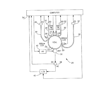
A non-invasive blood culture sensor station 20 for testing a blood sample
within a vial 22 is illustrated in Fig.l. Vial 22 is held within a holding
structure
24.
First and second light sources 26 and 27 are positioned at approximately
the same location adjacent the vial 22. A third light source 28 and a fourth
light
source 29 are spaced from the location of sources 26 and 27. A light sensor
31,
which is preferably a photomultiplier, is used as the high-sensitivity
photodetector. Preferably sources 26, 27, 28 and 29 and sensor 31 are
positioned immediately adjacent to vial 22, and most preferably in contact
with
the vial. In a preferred embodiment, sources 26, 27, 28, and 29, and light
sensor
31 are arranged adjacent to the cylindrical vial wall, all at the same
distance
from the vial bottom. In order to describe the location of the sources
relative
to the location of the light sensor, we use angles along the vial
circumference.
It has to be emphasized, however, that these angles are by no means "emission
or observation angles" as used in light scattering experiments. Rather, the
positioning of the lights and sensors is selected to emphasize the effects on
the
reemerging light such that the ratios finally calculated provide a good
indication
of the presence of bacterial activity. In the embodiment shown in Fig. l,
sources
26 and 27 are preferably located at an "angle of 180°" relative to the
light sensor
31. Preferably, source 29 is spaced from sensor 31 by an angle between
45° to
1U0°, with 85° being preferred. Further, source 28 is preferably
spaced from
7
~~'r ~3
.~ Y,d ~ 4.d ~ 1
docket 64,149-011
sensor 31 by an angle 100° to 180°, with 135° being
preferred. The light
sources are preferably LEDs. However, the positions of these light sources is
more exemplary. For example, sources 26 and 27 do not need to be located at
the same location.
A portion of the light being introduced from sources 26, 27, 28 and 29 is
guided from the light sources into an optical fiber 30, and through each
optical
fiber 30 into an input source monitor photodetector 32. All four
photodetectors
32 are connected to a computer 34 which controls the entire station 20. A
known computer may be utilized. As shown, computer 34 also controls the
power to light sources .26, 2?, 28 and 29.
Sensor 31 is connected to an AC/DC sputter 38, with the DC output of
the sputter 38 connected to computer 34. Computer 34 controls the high voltage
power supply 42 for sensor 31 so that approximately the same DC photocurrent
level is generated, independent of the amount of blood in the vial 22.
The AC output of splitter 38 is connected to a lock-in amplifier 4U, which
receives a reference signal from computer 34. The output of the lock-in
amplifier 40 is fed to computer 34.
When used to test an aerobic culture vial, light sources 26 and 27 are
utilized. Preferably, source 26 is operated with light having a wavelength of
500
800nm. Most preferably light source 26 is operated at a wavelength of 680nm.
Light source 27 is preferably operated with a wavelength of 805-1500nm.
Preferably, the wavelength of second source 27 is set at 875nm. It is
preferred
that a minimum difference of at least 100 nm be maintained between the
wavelengths of sources 26 and 27.
The first and second light sources 26 and 27 are turned on and off in a
periodic alternating mode, and the sensor 31 measures the intensity of the
light
reemerging from vial 22. In a most preferred embodiment of this invention, the
lock-in amplifier output signal is utilized within computer 34 to control the
intensity of second light source 27 by adjusting that intensity until the
first and
second light sources 26 and 27 cause identical intensities to be measured by
8
.:,.:..,.r.:,
~.~.S2'r'::'.~~~
~~~ ~~~~J~
docket 64,149-011
sensor 31. Once this condition is reached, the lock-in amplifier output signal
is
equal to 0.
The intensity introduced by the first light source 26, and the adjusted
intensity from the second light source 27 are measured through their
respective
fibers 30 and photodetector 32. A ratio of those two intensities is calculated
using the formula for U set forth above.
As an alternative to calculating the ratio U, the ratio R may be calculated
by measuring the other quantities required for such calculations as set forth
above. ~
In operation with anaerobic cultures, the third wavelength is preferably
in the range of 500-150Unm, with 680nm being a preferred wavelength.
Preferably the system is operated to vary the intensity of the light
introduced at
either the third or fourth light source 28 and 29 until the measured
intensities
are equal. The intensities introduced from sources 28 and 29 are measured and
computer 34 calculates the quantity W by the equation set forth above. Again,
as an alternative to calculating the ratio W, one may also calculate the ratio
quantity S according to the formula set forth above. It has been found that
the
calculated quantities or "ratios" show a clear distinction between a positive
and
negative sample vial.
Fig. 2 is a graph plotting the ratios R or U (these two values should
normally be identical, although they are calculated using different formulas)
vs.
the blood volume in a sample vial. This graph was prepared from tests on
standard vials containing negative controls, and other standard vials
containing
bacteria. As shown, the negative controls have ratios of above 30, and up to
S0,
depending on the blood volume. As also shown, all positive controls had values
below 20, and typically values below 10.
Given this large distinction between the ratios for positive and negatives,
once a calculated ratio has been reached for a particular sample, one will be
able to make a good prediction of whether that sample is a positive or a
negative by comparing it to a graph prepared experimentally such as that shown
in Fig. 2.
9
c~ 4 ~p ca : ~
~d .: ~.~ I ~ i.~
docket 64,149-071
Fig. 3 shows a similar graph prepared using a pediatric vial formula. The
pediatric vials typically include less blood volume, but still have a large
distinction in the ratios between the positive and controls. This is
particularly
true beyond 1ml of blood volume.
Fig. 4 shows experimental results obtained on anaerobic vials, and again
shows the differences between the negative and the positive vials. The
distinction between the two is clear, with all of the negative controls being
above
eight, and all of the positive being below three.
In preparing the experimental graph shown in Fig. 2, standard BACTEC'"'
vials containing a standard BACTEC'"' 6F aerobic medium available from
Becton Dickinson Diagnostic Instrument Systems in Sparks, Maryland were
utilized. Fig. 3 was prepared utilizing standard BACTEC'"' vials containing a
BACTEC'"' peds F aerobic medium also available from Becton Dickinson. Fig.
4 was prepared using standard BACTEC'"' vials containing a standard
BACTEC'"' LYTIC anaerobic medium available from Becton Dickenson.
In further modifications of this invention, photodetectors 32 could be
replaced by having all four fibers 30 being fed into one photodetector. The
photomultiplier sensor 31 could be replaced by a large-area photodiode,
followed by a logarithmic amplifier. Further, sensor 31 could be replaced by a
large-area photodiode, followed by an adjustable-gain amplifier. With such an
option, computer 34 would preferably control the adjustable gain of the
amplifier so that approximately the same output signal level is generated
independent of the amount of blood in the culture vial. In yet another
modification, the lock-in amplifier 40 could be removed with computer 34
taking
over the function of the lock-in amplifier. Finally, it is preferred that the
sensor
station be equipped with a bar code reader to identify a vial, and whether the
vial is aerobic or anaerobic, and initiate the appropriate operational mode.
In a most preferred method of utilizing the sensor station 20 according
to this invention, a vial 22 is inserted into station 20 for an initial test
immediately upon receipt by a laboratory. If that initial test determines that
the
vial is already positive, such is noted. If the initial test shows that the
vial is
r= .<
'~ q t '~ '~ '~ '~
.w r.~ i :.a v
docket 64,149-011
negative, that vial could be put into another sensor station of the "kinetic"
type
which makes ongoing measurements. In this way, one would be able to identify
"delayed" vials and make an immediate reading of whether the particular vial
is
positive.
Further, as is shown on the attached graphs, the amount of blood in the
vial will not effect the accuracy of the detection method. Although light is
the
preferred radiation used in this invention, it should be understood that other
types of electromagnetic radiation may be used.
A preferred embodiment of this invention has been disclosed, however, f
a worker of ordinary skill in the art would recognize that certain
modifications
would come within the scope of this invention. It would be possible, for
example, to locate part of the light sources on the side wall, and part of the
sources on the vial bottom, or to locate all sources and the light sensor on
the
vial bottom. It would also be possible to use vials with a non-cylindrical
cross-
section, and to distribute the sources and the detector over different surface
sections. For that reason the following claims should be studied in order to
determine the true scope and content of this invention.
11