Note: Descriptions are shown in the official language in which they were submitted.
~o 93/15690 ~ ~ ~ 7 7 il~ ~ PCI/US93/00767
ANNULOPLASl'Y AND SUTURE RINGS
Background of the IDvention
This invention relates to a prostbesis for use in the surgical correction
of certain mitral or tricuspid valve disorders. There are two atrio-ventricular
5 valves in the heart. That on the left side of the heart known as the mitral
valve, and that on the right side known as the tricuspid valve. Both valves
are subject to damage that requires that the valves be repaired or replaced.
C~linic~l experience has shown that repair of the valve, where this is
techni.~lly possible, produces better long term results than does valve
10 replacement. The mitral and tricuspid valve differ c;g~ir;~ntly in an~tc-my.
Whereas the annulus of mitral valve is somewhat "D" shaped, the annulus of
the tricuspid valve is more nearly circular.
The effects of vahular dysfunction vary. Mitral regurgitation has more
severe physiological consequences to the patient than does tricuspid valve
15 regurgitation, a small amount of which is tolerated quite well. ln pa~ie
with valvular in~l.rric;cncy it is increasingly common surgical practice to retain
the natural vahes, and to attempt to correct the defects. Many of the defects
are ~ccoci~tte~l with dilation of the valve annulus. This t~ t~tiQn not only
prevents comretçnce of the valve but also results in distortion of the normal
20 shape of the vahe orifice. Remo~lelling of the annulus is tberefore central
to most lecon~ ctive procedures on the mitral valve.
Many procedures have been described to correct pathology of the
vahe le~fle~c and their A~oci~te~l chordal tendinae and papillary muscles. In
mitral repairs it is essential to preserve the normal ~ t~nce between the two
25 fibrous trigones. The trigones almost straddle the anterior leaflet portion of
the annulus. Between the left and right fibrous trigones the mitral annulus
is absent (as described by Tsakiris AG. 'The physiology of the mitral valve
annulus" in The mitral valve - a pluridisciplinary ~I,roach. ed ~lmAncon
D. Publishing Sciences Group, Acton, Ma 1976, pg 21-26). This portion of
30 the mitral valve apparatus is formed by the change of the anterior potion of
the base of the aorta into the (so called) sub-aortic curtain, and hence into
the anterior leaflet of the mitral valve. A cignifi-~nt surgical diminution of
the inter-trigonal ~ nce could cause left ~/en~ ;ular outflow obstruction.
Thus it is highly desirable to m~int~in the natural inter-trigonal distance
WO 93/15690 PCr/US93/0076'-
~ i ~77~:~ 2
during and following mitral valve repair surgery. Consequently, when a
mitral valve is repaired (be it the posterior or anterior leaflet) the result isgenerally a re~luction in the size of the posterior segment of the mitral valve
annulus.
S As a part of the mitral valve repair it is either necec~ry to rlimini~h
(i.e. constrict) the involved segment of the annulus so that the leaflets may
coapt correctly OD closing, or to stabilize the annulus to prevent
post-operative ~ t~fion from occurring. Either is frequently achieved by the
implantation of a prosthetic ring iD the supra annular position. The purpose
of the ring is to restrict and/or ~iU~pOl l the annulus to correct and/or prevent
valvular in~l1fficiency. However, it is important not to over restrict the
annulus or an unacceptable valvular stenosis would result. As described
above, iD mitral valve repair, co~ iclion of the mitral annulus should take
place only in the area of the posterior section of the valve annulus.
Shortening of the posterior portion of the mitral valve annulus may be
a~comrlished in several ways. Firstly, by implanting a sub~ lially
inPyp~n~ le ring (smaller in size than the annulus). With this type of device,
the surgeon must accurately choose the size of ring that will just prevent
in~llfficiency, yet will not cause ~ignifi~nt valvular stenosis. Secondly, by a
using a contractible ring that may be p1ic~t~1 during implantation. This type
has the disadvantage that the surgeon must then accurately judge not only
the ring size to use, but also how to space the implanting sutures in the ring
and the annulus so that when implanted, insufficiency is minimi7erl, yet there
will be no ~ ir;c~nt valvular stenosis. Thirdly, and ~lefeldbly, by a
subst~nti~lly in~ nc~ e ring that may be contracted only in appropriate
segments (and not in the anterior portion). The natural inter-trigonal
distance should be m~int~ined, and the anterior leaflet should not be
~iimini~hed in circumference.
In tricuspid valve repair, coL~l~iclion of the annulus usually takes place
in the posterior leaflet segment and in a small portion of the adjacent
anterior leaflet. The septal leaflet segment is not usually required to be
shortened.
Various prostheses have been described for use in conjunction with
mitral or tricuspid valve repair. Each has disadvantages. The ring developed
~o 93/15690 ~ 1~ 2 ~ 7 ~:~ i PCr/US93/00767
by Dr. Alain Carpentier (U.S. Patent No. 3,656,185) is rigid and flat.
Although widely used, .;lili.;i~l of its infl~Yibility preventing the normal
alteration in size and shape of the mitral annulus with the cardiac cycle has
been widespread. The complication of left ventricular outflow tract
- 5 obstruction has been described in association with this device. This
complication can take the form of a decrease in the dimensions of the left
ventricular outflow tract, or systolic anterior motion of the anterior leaflet of
the valve. Both complications were reported by Geller M, Kronzon I, Slater
J et al. "Long-term follow-up after mitral valve recollslJ~Iction: incidence of
postoperative left vc~ll i.;ular outflow obstruction". Circulation 1986;74(supplI) I-99 - 103. They implanted Carpentier rings in sixty-five patients. All siYtysurviving patients were rest~ e~l 1 - 55 months postoperatively. All showed
a ~ ,.iri.~nt decrease in the dimensions of the left vcnlli~;ular outflow tract,and 6 patients (10%) also had systolic anterior motion (SAM). Another
complication of the C~e.llier ring has been inflow obstruction. This
complication ~ceoci~ted with its use in tricuspid valves was reported by
C~elltier et al. in nine of seventeen patients (Carpentier A, Deloche A,
Han~ni~ G, et al. "Surgical management of ac~uiled tricuspid valve disease".
J Thorac Cardiovasc Surg 1974;67:53-65). In addition, the Carpentier ring
has the disadvantage of not being of adjustable size. Thus the surgeon has
to accurately judge the correct size of ring needed to reduce the annulus size
and produce a c~ etçnt valve.
An open ring vahe prosthesis was des~,lil,ed in U.S. Patent No.
4,164,046 com~ ing a uniquely shaped open ring valve prosthesis having a
special velour exterior for c~ecli~lg mitral and tricuspid annuloplasty. This
ring was not adjustable in size during or following implantation. The fully
flexible armuloplasty ring described by Carlos D. Duran and Jose Luis M.
Ubago, "Clinical and Hemodynamic rclfolmance of a Totally Flexible
P~osll~etic Ring for Atrioventricular Valve Reconstruction" Annals of
Thoracic Surgery, (No.5), 458-463, (November 1976) could only be
shortened in the posterior segment by the placement of plicating sutures.
The judgement of the position, size and sp~cing of these sutures requires skill
and experience. However, inap~ liale suture placement in the anterior
segment could cause undesirable intra-trigonal shortening. Adjustable
w093/15690 ~4 2~ 4 Pcr/US93/0076~-
annuloplasty rings were described by Dr. William Angell (U.S. Patent No.
4,042,979) and Dr. Miguel Puig-M~c~n~ (U.S. Patent No. 4,290,151). Both
incorporate draw-strings capable of rednçing the size of the posterior portion
of the ring. The former contains a rigid or flexible member in the anterior
S leaflet portion of the ring. The latter ring is also adjustable but fuLly flexible.
With this device the use of a continuous implantation suture was
ecc,.~....ended rather than the more generally used inte~ ed sutures. With
the Puig-M~ n~ ring the use of intellllpled sutures would be likely to
interfere with the internal dlaw~ gs. However, should a colllilluous suture
10 be used for implantation, and the ring then contracted by the internal
dlaw~ gs, loosening of the cOll~inuous suture would be caused by the
reduction in the circumference of the annulus. A further disadvantage of
Puig-Massana's ring is that following the ti~htening of the dlaw~ gs, a
bulky knot is formed on the atrial surface of the ring. Hence, the knot lies
15 in the direct blood flow path into the inflow of the valve. Should a thrombusform on the knot it could later embolize. In addition, should the surplus
drawstrings be cut too close to the knot, there is the danger of the knot
becoming undone. Conversely, should ~iEnifi~nt surplus dla~ liugs tails
remain, abrasion of the valve leaflets could occur. The adjustable ring by Dr.
20 Ali Ahmadi has the disadvantage of being circular, which is not an
appropriate shape, particularly for the mitral annulus.
The rigid rings described above were probably conceived on the
~c~.. ption that the mitral annulus is "D" shaped and lies in a single flat
plane. That this was a misconception was shown by Levine, R.A., Triulzi
25 M.O., Harrigan P., and Weyman,A E. '~he relationship of mitral annular
shape to the diagnosis of mitral valve prolapse", Circulation 75, No. 4,
756-767, 1987. This work shows that the mitral valve annulus is a complex
and mobile structure and demonstrated that the mitral valve takes the form
of a central, elliptical portion of a hyperbolic paraboloid or saddle shaped
30 surface. It is dear that imposing a flat ring or even a segment of a flat ring
would distort the annulus and could cause left ventricular outflow tract
obstruction. The device which is the subject of this invention does not have
these disadvantages.
U.S. Patent 5,104,407, Lam et al, led and issued subsequent to
YY~O 93/15690 ~ .t 2 ~ ~ ~ 1 PCr/US93/00767
applicants' invention described herein describes an annuloplasty ring that has
a rigid portion extending out of the plane of the ring joined with a flexible
portion on each end of the rigid portion by a transition section in which the
rigidity gradually decreases as the ~lict~nce from the rigid portion increases.
~ S While Lam et al recognize that a planar annuloplasty ring fails to coJ~fo~
to the structure of the portions of the heart pr~Yim~te the mitral valve, the
! ~m et al structure does not provide for complete conro,.llation to varying
orientations and configurations of the heart. While some conformation is
permitted, the rigid structure and semi-rigid transition zones of Lam et al,
referred to as being a selectively flexible ring, forces the tissue to conform in
very large part to the configuration of the annuloplasty ring. It is an object
of this invention to provide an annuloplasty ring that differs structurally and
functionally from the Lam et al ring in that the ring is subst~ntially planar,
is adaptable to being sutured to generally annularly configured heart tissues
in a generally planar configuration and to configuring to the heart tissue
structure, and which provides distinct hinge-like structures at the end of a
stiffener, rather than the gradually less-stiff transition, i.e. selectively stiff,
structure provided by Lam et al.
Angell, U.S. Patent No. 4,042,979, describes a partially rigid
annuloplasty ring that ~ ises dlaw~lling means in the form of a ribbon
that is quite large relative to the ring, stiffener, and other structures and
which is ~ posed closely adjacent the outer periphery of the ring and, thus,
presents a difficuJty in suturing the ring to the heart tissue in that the
surgeon must avoid suturing the ribbon to the tissue. The ribbon is disposed
~dj~cent the outer periphery of the Angell annuloplasty ring c~u~ing the ring
to gather in bunched masses of irregular configuration when the ring is
contracted. In ~ddition, the rigid member of the Angell ring is secured only
~ to the ribbon and both the ribbon and the rigid member are free to float
within the ring rçsulting in an UnCt;l l~hll~ as to the precise disposition of the
rigid member in the ring and, consequently, an ulnc. l lah,ly in precise
positioning of the rigid member relative to the heart annulus. It is another
object of this invention to provide a ring which fixes the rigid member in the
ring, marks specific loc~tion~ on the ring for orienting the same and also
provides dlaw~ lgs that are secured away from the outer periphery of the
W O 93/15690 ~ PC~r/US93/0076--
annuloplasty ring thereby avoiding int~lfercnce in suturing and bunching or
gathering of the ring upon contraction.
Suture rings of many forms are used to secure heart valve prostheses.
Various forms of suture rings are depicted or described in the following U.S.
patents: US 3534411, US 3491376, US 4263680, US 5104406, US 4888009,
US 4865600" US 4702250, US 4477930, and US 4451936. It is an object of
this invention to provide a suture ring suitable for use on heart valve
prosthetic devices and the like for securing such devices in the heart or other
annular tissue.
Summ~ly of the invention
This invention relates to adjustable and flexible atrio-ventricular
annuloplasty rings cont~ining circumferential radiopaque markers with mitral
and tricuspid valve variations specific to their varying requirements. Certain
of the features of the invention are adaptable for use in m~mlf~cturing suture
rings for securing heart valve prostheses in the appropriate location in the
heart. A variant of the ring for use in the mitral region incorporates a
curved framework in the anterior segment. The framework member is to
maintain the intratrigonal and anterior leaflet ~ t~nce during implantation.
It is curved to prevent aortic oul~low tract obstruction. Two or more pairs
of drawstrings allow adjustment of four segments of the posterior portion of
the mitral valve annulus. The variant of the ring for use in the tricuspid
region incorporates a single dla~ llillg to allow adjustment of the posterior
left and right segment of ring at implantation. The flexible contractile body
of the ring common to both variants is of a biocu~lpatible cloth, pre~l~bly
of a braided polyethertetraphylate tubular material, joined and folded in a
particular manner that produces a eight walled body. The body is
subst~ntially oval in cross-section. The use of a braided material allows the
ring the ability to contract under the action of the ~lla~llmgs without
bunching.
Objectives of this invention include providing flexible, adjustable
annuloplasty rings specific for use in mitral and tricuspid valve repair,
providing an annuloplasty ring that may be adjusted in the required segments
of the annulus, providing a mitral annuloplasty ring in which the
inter-trigonal distance and anterior segment is maintained during
93/15690 ~ l 27, Pcr/US93/00767
implantation.
Other objectives include providing an annuloplasty ring that may be
adjusted in diameter by means of internal drawstrings during implantation to
~limin~te or minimi7P valvular regurgitation, providing an annuloplasty ring
that in preferred embodiments the ~lla~ ing tie knots do not lie in the main
blood flow path, providing an annuloplasty ring that will allow the surgeon
to correct certain technical errors that might have occurred during
implantation, providing a mitral annuloplasty ring that is flexible (in an
l-n~ ting manner) so as to follow the change in shape of the mitral annulus,
in the plane of the annulus, and providing a mitral annuloplasty ring that is
flexible about the posterior portion of its ~h~;uu~fele~lce, and that prevents
re~l,i.;lion of the left ventricular outflow tract.
Further objectives of this invention jnrlllAe providing a mitral
annuloplasty ring that is capable of selective adjustable lesl,iclion iD the
posterior leaflet segments, providing a tricuspid annuloplasty ring that is
capable of adjustable re~l~iclion in the posterior leaflet segment, providing
an annuloplasty ring that is techni~lly easy to use, providing an annuloplasty
ring that is capable of being implanted and adjusted in a relatively short time,and providing an annuloplasty ring that is radiopaque around its entire
cil.;uulfereuce.
Other objectives include providing a suture ring that can be securely
fastened to a heart valve or other annular prosthetic device for permitting the
device to be sewn to tissue, and provide methods of m~mlf~turing rings for
the aforesaid and other purposes.
In one facet, the invention is embodied in a suture ring for use in
surgery for securing a prosthesis in or ~dj~rçnt to an ~nnl~l~r organ structure
or stabilizing or shaping a generally annular organ portion cc,u-~ri~iug, in
combination: an elongate braided biocompatible ribbon having ends,
elongate edges and a central portion, the lateral cross-section of the ribbon
generally dçfining a V-shape, the edges extending outwardly from the center,
means sc iul,ng the lcsl,ecli./e ends of the ribbon together thereby configuringthe ribbon generally into an annulus, the central portion tlçfining the internalperiphery of the annulus, the edges extending outwardly from said internal
periphery; at least one drawstring c~lend,ng around at least a portion of the
WO 93/15690 '~ PCr/US93/0076'-
annulus and through the ribbon selectively to decrease the diameter of the
internal periphery of the annulus, the drawstring being disposed adjacent said
center, the edges extending outwardly from the dldw~lling; and means
securing the edges of the ribbon together, the edges of the ribbon defining
5 the external annulus periphery; the drawstring and ribbon-like member being
so constructed and configured that when the draw-string is drawn the internal
diameter of the annulus contracts and the width of the annulus increases
thereby substantially preventing the ribbon-like member from gathering into
irregular clumps as the internal diameter of the annulus contracts.
In another facet the invention is suture ring for use in surgery for
securing a prosthesis in or ~ ce.nt to an annular organ structure or
stabiiizing or shaping a generally annular organ portion com,olisiL~g, in
combination: biocompatible braided fabric tube defining an annulus, one
portion of the tube defining an interior periphery of the annulus and a
15 second portion of the tube ~lefining an exterior periphery of the annulus; and
at least one dla~ g extending around at least a portion of the annulus
and through the tube selectively to decrease the diameter of the internal
periphery of the annulus, the dlaw~lling being disposed in the tube adjacent
the portion of the tube that defines the interior periphery, the portion of the
20 tube defining the exterior periphery of the annulus being free of drawstringsfor being sewn to the organ structure; the dla~v~ g and tube being so
constructed and configured that when the draw-string is drawn the internal
diameter of the annulus contracts and the width of the annulus increases
thereby subst~nti~l1y preventing the fabric of the tube from gathering into
25 irregular clumps as the internal diameter of the annulus contracts.
In another facet the suture ring c~ lises bioco.ll~,atible braided
fabric tube ~le-fining an annulus, one portion of the tube defining an interior
periphery of the annulus and a second portion of the tube defining an
exterior periphery of the annulus; and at least two radiopaque thread
30 segments Iying side by side and extending around at least a portion of the
annulus for permitting loc~ting of the suture ring by x-radiation, the
radiopaque thread being ~ posed in the tube intermediate the portion of the
tube that defines the interior periphery and the portion of the tube defining
the exterior periphery of the annulus for permitfing the tubing to be sewn
O 93/15690 ~ i 2 7 7 ~ ~ PCI/US93/00767
through with a needle without interference.
The suture ring coln~lises, in one embodiment, a single length of
tubing comprising braided biocompatible fibers, said tubing having first and
second ends; one half of said tubing Iying inside the other half of said tubing
- 5 thereby forming a tube one half the length of said tubing, said tube having
third and fourth ends and having an inner tubing wall and an outer tubing
wall; means securing the first and second ends of the tubing together to form
an end-to-end tubing joint, the end-to-end tubing joint being so constructed
and positioned as to conl~ulise a portion of inner tubing wall spaced from the
ends of said double walled tube; means securing the third and fourth ends of
the tube together to form said tube into an annulus.
In another embodiment, the suture ring comprises an annulus formed
of tubing Co.~ g braided bioc.~l,lpatible walls defining an inner annular
periphery and an outer annular periphery; ùhaw~LIing means extending
through the wall of the tubing and inside the tubing around and pr~-Yim~te
to at least a portion of the inner annular periphery of the annulus; and at
least two colored marker sutures sewn into the tubing defining respective
portions of the annulus to be sutured, when used, adjacent respective
portions of the annular organ structure.
ln yet another embodiment the suture ring coluplises an annulus
formed of tubing comprising braided biocc.lllpatible walls defining an inner
annular periphery and an outer annular periphery; dlaw~l,illg means
COIll~ illg a plurality of drawstrings extending through the wall of the
tubing, each dlaw~LIing extending inside the tubing around and prn~nm~te to
at least a portion of the inner annular periphery of the annulus, the
respective dla-.~ll~gs ~lefining lespccli-/e portions of the annulus to be
sutured to respective portions of the annular organ structure, the drawstrings
being so constructed and configured with respect to the annulus as to permit
the user to pull and tie each pair of dlaw~llings in¢1ependently of each other
pair of dla~v~llings for contracting the annulus only in the portion of the
annulus defined by the respective dlaw~llillg. The respective pairs of
dla~ gs may be colored diL~e~ently from one another to pennit visual
ntifir~tion of each respective pair of dlaw~ gs.
The invention is also embodied in an annuloplasty ring for use in
wO 93/15690 ~ 10 PCI/US93/00767-
repairing a human heart valve annulus, said ring having an inner annular
periphery and an outer annular periphery and, between said peripheries, a
first face constructed and configured to lie, when in use, against the annulus
defined by the tissue aulluullding a human tricuspid heart valve, and a
S second face opposite the first face, the annuloplasty ring co,~ iaing a flexible
contractible portion, and at least one pair of drd~vaLIiugs for contracting saidcontractible portion, said drawstrings exiting the first face of the ring Iying
against the tissue annulus.
The annuloplasty ring may have an inner annular periphery and an
10 outer annular periphery coLlal.~cted and adapted for being sutured to the
human heart tissue and, Iying between said peripheries, a first face
constructed and configured to lie, when in use, against the annulus defined
by the tissue aullo~ ding a human tricuspid heart valve, and a second face
oppoaile the first face, the annuloplasty ring collllJIiàillg a flexible contractible
15 portion, and at least one pair of dlawallings for contracting said contractible
portion, said drawstrings exiting the first face prnyim~te the inner annulary
periphery for ring Iying against the tissue annulus and being spaced from the
outer annulary periphery.
The annuloplasty ring may be specifically for use in repairing a human
20 mitral heart valve having an anterior segment and a right and left posterior
segments. The ring colll~lises, in this embodiment, a braided fabric tube,
means connecting the ends of the tube to thereby form the tube generally
into an annulus and a stiffener wire extending subst~ntially the length of the
anterior segment, the sliL~euer wire having first and second ends, said ends
25 being configured to form loops on the respecli~e ends thereof. A first stringthe ends of which extend outwardly through tne tube walls at first and second
points respectively is provided. The first and second points are spaced from
the first and second ends of the wire. The string extends inside the tube a
point adjacent an end of the wire, at which point the string extends outwardly
30 through the wall of the tube, thence around the tube, thence inwardly
through the walls pr- Yim~te the said end of the wire, thence through the loop
in said end of the wire, thence outwardly through the walls, thence around
the first string outside the tube, thence inwardly through the walls and over
the first string, forming a knot to secure said end of the wire to the tube and
YYp93/15690 2l27~ PC,/US93,00767
to the first string to the first end of the wire, and thence into the tube and
along the tube. At the second end of the tube, the knot is repeated from the
direction of the wire with or without variation, e.g. reversaL in mirror image,
etc. From the second end of the wire the string extends along the inside of
S the tube a second distance and thence outwardly through the tube wall at a
second point said second distance from the second end of the wire, a first
end of the first string extending out of the tube wall prmr m~te the first point,
the second end of the first string extending out of the wall prmnm~te the
second point. A second string extends from outside the annulus proYim~te
10 a third point, through the tube away from the first end of the wire toward the
second point, outwardly through the tube wall and inwardly through the tube
wall prrYim~te a fourth point to secure the second string proxim~te said
fourth point, along the inside of the tube to pr~xim~te a fifth point, and
thence through the tube wall, a first end of the second string extending out
15 of the tube wall prnYim~te the third point, the second end of the second
string extending out of the tube wall proYim~te the fifth point. The third
point may be adjacent the first point and the fifth point may be adjacent the
second point, the second string form a knot at the fourth point if desired, and
the first ends of the respective strings colnl,Jise a first pair of drawstrings for
20 permitting contraction of the annulus between the first end of the wire and
the first point and between the first point and the third point, respectively
and the second ends of the re*)ccli~e strings co~ ,ise a second pair of
dla~lvsllillgs for permitting contraction of the annulus between the second end
of the wire and the second point and between the second point and the third
25 point~ respectively.
One or more strings may be provided extending from outside the
annulus prn~im?te said first point, through the tube away from the first end
of the wire toward the second point, outwardly through the tube wall and
inwardly through the tube wall pr~l~im~te a third point, that may, if desired,
30 be ah,lo~ tely eq~ t~nt from the first and second ends of the wire, to
secure the second string prnYim~te said third point, along the inside of the
tube to pr~ Yim~te the second point, and thence through the tube wall, a first
end of the second string f~ n~1ing out of the tube wall pr~ Yim~te the first
point, the second end of the second string eAlen.lil g out of the tube wall
Wo 93/15690 ~ 12 Pcr/US93/007~
prnYim~te the second point; the first ends of the respective strings co~ ,isi"g
a first pair of dlaw~lrings for permitting contraction of the annulus between
the first end of the wire and the first point and between the first point and
the third point, respectively; the second ends of the respective strings
S co~ g a second pair of dlaw~ gs for pellllillillg contraction of the
annulus between the second end of the wire and the second point and
between the second point and the third point, respectively. The second
drawstring need not permit contraction of the entire distance between the
first and third and/or second and third points, respectively. If, as is clearly
10 contempl~ted by the invention, a third drawstring is used the same result is
achieved with subst~nti~lly the same structure in the same way. Indeed, the
dlaw~ ngs my be embodied in a series of shorter d,aw~llhlgs. lt will be
understood, of course, that the greater contraction normally occurs between
the first and second points Iying opposite the portion wherein the stiffener
15 lies. Thus, while a minimum of two dlaw~Llings are required to obtain
optimum functional performance, any number additional dlaw~ gs would
be equivalent in that the same contraction can be obtained in the same way,
except in shorter segments of the annuloplasty ring.
The wire is preferably polished on all surfaces, the ends thereof are
20 r~dil.se-l and wherein the loops are formed without denting the wire in the
portions thereof that lie adjacent the ends of the wire.
More generally, the invention may be a suture ring for use in surgery
for securing a prosthesis in or adj~cent to an annular organ structure or
stabilizing or shaping a generally annular organ portion COl"p,iSillg, in
25 combination: an annulus formed of tubing co~ ing braided biocompatible
walls defining an inner annular periphery and an outer annular periphery;
dlaw~llhlgs extending through the wall of the tubing and inside the tubing
around and pr~Yim~te to at least a portion of the inner annular periphery of
the annulus; and stitching extending through the walls of the tube around the
30 annulus fixing the .llaw~ gs pr~ Yim~te the inner annular periphery; the
annulus and .I,a~ ~l.il gs being so co~.~llucted and configured that when the
dlaw~llings are tied the inner annulus contracts and the ~ Pnce between the
inner and outer peripheries increases thereby subst~ntially preventing
gathering of the tubing.
~0 93/15690 ~ Pcr/us93/00767
13
One facet of the invention is embodied in a stiffener wire for a mitral
annuloplasty ring com~lisillg a biocu~ atible wire in the configuration of an
arc substantially defined by a radius equal to the radius of the mitral valve
for which the annuloplasty ring is intended for use, the wire forming the arc
5 having an inner periphery and an outer periphery, the les~,ec~ e ends of the
wire being constructed to define at each end a generally circular passage
through a loop subs~ ;Ally tangential with the outer periphery of the wire,
the wire being smooth, free of sharp structures or edges, and free of
indentations.
As an annuloplasty ring for use in repairing a human mitral heart
valve having an anterior segment and a right and left posterior segments, the
ring may have a first face constructed and configured to lie, when in use,
against the annulus defined by the tissue ~ulluullding a human heart valve,
and a second face opposite the first face, the annuloplasty ring compJi~ing
15 said ring having a first face constructed and configured to lie, when in use,against the annulus defined by the tissue surrounding a human heart valve,
and a second face opposite the first face, the annuloplasty ring Cûlllp~ g:
a first portion con~llucted and configured to form a curved member that
el~cc!...paCcec sl~bsl~-,L;Ally the whole of the anterior segment of the human
20 valve annulus; a second portion co~sll ucted and configured to form a flexible,
contractible member that ellcc.lnl)asses sllbst~nti~lly the whole of the right
and left posterior segments of the human mitral valve annulus; and means in
the ring for selectively contracting, independently of one another, either the
right posterior segment or the left posterior segment, or both posterior
2S segments; the second portion being so constructed and configured and
connected respectively at first and second ends thereof to first and second
ends, respectively, of the first pGlLiOu, the first portion being relatively
subs~ ;Ally more rigid that the second portion, the first and second ends of
the second portion being so con~llucted and configured as to permit hinging
30 movement of the second portion relative to the first portion adjacent the
ends of the first portion to permit the ring to col~follll to the human mitral
valve annulus.
T~e invention may be in the form of an annuloplasty ring for use in
repairing a human mitral heart valve having an anterior segment and a right
~O 93/15690 ~ l 3 ~- PCI/US93/0076~-
14
and left posterior segments, said ring having a first face constructed and
configured to lie, when in use, against the annulus defined by the tissue
~ullounding a human heart valve, and a second face opposite the first face,
the annuloplasty ring col,lpli~illg: a first portion constructed and configured
S to form a curved member that c..c~ es sub:,l~..l;~lly the whole of the
anterior segment of the human valve annulus; a second portion constructed
and configured to form a flexible, contractible member that encompasses
sub~ ti~lly the whole of the right and left posterior segments of the human
mitral valve annulus; the first and second portions together consllucted and
10 configured such that the first and second portions, respectively, lie generally
in first and second planes; and means in the ring for selectively contracting,
independently of one another, the right posterior segment pr~Yim~te one end
of the curved member, the right posterior segment distal from the said one
end of the curved member, the left posterior segment proYim~te the other
15 end of the curved member, or the left posterior segment distal from the said
one end of the curved member. The second portion may be adapted to lie
adjacent the left posterior segment is contractible by means of a first pair of
drawstrings and the second portion that is adapted to lie adjacent the right
posterior segment is contractible by means of a second pair of drawstrings.
20 The drawstrings preferably exit the first face of the ring that is col.~llucted
and configured to lie against the tissue annulus.
The annuloplasty ring of this invention is, in one form adapted for use
in repairing a human mitral heart valve having an anterior segment and a
right and left posterior segments, said ring having a first face coll~LI ucted and
25 configured to lie, when in use, against the annulus defined by the tissue
~ull~unding a human heart valve, and a second face opposite the first face,
the annuloplasty ring colll~ g: a first portion collsllucted and configured
to form a curved member that encompasses sub~ t;~lly the whole of the
anterior segment of the human valve annulus; and a second portion
30 constructed and configured to form a flexible, contractible member that
er.co...~a~ses s~Jb~ tially the whole of the right and left posterior segments
of the human mitral valve annulus; and at least one pair of dlaw~llings in the
second portion, said portion being contractible by means of said dldwslliugs,
said dlaw~llings exiting the first face of the ring intended to lie against the
~O 93/15690 ~ ~ 2 ~ 7 D ~- PCr/USs3/00767
tissue annulus.
The annuloplasty ring may, however, be adapted for use in repairing
a human tricuspid heart valve, said ring having a first face con~l~ucted and
configured to lie, when in use, against the annulus defined by the tissue
surrounding a human tricuspid heart valve, and a second face opposite the
first face, the annuloplasty ring co~p~ ,g a flexible conl.~e~il,le portion, andat least one pair of dlaw~llings for contracting said contractib]e portion, saiddrawstrings exiting the first face of the ring lying against the tissue annulus.The tricuspid annuloplasty ring embodiment may ~~ ise an annulus
col"plised of multi-layers of braided tube formed from a single length of
tubular braid that is inv~gin~1ed to form a double walled tube having first
and second ends and inner and outer walls, a roll over fold formed at one
end thereof, and the two cut ends formed at the other end thereof, the two
walls of the tube being heat sealed together at the cut ends. In the prefel,ed
mPm1fPGtl1re of the invention, the tubing is cut and the two cut ends are
sealed together in the same operation by melting the polymer of which the
tubing is formed. The tube is then rolled so that the heat seal line lies
subst~ntiPI1y centrally in the inner wall of the tube and is then heat set into
a '~' configuration to produces an eight walled flexible co~,tla~;~ile member.
The invention is embodied in a method of manufacturing an
annuloplasty or suture ring co~lising the steps of: invPginPting tubing
braided of meltable, heat setable polymer fibers to form a tube of an outer
layer and an inner layer of tubing, a first end of the tube thus formed being
defined by an annular, inward fold of the tubing from the outer layer to the
inner layer; forming a second end of the tube by melting the inner and outer
layers of tubing to fuse said layers together in an annular seal between said
layers; and sliding said layers relative to each other defined new ends of
annular, inward folds of tubing and to space the annular seal distal from and
between the newly formed ends inside the outer layer of the tube. The
method may further co~lise heat setting the thus formed tube into a lateral
V configuration having a center heat set crease COIu~ illg four layers of
tubing.
More generally, the invention may be in the form of a suturable strip
suitable for use in the ~m~n~f~rtl-re of prosthetic devices colnl,l~il,g an
~ ~ r~
... . , ~ ~ , ,
- 16 - ~ ~ ~77~
elongate tube formed of an outer layer and at least one inner
layer of heat set polymeric fabric, the end of the suturable
strip being inward annular fold of said fabric, the tube being
heat set to define a ribbon the center of which is a heat set
bend comprising at least four layers of such fabric defining a
lateral cross-section of the strip into a generally V-shaped
configuration, the outer layer of fabric forming said strip
being free of joinders of the fabric.
In a specific application, one facet of the invention
is embodied in a stiffener wire use in annuloplasty rings
comprising flexure fatigue resistant biocompatible corrosion
resistant metal wire the central majority of the wire being
formed into an arc, the respective ends of the wire forming a
loop externally tangential to said arc, the ends of the wire
lying immediately adjacent portions of the wire spaced from the
ends, the loops and the arcuate central majority lying in the
same plane, the ends of the wire and all surfaces of the wire
being free of sharp structures, the portions of the wire
immediately adjacent the ends of the wire being free of
distortion or reduction in diameter.
The invention may be summarized broadly as an
annuloplasty ring comprising an annulus shaped and sized for
insertion in a human heart comprised of multi-layers of braided
tube, wherein each layer forms a wall, said annulus formed from
a single length of tubular braid that is invaginated to form a
double walled tube having first and second ends and inner and
outer walls, a roll over fold formed at one end thereof, and
two cut ends formed at the other end thereof, said inner and
outer walls of the tube being sealed together at said two cut
ends forming a seal line, said tube being rolled so that said
seal line lies substantially centrally in the inner wall of the
tube, said tube set to form a "V" cross-section configuration
to form an eight walled flexible contractile member.
66742-484
~ ~ ~ 7 7 Q ~
- 16a -
Other objectives and advantages of this invention
will be more apparent from the detailed description of the
device which follows.
BRIEF DESCRIPTION OF THE DRAWINGS
The present invention may be better understood and
the advantages will become apparent to those skilled in the art
by reference to the accompanying drawings, wherein like
reference numerals refer to like elements in the several
figures, and wherein:
FIGURE 1 depicts a plan view from the tissue annulus
aspect of the preferred embodiment of the ring intended for the
mitral valve.
FIGURE 2 depicts a plan view from the atrial aspect
of the preferred embodiment of the ring intended for the mitral
valve, portions depicting the internal structure of the ring.
FIGURE 3 depicts a side view of the ring intended for
mitra valve repair, a hinged, bent bi-planar configuration
being shown in broken lines.
FIGURE 4A depicts an end view of the preferred
embodiment of the ring intended for mitral valve repair.
FIGURE 4B depicts an end view of the preferred
embodiment of the
66742-484
B
~0 93/15690 2 1~ ~ 7 ~ ~ ~ PCI/US93/00767
17
ring intended for mitral valve repair, the rigid portion being bent at the hingeportions to lie outside the main plane of the ring.
FIGURE 5 depicts a cross-sectional view taken along line S-5 of
FIGURE 2.
S FIGURE 6 depicts a cross-sectional view taken along line 6-6 of
FIGURE 2.
FIGURE 7 depicts a plan view from the tissue annulus aspect of the
~refel,ed embodiment of the ring intended for the tricuspid valve.
FIGURE 8 depicts a plan view from the atrial aspect of the preferred
embodiment of the ring intended for the tricuspid valve, portions depicting
internal structure.
FIGURE 9 shows a cross-sectional view taken along line 9-9 of
FIGURE 8.
FIGURE 10 is an isometric view of the preferred embodiment of the
ring for mitral valve repair sewn onto the mitral annulus of the heart.
FIGURE 11 is a plan view of the ~,refelled embodiment of the ring
for tricuspid valve repair sewn onto a typically enlarged tricuspid annulus and
in~lfficient tricuspid valve of the heart.
FIGURES 12A, 12B, 12C and 12D depict the stiffener wire used in
the mitral valve, Figures 12A and 12B depiclillg the stiffener wire during
manufacture, with the end loops partially formed, Figures 12C and 12D
depict the ~,lifrellcr wire in a further stage of ~n~nlJf~cture, the loops and the
wire Iying in the same plane.
FIGURE 13 is an exploded pel~c~ e view depicting a jig for
forming the ~ ener wire.
FIGURES 14A and 14B depict, respecli~ely~ a side elevational view
and a bottom plan view of the bending tool of FIGURE 13.
FIGURE 15 depicts the tie of the d~a.v~lling to the stiffener wire
before tightel~ing the same into a knot.
FIGURES 16A, 16B, 16C, 16D and 16E depict ~roE,les~i~/e steps in
the formation of an inv~n~ted braided tube used in forming the ring.
FIGURES 17A, 17B, 17C, 17D and 17E depict the tube at various
stages during the form~tion of the inV~gin~te~l tube used in f~ ing the ring,
FIGURE 17E being a cross-sectional view of the tube as depicted in
wo 93/15690 ~ 18 PCr/US93/0076~--
FIGURE 17D, the section taken along lines 17E-17E in the direction of the
arrows.
FIGURE 18 is an exploded perspective view of the jig Ying the
inv~gin~ted tube into a V configuration for being heat set in that V
configuration.
FIGURE 19 is a perspective view of the heat set V-shaped inv~gin~te-l
tube before being formed into an annulus.
FIGURE 20 depicts a suture ring suitable for use on prosthetic heart
valves and other prosthetic devices. While a dlaw~ lg arrangement is not
required for the suture ring, such an allaLl~ent may be used, if desired, to
secure the suture ring to the valve ring.
FIGURE 21 depicts a cross-section of the suture ring of Figure 20.
FIGURE 22 depicts an enlarged view of the interior con~LIuction of
a portion of the suture ring of Figure 20 where the .I-aw~ gs exit, one-half
of the ring fabric structure being cut away to expose the interior.
DESCRIPTION OF THE PREF~.PRIil- EMBODIMENTS
The following desc.i~lion of the prc~l-ed embodiments of the
invention are exemplary, rather than limiting, and many variations and
adaptations are within the scope of the invention.
In one facet, the invention is directed to adjustable and flexible atrio-
ventricular annuloplasty rings cont~ining circumferential radiopaque markers
with one pre~.led embodiment for use in mitral valve and a second
,refellcd embodiment for use in tricuspid valve repair. Adjustment of the
ring diameter is achieved by means of internal draw~ gs. To avoid the
presence of a bulky knot on the inflow aspect of the rings, the drawstring exit
points are ~-efe.ably located on the face of the ring which lies adjacent to
the tissue annulus. Hence, when the dlaw~llings are tightened and pairing
ends are tied together, the knots are formed between the annulus and the
ring, out of the direct blood flow.
In the plefeJIed embodiment of the invention intended for use in the
mitral valve, the flexible, contractile portion of the prosthesis is formed to fit
about the base of the posterior leaflet of the valve. A plurality of pairs of
~ a~ gs are loc~ted in the posterior segment of the ring to allow
adjustment of segments of the posleli~ portion of the mitral valve annulus.
~0 93/15690 ~ 7 13 ~ PCI/US93/00767
19
A curved framework member located in the anterior portion of the prosthesis
is to m~int~in the natural geometry of the anterior segment during
implantation. It is shaped to follow a curved path on an inclined plane on
the sub aortic curtain above the so called annulus of the anterior leaflet.
5 Colored trigone markers in the anterior segment are used as sizing and
implanting guides. When the mitral annuloplasty ring is secured into position
about the valve, any or all of the dlavv~lriJ~gs located in the posterior segment
of the valve annulus may be tightened if required to halt or minimi7e any
residual valvular inc1~fflrien~y. D~aw~lling tighle~ g may be made
10 individually or together in pairs to minimi7~ any rem~ining incl~fficiency.
This fine tuning capability allows a larger, rather than a smaller ring to be
implanted and then the si_e optimally re~luce-l
In the prefe~led embodiment of the invention intended for use in the
tricuspid valve, the prosthesis is flexible around its circumference and is
15 formed to fit about the base of the valve leaflets. A contractible portion ofthe prosthesis is formed to fit about a sul~ lial portion of the base of the
posterior segment, and may extend into a incub~ portion of the base
of the anterior segment of the valve annulus. This contractible segment
incorporates a pair of dla~ gs to allow adjustment of a subst~ntial
20 segment of posterior annulus and an incub~ t;~1 segment of the anterior
annulus at impl~nt~tion- When the tricuspid variant is secured into position
about the valve, the d~a..~ ings loc~ted in the posterior segment of the valve
annulus may be tightened if required. Tightening may be individually or in
pairs to minimi7~ any rem~ining in~--rri.,c~cy. This fine tuning capability
25 allows a larger, rather than a smaller ring to be implanted and then the si_e optimally reduced.
The flexible contractible body of the ring common to both variants is
of a bioco-~ ,p~tihle doth, preferably of a braided polyethertetraphylate
tubular material. During construction of the ring, the tubular braid is cut to
30 length and inV~gin~te~l to form a double walled tube having a roll over fold
at one end, and the two cut ends at the other. The two walls of the tube are
heat sealed (welded) together close to the two cut ends and at an a~ iate
~iict,~nce from the folded end using a heated knife. This heat seal forms a
circumferential weld around the tube. The tube is then rolled so that the
WO 93/15690 ~ Pcr/US93/00767--
weld line will lie suh~ ly centrally in the inner wall of the tube. The
tube is then heat set into a '~' configuration. This configuration produces
an eight walled flexible contractile member when the annuloplasty ring is
completed. Two of the many steps in the completion of the ring include the
5 sewing of the folded ends together to form a radial seam, and sewing of the
apices of the '~' together to form a ~ ilculllçelc~llial seam. Various
- components, such as dla~ gs, stiffener and radiopaque markers are
conveniently placed within and/or sewn in the '~' form before the
circumferential seam is completed. This construction method produces an
annuloplasty ring that is relatively simple to m~nl1f~ture~ yet contains
drawstrings to provide adjustability, radiopaque markers for postoperative
~c~çccment, and a semi-flexible member in the anterior portion of the mitral
variant to m~int~in the natural geometry of the intratrigonal and anterior
leaflet ~ t~r~ce. At the same time it provides adequate strength and
flexibility, yet permits a low needle penetration force for convenience of
implantation. A particular advantage of this construction is that there are no
portions of the textile material that might fray, and that the weld line is so
placed within the ring is both inco-,syi~ ous and not subject to undue
stresses. The body is s~ ,I;A11Y oval in cross-section.
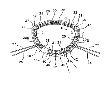
Refellil,g to the drawings wherein like numerals indicate like elements
there is shown in FIGURE 1 and FIGURE 2 plan views (from the annulus
and atrium aspects le~ecli~ely) of the ~re~l.ed embodiment of a flexible,
adjustable annuloplasty ring intended for the mitral valve de~i~Atetl as 10.
FIGURE 3 shows a side view and FIGURE 4 shows an end view of the
I,re~l. ed embodiment of the variant of the ring intended for the mitral valve.
FIGURE 5 shows a cross-sectional view taken along line 5-5 of FIGURE 2.
FIGURE 6 shows a cross-sectional view taken along line 6-6 of FIGURE 2.
The device is composed of a tubular body of textile nature 11, which
has its folded ends sewn together at seam 12 to form a ring. The ring has
three segments, the anterior segment 13, the right posterior segment 14, and
the left posterior segment 15. In the interior of tubular body 11 are provided
filiform strings 20, 21, which have ~AIel..al portions 22, 23, 24, 25. These
strings, which are preferably of a braided polyester surgical suture, emerge
from the annulus face of the ring at exit points 26, 27, 28, 29 res~,ecLi~ely.
93/15690 Pcr/uss3/oo767
21
The ~liC-t~nce between exit points 26 and 28 are ayp~ t-ply 3 mm. The
distance between exit points 27 and 29 is similar.
The strings are anchored to the ring at points 30, 31, and 32. Points
33 and 34 are colored markers sewn onto the upper (atrial) surface of the
ring that are intended as guides for implantation. These points are intended
to lie adjacent to the right and left fibrous trigones of the mitral annulus.
Also at implantation, point 31 will ayyr~ tely collesyond to the junction
of the anterior leaflet and the right coTnmicc-llral leaflet of the mitral annulus.
Point 32 will approYim~tely collesyolld to the junction of the anterior leaflet
and the left commic-c-l~ral leaflet. An internal curved flexible stiffener
member 35 spans the area corlcslJonding to the anterior segment of the
mitral annulus from points 31 to 32, and has a closed loops 36, 37 at its ends.
Dlaw~llillg 20 is passed through these loops and through the walls of body
11 to form part of the anchor knots at 31 and 32. Drawstring 21 is passed
through the walls of body 11 to lie externally for a short ~ nGe 30 and is
tied internally to form the anchor knot laying under e~.lcll~al portion 30.
The framework or stiffener member 35 is yrefe~ably of a
biocull,patible corrosion resistant metal wire with good flexure fatigue
rÇcict~nce such as Carpenter MP35N alloy or Elgiloy.
Refclence is made briefly to Figure 12 through 14. A wire 35 of the
material described, e.g. Carpenter MP35N, 0.028" diameter, or and
equivalent, is first inspected. Next, the wire is cut to the desired length, with
an abrasive saw, or an equivalent device, burnished to a.7sure that it is
smooth, and secured in a jig such as is depicted in Figure 13. The jig
co,llylises a base plate 310, a lorl~ing bar 311, a wedge plate 312 having an
incline surface 313, and a support plate 314 are secured to the base plate.
A lock plate 315 is secured to the ~ ypol l plate 314 and inter acts with lock
bar 316, that has a beveled corner 317 to receive and lock firmly in position
the wire 35. The wire 35 PYtpn~ls to the edge of the base plate which serves
as measure of the length of wire to be formed into a loop and lies ~dj~cçnt
a mandrel post 318 that is less than the diameter of the loop to be formed
in the end of wire 35. A bending tool 319, coluyli~ing a knob capable of
being gripped and turned, with a duwllvva~dly extending portion 320 having
formed centrally therein an aperture 321 sized to slip snugly over mandrel
WO 93/15690 ,~ 22 PCr/US93/0076' -
post 318 and an engaging post 322 that is spaced from the aperture a
distance slightly larger than the diameter of the wire 35. The bending tool
is fitted over the mandrel post, the wire 35 being received between the
mandrel post and the eng,q-ging post. The bending tool is turned thereby
5 bending the wire 3S into a semihelical loop. It is nececcqry to bend the wire
into a smaller loop than the llltim~tely desired loop bec-qllce the wire
rebounds slightly from its Illtim-q-tely looped configuration. When the bending
tool is removed, the wire, which has been burnished to assure that it is
smooth, assumes the configuration shown in Figures 12A and 12B, with the
10 loop, such as loop 36a or 37a, spiraling partially out of the plane of the wire.
The end of the wire, e.g. 36b or 37b, which has been smoothly cut with an
abrasive saw and rolln~led and burnished to remove all sharp edges, burrs,
etc., is then bent d~ .valdly, as shown in Figure 12B so that the loops lie
in the same plane as the wire, as shown in Figure 12C and 12D, the ends
15 Iying closely adjacent the wire a short ~lict-q-nce from the ends. The loop is
bent to assure that the end of the wire, as bent, depicted in Figures 12A and
12B, does not overlap the portion of the wire to which is closely qdj-qcent.
When the loop is bent into the plane of the wire, the wire in that portion is
not dented, distorted or deformed. As a final step, before final inspection,
20 the ~ [e-ler 35 is burnished again to assure that it is perfectly smooth, free
of indentations or deformations that may weaken it, and free of sharp edges
or other structures that might abrade the fabric of the ring.
A colored demarcation suture line 38 on the upper (atrial) surface of
the ring body indicates to the surgeon a line, outside of which the implanting
25 sutures must be placed to avoid inlelference with the internal dla~ ,ings or
the ~ [cller member. A collcs~onding, but prefel~bly uncolored,
demarcation suture line 39 is ~is~qted on the lower (annulus) surface of the
ring body. A flexible radiopaque member 40, is contained within body 11, in
the posterior region between dla~v~lJillg anchor points 31 and 32. This
30 member, in conjunction with metallic stiffener 35, forms a circumferential
X-ray marker. Member 40 may conveniently be composed of a single
c~ . ous length of 0.020" diameter extruded silicone rubber iulplegnated
with 55% Rqri~lm Sulfate and 6~o Tulllgalen. Material of this composition
and diameter is sufficiently radiopaque, but does not unduly impede the
~o 93/15690 ~ 7 7 û 1 Pcr/US93,00767
passage of the needles of the implanting sutures. It has elements 41, 42,
"hairpin" bends 43, 44, and ends 45, 46. The "hairpin" bends 43, 44, lie
adjacent to loops 36, 37 respectively, and ends 45, 46 lie adjacent to seam 12.
FIGURE 5 shows a cross-sectional view taken along line 5-5 of
FIGURE 2. Cloth layers 100, 101, 102, 103, 104, 105, 106, 107 are formed
from a singular tubular braided length of material folded and joined as
previously described. The four cloth layers, when folded and heat set, form
an upper (atrial) surface 100, and a lower surface 107 that will lie on the
natural annulus. Colored demarcation stitch 38 passes from cloth layer 100,
through layers 101, 102, 103 and hence back to layer 100. The second
demarcation stitch 39, which is p~efelably uncolored (white), is similarly
passed from cloth layer 107, through layers 106, 105, 104 and hence back to
layer 107. Demarcation sutures 38, 39 p~Ccing through their respeeLi~/e cloth
layers ~leline~te channel 108 which contains d~w~LIing 20 (or 21).
Radiopaque marker member portions 41, 42 are enclosed between cloth
layers 103, 104. A helical, contilluous, e,ir~iunl~erential sewn seam 109 joins
cloth layers 100, 101, 102, 103 to colle~yonding cloth layers 107, 106, 105,
104.
FIGURE 6 shows a cross-sectional view taken along line 6-6 of
FIGURE 2. Framework member 35 is ret~ined along its length against the
fold 110a of the cloth layer 103, 104 by a co..L;.~ous helical thread 111,
and/or by suture ties 38 and 39, for example. The fold 110a is a sin~le layer
of tubing Iying innermost in the four-layer braided fabric consJuction, the
exterior bend 110k forming the outermost layer of said four-layer
25 COIISJ uction
FIGURE 7 and FIGURE 8 show plan views (from the annulus and
atrial aspects respccli~ely) of the yre~l~ d embodiment of a flexible,
adjustable annuloplasty ring intended for the tricuspid valve deci~te~l as
200. FIGURE 9 shows a cross-sectional view taken along line 9-9 of
30 FIGURE 8. The device is composed of a tubular body of textile nature 201,
which has its folded ends sewn together at seam 202 to folm a ring. In the
interior of tubular body 201 is provided a filiform string 203 which has
external portions 204, 205. This string, which is p.efelably of a braided
polyester surgical suture, emerges from the annulus face of the ring at exit
WO 93/1569~ PCI/US93/007~'-
points 206, 207. The ~ t~nce between exit points 206 and 207 is
dyyr.~ tely 3 mm. The string also emerges from, passes around the body
(208, 209) and reenters the body 201 at the dldw~ lg anchor points. The
string is anchored in the ring by internal looped knots adjacent to external
loops 208, 209.
A colored marker 210 is sewn onto the upper (atrial) surface of the
ring. This is a guide to the surgeon, in~ ting the point that should be
positioned ~dj~cçnt to the junction of the septal and anterior leaflet at
implantation. A colored demarcation suture line 211 on the upper (atrial)
surface of the ring body in(lir~tçs to the surgeon a line, outside of which the
implanting sutures must be placed to avoid intelrelence with the adjustable
segments of the internal dlaw~ g 203. A co-re~yonding, but yreferably
uncolored, demarcation suture line 212 is sitU~te~ on the lower (annulus)
surface of the ring. A flexible member 213, is contained within body 201.
This member forms a circumferential radiopaque marker. It may
conveniently be composed of a single length of 0.020" diameter extruded
silicone rubber impregT~t~l with 55% Barium Sulfate and 6% Tungsten.
This member 213, having ends 214, 215 Iying adjacent to seam 202 passes
twice around the circumference of the ring to form concentric members 216,
217.
FIGURE 9 shows a cross-sectional view taken along line 9-9 of
FIGURE 8. Cloth layers 300, 301, 302, 303, 304, 305, 306, 307 are formed
from a singular tubular braided length of material folded and joined as
previously described. The four layers, when folded and heat set, form an
upper (atrial) surfaces 300, a lower surface 307 that will lie on the natural
annulus. Colored demarcation stitch 211 passes from cloth layer 300,
through layers 301, 302, 303 and hence back to layer 300. A second
demarcation stitch 212, which is y~erelably white, is likewise passed between
cloth layer 307, through layers 306, 305, 304 and hence back to layer 308.
Demarcation sutures 211, 212 passing through their resyecli~e cloth layers
tlçlir~e~tç channel 308 which a~ s Law~llmg 203. Radiopaque marker
member elements 216, 217 are enclosed between cloth layers 303, 304. A
helical, co..l;..~ous, circumferential sewn seam 309 retains cloth layers 300,
301, 306, 307 together.
YYD 93/15690 PCr/US93/00767
In the embodiment of the annuloplasty ring spccifically adapted for
use in repairing a human mitral heart valve having an anterior segment and
a right and left posterior segments a braided fabric tube is connected to forrn
the tube generally into an annulus. The stiffener wire extending s~bst~nti~lly
S the length of the anterior segment, the ~ ener wire having first and secondends, said ends being configured to form loops on the respective ends
thereof. As depicted in Figure 15, a first string 20 extends from outside the
annulus through the wall of the tube at a first point 20a a first distance from
the first end of the wire into the tube. Adjacent the first end of the wire the
string extends outwardly through the tube wall at 20c, through the four-layer
wall at 20d, thence around the tube as in~1ic~te~1 at 20e, thence inwardly
through the four layers prnYim~te the first end of the wire, 20f, thence
through the loop 37 in said first end of the wire 35, thence outwardly through
the walls, 20g, thence around the first string outside the tube, 20h, thence
inwardly through the walls, 20i, and over the first string at again, 20j forminga knot to secure the string 20 and the first end loop 37 of the wire 35 to the
tube pr~Yim~te the first end of the wire. The string 20 then extends through
the wall at 20k and along the length of the tube. At the second end of the
tube, knot is repe~tecl from the direction of the wire, i.e. a mirror-image of
the arrangement just des~;libed is formed securing the other end of t_e wire
and the string to the tube prnYim~te the second end of the tube. As will be
apparent, the sc~ ement just described can be ~e~nmr1i~hed from either
direction, relative to the end of the wire, and may be the same or reversed,
e.g. a mirror image knot, at the leipe~ /e ends of the wire. Other knot
securements may also be used. From the second end the string extends along
the inside of the tube a second tli~t~nce and thence outwardly through the
tube wall at a second point said second distance from the second end of the
wire, a first end of the first string extending out of the tube wall prn~rim~te
the first point, the second end of the first string eYten~ling out of the wall
pr~-Yim~te the second point 20b. The points 20d, 20g, and 20i may be
coincident, i.e. a single hole may define all of these points.
One or more strings may be provided eYter~Aing from outside the
annulus prnYim~te said first point, through the tube away from the first end
of the wire toward the second point, oulwardly through the tube wall and
W0 93/l5690 ~ 26 Pcr/us93/oo76~-
inwardly through the tube wall pr-~Yim~te a third point, that may, if desired,
be appr~Yim~tely equi~ t~nt from the first and second ends of the wire, to
secure the second string prnYim~te said third point, along the inside of the
tube to pr-.Yim~te the second point~ and thence through the tube wall, a first
S end of the second string extending out of the tube wall proYim~te the first
point, the second end of the second string extending out of the tube wall
pr~ Yim~te the second point; the first ends of the respective strings COlllpliSiLlg
a first pair of d~dw~ ngs for p~ .g contraction of the annulus between
the first end of the wire and the first point and between the first point and
10 the third point~ respectively; the second ends of the resl)e~;live strings
compli~ing a second pair of dl~w~ gs for permitting contraction of the
annulus between the second end of the wire and the second point and
between the second point and the third point, lcspecli~ely. The wire is
pre~elably polished on all surfaces, the ends thereof are r~ e-i and wherein
15 the loops are formed without denting the wire in the portions thereof that lie
cçnt the ends of the wire.
FIGURE 10 shows an isometric view of the plefclled embodiment of
the ring for mitral valve repair sewn onto the mitral annulus of the heart (the
left atrium is removed for clarity of illustration). The heart is shown during
20 ~/e~ ic~llar systole (i.e. the mitral valve is closed and the left vclllricular
outflow tract is pressurized). The annuloplasty ring 10, is positioned such
that colored markers 33, 34, are coincident to the right fibrous trigone 401
and left fibrous trigone 402 of the mitral valve apparatus. The anterior leaflet403 is shown co~liag to the posterior leaflet 404. Seam 12 will lie
25 appr-~Yim~tely at the midpoint posterior portion of the annulus. Dla~ g
anchor point 31 is loc~te~l on the annulus a~l..-;...~tely at the junction of
the anterior leaflet and the right cu.-....;~ .al leaflet, 405. Likewise,
d~aw~ g anchor point 32 is located on the ~nn~ s apprnYim~tely at the
junction of the anterior leaflet and the left commi~ural leaflet, 406.
The curved anterior portion of the ring 13 containing the internal
curved framework member spans the anterior segment of the mitral annulus
403 from points 31 to 32. As manufactured, the plane of segment 13 lies in
the same plane as the ring, as shown in solid lines in Figure 3 and as
depicted in Figure 4A. The fleYible ring forms a hinge immediately adjacent
~1~ 7~ ~
Wo 93/15690 ~ Pcr/US93/00767
27
the ends of the framework member permitting the framework member to
hinge or bend outside the plane of the ring up to an angle of appr-~Yim~tely
85~ relative to the plane of the remainder of the ring. Depending on the
particular application of the ring, the framework may, during some periods
of time, hinge such that the plane in which the framework lies is at an angle
typically of about 45~ and up to 85~ relative to the plane of the remainder of
the ring. In some applications, the ring, incl~ ing the framework portion,
will lie subsl~,lially in the same plane. The angle, if any, in which the
framework lies is not a function of the annuloplasty ring per se but rather of
configuration of the heart, or other organ, to which the ring is applied and
to the method the surgeon uses for appl,ving the ring. A colored demarcation
suture line 38 on the upper (atrial) surface of the ring body indicates to the
surgeon a line, outside which the implanting sutures 407 must be placed to
avoid interference with the internal dlaw~l~ings or the stiffener member
Numerous interrupted sutures 407, are used to fix annuloplasty ring to the
mitral valve annulus and to the sub aortic curtain 408. External portions of
the ~llaw~llh gs 22, 23, 24, 25 may be tightened and tied to the adjoining
dlaw~ ng to constrict the ring where required to correct or minimi7e
valvular in~vfficiency. The act of d~àwi~g in either or both d~a~ lings 22,
24 and or 23, 25 will cause the ring to contract between the draw~l~in
anchor points 30 and 31 or 30 and 32 rc;spc~ ely. The amount of
contraction will depend upon how much each d~aw~llh~g is tightened, and
whether only one or all dlavv~l~ings are tightened. By such means the
circumference of the annulus may be further reduced to correct or minimi7e
any r~ ining valvular ~ urr;- ;c-.~ following ring impl~nt~tiQn. It is
emph~i7~l that, as to the present invention, there is no ;;gniri~nce to the
showing or one or two or three pairs of dlaw~hings, as any number of
dla~.~hil,gs are coL.telllplated by this invention.
FIGURE 11 shows a plan view of the pJefe.led embodiment of the
invention for tricuspid valve repair sutured in place in the typically enlarged
tricuspid annulus (as described by Bex JP and T ~,c..~ te Y. '~ricuspid valve
repair using a flexible linear reducer", J Cardiac Surg, 1:151, 1986). The
tricuspid valve has an anterior leaflet 501, a posterior leaflet S02 and the
septal leaflet 503. The junction of the septal and anterior leaflets is 504, the
Wo 93/15690 ~ PCI/US93/00767
28
junction of the anterior and posterior leaflets is 505, and the junction of the
posterior and septal leaflets is marked 506. The dotted line 507 shows the
circumference of the annulus before pathologic dilatation.
The annuloplasty ring 200, is positioned such that colored marked 210
S is ~p~ t~-ly coin~id~nt with junction 504. Numerous inte~ upted sutures
508, are used to fix annuloplasty ring to the tricuspid valve annulus. The
adjustable segment is delineated from dla~ gs anchor points 208 to 209.
Typically, this adjustable segment will apprnYim~tely straddle a s-lbst~nti~l
portion of the posterior leaflet 502, as well as the junction of the posterior
and anterior leaflets 505. It may also straddle an incllb~ l portion of the
anterior leaflet 501. The act of drawing in either or both drawstrings 204,
205 will cause the ring to contract between the dlaw~ g anchor points 208
and 209. The amount of contraction will depend upon how much the
dla~LIh~g is tightened, and whether only one or both dlaw~ gs are
tightened. By such means the enlarged c ;I. u~l,rerence of the annulus may be
reduced to that shown by dotted line 507. Following a~r~liate reduction
the dlaw~Lling pairs are tied using a surgeon's knot which will lie between the
ring and the annulus, out of the bloodstream.
Referring now to Figures 16A ~ 16E, the initial steps in m~nllf~tllring
the braided ring is deccribed. A pre-washed length of heat-setable, meltable
braided fiber tubing 350, e.g. Atkins & Pearce braided polyester tape, is cut
to the desired length, e.g. 250 - 290 mm, and the cut length is slid over a
mandrel, rolled back onto a pusher rod 352 so as to form a double walled
tube, having an inner wall 356 and an outer wall 354, appr- Yim~tely half the
length of the original tubing. The tube has an inward fold 358 from outer
wall 354 to inner wall 356 fnrming one end, the right end as depicted in
Figure 16A, the other ends of the tubing 360 and 362 lying generally adjacent
each other.
Referring to Figure 16B, the double walled tube 370 is cut to a desired
length, e.g. 112 - 133 mm, at 364 with a heated blade that cuts by melting the
fibers and fusing the fibers together to form a fused end, the inner and outer
walls being joined in an annular fused joint at 364.
Referring to Figure 16C, temporary sutures 366 and 368 are secured
only through the outer layer 354 a desired distance, e.g. 56 - 66mm from the
7 ~
~yO 93/15690 ~ V ~. PC~r/US93/00767
29
end of the tube. The fused joint 364 is then rolled into the inside of the tube
so as to turn a portion of the tube inside out, the temporary sutures being
used to pull the layer through which they extend to roll the tube inside out
to position the fused joint in the inside wall, preferably in the center of the
5 inside wall of the tube 370. To clarify, the tube as shown in Figure 16B, is
rolled inside out so the the sealed-cut ends are on the right as shown in
Figure 16C, the sutures are Ptt~chetl and the tube is further rolled partially
inside out until the sutures are at the right end as shown in Figure 16D with
the heat-sealed joint between the orignal ends of the tubing inside the final
10 two-lay tube as shown in Figure 16B
The steps in forming the final tube are depicted in Figures 17A - 17B
and depict the steps of one facet of the invention, namely the method of
mPnl~fPcturing an annuloplasty or suture ring colllplismg the steps of
inV~gin~sing tubing 350 braided of meltable, heat setable polymer fibers to
15 form a tube 370 of an outer layer and an inner layer of tubing, a first end of
the tube thus formed being defined by an annular, inward fold 358 of the
tubing from the outer layer to the inner layer; forming a second end 364 of
the tube by melting the inner and outer layers of tubing to fuse said layers
together in an annular seal between said layers; and sliding said layers
20 relative to each other defined new ends of annular, inward folds of tubing
and to space the annular seal 364 distal from and between the newly formed
ends inside the outer layer of the tube.
This double wall tube may be used in the devices of this invention, as
a suture ring for heart valves and in any other device or method wherein a
25 fabric suture strip, ribbon or ring is used to secure a prosthesis to tissue or
to secure tissue to tissue.
RefelliJlg to Figures 18 and 19, the method, as used in mPking the
aforementioned suture or annuloplasty rings, further cc,mplises heat setting
the thus formed tube 370 into a lateral V-shaped band 380 having a center
30 heat set crease CO~ lg four layers of tubing.
Reference is made specifical~r to Figure 18 which depicts, in exploded
view, the fixture for heat setting the tubing 370 into a V-shaped band 380.
The double walled tube 370 is slipped over a V-shaped mandrel 372 which
may be of metal or high temperature resistant polymer, e.g. polytetrafluoro-
W0 93/15690 c~ L 30 Pcr/US93/00767
ethylene. The mandrel 370 carrying on it the tube 372 is clall,ped betweenforming tools 374 and 376 which define a V-shaped opening the size and
shape of the desired V-shaped band. A pair of bolts, C-clamps, or any other
clamping device may be used to secure the forming tools together. Bolt are
5 preferred to maintain aligmnent of the tools. The clamped tools with the
mandrel and tube are placed in an oven, or otherwise heated, to a
temperature sufficient to heat set the polymer of which the tubing is formed
without fusing it. In the case of polyester, temperatures in the range of 100 -
110~C. are quite suitable in most instances. After a sufflcient period, usually
10 about ten minutes, to heat form the tubing, the cld.ll~ed mandrel, with the
tube in place, is first cooled to set the tube into a V-shaped band or tape 380
and then removed.
The V-shaped band may be secured end-to-end to define a suture ring,
such as, for example, the suture ring 382 depicted in Figures 20 - 22. The
suture ring 382 is formed of a length of V-shaped band or ribbon 380 secured
end-to-end in any desired manner. Such a ring may be used for heart valve
prostheses, for ~ mple If desired, a ~a~ g 384 may extend through the
tube, and through the tube walls, around the inner periphery of the suture
ring. Pairs of demarcation seams 386 and 388 are sewn around the ring to
20 m~int~in the drawstring in place and stabilize the ring. These demarcation
seams may be of di~lelel-t colors thereby identifying the respective sides of
the ring. The ring is closed at a first outer periphery by dosure suture 390.
ln use, the dl~w~ll~g may be a heat-shrink type monofilament or
polyfilament string. The ring is placed on the prosthesis, the drawstring 384
25 tightened and tied. The prosthesis may then be heated r~llcing the
drawstring to form an extraordinarily tight and strong securement of the ring
to the prosthesis. The use of a heatshrink dlà~ g is not necçsc~ry, of
course. In some applications, where x-ray identification or location of the
suture ring is desirable, a radiopaque fiber is wrapped two or more times
30 around the ring or placed such that two or more fibers lie ~1jacçnt each
other.
The invention, as described, may be in the form of a suture ring for
use in surgery for securing a prosthesis in or adjacent to an annular organ
structure or stabilizing or shaping a generally annular organ portion. The
wO93/15690 ~ ~ 7~ PCr/US93/00767
31
suture ring comprises an elongate braided bioc.~ atible ribbon 380 having
ends, elongate edges and a central portion, the lateral cross-section of the
ribbon generally r~fining a V-shape, the edges ~Yten~ling oul~aldly from the
center, means securing the respective ends of the ribbon together thereby
configuring the ribbon generally into an annulus, the central portion rl~ofiningthe internal periphery of the annulus, the edges extending outwardly from
said internal periphery. The ring may be provided with at least one
d~ g 384 extending around at least a portion of the annulus and
through the ribbon selectively to decrease the diameter of the internal
periphery of the annulus, the dla~ ing being disposed adjacent said center,
the edges extending outwardly from the drawstring. The drawstring may be
omitted and the ring secured to a valve using one or more wraps of heat-
shrinkable material, e.g. polyester, to tye the ring to the valve. Means are
provided for securing the edges of the ribbon together, the edges of the
ribbon dPfining the external annulus periphery. The dravv~lling and ribbon-
lilce member are so constructed and configured that when the draw-string is
drawn the internal diameter of the annulus COUtla~ and the width of the
annulus increases thereby s~ lly preventing the ribbon-like member
from gathering into irregular clumps as the internal diameter of the annulus
contracts.
The ability of the ring to contract in its interior annulus without
bunching, gathering or folding is a great advantage. The construction
described permits the excess material to expand laterally outwardly as
ne~eSspry without bunrhit~g.
The method of use of is as follows. The left atrium is exposed
following a mid-line sternotomy, and cardio-pulmonary bypass established
using conventional techniques. A left ve~ icular drain may be inserted into
the apex of the left ventricle. The aorta is cross-clamped and a cold
cardioplegia solution is pllmperl into the aortic root c~nCing the heart to be
cooled and in~luçing cardiac arrest. Alternatively, retrograde cardioplegia may
be given via the coronary sinus. An incision is made into the left atrium,
exposing the damaged mitral valve. The teçhnique so far described is a
general descli~,lion of mitral vahe exposure, and is not speçific to the
particular device.
wO 93/15690 Pcr/Us93/00767
i ~ c~ r~ 32
Surgical techniques of implantation vary between centers. The
surgeon will then carefully examine the mitral valve mech~ni~m to asccllaiu
if the valve is amenable to repair, and if so, what the mech~ni~m of repair
will be. For example, the lesion may be at the level of the annulus, leaflets,
S chordae, or papillary Illusclcs. The appr~liale surgical repair procedure will
depend upon the pathology found. In cases where the annulus is enlarged,
or where a segment has been removed from the mitral valve leaflets, the
mitral valve annuloplasty ring will be implanted as part of the repair
procedure. First the appropriate size ring is chosen using the sizer set
supplied for this purpose. Interrupted sutures are placed at the fibrous
trigones, between the trigones, and around the rem~ining ~ ;u~llîerence of
the annulus. The sutures are passed through collcsponding portions of the
annuloplasty ring, outside the line of demarcation, marked by the colored
suture line on the atrial side of the annuloplasty ring. The ring is then
pushed down the sutures until it lies on the annulus. Each interrupted suture
is tied and cut. Mitral valve competency is then ~ce~ced by suitable means
(e.g. by the injection of a saline solution through the left ~,enlli.;ular drain,
or by removing the aortic cross-clamp and temporarily rendering the aortic
valve incompetent). If the mitral valve is ~ gilant the areas of reflux are
noted. If leakage occurs in the right posterior leaflet, this area of the ring
is contracted by gently pulling on one or both of the dlàw~LIiugs in this
region of the ring. It may be nece~ to tighten one or both draw~llhlgs,
pairs ~epen~ling upon the individual valve pathology. Mitral valve
c<3..... ...pe~ence is again ~ec~e-l and if ne~e~ y further adjustments made.
When the surgeon is satisfied that an o~tilllun, mitral valve
competence has been achieved without coul~ cing mitral valve opening
area the d~aw~ gs are tied as pairs. The surplus portions of the
dlaw~lliugs are cut close to the knots. Mitral valve ~,...pe-t~n~e is then
co~irllled. At this stage, should any slight reflux be p~eseul, it can be
30 elimin~terl or minimi7ed by placing one or more plic~ting sutures at the
a~propliate portion of the annuloplasty ring. However, should a dlàw~ g
have been inadvertently over-tightened, it may be cut below the knot to
release t}~e col.sllicLing segment. That portion of the ring might then need
to be shortened by the placement of sufficient plic~ting sutures. The left
WO 93/15690 ~ i~ 2 7 7 ~, Pcr/us93/oo767
33
atrium is then closed, air removed ~om the heart, warm blood allowed to re-
perfuse the coronary tree, the heart defibrillated if necessary, and the patientslowly weaned from cardiopulmonary bypass. Impl~nt~tion of the tricuspid
ring follows a similar course with variations appropriate to the di~erent
z~n~tomy and the corresponding differences in the tricuspid and mitral
annuloplasty rings.
WO 93/15690 ~ ~ ~ Pcr/us93/oo767
References Cited
UNITED STATES PATENTS
3,491,376 01/1970 Shiley
3,534,411 10/1970 Shiley
3,656,185 4/1972 Carpentier
4,042,979 8/1977 Angell
4,055,861 11/1977 Carpentier et al.
4,164,046 8/1979 Cooley
4,217,665 8/1980 Bex et al.
4,263,680 04tl981 Muller, et al.
4,290,151 9/1981 M~.c~n~
4,339,831 7/1982 Johnson
4,451,936 06/1984 Carpentier, et al.
4,606,911 7/1986 Ahmadi et al.
4,702,250 10,1987 Levy, et al.
4,477,930 10/1984 Totten, et al.
4,865,600 09/1989 Carpentier, et al.
4,888,009 1V1989 Lederman, et al.
4,917,698 4/1990 Carpentier et al.
5,104,406 04/1992 Curcio, et al.
5,104,407 04/1992 Lam et al
PUBLICATIONS
Tsakiris AG. "The physiology of the mitral vahe annulus". in The
mitral valve - a plulidisciplinary approach. ed ~lm~n~on D. Publishing
Sciences Group, Acton, Ma., pg 21, 1976.
Geller M, Kronzon I, Slater J et al. "Long-term follow-up after mitral
vahe recou~ll uction: incidence of postoperative left veL~ iular outflow
obstruction". Circulation, 74:I-99, 1986.
Carpentier A, Deloche A, H~n~ni~ G, et al. Surgical management of
ac~ui~ed tricuspid valve rli~e~e J Thorac Cardiovasc Surg, 67:53, 1974.
Duran CD, and Ubago JLM. "Clillical and Hemodynamic
Pelfo...~nce of a Totally Flexible ~lo~lLetic Ring for Atrioventricular Valve
Re~one~uction" Annals of Thoracic Surgery, ~:458, 1976.
Levine, R.A., Triulzi M.O., Harrigan P., and Weyman,A.E. "The
WO 93/15690 ~ ~ ~ 7 ~ ~ ~ Pc~/us93/00767
relationship of mitral annular shape to the diagnosis of mitral valve prolapse",Circulation, 75:756, 1987.
Bex JP and Leco~pte Y. 'Tricuspid valve repair using a flexible linear
reducer", J Cardiac Surg, 1:151, 1986.