Note: Descriptions are shown in the official language in which they were submitted.
WOg4/1~96 - PCT~S93/1~33
212~427
ORGAN ~UPPORT 8Y8TEM
This application incorporates herein by reference U.S.
Patent Application No. 07/524,Q75, filed on May 16, 1991, U.S.
Patent Application No. 07/965,448 (the Continuation-in-Part of
U.S. Patent Application No. 07/524,0753, filed on October 23,
1992, and PCT Publication No. WO 91/18087~
The present invention relates to an organ support system and
method for sustaining a patient, and more particularly to an
organ support system having a cell line which mimics or supports
the function o~ a specific bodily organ e.g., liver, kidney, etc.
: 15 The embodiment of the invention discussed in detail below is
directed to the liYer~ but it i3 envisio~ed that the support
system can be used for other organs. The components of the system
include a hollow fiber cartridge, biologically active cells which
~: aould be a continuously cultured cell line, and a pumping system.
~: ~0
; ollo~ Fik r Cart~l~a ~
: ~ Briefly, hollow fiber cartridges consist of a tube which
: ~ ~ontain~ a plurality ~f hollow fibers. The hollow fibers can be
.
~ made o~ ~ numbar of 3ubstances such as pclysulfone or cellulose
:
2S acetate, and ~ay vary in diameter. ~he cartridge has two spaces;
an intracapillary space ~ICS) and an extracapillary space tECS).
~h~ ICS is the ~pace comprised of the interior of the ibers, and
i~ accessed through the end ports of the cartridge. The ~CS is
- the space between the outside of the fibers and the shell of the
cartridge, and is accessed through the side ports as shown, for
example, in ~igure 1. These two spaces are the basis of
WO94/1~g6 ~1 PCT~S93/1~33
9~Cl
hemodialysis; a continuous stream of blood passes through one
space and is dialyzed against a continuous stream of fluid (i.e.,
- a dialysate) which passes through the other space. The nature
of the membrane dictates the type of exchange which takes place
betw~en these two streams, but transfer of water and small-to-
medium-sized molecules is usually the goal. Blood is usually
passed through the intracapillary space since flow is less
turbulent, and clotting is reduced. The system would function
in the reverse orientation, i.e., blood in the ECS, dialysate in
the ICS. The description in this application refers to the
conventional orientation of blood flow for convenience, but it
is recognized that the system may work equally well in the
reverse orientation.
AC~aRO~ND OF T~ INVENTION
It is known that the acute loss of ~ore than 60% of liver
function is a serious risk to survival. It is also known that
patient~ with chronic liver insufficiency may have periods when
a metabolic stress such as surgery or an infection places them
ZO in liver failure. The liv~r serves to remove impurities from the
blood and either recycles the~ to useful compound~, or converts
the~ to harmlass waste products which are excreted by the
kidneys. Without a properly functioning liver, the body is
unable to maintain its normal metabolic balance, and many organs
cease to function because of the build-up of toxins or because
: the liver is no longer synthesizing important nutrients. The
.,
functions of the liver are not completely known, but are such
that simple removal of toxins from the blood by hemodialysis or
W094/1~96 ~12 9 4 2 ~ PCT~S9311~33
he~operfusion does nvt alleviate the patient's condition.
Remo~al of toxins by these methods may improve one or more
aspects of the patient's condition such as acid-base balance or
mental status, but the overall condition is unaffected, and
mortality is not improved.
Even though the liver is the only organ capable of
regeneration, severe liver failure does not provide the optimum
metabolic circumstances for such regeneration to take place.
Faced with a rapidly deteriorating patientj. the only successful
treatment to date has been the removal of the failing liver and
transplantation with a donor livex. There are, however, several
major concerns with liver transplantation including the
procurement of a matching organ within a useful time frame, the
transport of the organ to the patient, major surgery which
carries a 10-20% mortality, the continuing danger of rejection
of the transplanted organ, and the expenses involved in the
operation and subsequent medical care of the patient.
. In view. o~ the foregoing concerns, potential uses for a
liver support system include supporting a patient until recovery
from a metabolic stress, sustaining a liver transplant candidate
until a ~ui~able organ is available, and supporting a patient
a~ter transplantation until the grafted liver is functioning
adequately and can fully sustain the pa~ient. The solution to
the problem is a metabo~ically-active liver assist device, i.e.,
25~ one containing ~unctioning liver cells. Implementation of such
a device raises several problems which have not previously been
encountered in extra~orporeal bl.ood therapies. These problems
include inter alia:
W094/1~96 ~ 9 ~ ~ PCT~S93/1~33
The need for a continuous oxy~-en supply to maintain
cell viability;
~ The need to maintain a positive pressure gradient from
the ICS to the ECS to prevent cells from migrating into the ICS
in the event of a fiber rupture; - `
The need to perfuse the ECS in order to reduce the
concentration of clotting factors, thus reducing the likelihood
of blood clotting in the cartridge;
The need to monitor the fluid in the ECS to assecs the
co~tinuing viability of cells in the ECS;
~he need to return fluid from the ECS to the patient's
blood stream in order to supply proteins which are secre~ed by
the cells; and
^ The need to temporarily support the metabolic
reguirements of a eartridge whlle the need for ~urt~er treatme~t
is evaluated.
E8~ 0~por-a~ ~log3~ rapi-s
A nu~b~r of blood purification systems are available. In
20 each instan~e, blood flow and control of th~ overall.operation
are of ~ritical importance, and the pu~ping sy t~m has be~n
de~ig~d to addres~ th~ specific needs of the procedure. None
of these conventional systems, however, address~s the specific
n~eds outlined in the paragraph above. Their shortcomings ar~
di~us~ed b~low.
W09411~96 . PCT~S93/1~33
212~7
~aofl~ly~l~
Hemodialysis is a form of extracorporeal blood treatment in
which blood flows of up to 25% of the cardiac output are
employed. It is by ~ar the most widely practiced extracorporeal
procedure involving about lO0,000 patients and requi~ing about
15,000,000 treatments annually in the United States. The
treatment has been roukinely practiced for the past 25 years with
an ever-expanding and lo~ger surYiving patient population. The
most widely used form of hemodialysis is chronic intermittent
he~odialysis (CIHD), in which the blood is purified by using a
dialysate. ~ialysate is a salt solution designed to promote
diffusion of toxins from the patient to the dialysate while
restoring salt and acid-base balance to the patient's blood. In
CI~D, the patient's blood is exposed through a membrane to a
consid~rable quantity of dialysate. In CIHD methods and
appara~u , th~ dialysate is typically prepared on-line from salt
concentrates and water. Typically, the water u~ed in the
dialy~ate is pr~pared by reverse osmosis. Since the dialysate
is always separa~ed ~rom the blood stream by a ~emi~permeable
membran~ (which do~s not admit micro-organisms), it is neither
nece~sarily sterile nor pyrogen-free. Thus, the fluid from the
ECS can~ot be recircul~ted to the patientls blood stream.
Conv~ntional h~odialysis a~so xequires careful management
of f lu~ d balance. One of the most importan~ issues in
hemodialysis i~ the control of ultrafil ra~ion, ~he removal of
- excess f~.uid from the patient. Removal of excess fluid or
insufPicient fluid rom the patient may. be fatal. Hence, a
considerable portion of conventional hemodialysis hardware and
W094/1~96 ~9 ~ ~ ~ PCT~S93/1~33
softwar~ systems is devoted to monitoring, controlling, and
as~uring appropriate patie~t fluid removal at all times.
Another shortcoming in con~entional dialysis opPrations is
that the conventional systems operate on a given patient for a
period of about 4 hours. Continuous veno-venous hemodialysis
(CVVHD) is a technique in which therapy is continuous for several
day~. However, CVVHD has the same shortcomings as CIHD; fluid,
ele~troly~e, and acid-base balance are the goals as well as the
source of greatest concern in terms of side effects, and the
fluid from the ECS cannot be returned to the patient's blood
stream.
Yet another shortcoming is the inability of existing
hardware to sustain a metabolically-active device once blood flow
i~ diverted from the device, e.g., so that the function of the
15 patient ' s 1 iver can be assessed.
: ~srooal ~op~r~u~io~
O~e example of the ~onventional sy~ems is a portable
hepatic-assist ~ethod and apparatus disclosed in U.S. Pa~ent No.
4,209,392. Thi~ syste~ ~mploys a hemofiltration membrane (plasma
~ep~ratsr) having a plurality of microporou~ ~embranes with an
averag~ pore diamQter of approximately 5 to 50 microns, and a
sterilizable disposable sorbent cartridge for adsorption of
h~patic toxins. Blood from th~ patient is passed through the
plasma ~eparator, and the ~luid portion of the arterial blood
containing substantially all hepatic toxins i5 removed from the
blood. Thereafter, the hemofiltra~e is passed ~hrough the
activated charcoal-type sorbents cartridge, and the detoxified
W094/1~96 2 ~ ~ 9 4 2 l PCT~S93/1~33
hemofiltrate is filtered throu~h a fine submicron particula~e
filter via a valve regulator to r~move any bac~eria, sorbents,
and pyrogens, and is passed to a detoxified hemofiltrate
reservoir. The detoxified hemofiltrate is pre~erably heated,
checked for proper pH and electrolyte levels, and t~en either
returned to the patient's blood or recirculated in the closed
loop hemofiltrate circuit.
This conventional device, while providing a clo~ed loop
system, also has several drawbacks. For examplej a plasma
~eparator is required, and the method is directed to operating
under the concept of plasma separation. This is a problem
because plasma separation is not typically performed continuously
for more than 4-6 hours. In addition, plasma lacks the oxygen
carxying eapacity of whole blood~ M~tabolically-active cells
will becom~ anoxic under these circumstances. This problem will
be further ~xaoerba~ed by ~he us~ of a closed hemofiltration loop
which will allow further oxygen depletion of the plasma.
Additionally, the hQmofiltrate is mixed with a phy~iological salt
solution and is stored temporarily in a reserYoir which is needed
to replenish the blood to ~he pa~ient. Yurthermore, the pore
size o~ the hemo~iltrate membrane is fairly large, and is on the
order o~ O~l to 0.5 microns. This type of fiber p~rmits the
pa~sage o~ im~unoglobulins which are potentially harmful to the
living cells in the ¢xtracapillary space. Finally, the system
2~ is not able to support a met~bolically-active ~evice once blood
~low is di~rted from the device.
.
W094/1~96 9 ~ ~1 PCT~S9311~33
Pl~8~aD~i ~
Another conventional type of device is a plasmapheresis
machine which can also be utilized with a cartridge.
Plasmapheresis involves the separation of blood into a plasma
fraction (ultrafiltrate) and a cellular component fra~tion (red
cells, white cells, and platelets) which make up approximately
45~ of the blood volume. The treatment is performed in patients
who have toxic substances circulating in the plasma fraction of
their blood. The ultrafiltrate is drawn into the extracapillary
space of a cartridge at a rate of approximately 50-lO0 ml/minute,
and the cellular components are returned to the patient with a
replacement fluid. The plasmapheresis system has several
shortcomings which preclude its use as a support system for
cellular-based therapies. Firs^, plasmapheresis is designed for
1uid removal, but not for return of the ultrafiltrate to the
patient. Second, an in-line filter is not part of the system
since cellular elements ~n the ultrafiltrate pose no threat.
Third, it is not designed to allow sampling of the ultrafiltrate.
Fourth, there is no need to assure a continuous positive pressure
Z0 qradient fro~ the ICS to the ECS since there is no risk of cells
washing b~ck into the blood stream. Fifth, the system cannot
attain flow rates sufficient to sustain a large mass of living
cells. Sixth, the ~ystem cannot support a metabolically-active
device once blood flow is diverted from the device.
:~ i
W094l1~9fi 2 l 2 g ~ 2 7 PCT~S93tl~33
Ultr~ rat~o~
A closely related method to plasmapheresis is
ultrafiltration, as mentioned above, which can be used on a
continuous basis for, or in combination with, dialysis.
Ultrafiltration relates to filtering out the macrom~lecular
sub~tances having molecular w~ights higher than approximately
10,0~0, and generally at least 40,000 - 50,000, and which
includ~s blood cells ~nd the like from the remzining
ultrafiltered a~ueous portion o~ the blood. Ultrafiltration
diff~rs from plasmapheresis in that the blood is not separated
into plas~a and cellu~ar components, but instead in~o
macromolecular fractions which include the cellular compnnents
and portions of.the plasma, and a low molecular fraction which
must be removed as waste. This process reguires ~hat the flow
rates of the ultrafiltrate be carefully controlled. T~is system
i not suited to the purpQ~e of the organ support ~ystem for the
~a~e reasons mentivned ~ove regarding pla~mapher~sis.
As discu~ed abo~e, the foregoing con~entional systems and
~ethod have several drawbacks which ~ake them unsuitable for an
organ ~upport ~y~te~.
In view o~ the for~going problems of the conventional
methods, an obj~ct oP the present invention is to provide a new
` 25 and improved support system and method for sustaining a bodily
., organ such a~ a liver having high flow rates and which can be
moni~ored for patient safety.
~ ~ PCT/US93/12333
A second obj ect of the present invention is to provide a
means of implementing treatment with an organ support system
which maintains viability of cells in a cartridge during
treatment .
A third obj ect i~3 to provid~ a system in which ~ pressure
gradient from the ICS to the ECS is maintained continuously
during therapy.
A fourth o~ject i~ to provide a support system which is
designed such 1:hat a dialysate is not required to detoxify blood
l O and the l ike .
A fifth object is to provid~ a closed loop system in which
there is no appreciable shifting irl the patient's balance ~other
than the fluid recircula ed in the extracorporeal circuit) and
which allows continuous and accurate measurement and control of
the volume of fluids removed from the patient.
A sixth obj ect of the invention is to provide an organ
support system which can be operated continuously.
; A seventh object is ~o providle an apparatus in whi::h blood
returned to the patient i~ sterile and pyrogenfree.
2 0 ~ eighth obj éct is to pro~ide an apparatus in which an
ultraf iltrate is return~d to ~he patent ' s blood stream in a
sterile and pyrogen free manner.
A ninlth object is to provide an organ support sys~em which
is regulated in such a manner as ~o assure that treatm~nt is
automatically discontiml~d in the event that an untoward everlt
oc~urs .
W094/1~96 2 ~ 2 9 1 2 ~ PCT~S93/1~33
A tenth object is to provide an organ support system which
does not require continuous human monitoring other than to
respond to an alarm.
An ~leventh object is to provide an organ support system in
which oxygenation of a biologic~ally active devi~e can be
monitor~d.
A twelfth object is to provide an organ support system which
is capable of suppoxting a metabolically-active device (such as
an artificial organ) during varying periods of disconnection from
the patient in order to allow such activities as testing of the
patient's own organ function.
According to the present invention, the above objects are
accomplished by an organ assist and support method and apparatus
having a closed loop system and designe~ for us~ with an organ
~e.g., liver) assist device including cells placed in a hollow
fiber or similar cartridge in which blood flows from the patient
through the cartridge and returns to the patient. A small
fraction of the blood flow is continuously ultrafiltered and
pa$~ed through the cell space, is ch~cked to determine integrity
of the fi~ers of the cartridge, is filtered to rem~ve any cells
poten~îally harmful to the patient, and is then returned to the
blood ~tream. This dual flow path with saPety che~ks, return of
fluid ~rom the ECS to the patient's blood stream, and a mechanism
for preven~ing c~lls from returning ~o the patient are some of
~5 the unique aspects of the invention.
~ ore pecifically, the apparatus includes an organ assist
device, an access ("arterial") line having one end coupled to the
patient and a second end coupled to an input of the cartridge to
WO94/1~96 2 ~ ~ 4 ~ ~ PCT~Sg3/l~33
return the treated f luid thereto, a cell line having one end
coupled to the cartridge and a second end coupled to the second
line, the cell line including a mechanism for detecting leaks in
the cartridge and preventing loose cells from returning to the
patient's blood, and a control system for controlling opara~ions
of the organ support ~ystem.
The method for treating blood or body fluids of a patient
according to the in~ention is adapted for use with a fluid
modifying (e.g., detoxifying) device, and includes: removing the
fluid from the patient; passing the fluid through a fluid
modiPyi~g device adapted to the condition being treated, ~he
fluid modifying device having a semi-permeable membrane and a
mol~cular weight cutoff of between 10,000 and ~S0,000 and
preferably 70,000; w~thdrawing a flow of fluid from the extra-
~5 capillary space of the fluid modifying device to determine
whether the fluid ~rom the extra-capillary space has been
modified; and returning the fluid having been modified to the
patient, wherein the organ assi t device is capable of filtering
protein~ ha~ing ~ ~ol~cular weight of be~ween 10,000 and 250,000,
and pr~er~bly b~tw~en 60,000 and 80,000, the fluid being passed
through the de~ice ~nd being modif ied by both the dif ~usion of
~olecul~ acr~ss~ the se~i permeable membrane, and by the passage
o~ ultra~iltrzlte acro s the membran~s into th~ ECS. The
ultraf iltrate which i8 returned to the patient is supplemented
with synthetie products of the cells in the ECS.
Thla or~an suppoxt ~ystem is designed to be opera~ed in an
int~nsive c:are setting, and is an ex~racorporeal system in which
the patient's blood is accessed and delivered to the therapeutic
12
wo 94"~96 2 1. 2 9 4 2 ~ PCT~S93/1~33
d~vice (e.g., the artificial liver cartridge) through plasti~
tubing similar to that used in artificial kidney treatment,
therapeutic plasma exchange, open heart surgery, standard
intravenous methods, etc. Additionally, the pressure of the
tubing and the blood flow therethrough can be moni~ored at
various points in the extracorporeal circuit. These pressure
monitors are similar to those used in hemodialysis and
therapeutic plasma exchange systems.
The control system provides flow control through pumps,
monitors the pressures, and monitors the patient return (venous)
line to ensure that air is not pumped to the patient. The
control system also includes the operator interface where the
flow rates and alarm levels are set and where the measured
pressures are displayed.
Since the apparatus does not utilize a dialysate, none of
the issues attendant thereto, particularly preparation, quality
monitoring and flow control, is of concern.
Additionally, since the system has a closed loop
configuration in which the patient has first and second lines
connected thereto with the organ assist device and cell line
therebetween, there is no appreciable shifting of patient fluid
balance, ~nd the control of patient fluid ~alance is not an
issue. As mentioned above, in conventional hemodialysis
machines, one of the crucial treatment issues is the control of
the removal of excess fluid from the patient. ~ considerable
portion of the conventional hemodialysis hardware and software
is de~oted to monitoring, con~rolling and assuring appropriate
fluid removal. The method and apparatus of the invention does
13
W094/1~96 2~ 2 ~ 4~ PCT~Sg3/l~33
not involve any appreciable shifting of t~e patient's fluid
balance. Con~equently, these hemodialysis issues, which pertain
to fluid ~alance, are not of concern.
Further; the apparatus may be operated nearly continuously
and without human supervision, for several days in a~ intensive
care unit or other specialized setting.
~I2F ~BCRIPTIO~ OF T~ D~IN~8
Figure 1 is a schematic view of tubing connections for an
organ assist device o~ the organ support system according to the
i~ention;
Figure 2a is a schematic view of the blood circuit of the
tubing et u~ed in the system shown in Figure l;
Figure 2b i5 a sch~matic view of the ultrafiltrate circuit
of the tubing set us~d in the eystem shown in Figure l;
Figure 3 is a schematic o f the combin~d tubing sets
as~bl~d ~or us~ with the organ support system shown in Figure
l;
Figure 4 i~ a front view of the eontrol sys~em of the
: 20 in~ention;
FigNre 5 i8 a ~chematic view of an organ assist device used
in t*le inYention;
~igure 6 is a schematic view of a tubing modification which
allow~ a cartridge to be oxygenated;
Figure 7 illustrates the overall blood circuit;
Figure 8 illustrates the ar~erial line of the blood circuit
leading between the drip chamber assembly and the cartridge;
W094/1~96 21 2 ~ 4 X I PCT~S93/1~33
Figure 9 illustrates the three-port (e.g., Y shaped) tubing
connection of the blood circuit;
Figure l0 illustrates the overall ultrafiltrate circuit;
Figur~ ll illustrates the ultrafiltrate line of the
S ultrafiltrate circuit;
Figure 12 illustrates the filter line o~ the ultrafiltra~e
circuit: and
Figure 13 is a graphical illustration of the results of
utilizing the organ assist system according to -the present
invention with a 12-year old girl, a~d, specifically, the
increa~e .in her Galactose Elimination Capacity (GEC) during
treatment with the system according to the present invention.
~ D W ~ PTION OF ~ PR~R~ ~M~ODI~RNT
A preferred embodiment of the invention is described
hereinbelow with re~erence to Figures 1-5~ An external organ
te.g., liver) assist device l for modifying (e.g., regulating,
d@toxifying, etc.j the bodily fluid (e.g., blood) of a patient
200 ha~ing either two indi~idual venous ca~heters (unreferenced)
or a d~ubl~ lu~en ~nous ca~h~ter 300 or the like connected
th~reto, has an input coupled to an "arterial" line 2 leading
fr~m th~ patient 200 to receive the blood from the patient. An
output of the organ ~ssist d~vice l i5 connected to a "venous"
line 3 returning the modified body fluid to the patient.
"Arterial" and "venous" are used to designate access and return
linss. The use of a double lumen catheter i~dicates that access
and return are to the same blood vessel. This nomenclature is
co~monly used in the description of extracorporeal circuits, and
W094/1~96 PCT~S93/1~33
is used for convenience. The venous vessels typically used with
the double lumen catheter are the femoral, subclavian, or
internal jugular. It is noted that the use and operation of the
double lumen catheter is well known. Further, it is noted that
the organ assist device has been employed previously without any
external elements used to pump blood through the device.
Specifically, the organ assist device has been employed with a
68-year-old patient in which the patient's arterial flow was used
to pump the blood through the device.
An example of the external organ assist device which is
preferably used with the system is a ce.ll line commercially
available from Baylor College of Medicine and designated as C3A.
The cell line is designed to be inserted into a hollow fiber
cartridge to for~ the external organ assist device cartridge. As
discussed below, the organ assist device in this embodiment is
an extracorporeal liver assist device ~LAD). As used herein,
fiber" preferably means a cylindrical fiber made of a semi-
per~eable material such as ~ellulose acetate and having an
internal diameter of approximately 200 ~ and a wall thickness of
approximately 30 ~.~Now~ver, the fiber may have other shapes ~nd
other internal diameters and wall thicknesses. The
charact~r~stics of the hollow fiber cartridge include an outer
shell wh~ch contains a plurality of fibers, and which provides
ind~pendent access to the ECS and the ICS.
Looking at the liver assist cartridge and the cell line
used therein in greater detail, the cell lines are liver cell
lines derived from a hepatoblastoma that retain most of the
characteri tics of the human hepatocyte. As used herein,
16
W~94/1~96 212 9 4 2 7 PCT~S93/1~33
~hepatoblastoma" is a liver tumor of unknown etiology, but is
presumed to be the result of inactivation of a tumor suppressor
gene. "Hepatocyte" means a normal human liver cell which
performs the metabolic functions which are typical of the normal
human liver. The cell lines are able to mimic the li-~er both
qualitatively and quantitatively. The cell lines express near
normal levels of several central metabolic pathways, including
glycolysis, gluconeogenesis, glycogenesis and ur~ogenesis.
Additionally, these cells synthesize near normal levels of
albumin and other serum proteins, contain high levels of liver
specific transcription factors, and exhibit the structures and
polarity characteristic of the human hepatocyte.
The cell lines are derived from a known hepatoblastoma cell
line, HepG2. By "derived," it is intended that the cell line is
obtained or cloned from NepG2 by a defined selection method. The
HepG2 line is a human hepatoblastoma cell line which exhibits
certain characteristics of normal human hepatocytes. The cell
line is disclosed in U.S. Patent No. 4,393,133 and is available
from the American Type Culture Collection (ATCC), Rockville, MD,
~s ATCC No. HB8065. Characteristics of the cell line have been
discus~d in publicstions including Darlington et al., In Vitro
Cellul~r and DeveloDmental BioloaY 23;349-354; Kelly et al. In
Vitro ~ellul~r ~nd Developmental Bioloq~ 25:217-Z22;
Darlington, G.J., Meth. EnzYmol. 151:19-38 (1987); Thrift, R.N.,
et al., J. Lit~. Res. 27:236-250 ~1986). Unlike most other
human liver lines, HepG2 does not carry any human hepatitis B
,.
virus (HBV) genetic sequences. Thus, the cell lines of the
WO 94/14496 r~ PCT/US93/12333
,t ?~
invention, clonally deri~ed from HepG2, do not carry any Hsv
genetic ~equences.
The cell lines may be obtained from the HepG2 line by
selecting for cells which show: (1) strong contact inhibition;
(2) high ~xpression of albumin; (generally at leas~ about 20
g~mg total cell protein~4 hr, more generally a~ least abou~ 25
g/ml total cell protein/24 hr); and (3) high albumin to
alphafetoprotein ratio at confluence (generally a ratio of at
least about 15, more g~n~rally at least about 25).. This is
discus~ed in greater detail in U.S. Pat2nt Application No.
07/524,075 and PCT W0 91/18087, both incorporated her~in by
reference. A preferred cell lin~ is C3A, which is described more
fully below. This cell line has been deposited at the American
Type Culture Collection under ATCC No. CRL-10741.
The selected cell lines synthesize levels of human albumin
and other seru~ protei~s that are similar to levels produced by
normal hu~an hepato~ytes and demon~trate regulation of gene
expression as i8 pr~dicted for developing or regenerating normal
hepa~o~ytes. A~ indicated, such cell lines are cloned by
selection for high ~lbumin production and a high albumin to
alphafetoprotein (AFP) ratio when the ~ells reac~ confluence.
Th~ ter~ ¢onfluence refers to the cell density in culture when
the cells begin ~o con~act on~ anoth~r and cover mos~ or all of
the available growth surface.
2~ In the precon~luent phase of growth, selected ~ells behave
like a r~generatiny liver. They have a rapid doubling time
(abou~ 24 hr) and express a number of fetal proteins, including
AFP, aldolase A/C and pyruvate kinase K. Upon reaching
WO94/1~96 2 1 2 ~ ~ 2 7 PCT~S93/1~33
confluence, the cells assume an adult phenotype wherein cell
division slows dramatically ~doubling time >200 hr) and
expression of fetal proteins is extinguished. Cells expressing
an adult phenotype become predominant, as evidenced by production
of albumin, aldolase B, and pyruvate kinase L, and development
of histologic features of a normal liver.
The cell lines of the invention have several distinct
advantages over hepatoma cell lines known in the prior art. They
are extremely well differentiated. Consequently, they
constitutively express liver-specific biological activities at
a level suf f icient to support a subject in hepatic failure or
insufficiency for either short or long periods.
The term "constitutively" refêrs to the fact that these
cells normally express liver-specific biological activities
without any need for particular forms of induction. Once these
cells reach confluence, when they grow to fill the available
æurface, they maintain normal liver-specific biological
activities.
The term "liver-specific biological activity' as used herein
refers to a number of physiologica}~biochemical reactions which
take place specifically in hepatocytes, as well as in the cells
o the present invention. Also intended by this term are the
proteins, protein complexes, lipids and lower molecular weight
products which these cells synthesize and secrete.
Hepatocytes perform multiple finely-tuned functions which
are critical to homeostasis. Of the variety of cell types in the
mammalian body, only hepatocytes combine pathways for synthesis
and breakdown of carbohydrates, lipids, amino acids, proteins,
2~ PCT~S~3/1~33
nucleic acids and co-enzymes simultaneously to accomplish a
unique biological task. The key "liver-specific" biolo~ical
function~ include~ gluconeogenesis; (2) glycogen synthesis,
storage and breakdown; (3) synthesis of serum proteins including
albumin, hemopexin, ceruloplasmin, the blood clotti~g-factors
(including Factors V, VII, IX, X, prothrombin and fibrinogen),
~l-antitrypsin, antithrombin III and AFP; (4) ccnjugation of bile
acids: ~5) conversion o~ heme to bile pigments; (6) lipoprotein
synthesis; ( 7 ) vitamin storage and metabolism; (8) ~holesterol
synthesis; (9) ammonia ~etabolism, including urea synthesis and
glutamine synthesis; (lO~ amino acid metabolism, including
metabolic conversion and re-utilization of aromatic amino acids;
and ~ll) detoxification and drug metabolism.
The cells of the invention are believed capable of
per~orming all classes of the 7'liver-~pecific" biological
functions. All functions have been tested except for classes 4
and 5. Exemplary functions include the ability to perform
a~monia m~t~boli~m, a~ino acid ~etabolism, detoxification, and
protein production, ~ pscially of coagulation fac~ors. These
four groups of liver-~pecific biological functions are of
particul~r i~portance where the cell are to be used in a liver
as~ist devlce (LAD).
For 5upport of ~ubjects in the form of relatively short term
LADs, such as patients with ~ulminant hepatic failure (F~F),
25 pati~nts awaiting liver transplantation, or patients with
nonfunctioning liver grafts, the four groups of liver-specific
biological functions noted above are belived to be of central
importance. However, notwithstanding the above, there may ~e
WO 94J14496 PCT/US93/12333
2129427
others of equal or greater importance. The other functional
defieits can be provided by other means (such as by provisior! of
glucose and monitoring of glucose levels) or do not require acute
attention (for example, conjugation of bile acids or bile pigment
production, or drug metabolic activity).
The levels of liver-specific biological activity "sufficient
to support" a subject suffering from hepatic failure or
insufficiency are those which will result in normal or near
normal levels of serum proteins, coagulation factors, amino
acids, and other metabolites produced in or metabolized by the
liver. These improvements may be measured biochemically or by
an improvement in patient's clinical status. These various
~olecules, metabolic and clinical parameters and products and the
physiological as well as pathological ranges of their
concentrations or levels are well known in the art and are set
forth, for example, in Zakim & Boyer, Hepa~lo~y: A ~extbook of
er Disease, W.B. Saunders Company; Harcourt, Brace,
Jovanovich, Inc., Philadelphia, L~ndon, Toronto, Montreal,
Sydney, Toky~ 199o), which is hereby incorporated by reference.
O~ce ~ p~rticul~r cell line has been selected based upon the
initial criteria of strong contact inhibition, high expression
of albu~in, and a high albumin/alphafetoprotein ratio at
confluence, the cell line can then be tested for the performance
of liver-specific biological functions. Thus, tests as described
below can be perform~d to examine the me~abolic functions of the
~:~ cells, particularly in an en~iro~men~ in which the cells can be
used as a liver assist device. Metabolic ~unc:tions tested
~ ~2 9 PCT~S93/1~33
include oxygen dependence, glucose and urea synthesis, bilirubin
uptake and conjugation, and clotting factor biosynthesis.
The liver is an extremely aerobic organ and accounts for 20%
of the body's oxygen consumption. Like the liver in vivo, it is
noted that the cultures of the invention require ~xygen ~or
high-level liver-specific function (see U~S. Patent Application
No. 07/524,075). Provision of adequate oxygenation may stimulate
both growth and differentiated function in selected cells. The
effect of oxygen on selected cell lines may be tested in several
ways, including the following:
(1) The growth rate of the cells in continuously perfused
cell culture may be examined in increasing concentrations of
dissolved oxygen (4-20%). Growth rate can be examined in a
standard medium containing high concentrat.ions of glucose and in
~5 glucose-free m~dium containing lactate and amino acids as the
only carbon source. As gluconeogenesis is exceedingly
oxygen-sensitive, one would expect cell growth to be more
dramatically affected in the glucose-free medium as compared to
cells in the pre~enc~ of glucose.
(2~ Indicatoræ of metabolic acti~ity may also be measured
-~ in the cells at different concentrations of oxygen. Such
metabolic activities include total oxygen consumption, energy
charge, redox state ! and the ratio of glucos~ consumption to
oxygen consumption.
The logical extension of these experiments is the
application of the patient treatment. Since the cell function is
associated with an adequate oxygen supply, the continuous or
inte~mittent monitoring of the blood flowing through the device
22
W094/1~96 2 ~ 2 ~ 4 2 7 - PCT~S93/1~33
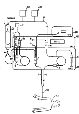
may be performed. Accordingly, a device 350, as illustxated in
Figure l, for monLtoring oxyge~ tension of the blood flowing
through the extracorporeal blood line may be employed. For
example, a commercially available 2 sensor may be used.
Similarly, other para~eters such as temperature or the ~ike may
~e monitored as desired. The monitoring device may be coupled to
the auxiliary monitoring unit 420 to alert the operator of
un atisfactory levels. I~ is envisioned that external monitorinq
devices could be developed for non-invasiv~ detection. While the
monitoring device 350 is shown coupled to the arterial line 80
in Figure 1, the monitoring device(s) may be employed at any
position in/on ~he extracorporeal blood line.
Glucose and urea ~ynthesis are the primary means of removing
excess amino acids and ammonia from the blood. Amino acid
catabolism r~sult~; in the libera~ion of carbons which are shunted
into the citric acid cycle and thence to glucose. The nitrogen
released during this process is used in the synthesis of urea.
Therefore, a selected cell line must synth~ize both glucose and
urea. ~ethods for m~urs~ent of glucose and urea are known in
the art, for ex~mpl~ ~ee ~ersh~er et al., in ~e ~bL9~ LD Y~ 5i~
~ DEii~ H-U- Bergmyer, ed., 3rd ed., Verlag Chemie, Weinheim,
Vol. VII, pp. 59-67 gl983).
El~vated serum bilirubin is 3 highly visible indicator of
liver disease. While not generally toxic in adults, high
circulating l~vels of unconjugated bilirubin may produce brain
damage a~d even death in neonates. This condi~ion is known as
kernicterus because of the typical yellow appearance of the brain
stem nuclei at postmortem examinations. The ability o~ the
23
W094/1~6 ~ PCT~S93/1~33
selected cell lines to metabolize bilirubin may be examined, for
example, using oxygenated monolayer cultures. For this test,
erum fro~ patients with hyperbilirubinemia can be incubated with
oxygenated cel}s to determine whether the cells are able to
conjugate the bilirubin. Direct binding studies may-be carried
out using [3H]-bilirubin in the presence and absence of unlabeled
competitor in order to de~ermine V~x and Xm.
The cell lines are also tested for clotting factor
biosynthesis. Many of the clotting factors are synthesized by
the liver, and the development of a severe coagulopathy is an
o~inous sign in FHF. Although all of the vitamin K dependent
gro~p is affected, antithrombin III ~AT III) has been identified
as the most significant deficiency. The cell lines are tested
for the ability to synthe~i2e fibrinogen, prothrombin, factors
VII, and X, and AT III. The levels of production of these
factvrs may be guantitated using commercially available
antibodies.
~ he properties of the cell lines make them particularly
useful in liver assist de~ices (LAD). For the most part, the
cells ~ay be u~ed in any deYice which provides a means for
culturing the c~lls, a~ well as a means for separating the cells
fro~ blood which will be passed through the d~vice. Membranes
or capillarie~ ar~ available in the literature for use which
allow for the crossoY~r of toxic solut~s from the blood to the
cells as well as the diffusion of vital metabolites provided ~y
the c~lls across the membrane into the blood. The permiselective
or semipe~meable membrane additionally provides a mechanical
barrier against the immune system. For the most part, a membrane
24
WO~4/1~96 PCT~S93/1~33
2 1 2 9 4 2 i~
or capi~lary is used which features a molecular weight cutof f
from about 10,000 up to about 250,000, and generally about 60,000
to 80,000 (preferably 70,000).
Generally, the c~lls are grown in the liver assist device.
After growth of the cells, the subject's blood is passed through
the device, and dissolved molecular cpecies (e.g., bilirubin)
diffuse through the membrane and are taken up and metabolized by
th~ cells. For the mo~t part, the devices are base~ primarily
on extracorporeal blood processing. Generally, the.devices are
designed to house ~h~ cells in a blood-perfused de~ice attached
to ~he blo~d stream. Typically, the device is attached to the
blood stream by v~in, as discussed below in more detail.
Several designs of livçr assist devices are known in the
literature. For example, devic~s have been described by Viles
et al., U.S. Patent Nos. 4,675,002 and 4,853,32~: Jauregui, Great
Britain Patent ~o. 2,221,857A; Wolf et al., Inte~na~io~al J~ of
Artificial V ans 2:97-103 (1979); Wolf et al., ~e~::national J.
L Q~ 45-51 (1978); and Ehrlich et: al., In Vitro
14 : 443-~a50 ~ 1978 ), which disclosures are h~rein incorporated by
20 refererlce. Pr~ferr~d d~vices include the hollow fiber cartridge
and si~ilar pQrfusion devices.
Bioreactors, such as hollow f iber bioreactors, may be
utilized as liver assist devices. Such bioreactors, such a~; the
Anchornet series, ar~ known in ~he literature and are available
co~mercially. See, ~or example, Heifetz et al. t BioTechnioues
: 7:192-199-(~989); and ~ono~rio, D.M.. ~ A~r. Biotech. Lab. Sept.
1989, Publication #940, which disclosures are herein incorporated
by reference. C~mmercially available dialysis cartridges such
W094/1~96 PCT~S93/l~L33
2 ~ 4 2 r~
as Althin CD Medical, Inc. (of Miami Lakes, Florida) Altraflux
may also be used.
The cells of the invention, when grown in a hollow fiber
cartridge or similar perfusion device with capaLcities for high
S numbers of cells, can function as a perfused liveP,-allowing
accurate a~se~sment of human liver metabolism and replacement of
liver-specific biological activities. ~bLerefore, a perfusion
device containing a culture of the disclosed cells is capable of
functioning as a liver assist device. In the preferred
e~bodiment of this invention, the LAD is extracorporeal,
referring to its connection to the circulaLtion outside the body~
An ~Oxtracorpor~al LAD (or EL~D) is particularly useful for
providing temporary liver ~uppo~ for subjects sufering from
FHF. It is e~visioned that the LAD could also be implanted in
the body, that is, "intracorporeal." This embodime~t may be
advantag~ous as a long~r term LAD.
For u~ in a liver assist device, th~ c~lls are gen~rally
grown on the ~mbrane ~r porous support which may be formed of
cellulos~ acetate. For the most part, th~ cell~ attach to the
~upport upon gro ~ ~. ~owever, it is recognized that linkage
~aterial8 may ba pro~id~d ~o at~ach the cells to a support.
Suitabl~ linkage ~ateriaLls are known in the art. S~e, for
example, J~ure~ui, Gr~at Britain Patent No. 2,221,857A.
~ollow fiber cartridges are ~wo-chamb~r units which
2~ reproduce the three-dimensi~nal characteristics of nonmal organs
(Knazek, R.~., E~9~ Y9~. 33:197~-1981 (1974); Ku, R. et al.,
Biotechnol. Bioenq. 23:79 95 (1983)), which references are
hereby incnrparated by reference. Culture or growth medium is
WO 94/14496 PCT/US93/12333
2129427
circulated through the capillary space a~d cells are grown in the
extracapillary space (Tharakan, J.P. et al., Biotechnol. Bioenq..
28:1605-1611 (1986). Such hollow fiber culture systems have been
disclosed as useful ~or culture of hybridoma cells lines for the
production of monoclonal antibodies (Altshulter, G.L: et al.,
~iQ~echnol. Bioenq 28:646-658 (1986); Heifetz, H~H. et al.,
.
~Bio~echnioues 7:192-199 (1989); Donofrio, D.M., Amer. Biotech.
Lab., Sept. 1989, Publication #940)). Further, a number of other
~ell types, including the liver cell lines PLC/PRF 5 and Reuber
hepatoma, (McAleer, W.J. et al. J. Virol. Meth. 7:263-271
(1983); Wolf, C.F.W. (1982)) and pancreatic islet cells (Araki,
Y. et al. piabetes 34:850-854 (1985)) have been cultured in this
manner. Cells could conceivably be grown inside the fibers.
Once a device has been chosen for use as a liver assist
device, ît is provided with the appropriate medium and an
inoculation of cells. Generally, cells are grown in a complex
medi~, for example, in a 3/1 mixture of Eagle's MEM with Earle's
8alt8 ~Gibco) and Waymouth'~ M~B 87/3 (Gibco) 30 containing 10%
defined/supplemented calf serum (Hyclone). The devices are then
maintained at 37 degrees C with constant recirculation of medium
~nd con~tant inflow of fresh medium. Each cartridge growth
circuit includes a membrane oxygenator which maintains oxygen
saturation of the medium. For use with a hollow fiber cartridge,
u ing a ~2m2 hollow cartridge, the cartridge is provided with 150
~l/min of recirculated medium with a constant inflow of about
0.3-1.0 ml/min. A 2m2 cartridge is generally inoculated with
about 1 x 10⁹ cells.
WO94/1~96 PCT~S93/L~33
4?~
The f~nction of the cells in the device can now be tested
for the capability of the device to function as a liver assist
device. This includes measurements of essential liver bi~logical
~unctions as discussed above.
For the most part, it will not be necessary- to add
additional oxygen to the membrane oxygenator. However, the
oxygen tension in the cultures can be determined and additional
oxygen added i~ necessary.
In order to vary the oxygen tension in cultures of the
selected cell lines to determine the optimum oxygen level, cells
ean be ~rown in a continuous perfusion apparatus. The apparatus
will consist o a recirculation pump, medium bottles, and a lid
that fits on a standard 6-well culture dish. ~he medium is
~ontinually recycl~d ov~r the surface of the cell~ and back into
lS the medium container whers it can be ~as ed. ~edium will be
ga~ed with pr~parations containing betw~en 4% and 20% oxygen,
5% ~2 and the remaind~r ~itrogen. In this way, ~he c~lls can be
~aintained in th~ appropri~te at~osph~re such that the effect of
the gas ~ixtur~ can be determined. ~rowth rate may be determined
by ~onitoEing totai cell protein per we}l.
ATP, ADP and AMP will be measured as described by Lundin et
- nzvmol. 133 : 27 - 4 1 (1986), using firefly
lucifer~e. The ratia.-~of N~D/NADH can be calculated from the
xatio of lactate ~o pyruvate across lactate dehydrogenase and
from the ratio o mal~te to oxaloacetate across malate
dehydrogenase. The concentra~ions of the~e ~etabolites can be
determined as ~aught by the methods et ~orth in Methods of
Enzymati~ An~lysis, H.U. 8ergmyer, ed., 3rd ed., Verlag Chemie,
28
W09411~96 212 9 4 ~ 7 PCT~S93/1~33
Weinheim~ Vol. VI, pp. 570-588. The ratio of NADP/NADPH may be
calculated from the ratio of isocitrate to alpha-ketoglutarate
across isocitrat~ dehydrogenase and from the ratio of malate to
pyru~ate across malic enzyme. The determination of these
metabolites is also ~et forth in Methods of Enzymatic.A~alysis.
Energy change may be calculated from the equation (ATP + 0.5
ADP)/(ATP + ADP + 5 AMP). Besides looking at the oxygen
dependence of the liver assist device, the devices will also be
characterized with respect to their ability to cimulate an
isolated, perfused human liver. This includes testing the device
for glucose and urea synthesis, bilirubin uptake and conjugation,
and clotting factor biosynthesis as described above. Urea may
be quantitated using a coupled glutamate dehydrogenase/urease
assay. Gluco~e may be determined using a dye-coupled glucose
oxida~e assay. The assays for urea and gluco~e dstermination are
found in M~h~ds of Enzym~tic Analysis. As discussed earlier,
the v~riouC vitamin K dependent clotting factor~, prothrombin,
factors YI}, IX and X, as well as antithrombin III, can be
determined using a solid phase radioimmunoassay as described by
Kelly et al., In ~i~ro Cell Dev. Biol. 25:217-222 (1987).
Antibodies for the immunoassay may be obtained from DAK0, Inc.
In a preferred L~D embodiment, the cell line C3~ (as
mentioned ~bove, commercially available from Baylor College of
~edicine) iæ provided in a hollow fiber cartridge (as also
mentioned above, commercially available from Althin CD Medical,
Inc.) for use as a liver assist device. The device comprîses
hollow fiber capillaries contained within a plastic housing. A
~eal around the ends of the fibers provides two spaces (an ICS
29
WO94/1~g6 ~I PCT~S93tL~33
?~ :`
and an E~S). Media are circulated through the ICS and cells are
grown in the ECS.
For the growth of cells, cells are seeded into the
~x~racapillary spac~ and supplied a constant inflow of fresh
5medium. 2m2 c~rtridges are inoculated with an effectiv2 number
of cells~ usually about 1 x 10⁹ cells, and grown to confluence,
usually about 28 days.
The medium supplied is generally a complex medium, as
mentioned above, usually a 3/1 mixture of Eagle's MEM and Earle's
10salts con~aining 10% defined/supplemented calf serum. This
provides nutrients for cell growth. Thus, the cells grow on the
outer sur~ace of the capillaries. The hollow iber cartridge
containing the conflue~t cells is capable o~ functioning as a
liver assist devi e for s~lpporting a subject suffering from
15hepatic failure or insufficiency.
The e:ell linas may also find use as bioartificial livers or
liver ~;upports. In thi~ ~anner, the cells are encapsula~ed or
grown in hollow fiber capillary membranes for use as a
bioartificial organ. ~he cells are encapsulated in biomaterials
20such as algin~te-polyly ine membranes, as taught by Cai et al.,
12 : 38~-393 ; Sun et al ., ~rans. ~ Soc.
i ~ _c~ 86); O'Shea et al.,
804, :133-136 (1984): Sun et al. ~ J.
Controll~_ Rel~;e 2:13~ 141 (1985): and U.S. Patent No.
254,391,90~. The encapsula~ted cells and vehicle capsules are ~hen
injected intraperitoneally into a subject (along with other
insertion devices such as straws, bags, etc.).
WO94/1~96 212 9 ~ ~ 7 PCT~Sg3/~U33
The.novel cell lines are useful for studies of human liver
metabolism as well as the study of liver specific gene
regulationO The cell line is originally derived from a human
hepatoblastoma, not from a human hepatoma as is the usual case
with human liver cell lines. The cell lines are useful for
studying all liver functions, including metabolic functions and
liver specific gene e~pression. They also provide a useful in
v tro liver model.
The cells and cell lines may also be used for s~udying the
metabolism and/or toxicology of drugs or other pharmacological
compositions. m e cells, grown on a membrane or liver assist
device, serve as a prototype artificial liver. Thus, the
clinical effects and ~etabolic byproducts o~ various drugs or
compounds can be asse~sed in an in vitro model. The cells grown
lS in liver assist devices are also useful for the production of
serum proteins. As indicated the cells exhibit liver specific
biological activity and synthesize serum proteins, isoenzymes,
clotting factors and the like. Accordingly the cells can b~
utilized as an inL~Li~~ factory for these proteins. In this
manner, the supernatant fluid is recovered frsm the cell culture
and the plasma proteins isolated and purified. For con~enience,
the cella may be grown on a semipermeable me~brane which allows
: ~or d~fu~ion of serum proteins across the membrane where they
are isolated and purified for fur~her use.
A~ the cells are capable of functioning as a liver model,
they are also useful for studying viral hepatitis. This is
~: particularly true as the cell lines are not transformed by
hepatitis B virus (HBV~ and do not carry any HBV sequences.
31
W094/1~96 ~ PCT~593/L~33
The liver cells disclosed in the pr~sent invention have
advantages over other systems known in the art, such as ~he
isolated perfused rat liver (IPRL). The cultures are permanent.
That is, they have an ind~finite life-span, ther~by allowing the
effects of long-term exposure to be studied in an experimentally
rigorous situation~ Monolayer cultures of the permanent cell
lines are typically maintained for seYeral months and a liver
assist device pr~pared according to the methods of the invention
functions normally over at least an indefinite perisd., generally
eight to twelve weeks, as determin~d by albumin production and
glucose utilization. LADs have been maintained for 6 - 8 months
by the pre~ent inventors. Use o~ the culture methods of the
invention reduces the need for the regular sacrifice of animals
reguired ~or liver perfusion, which comports with current U.S.
goYernment goals (NIH Guide for Grants a~d Contracts, suEra).
Finally, ~he cartridges containing cultur~d cells of the
invention reflect human ~etaboli~m more clo~ely than the isolated
p~rf u~d livers f~om other cpecies.
Ths inventive ~ystem, particularly~the use o~ a hollow
~iber-ba~ed ~y~t~m,l off~r~ several advantag~s as liver assist
device~. cartridges ~upport the growth of v~ry high density
cultur~. Bas~d on the extracapillary volume, 200q o~ cells can
be grown in a 2m unit. The uni~ i~ capable of achieving
sufficient cell mass to provide liv~r suppvrt ~o a ~ubject
suffering from liver failure.
Cartridg~ grown cells are polarized and their growth
approximates normal liver structure. The cells receive nutrie~ts
from the ICS and secrete complex metabolic products in~o ~he ECS
WO94/1~96 PCT~S93/1~33
21~4~7
and back to the ICS. The ECS can be perfused to prevent
accumulation of toxic products although no toxic product has been
identified at the present time and perfusion of the ECS has not
been necessary. The rontinual flow of media and an in-line
oxygenator may be employed as di~cussed below to pro~ide a more
constant supply of oxygen and energy. Thus, the organ (e.g.,
liver) assist device is inserted in-line with the patient's blood
flow to ~odify (e~g., metabolically regulate, filter, detoxify,
etc.) the blood.
Figure 5 shows an example of a cross-section of two fibers
illustrating their ICS la and their ECS lb with cells growing on
their outer suraces~ Thus, media are circulated through the ICS
and cell are grown in the ECS of the cartridge.
To monitor the integrity of the org~.n assist device 1 which
has been placed in a hollow fiber cartridge, a recirculation
tubing set, as shown in Figures 1, 2b, 3, 6 and 10, has a first
end connected to the device 1, and a small volume of fluid is
withdrawn from the extracapillary space of the device 1. The
~luid in the extrac~pillary space can be checked for hemoglobin
2.0 or the like which would indicate a leak in one or more of the
hollow fibers of the cartridge. To prevent any cells from
returning to ~he patient, a filter ~echanism or the like may be
installed on the fluid line from the extracapillary space. Any
of a variety of filters may be employed to include a 0.45 ~m
filter. Additional filters may be placed in the extra-capillary
space fluid line for additional safety. For example, a tandem
filter set may be employed. T~e tandem filters may be
commercially available from Arbor Technologies or Gelman
33
wo 94"~96 212~ ~ 2 1 PCT~S9311~33
Corporation, both of An~ Arbor, MI. The re~irculation tubing set
has a second end connected to the outlet tubing of the organ
assist device l.
As shown in Figures 1 and 4, a control system 4 including
three modules controls the overall system operation. T~e control
system may include the three modules in a single integrated, or
a~ separate modules, as shown in Figures l and 4. One of the
three modules comprise a dual pump system which is the primary
control module 410. Module 4lO is commercially avail~ble (e.g.,
a BS~-22 Dual Pump Blood Safety Module commercially available
from CG~, Inc. of Lakewood, Colorado).
Another module of the control system is an auxiliary
~onitoring unit (~MU) 420 which is designed to ~onitor pressures,
accept alarm settings fro~ the operator by a kexpad or the like,
and, in turn, notify the operator if certain alarm limit~ are
reached~ T~e primary control module 410 and the AMU 420 are
mounted together so that relative motion therebetween is
pr vented. The primary control module 4lO also ~ay have one or
~or~ alarm units associated th~rewith. In certain casss, as
disc~s~ed below, t~e A~U Gan initiate a system shutdown ~larm.
Th~ third ~odule of the control system is a Venous PressurP
Monitor ~P~) 430 which monitors the pressure in the venous
return to ~he patient in an extracorpor~al circuit during
treatment~ Th~ VPM, also commercially available ~rom CGH, Inc.,
may includ~ two types of alarms. A first type of alarm has a
li~its window such that the alarm is triggered when the pressure
value is 40 mmHg or lower or 70 mmHg or greater than the selected
value. A second alarm is a so-called "out-of-range alarm" in
34
WO94/1~96 PCT~S93/1~33
2129~27
which the alarm is trig~ered when the pressure value is higher
than +450 mmHg or lower than +10 mmHg. When an alarm is
activated, the blood pump stops. The VPM includes pressure
transducing elements and a power supply.
Referring to Figure 4 and looking at the control-system in
greater detail, the primary control module 410 operates on a
normal electrical supply, and includes a blood pump having a
maximum flow rate of 700 ml/minute, an ultrafiltrate pump having
a maximum flow rate of 2 l/hour, a Heparin ~or similar
anticoagulant) pump having a maximum flow rate of up to
approximately 10 ml/hour, and pressure monitors and alarms
connected to the pressure monitors. The primary control module
has mounted thereon a drip chamber holder (Pv) 411 for holding
a first (venous) drip chamber 1~, a drip chamber holder (P2) 412
for holding a second, e.g., ultrafiltrate, drip chamber 12, and
dual blood (e.g., ultrafiltrate and arterial~ pumps 413. As
illustrated in Figure 4~ an arterial pressure sensor 9 may be
provided on t~e primary control module. Additionally, an
atmospheric pressure monitor (unillustrated) may be provided.
The AMU 420: may contain a plurality of commercially
available pres~ure transducers capable of withstanding gauge
pressur~s from approximately 1 atmosphere negative to 3
atmospheres positive. The operating range of pressures from
approximately 100 mm~g negative to 200 mmHg positive is
preferably accurate to within + 5 mmHg + 2% of the reading. The
repeatability within ~he operating range when this operating
range has not been exceeded is preferably within approximately
+ 2 mmHg.
WO94/1~96 , PCT~S93/1~33
As shown in Figure 4, the AMU includes a holder 422 for ~he
device, a drip rhamber holder (P1) 423, a second drip chamber
holder (P3) 424, a~ well as a display/user interface and a
control section. The user interface can be a relatively simple
LCD display, e.g., four lines of 32 characters each, and is
formed to allow easy ~etting of alarm levels by means of the
keypad and by presentation of the relevant readouts.
The AMU has appropriate electronics to allow the four
transducers to be calibrated at the same two pre~sures, i.e. a
common source can be applied simultaneously to all four pressure
transducers ~nd maintained for a duration sufficien~ to establish
and store readings for each transducer at this common es~ablished
pres~ur~. For exa~ple, the two reference pressures may be
establi~hed by an atmospheric refer~nce and a mercury manometer
(or oth~r acceptable secondary standard). The AMU 420 is capable
of accepting inputs for upper and lower alarm limits on each of
four dif~erential pressures which are important to the device
operation. The AMU accepts the inputs via the keypad. These
pressure dif~erential~ are as follows:
Pv ~ P2, the minim~m transmembrane pressure for the
therap~utic d~vice;
Pl - PV, the blood pressure drop within the therapeutic
devic~;
P~ - P2~ the maximum transmembrane pres~ure for the
t~erapeutic device; and
P3 - PV ~ the transfilter pressure drop.
The ~MU 420 is also capable of generating alarms according
to th~ alarm set in the manner discussed below.
36
wO94/l~s6 PCT~S93/1~33
2129A27
PV - P2 ~ generate AMU audible alarm with system shutdown
and AMU panel display suggesting that the potential exists for
infusion of cells into the blood stream. Action such as
increasing Pv by increasing blood flow, or decreasing P2 by
increasing plasma flow may be instituted to achieve Pv - P2 >
Pl - PV rising > 200mmHg generate AMU audible alarm with
display panel suggesting that if the blood flow has not been
increased, then possible clotting should be investigated.
Pt - P2 rising > SmmHg generate AMU audible alarm with
display panel the same as described ab~ve with regard to P~ ~ Pv
and Pv ~ P2 <
P3 - PV rising > 250mmHg generate AMU audible alarm with
diæplay panel suggesting that a transmembrane leak or cellular
sloughing be investigated.
~5 P3 - PV rising > 5mmHg in 30 seconds, same response as
P, - P2 ri~ing > SmmHg with the additional information that the
rate of pressure rise was exceeded.
P3 - PV exceeding maximum value. Stop entire system. This
AMU alarm will also stop the system. It is envisioned that any
~; 20 one or more of tbe above~alar~s.can be programmed to shut down
the sy~tem. Addi ionally, a temporary manual override may be
employ~d to allow pressure readjustment.
There may.be a mute capability on the audible alarm, and the
system may also be provided with a distinguishing visible alarm
(e.g. a flashing light).
The AMU 420 operates from the power supply available from
the primary control module 4lO and additionally has a battery
backup ~r the like to retain calibrations therein. If the
W094/1~96 21~ PCT~S93/~33
battery backup fails, then the unit automatically displays that
recalibration of the pressure transducers is required. The AMU
also ~ay be provided with a device for adjusting the monitoring
chamber levels during operation of the support system such as a
s syringe connected to the transducer line of the drip c~amber.
Regarding the organ support system tubing sets discussed in
greater detail bslow (and illustrated in greater detail in
Figures 7 12), the tubing set design is contingent upon the
relative positioning of the AMU 420 and the primary control
module 410, and is easily adjustable in terms of appropriate
lengths and connections to compensate for different designs such
that numerous configurations based upon this disclosure are
believed to be within the grasp of the ordinarily skilled
artisan. Generally, the tubing sets include four portions. The
portions which connect to the patient, both artarial and venous,
are commercially available, e~g., from CGH Medical, Inc. There
ar~ four additional lines, as shown in Figures 103 and 6-12 and
as described below, which are uniquely for the organ support
system.
~ 20 The tub~ng sets comprise extruded polyvinylchloride (PVC~
:~ tubing or the like of the grade typically employed in systems
utilized in hemodialysis, therapeutic plasma exchange, and open
: heart surgery~ The pump segments of the tubing preferably are
designed to operate at a blood flow rate of approximately lOo
ml/minute to 500 ml/minute, and preferably 250 ml/minute, for
approximately 120 ~ours without developing failure resulting in
loss of blood by the patient. The mo}ded parts utilized in the
tubing sets comprise rigid PVC, L2xan HP resin or other like
38
WO 94/144g6 PCT/US93/12333
212~27
material and are designed to exhibit long term high strength
bonds to PVC tubing in an environment consistent with uses
described above. The sterilization method for the tubing sets
includes ethyiene oxide (EtO) composed of a mixture of EtO and
other gases or the like to yield sterilization of the tubing
sets. Possible designs of the tubing sets are shown in Figures
1-3 and 6-12, ~nd are described below. However, numerous other
configurations are envisioned, and thus the configurations shown
in the drawings and described herein are merely representative,
and not exhaustive.
Referring to Fi~lre 1 and examining the structure and
operations of the present invention in greater detail, the
arterial line 2 is shown through which blood is delivered from
a double lumen venous catheter (or the like) from the patient.
An anticoagulant, e.g., Heparin or the like, is delivered to the
arterial line 2 by a syringe 14. Urea, clotting factors, other
:: hepatocyte derived proteins or conversion products, etc. may also
be added to the blood. The blood enters an arterial drip chamber
10 (Pl), where the precolumn pressure is monitor~d by the AMU.
,
Blood passes out of the drip ch~mber and into the organ assist
device 1 po~itioned in a cartridge. A ~ilter 250 or the like
(eOg., a c~m~ercially available l-mm mesh filter) may be
poæitioned between the drip chamber 10 and the device 1 to
pr~vent clogging of the device. The organ assist device l has
an inlet tubing set:to which the blood from the ar~erial line,
with or without the anticoagulant, is delivered. The cartridge
process~s the blood.
39
W094/1~96 ~ PCT~S93/1~33
Specifically, during the passage through the carkxid~e,
molecules and proteins with a molecular weight cutoff of between
10,000 and 250,000 (and preferably 60,000 to 80,000) are able to
diffuse across the cellulose acetate fibers and are exposed to
the C3A cells. No cellular material from the blood comes into
direct contact with the C3A cells. Small molecules and proteins
less than the molecular weight cutoff pass back into the blood.
The cartridge delivers the processed (e.g., modified or
detoxified) blood to a venous drip chamber 11, which may be part
of an air-in-blood detector, and to the venous line 3. The AMU
monitors pressure in drip chamber 11 and displays it as venous
pressure. The venous pressure is also independently monitored and
displayed by the VPM 430. The AMU also displays the column
pressure (~l-Pv) and the primary control module (the BSM-22)
monitors for air in the chamber. A flow of blood is drawn from
:~ the cartridge and circ~lated through a recirculation tubing set
to check for the integrity of the cartridge and to ensure that
the blood has been processed (e.g., detoxified) to an appropriate
level.
Specifically, 8i~ultaneously with blood flow through the
: cartridqe, plasma is ultrafiltered through the cellulose acetate
fib~rs o~ the device 1 and into the cell side of the cartridge,
where it come in direct contact wit the C3A cells. An
ultrafiltrate pump draws pla~ma across the cellulose acetate
fibers of ~he device 1 and into the ultrafiltrate cham~er 12
(P2). The AMU monitors pressure in this chamber znd displays the
membrane pressure ~Pl-P2).
wos4/l~s6 PCT~S93/1~33
212~7
Ultrafiltered plasma passes into a second ultrafiltrate drip
chamber 12a (P3) and through a cell filter element 5, e.g., a .45
~m filter, which is provide~ to ensure that cells or large
molecules do not leak to the patient. Thus, the ult~afiltrate
5drip chambers are interposed between the outlet of the cartridge
and the inlet of the filter as desired. The AMU monitors
pressure in the second drip chamber 12a (P3) and displays the
filter pressure (P3-Pv).
Pump elements, as described above, may be provided to pump
10the Heparin at a desired flow rate e.g., 1-10 ml/min, and
preferably 1-3 ml/minute. A concentrated form of the Heparin may
be used in which case ~he flow rates may be adjusted accordingly.
A pressure sensor 9 is situated in-line between ~e arterial line
acce ~ and the Heparin inlet to the arterial line 3. An arterial
15drip chamber 10 is provided between the Heparin inlet and the
cartridge, and a venous drip chamber 11 (associated with an air-
: in-blood detector if d~sired) is provided on-line with the venous
line 3 between the outlet of the cartridge and the double lumen
venou~ c~theter.
20Referring to Fi~ure 2a, a blood circuit of the tubing set
u~ed in ~i~ure 1 i~ shown~ in which an ar~erial connector has a
; polyvinylchloride (PVC) tubing with a predetermined diameter
eOg~, 3fl6~, to ensur~ th~ desired flow rate, connected thereto.
A ~econd end of the ~ubing is connected to the arterial drip
25chamber 10 which is conn~cted to a second similarly constituted
PVC tubing connected to an arterial device connector.
Referring to Figure 2~, the ultrafiltrate circuit of the
tubing set used with the sys~em shown in Figure 1 is shown.
41
W094/1~96 ~t l'!j~ PCT~S93/1~33
Specifically, a filtrate connector is connected to an input end
of the first ultrafiltrate drip chamber 12. The second
ultrafiltrate drip chamber 12a is connected to the first
ultrafiltrate drip chamber by a tubing. An outlet of the second
ultrafiltrate drip chamber has a 3/16" PVC (or the like~ tubing
connect0d to the filter 5 te.g., a single filter or a double
filter). The filter S senses and contains any leakage of cells
from the ~xtra~capillary space of the or~an assist device 1.
Eithex of these filters (i.e., the single or the double filter)
may be connected to an alanm unit of the system's control module.
As shown in Figure 6, the outlet of the filter 5 may be
connected to a first three port (e.g., Y-shaped or T-shaped)
tubing fitting having a fitting for an oxyqenator line at one end
for conn~ction to an oxygenator 60 discussed in detail below.
For conv~nience, a Y xhaped connector is illustrated in the
drawings and describ~d hereina~ter. A se~ond Y-shap~d tubing
fitti~ ~ay be connected to the first Y tubing fitting, and may
include a ~e~ous t~king fitting at a ~irst end and a venous
connection devic~ at ~ second end.
The tubing and connections th~reof are preferably capable
of withatanding positive pres~ure (lumen to sx~erior) of 3
at~ph~s (2,300 m~Hg) and negative pressur~ of ~75 atmospheres
without uffering catastrophic ~ailure or developing leaks
be~ween the interior and exterior of the tubing ~et. This design
results from the ~onsideration that the typical pumps a~d t~bing,
used ~or extracorporeal treatmen~, r~ach their delivery limits
at about .7 atmospheres negative pressure and 1.5 atmospheres
42
WO94/1M~6 PCT~S93/1~33
~ ~ 2 .~
positiva..pressure. The pressure limits established bracket these
limits and provide a reasonable safety margin.
The recirculation flow, e.g., the extraction flow rate, for
the recirculation tubing set is between 5 and 120 mls/minute, and
preferably from 20 to 80 ml/minute. The paramet~rs of the
extraction flow rates are based on the consideration that by
using such a flow, it is ensured that a broken fiber will not
result in flow from the extracapillary space to the
intracapillary space within the therapeutic device.- This flow
can also be defined in terms of a fraction of the blood flow. For
example, the extraction flow rate is within a range of from 5%
to 30% of the blood flow rate, and preferably from 10% to 20% of
the blood flow rate. The operator is preferably provided with
a table of recirculation flow rate~ correlated with blood flow
rates, or alternatively it is envisioned that such could
preferably be stored in a memory of the controller.
The arterial line 2 has an interlock with an arterial
pressure alarm. This ~eature may be included in the AMU. The
venou6 tubing set al80 has a unique integration with the primary
control module 410 e.g., the BSM-22. In the c~se of the venous
line, thi~ ~ay include the air-in-blood (AI~) detector system.
However, instead of this configuration, the AIB can be added to
the A~U.
As mentioned above, pressure sensors may also be employed
in the system for added safety. For example, as shown in Figure
1, the pressure sensor g may monitor~the pressure of the arterial
blood ~eing pumped from the patient to the device 1.
Additionally, a pressure sensor may monitor pressures at ~he
W094/1~96 PCT~S93/1~33
inlet t ~ connected to device l after Heparin or a like ant1-
coagulant is pumped into the arterial line. Other pressure
sensors 8 may be included at the outlet venous line to measure
the return o~ fluid to the patient, as well as in the
recirculation ~ubiny set at various locations for added-safety.
Thus, th~ pressure sensors allow for the monitoring of both the
access and return pressures of the patient, and the pressure
across the device to detect plugging or rupture problems thereof.
Furthermore, pressure sensors on each side of the filter can
monitor for any release of cellular or large particles from the
device and pressure ~ensors on the ECS can monitor a rise in the
ECS pressure which will result in flow of fluid from the ECS to
the ICS.
A hemoglobin detector 13, shown in Figure l, may be utilized
to i~dicate any leaks in one of the hollow fibers of the d~vice
1. The hemoglobin detector ~an also serve to indicate any loss
- of cells or particles from the extracapillary space as these
ce1ls ~catter the light and reduce the monitor's output
corre~pondingly. Further, the hemoglobin monitor can be coupled
to various alarm circuits to indicate that operator atten~ion is
reguired. The preCsure sensors ~ can be incorporated into
similar ~l~rm systems, or have an alarm system dedicated thereto.
Both ~he h~oglobin detector and the pressure sensors, as
discussed above, can be coupled to the controller, and can be
u~ed to shu~ down one or more pumps of the closed loop system.
The optical hemoglobin detector is preferably capable of
d~tecting blood losses to the recirculation line of l part packed
red cells in 60 parts of plasma. This detection method should
44
wo 94"~96 2 ~ 2 9 ~ 2 7 PCT~S93tl~33
preferably operate for both losses which result in intact red
cells in the detector or for the specified quantity of cells
totally hemolyzed.
If an optical cell or the like is required to accomplish
S detection of the hemoglobin, then the connections to t~e optical
cell must be compatible with the selected tubing of the
recirculation line. The optical cell should be reliably mounted
into the electromechanical equipment to permit manipulation of
tubing s~ts and equipm~nt without compromising either accuracy
or reliability of operation. The optical cell is conventional,
and is believed to be commercially available. Detection of the
leakage of red blood cells into the extra capillary space may be
p~rfo~med by measuring the pressure differential across the
filter.
15An oxygenator 60, as shown in Figure 6, may also be provided
with a corresponding oxygenator shunt. The oxygenator and the
oxygenator shunt are com~rcially available from Unisyn Fibertec,
Inc. and Li~e~ed, Inc., respec~i~ely. Th~ o~ygenator shunt
allows the inclusion of an oxygenator to permit assessment of the
patie~t's ability to be weaned from the sy~tem without
compromi~ing ~he possibility of returning to the system. This
: is readily achi~ved by proYiding attachment ports for the shunt
50 that it can rapidly be attached. T~e oxygenator performs the
funrtion of providing oxygen to the cells and allows the patient
to be weaned by bypas~ing the organ assi~t device when the shunt
is connectedO Thus, the patient may be functionally, but perhaps
not physic lly, detached from the system. The required supply
o~ nutrient~ and the "respiration" for the cell line must come
W094/1~g6 2 ~29 ~ PCT~S9311~33
from other sources such as fresh culture media and an oxygenator.
This capability will require that the patient lines be flushed
of ~lood and then be loaded with tissue culture fluid. Presh
tissue culture fluid could also be infused continuously as is
done during preparation of the cartridges. ~ -
One method of attachment of the oxygenator 60 is shown in
Figure 6, in which a first conn~ction is a part of the arterial
line 2, and is connected to a connector of the oxygenator. A
second end of the oxygenator is connected to part of the
recirculation line set which is shown in greater detail in Figure
3. The setup 2nd connection of the oxygenator to the inventive
configuration is believed to be well within the grasp of one of
ordinary skill in the art.
Further~ore, the y~tem configuration can be modified to
include an arteriovenous fistula in which the pump connected to
the arterial line is obviated. Further, the configuration can be
~dapted for us~ with a singl~ needle acce~s by adding a reservoir
at ei~her end o~ the cartridge and including a blood pump on the
return line.
Th~ blood ~low rate may ~e adju~table within the range of
0 to 500 mt /minute. The rationale for this is several fold~
It is w~ll established that continuous hemodialysis is ~ffective
at blood flows of 150 mls/minute. This is to be contrasted with
the resting normal renal ~low rate of about 1,000 mls/minute.
It is beli~ved tha~ the liver has less re erve capaci~y than the
kidneys, and hence the maximum flow rate is a higher fraction o~
the resting normal hepatic blood flow rate of a~out 1,500
mls~minute. It is also well established that such extracorporeal
46
;r~r, ~
W094/1~96 2 ~ 2 ~ ~ 2 ~ PCT~Sg3/l~33
flow rates are achievable with standard blood access devices,
e.g. single or dual lumen subclavian catheters. With higher
blood flow ratPs, the therapeutic effect may be enhanced.
~fficacy of this de~ice has been demonstrated at flows of 75-lO0
ml/minute (see, e.g., Kelly et al., "Assessment- of An
Extracorporeal Liver A~sist Device," Artificial Orqans, 1992,
vol. 16, pp. 5-9: Sussman et al., ~e~ersal of Fulminant Hepatic
Failure Using an Extracorporeal Liver Assist Device," HePa~oloqY,
1992, ~ol. 16, pp. 60-65.).
Referring to Figures 7-12 and examining the organ support
system tubing sets in greater detail, Figure 7 ~hows the overall
blood circuit, and Figure 10 illustrates the overall
ultra~iltrate circuit. As mentioned above, ~he arterial line
leading to the co~n~ction at the first drip chamber 10 is
commercially available from CGH, Inc. Likewise~ the venous line
l~adi~g from the second drip chamber 11 ba~k to the patient is
also co~mercially available from CG~, Inc. The arterial line
lead~ng from the first drip chamber 10 to the organ assist device
and th~ Y-sh ped connector tubing are unique to this invention.
Lik~wise, the ultra~iltrate circuit is unigue to this invention,
and include~ ~n ultrafiltrate line and a filter line, as shown
in greLat~r detail in Figures 11 and 12.
Turning to Figure 8, the arterial line 80 i~ illustrated
l~ading between the first drip chamber (D/C) 10 and the organ
a~ist de~ice. ~he arterial line includes a male DIN connector
81 and a prot~ctive cap 82 press-fitted thereto. A reverse flow
D/C ass~bly 10 is coupled to a PVC tubi~g B3a. The reverse flow
D/C assembly 10 has a second end coupled ~o a PVC tubing 84a and
47
WO94/1~96 ~ PCT~S93/1~33
a second PVC tubing 83b~ PVC tubings 83a, 83b may have an outer
diameter of .262 mm and an inner diameter of .187 mm. PVC tubing
84a may have an outer diameter of .125 mm and an inside diameter
of .062 mm. PVC tubing 84a is coupled to a female locking luer
connector 85. A male locking luer cap 86 is twisted onto the
female locking luer connector. A locking dialyzer connector 87
is coupled to the second PVC tubing 83b. The connector 87 is in
turn secured by a dialyzer cap 88 which is press-fitted to the
second PVC tubing 83b.
Figure 9 shows the Y-connector line 90 and its arran~ement.
A Y-connector 9l has a first end coupled to a PVC tubing 92a
which in turn is coupled to a locking dialyzer connector 93
having a dialyzer cap 94 press-fitted thereon. A second end of
the Y-connector 9l is attached to a second PVC tubing 92b which
in turn is coupled to a ~ale DIN connectos 95. Connector 95 has
a protective cap 96 pr-ss-fitted thereto. A third end of the Y-
conne~tor 9l has an arrangement si~ilar to that of the second
end. All the junctions are suitably bonded together, e.g., by
cyclahexanone bonding, etc.
Referring to Figure lO, an overall sche~atic of the
ultrafiltrate circuit is shown. As mentioned above, the two
lines ~king up ths ultrafiltrate circuit include the
ultrafi}trate line (shown in greater detail in Figure ll) and the
filter line (æhown in greater detail in Figure 12~.
Turning to Figure ll, the ultrafiltrate line llO is coupled
to an outlet of the organ assist device, and includes a Hansen-
style dialysa~e connector llOa which in turn is coupled to a
duraclamp lll which is slidably received on a length of PVC
4~
WO 94/l4496 212 9 ~ 2 ~ PCT/US93/12333
tubing 112. The PVC tubing 112 may have an outer diameter of
. ~62 mm and an inner diameter of .187 mm. A ~:ec:ond end of the
P~JC tubing 112, which may include an injection site
(unreferenced), is coupled to the drip chamber assembly 12 which
5 may be a three-port drip chamber assembly. Drip cham~er- 12 also
has input thereto a PVC tubing 113a which in turn is connected
to a female locking luer connector 114a which has a male locking
luer cap 115a ~;ecured thereto. A second PVC kubing 113b is also
input to the drip chamber. PVC tubings 113a and 113b may have
an outer diameter of .125 mm and an inner diameter of . 062 mm
PVC tubing 113b i5 coupled to a slide clamp 116. A second end
of the PVC tubing 113b is connected to a female locking luer
connector 114b which has a male locking luer cap 115b secured
thereto .
The outlet: of the drip chaD~er 12 is coupled to a PVC tubin~
112b which in ~urn is coupled to a pump segment 117 by a pump
connector 118 which in t:uæn is coupled to a PVC tubing ll~c. The
outlet of 1:he PVt:~ tubing 112c is coupled to an inlet of a drip
cl~a~ber l~!A. Drip chamber 12A may be a reverse-flow drip chamber
a~se~bly. The ou~le~ of i:he I~/C assembly l~A is coupled to a PVC
tubing 113c: h2lving a female locking luer }14c and a pro'cective
cap tl5C thereon. Additionally, a PVC ~ubing 112d is coupled ~s~
the outlet o~ the drip chamber 12~ and includes a fistula loc:king
hub 119 and a fistula locking luer 120 coupled thereto~ ~ vented
luer slip 121 is press-fitted to th~ locking luer 120.
Turning 'co Figure 12, the filter line 130 is shown having
a vented luer cap 1302l which is coupled to the ultrafiltrate line
by being press-fi'cted thereto. The cap 130a is coupled to an
49
WOg4/l~96 ~rl PCT~S93/1~33
inlet of the filter 5. The outlet of the filter has a fir~t end
of a PVC tubing 131 stretched thereover. A second end (i,e., the
outlet) of the PVC tubing 131 is in turn coupled (e.g.,
cyclahexanone bonded) to a locking dialyzer connector 132.
S Connector 132 has a dialyzer cap 133 press-fitted there~n. The
locking dialyzer connector is adapted to be conn~cted to one of
the inputs of the Y-connector described above.
The configurations of the tubing ~ets are merely
representative, and can be modified as necessary. Along these
lines, certain modifications may ~e possible for long-term
dialysis. One possibility would be to make portions of the tubinq
sets easily re~ovable (i.e., modular) and interchangeableO For
example, the ultrafiitrate line and the PVC tubing line between
the drip cha~ber 10 and the organ assist device 1 may be replaced
a~ nece~ary. Further, the PVC tubing line between the drip
chamber 11 (PV~ and the patient may be repl~ced ac necessary.
Ad itionally, it is en~isioned ~hat m~sh filters or the li~e may
be positioned at the outl~t of the drip chamber 10 ~o prevent
clots from e~bolizing to the organ assist device 1.
To e~tablish operation of the syste~, ordinary m~dical
pro~edures are conducted, and equipment setup i~ belie~ed to be
w~ll wi~hin th~ grasp o~ the ordinarily skilled artisan.
Briefly, the op@rator responsible for the setup of th~ equipment
will loa~ the ~lood tubing set onto ~he control unit,
appropriately thread the pump headers into the pumps, attach the
prassur~ ~onitoring tubing to the pressure ~onitor connections,
set the alarm settings to the values appropria~e to ~he priming
mode, ill the anticoayulant (e.g., Heparin) syringe with the
~0
WO94/1~96 PCT~S93/1~33
212~'~27
prescribed Heparin dosage, attach the Heparin syringe to the
tubing set, secure the Heparin syringe to the control unit, and
attach the priming solution to the tubing set arterial
connectors. The priming solution ~ay be normal saline.
For blood access, thP physician in charge of the-procedure
will establish an appropriate procedure and perorm the blood
access. This blood access must be capable of delivering the
blood flow rate mentioned above required to achieve the desired
therapeutic input upon the patient. This blood access must be
appropriately an~icoagulated by Heparin or the like as discussed
above~ The principles of operation of the therapeutic device
depend upon unhi~dered pa~sage of certain blood borne materials
to the extracapillary space and similar passage of solutes from
the extracapiilary space to the blood. Compromising this
carxying capacity due to inadeguate anticoagulation i5 to bG
a~oid~d. Of particular concern at ~he initiation of circula~ion
is coagulation created by ~tasis within the ac~ss during
prep~ration.
The first connection to be made is the patient acce~s line
e.g., arterial liné. The priming solution is ported into the
tubing ~et~ arterial connection at a rate sufficient to ensure
that the return li~e and return l~ne connection are free of
trapped air, ~hen the csnnection is made, flow o~ priming
colution is halted so that the physi~ian can manipulate the
tubi~g to ensure that there is not an unacceptable amount of air
at the connection. The arterial line is then connected.
To initiat~ the procedure, the bypass pump i~ started at the
protocol flow rate and the recirculation (e.g., ~ypass) loop is
wo94/l~s6 ;;~ PCT~S93/L~33
2129~27
visually checked for drip chamber levels, leaks, and evidence of
blood cell leaks. The recirculation pump is started. The venous
line is unclamped, and the Heparin pump is started at its initial
setting. The blood pump is started at a low flow rate and the
S various attachment psints are checked for leaks. The pressure
monitoring chamber levels are examined and adjusted if necessary.
To continue the procedure, the operator or attendant personnel
should periodically examine the fittings for leaks, the bypass
tubing set for evidence of blood cell accumulation, and the
monitoring chambers for appropriate levels. The monitoring
chamber levels should be readjusted if they vary by more than 0.5
cm from the nominal level, the nominal level being 50~ or higher
of the drip chamber. Frequent adjustment of a given monitoring
chamber level should motivate the operator to thoroughly examine
the tubing for minute leaks. The Heparin syringe should be
monitored for the amount remaining and replaced as appropriate.
When the procedure is to be terminated, and the setup broken
down, the blood pump, the recirculation pump, and the Heparin
pump are stopped in turn, and the arterial access clamped. The
blood remaining in the system is returned to the patient per
protocol using either fluid or air displacement, and the venous
access clamped. At this point, the control unit with the
attached tubing set and therapeutic device can be removed from
the ICU or area in which it has been used.
Other components of the system may include automated
blood-in-filtrate detectors, or individual dedicated monitoring
chamber level adjustments.
52
W094/1~96 21 2 9 ~ 2 7 PCT~S9311~33
The major safety issue in using the system is rupture of the
membranes. There are two issues concerning a ruptured membrane.
A first i sue is the possibility of the cultured cells detaching
from the walls of the devire and infusing into the blood stream.
Secondly, the loss of blood by the patient is an issue.
Regarding a potential cell line leak to t~e blood system,
the unit is to be operated with the Pv - P2 positive, i.e., thP
flo~ will always be in the direction from blood space to the
extracapillary space. Fiber rupture is extremely rare (i.e.,
approximately less than l/lOO,OOO cartridges). With the integrity
of the fibers intact (i.e., in the absence of a fiber rupture),
this ~low causes a modest ultrafiltration which passes through
the recirculation pu~p and through the filterts) of the
recirculation loop. A rise in the pressure ~radient will trigger
an alaxm and stop th~ blood pump until corrective action is
taken. When blood pumping ceases, the therapeutic device is
isolated from the patient. The therapeutic device can then be
replaced along with the recirculation line. Thus, no cell~ will
have entered the patien~s blood stream unless the filter or
filters-have failed.
Re~rding the safety asp~cts of a membrane rupture
re~pe.ting potential patient blood loss, a concern in CIHD
described bri~fly abov~ is that a blood leak to dialysate may
result in unacceptable blood loss. This is largely due to the
high dialysate flows (on the order o~ 500-l,OOO mls/minute)
which, if replaced by the patient's~blood, would result in rapid
exsanguination of the patient. This concern has led to the use
of hemoglobin detectors in the dialysate pathway.
53
W094/1~96 PCT~S93/1~33
21 2~ ~?~7`
In the present invention, this is not a major concern since
the dialy~ate ultrafiltrate flow is on the order of 20-80 ml/min;
the ultrafiltrate is returned to the patient and hence no net
blood loss; and the cell filter is occluded with as little as .1
~l/min. Hence, blood loss is not a major concern and does not
have the same significance as in CIHD.
Described above is the present invention which relates to
an organ support sys~m and method for sustaining a patient. As
mentioned above, ~he embodiment of the invention discussed in
detail is directed to the liver, but it i~ envisioned that the
support system can be used for other organs. The organ support
system has a cell lin~ which mimics the function of a specified
bodily organ e.g., liver, kidney, etc. The cell line is placed
in a hollow cartridge and blood from a pa~ient is pass~d through
the inter-Gapillary spac@ of the cell line allowing ~olecules ~o
pass through the ~mi-peEmeable membrane for conversion and
d~toxification in the extra-capillary sp~ce. The sy~tem can be
~asily checked ~or leaks and modification (e.g., r~gulation,
detoxification, etc.) ~els of the blood, while ensuring safety
of the patient~
The present invention o~fers many advantages over the
conventional systems. For example, the inventive sys~em has a
closed loop configuration, and does not reguire the use of a
dialysate. Thus, pa~ient fluid balance is not an issue since
there is no appreciable shifting of the fluid balance.
Additionally, the present invention is directed to fluid
modification, e.g., separation of molecules/cells, as opposed to
only plasma separation. Inde~d, many treatmen~ systems require
WO9411~96 PCT~S93/1~33
2129427 ` -
pla ma ~paration prior to cell diffusion. Thus, the present
invention avoids the problems of plasma separation and can be
configured to exclude the presence of antibodies in the ECS.
Further, the pore size of the device allows proteins on the order
of l0,000 to 250,000 (molecular weight~, and preferably-70,000.
Thus, the pore size of the fibers will admit molecules from
l0,000 to 250,000, and preferably 70,000.
Further, the flow rates utilized with the inventive
configuration are higher than those employed in the c~nventional
systems, r~sulting in the detoxification of the patient's fluids
b~ing per~ormed much more quickly than in the conventional
~ystems. Still further, as mentioned above, the in~ention
provide~ a much safer apparatus, and one in which the patient is
~uch less likely to r~ceive toxic flu~d products which have been
recirculated.
Oth~r advantages of the present invention include providing
a ~ew and improved .upport system and method for sustaining a
bodily organ ~uch a~ a liver having high flow rates and which can
be monitored ~or patient safety; providing a device for
2n impl~m~nting treatment with an organ support system which
maintains ~iability of c~lls in a cartridge during ~reatment;
providing a ~y~tem in which a pressure gradient from the ICS to
the ECS i ~aintained continuously during therapy; providing a
support syst~m which is designed such that a dialysate is not
re~uired to regulate blood and the like; providing a closed loop
system in which there is no appreciable shifting in the patient's
balance rother than the fluid recirculated in the extracorporeal
circuit) and which allows continuous and accurate measurem nt and
~5
wos4ll~s6 PCT~S9311~33
~?~
control the volume of fluids removed from the patient;
providing an organ support system which can be operated
continuously; providing an apparatus in which blood returned to
the patient is sterile and pyrogen-free: providing an apparatus
in which an ultrafiltrate is returned to the paten~'s blood
stream in a sterile and pyrogen-free manner; providing an organ
support system which is regulated such that treatment is
automatically discontinued in the event that an untoward event
occurs; providing an organ support system which does not require
continuous human monitorinq other than to respond to an alarm:
and providing an organ support system which is capable of
supporting a metabolically-active device (such as an artif icial
organ) during varying periods of disconnection from the patient
in order to allow such activities as testing of the patient's own
organ function.
The present in~ention will be further described by way of
the following Example to illustrate aspects of this invention.
The Example is not intended to limit the scope or applicability
of this invention.
56
WO94/1~96 212 9 ~ S 7 PCT~S93/1~33
~8T ~E8~T8
10 :1
Recovery from 5yncytial Giant-Cell hepatitis (SGCH)
following treatment by the inventive system as described above
and using an extracorporeal liver assist device-has been
illustrated, as discussed below.
SGCH has been reported as a cause of severe hepatitis with
little hope of spontaneous recovery. However, the inventive
system was u~ed with a twelve year-old girl having a viral
syndro~e which was initially treated with ibuprofen and
promethazine HCl. Multisystem disease developed over the
following week with persistent fevers~ eosinophilia, pn~umonitis,
dermatitis, myo~itis, pancreatitis, nephritis, and hepatitis.
Th~ p~tient d~eloped hspatic encephalopathy on day 19 of her
illnes~, and became comatose one week lat~r. Liver biopsy
revealed v~cuolated hepatocytee, syncytial cells9 and cytoplasmic
inclusio~ bodies con ictent with a paramyxo~irus inf~c~ion. The
pati~nt W2~ coupled to and trsated with an organ support system
aecording to the in~ention, at Texas Children'~ ~ospital,
HoustQn, Texas.
Sp~cifically, the system used included an Auxiliary
~onitoring Unit, a Venous Pressure ~odul~, and a Primary Control
odule (BSM-22) with a~sociated pump circuits, and art~rial,
venous, and recirculation tubing sets connected with an
extracorporeal liver assist device (E~AD) containin~ 200 g of
cultured human hepatocytes. Equipmen~ se~up was as described
~boYe. Specifically, the patient was conne~ted to a double lumen
cathe~er and the system incorporated ~he organ assist device
wo 94"~96 ~9 4~ PCT~S9311~33
(i.e., the ELAD having a C3A line, commercially available from
Baylor College of Medicine, and housed in an Althin CD Medical,
Inc. Altraflux cartridge). The ELAD containing the 200 g of
cultured human hepatocytes had an input coupled to an arterial
line leading from the patient and an output connected to a venous
line returning the modified blood to the patient. Blood flows
were l90ml/min. and a Heparin line was connected to the arterial
line at a flow of 2ml/min. (concentration o Heparin was 200
units/ml.
The patient was treated with the system employing the ELAD
for 58 hours, and the system/ELAD had an immediate effect on her
Galactose Elimination Capacity (GEC) which increased from 4 to
ll ~mol/min/kg, as shown in Figure 7. Steady improvement was
seen over the next two days, GEC was l6~5 at the conclusi~n of
~15 treat~ent, and increased to near-no~mal levels by lO days. Her
mental status lagged behind the GEC, but returned to normal.
Cholestasis and elevated transaminases were evident at discharge,
but these continue to improve and are normal at the 6-month time
point.
The following conclusions can be made based upon the test
results above.
- 1. Survival was highly unlikely in view of the etiology
of the patient's disease, her advanced encephalopathy, and her
low GEC.
- 2. The metabolic capacity of the system in general and the
EL~D in specific was demonstrated by a significant increase in
the GEC of the patient during the course of treatment.
58
~, . . ,, .. , ... ,, ,, . ,, ~ .. . ~ .. ..
WOg4/1~96 21 2 9 ~ 2 7 PCT~S93~1~33
- 3. Reco~ery of the patient's own liver function was
documented by serial GEC assays, and by her eventual recovery.
- 4. Recsvery from advanced stages of FHF is possible if the
patient is supported through the critical phase of the illness.
While certain preferred embo*iments have been shown and
described, many changes and modi~ications within the spirit of
the invention will be apparent to those of working skill in this
technical field. Thus, the scope of the invention should be
con idered as limited only by the appended claims.
59