Note: Descriptions are shown in the official language in which they were submitted.
W093~15229 2 ~ 2 3 4 !~1 4 PCT/US93/01281
~E
AMPLIFICATION O~ ASSAY REPORTERS
BY NUCLEIC ACID REPLICATION
~E~
This invent on relates to a method for the
amplifie~ detection of an analyte in fluid, wherein
amplificati~n is achieved by replicating a target
nucleic ac'd se~uer.ce ~hic~i ~a~ been immobilized in
respcnse to analyte.
BAC,~BQUND OF TH~;~TIO~I
The introduction of immunoassays in the l960s and
1970s greatly increased the number of analytes amenable
to precise and accurate measurement. Radio immunoassays
(RIAs) and immunoradiometric (IRNA) assays utilize
radioisotopic labeling of either an antibody or a
competing antigen to measure an analyte. Detection
systems based on enzymes or fluorescent labels were then
developed as an alternative to isotopic detection
systems. D. L. Bates, ~ , 5~7~,
204 (1987), describes one such method based upon enzyme
amplification. In this method a secondary enzyme system
is coupled to a primary enzyme label, for example, the
primary enzyme can be linked catalytically to an
additional system such as a substrate cycle or an enzyme
cascade. Enzyme amplification results from the coupling
of catalytic processes, either by direct modification or
by interaction with the prod~ct of the controlling
enzyme.
U.S. Paten~ 4,668,621 describes utilization of an
enzyme-linked coagulation assay (ELCA) in an amplified
immunoassay using a clotting cascade to enhance
sensitivity. The process involves clot formation due to
thrombin activated fibrin formation from soluble
fibrinogen and labeled solubilized fibrinogen.
Amplification of the amount of reportable ligand
WO93/15229 212 9 ~ 4 4 PCT/US93/01281
attached to solid phase is obtained only by combining
use of clotting factor con~ugates with subsequent
coagulation cascade reactions.
Substrate/cofactor cycling is another variation of
enzyme-mediated amplification, and is based on the
cycling of a cofactor or substrate which is generated by
a primary enzyme label. The product of the primary
enzyme is a catalytic activator of an amplifier cycle
which responds in proportion to the concentration of
substrate and hence the concentration of the enzyme
label. An example of this type of substrate cycling
system is described in U.S. Patent 4,745,054.
Vary et al., Cli~ C~m., ~2, 1696 (1986) describes
an enzyme amplification method suited to nucleic acid
detection. This method is a strand displacement assay
which uses the unique ability of a polynucleotide to act
as a substrate label which can be released by a
phosphorylase.
Bobrow et al., J.~s:__J ~YlL-ls~ob~ 12~, 279
~1989) discloQes a method to improve detection or
quantitation of an analyte by catalyzed reporter
deposition. Amplification cf the detector signal is
achieved by activating a con~ugate consisting of a
detectably lab,eled sub8trate specific ~or the enzyme
system, wherein ssld con~ugate then reacts with the
analyte-dependent enzyme activation system to form an
activated conjugate which deposits wherever receptor for
the con~ugate is immobilized.
Nucleotide hybridization assays have been developed
as a means for detection of specific nucleic acid
sequences. U.S. Patent 4,882,269 discloses an amplified
nucleic acid hybridization assay in which a tarqet
nucleic acid is contacted with a complementary primary
probe having a polymeric tail. A plurality of second
signal-generating probes capable of binding to the
WO g3/15~Z9 ~ 1 2 9 ~ ~ ~ PCr/llSg3/01281
polymeric tail are added to achieve amplified detection
of the target nucleic acid. Variations of this
methodology are disclosed in PCT Application WO 89/03891
and European Patent Application 204510, which describe
hybridization assays in which amplifier or multimer
oligonucleotides are hybridized to a single-stranded
nucleic acid unit which has been bound to the targeted
nucleic acid segment. Signal amplification is
accomplished by hybridizing signal-emitting nucleic acid
bases to these amplifier and multimer strands. In all
of these disclosures amplification is achieved by
mechanisms which immobilize additional sites for
attachment of signal-emitting probes.
In contrast, the present invention utilizes a
fundamentally different concept in achieving signal
amplification. In response to analyte, a target nucleic
a~id sequence is immobilized and replicated using
nucleic acid replication techniques. Signal enhancement
is achieved by generating and detecting replicates of
the target sequence.
U.S. 4,994,368 discloses a nucleic acid
hybridization assay which accomplishes detection of
?olynucleotide analytes by producing replicated copies
of a primary polynucleotide sequence. The target
9equence of interest is first restr~cted to provide a
free 3' OH end, and then is hybridized to a
complementary binding sequence located at the 3' end of
two or more template sequences in a single-stranded
pattern polynucleotide. Chain extension is performed on
the target sequence, and this extension product is then
cleaved into fragments which are subsequently hybridized
with single-stranded pattern nucleotide. The
polymerization, cleavage, rehybridization,
polymerization cycle is repeated until a detectable
number of copies have been obtained. In a similar vein,
W093/15229 ~ 1 2 9 4 4 4 PCT/US93/01281
PCT a~plication WO sO/0345 describes a nucleic acid
detection assay wherein the reporter molecule is an
adduct comprising 1) an oligonucleotide probe sequence
which is complementary to the targeted site; 2) a primer
sequence capable of initiating primer extension; and 3)
a sequence segment which is complementary to the primer
sequence. As initially added to the test nucleic acid
sample, the adduct assumes a hairpin structure which
renders the primer inactive. Upon hybridization of the
adduct to a target sequence in the sample, however, the
adduct becomes activated and its primer sequence becomes
available for initiating a primer extension product.
The art methods differ from that of Applicants' in that
the art uses significantly different and more cumbersome
approaches to producing multiple copies of a detectable
nucleic acid. Also, these methods are limited to t.he
detection of nucleotide sequences, while Applicants'
method is applicable to a wide range of analytes.
The use of RNA as a reporter for immunological
assays has been descriked in the literature.
WO 87/06270 teaches the use of an RNA capable of being
autocatalytically replicated by an RNA-dependent RNA
polymerase as a reporter for assaying biopolymers by
immunoa~qay or by nucleic acid probe hybridization.
S~milarly WO 91/17442 describes various
protein/nucleic acid hybrid probes wh~ch can be used to
amplify the detectable signal in immunoassays. Signal
is amplified by a method comprising first immobilizing
an antigenic analyte on a solid substrate, binding to
the analyte a protein/nucleic acid hybrid probe
comprising a double-stranded RNA T7 polymerase promoter-
operably connected to either a single-stranded or
double-stranded nucleic acid template, removing any
unbound probe, transcribing multiple copies of RNA
oligomers and detecting and quantifying the transcripts.
WO93/15229 21 2~ PCT/USg3/01281
Template replication is on the order of 1ol to 104
copies per template.
The above methods are useful for enhancing the
level of detection of analytes by immunoassay, however,
both methods suffer from significant restrictions. For
example, both methods rely on the use of RNA-dependent
polymerases for nucleic acid replication which gives
inherently less amplification than other nucleic acid
amplification methods such as Polymerase Chain Reaction
~PCR) or Ligase Chain Reaction (LCR), and does not
result in a molecularly-defined product. It is well
known in the art that PCR, for example, will give
amplification on the order of 106 to 1014 copies per
target of discreet length. Furthermore, the above
methods are not easily adapted to the detection of more
than one analyte in a sample.
Sano et al. 55~ , 2~, 120, (1992) describes an
antigen detection system, termed Immuno-PCR, in which a
specific DNA molecule is used as a reporter. A
streptavidin-protein A chimera was used to attach a
blotinylated DNA to antigen-monoclonal antibody complex
that had been immobilized on microtiter plate wells. A
segment of the complexed DNA was amplified by Polymerase,
Chain Reaction.(PCR) and the PCR products were analyzed
by gel electrophoresis. This method is limited by the
need for mu~tiple reagent additions and extensive
washing requirements.
SU~By OF THE; INVENTION
Applicants disclose a sensitive method for
detecting an analyte by amplifying the detectable
response of an analyte-dependent repcrter system. The
amplification is achieved using nucleic acid replication
of a taIget nucleic acid sequence after said target
sequence has been immobilized in response to the
presence of an analyte.
WO93/15229 ~ 9'I~4 PCr/USg3/0~
The invention is an amplified detection method for
the detection and quantitation of an analyte in a fluid
sample. The method comprises first immobilizing an
analyte to form what Applicants have termed an "analyte-
S dependent reporter system" (ADRS). The ADRS will becomprised of a target nucleic acid sequence which has
been immobilized in response to the presence of analyte
in the sample. Next, the immobilized target nucleic
acid sequence of the ADRS is contacted with a nucleic
acid replication composition under conditions wherein
the target sequence may be replicated, and replication
of the target is carried out. And finally, the
replicated target nucleic acid sequences are detected,
whereby the presence of analyte may be determined.
Applicants' amplified detection method may be
specifically designed to be practiced in a number of
different way~. For example, Applicants have presented
four possible v~riations of the method ~see Figures 1,
2, 3, and 6).
In a preferred embodiment of ~pplicants' method,
reference nucleic ac~d sequences which are different
from the target sequences would be included with the
ADRS at one or more steps and replicated concurrently
wlth the target QequenceQ under the assay conditions of
that method. These reference sequences will be designed
to generate sequences which are detectably distinct from
the replicated target sequences, and will therefore
serve as measures of internal control for each
particular assay methodology.
In another preferred embodiment, which is
illustrated in Figures 4 and 5, the method can be used
to detect several analytes within one sample by varying
the length of the "variable segment" of the target
nucleic acid used in the reporter conjugate for each
analyte. In this way, the same primeFs may be used for
WO93/15229 ~1 2 9 4 ~ 4 PCT/US93/01281
all replications within one sample, and size separation
of the replicated nucleic acid targets will enable
convenient detection of multiple analytes in one sample.
In still another embodiment, which is illustrated
in Figure 6, a convenient variation of the method is
disclosed wherein a ligand reporter conjugate is used to
compete with sample analytes for binding sites on the
capture reagent. Target segments on the unbound ligand
reporter conjugates may then be replicated, indicating
the presence of analytes in the sample.
Figure l illustrates the "Direct Target Deposition
Method" of amplifying the response of an analyte-
dependent reporter system.
Figure 2 illustrates the "Catalyzed Target
Deposition Method" of amplifying the response of an
analyte-dependent reporter system.
Figure 3 illustrates the "Catalyzed Indirect Target
Depo~ition Method" of amplifying an analyte-dependent
reporter system.
Figure 4 illustrates the use of the "Direct Target
Deposition Method~ to detect more than one analyte per
sample . .
Figure 5 illustrates a variable length reporter
con~ugate designed for use in the "Direct Target
Deposition Method" to detect more than one analyte per
sample.
Figure 6 illustrates the "Competitive Binding
Method" of amplifying an analyte-dependent reporter
system.
Figure 7 is a photograph of an electrophoretic
agarose gel used to separate the replicated target
sequences in a "Direct Target Deposition" assay of the
instant in~ention.
.
WO93/15229 2 1 2 9 ~ ~ ~ PCT/US93/01281
Applicants disclose a sensitive method for
detecting an analyte by amplifying the detectable
response of an analyte-dependent reporter system. The
amplification is achieved using nucleic acid replication
of a target nucleic acid sequence in response to the
presence of an analyte.
The present invention provides a method for
amplifying the response of an analyte-dependent reporter
complex ~ADRC). The ADRC is formed in response to
analyte and contains the bound analyte. The ADRC may
directly immobilize a target nucleic acid, or may react
w~th other reagents to ultimately result in target
nucleic acid sequences which are immobilized onto
receptors. The resulting product is a nucleic acid
replication system which is capable of forming many
copies of a target nucleic acid sequence when the proper
replication reagents are added. The whole immobilized
replication system ~s referred to ~y Applicants as the
analyte-dependent reporter system (ADRS). Copies of the
resulting amplified nucleic acids from the ADRS may be
detected, providing a means which is useful for the
detectlon and measurement of analytes in test fluid.
The term "analyte" will refer to a substance to be
detected or assayed by the method of the present
invention. Typical analytes may include, but are not
limited to proteins, peptides, nucleic acid segments,
molecules, cells, microorganisms and fragments and
products thereof, or any substance for which attachment
sites, binding members or receptors (such as antibodies)
can be developed.
The term "analyte-dependent reporter system" (ADRS)
refers to an immobilized system which is formed in
response to the presence of an analyte. The system will
contain an immobilized nucleic acid target sequence.
O93/15229 2 1 2 9 ~ ~L 4 PCT/USg3/01281
g
The analyte is subsequently detected or quantitated by
detection of amplified copies of this target nucleic
acid sequenc~.
The term ~analyte-dependent reporter complex"
S (ADRC) refers to one component of the above system. The
ADRC refers to an immobilized analyte capture reagent,
an analyte, and a reporter conjugate.
The term "immobilized capture reagent" refers to
any substance capable of binding an analyte, such as an
antibody, receptor, lectin, nucleic acid or binding
protein, which has been immobilized by attachment to an
appropriate support. Also, in some instances, the
immobilized capture reagent may simply be comprised of a
solid support matrix to which an analyte may bind
without the aide of an intermediary substance.
The term "reporter c~njugate" refers to either:
1) a conjugate comprising a target nucleic acid sequence
coupled to one member cf a binding pair such as an
antibody, lectin, receptor or binding protein or other
moiety which can bind to analyte ~as in Figure l); or
alternatively 2) to a con~ugate comprising an enzyme
coupled to one member of a binding pair such as an
antibody, lectin, receptor or binding protein (as in
Figures 2 and 3).
The term "target nucleic acid sequence" or "target
~equence" or "target" refers to the template nucleic
acid within the ADRS which will be replicated to
generate replicated nucleic acid target sequences.
The term "nucleic acid replication composition"
refers to a composition comprising the ingredients
necessary for performing nucleic acid replication.
Applicants contemplate that replication may be
accomplished by any of several schemes known in this
art, including but not limited to the polymerase chain
reaction (PCR); or the ligase chain reaction ~LCR). If
W093/15229 - PCT/US93/01281
21~g~4~ 10
PCR methodology is selected, the replication composition
would include for example, nucleotide triphosphates, two
primers with appropriate sequences, DNA or RNA
polymerase and proteins. These reagents and details
describing procedures for their use in amplifying
nucleic acids are provide~ in U.S. Patent 4,683,202
~1987, Mullis, et al.) and U.S. Patent 4,683,195`(1986,
Mullis, et al.), which are hereby incorporated by
reference. If LCR methodology is selected, then the
nucleic acid replication compositions would compriæe,
for example, a thermostable ligase, e.g., T. aqu~ticus
liga~e, two sets of adjacent oligonucleotides wherein
one member of each set is complementary to each of the
target strands, Tris HCl buffer, KCl, EDTA, NAD,
dlthiothre~tol and salmon sperm DNA. See, for example,
Tabor, S. and Richardson, C. C. ~1985) Proc. Acad. Sci.
USA 82, 1074-1078), which is hereby incorporated by
reference.
The term "replicated target sequence" refers to the
copies of the target nucleic acid sequence produced in
the replication process.
The term "nucleic acid replication substrate"
refers to a con~ugate comprising a target nucleic acid
sequence connected, optionally via a spacer, to a moiety
capable of activat~on by an enzyme.
The term "activated nucleic acid replication
intermediate" refers to the product obtained from
reaction of the nucleic acid replication substrate with
the enzyme of the ADRC.
The term "deposited nucleic acid replication
product" refers to the product resulting from deposition
of the activated nucleic acid replication intermediate
onto a receptor.
The term "deposit" or "deposition" means directed
binding to an immobilized receptor. Such deposition may
wo 93/~s22g 2 I 2 9 4 ~ ~ PCT/US93/Ot281
result for example, from formation of a covalent bond,
direct binding to a solid matrix, or from a specific
binding pair interaction.
The term "receptor" means any site which will bind
to an activated conjugate of the ADRS, either through
the formation of a covalent bond or through a specific
binding pair interaction. For example, in Schemes II
and III (see Figures l and 2), the receptors for the
activated conjugates of step 2 may be located on the
same support which immobilizes the ADRC ~as shown); or
alternatively, the receptors for the activated
con~ugates of step 2 may be located on different
insoluble supports.
The term "binding substrate" refers to a conjugate
comprising a first member of a binding pair species,
optionally a spacer, and a moiety capable of being
activated by an enzyme.
The term "binding pair" includes any of the class
of immune-type binding pairs, such as antigen/antibody
or hapten/anti-hapten systems; and also any of the class
of nonimmune-type binding pairs, such as biotin/avidin;
biotin/streptavidin; folic acid/folate binding protein;
complementary nucleic acid segments; protein A or
G/immunoglobulin8; and ~nding pairs which form covalent
bond~, Quch as sulfhydryl reactive groups including
maleimides and haloacetyl derivatives, 'and amine
reactive groups such as isotriocyanates, succinimidyl
esters and sulfonyl halides.
The term "activated binding intermediate" refers to
the product obtained from reaction of the binding
substrate with the enzyme of the ADRC.
The term "deposited binding product" refers to an
activated binding intermediate which has been deposited
onto a receptor.
WO93~15229 PCT/US93/01281
2129 4 ~ ~ 12
The term "nucleic acid replication conjugate"
refers to a conjugate comprising a second member of a
binding pair species, optionally a spacer, and a target
nucleic acid sequence.
The term ~nucleic acid replication binding pair
complex~ refers to the complex formed between the
deposited binding product and the nucleic acid
replicat~on conjugate.
The term "replication control" refers to a mixture
comprising a reference nucleic acid sequence and a
cognate set of primers which have been designed to
facilitate nucleic acid replication of the reference
sequence.
The term "reference nucleic acid sequence" or
"reference sequence" refers to a template nucleic acid
that is different from the target nucleic acid sequence.
The reference sequence may be incorporatec within the
ADRS and additionally replicated to serve as an assay
control which improves quantitation.
The term "reference nucleic acid conjugate" refers
to a con~ugate comprising a reference nucleic acid
sequence, optionally a spacer, and the analyte or
analyte equivalent.
The term "signal-generating nucleic acids" refers
to any nucleic acid which has been modified or labeled
with a moiety capable of detection via enzymatic means
or energy emission; including, but not limited to,
fluorescent moieties, radioactive tags, or light-
emitting moieties.
The term "primer" refers to a nucleic acid sequence
that is complementary to a portion of at least one
strand of the targ~ted nucleic acid and whose purpose is
to sponsor and direct nucleic acid replication of the
targeted sequence. Primers are designed to be
complementary to specific segments of the target or
WO93flS2~9 PCT/US93/Ol~l
212~4~
13
reference sequences, and may be used in combination with
another primer, thus forming a "primer set" or "primer
pair--. ~equirements for primer size, base sequence,
complementarity and target interaction are discussed in
S the primer section of the detailed description of the
invention. The term "primern, as such, is used
generally herein by Applicants to encompass any
sequence-binding oligonucleotide which functions to
initiate the nucleic acid replication process; such
replication processes may include, for example, PCR, LCR
or other enzymatic reactions which employ single rather
than multiple oligonucleotide initiators.
The phrase "replicated nucleic Acid sequences" or
"replicated sequences" refers to the nucleic acid
replication products produced within the ADRS assay
scheme, and is used within this context to include both
replicated target sequences and replicated reference
sequences.
The term n ligand" will refer to one member of an
analyte specific binding pair such as a molecule,
protein, peptide, nucleic acid segment, therapeutic
agents, polypeptide, toxin, nucleotide, carbohydrate,
cell, microorganism, antibody, lectin, receptor, binding
protein or chemical agent that is either identical to or
structurally related to an analyte, and is capable of
binding to the second member of the analyte specific
binding pair. The ligand may be structurally modified
to enable chemical attachment.
The term "ligand reporter conjugate" refers to a
conjugate comprising a target nucleic acid sequence
coupled to a ligand. qhe ligand may be coupled either
at the 3' end, the 5' end or at any position between the
3l and 5' ends of the target. Additionally, the ligand
is capable of competing with sample analytes for analyte
binding sites on an immobilized capture reagent.
~' ` .
W093/15~29 PCT/US93/01281
217,~
The term ~immobilized analyte complex~ refers to a
complex formed between an analyte and an immobilized
capture reagent.
The present invention provides an amplified
detection method for the detection and quantitation of
an analyte in a sample. The method comprises, at step
i)immobilizing an analyte to form what Applicants have
termed an "analyte-dependent reporter system" (ADRS).
The ADRS will be comprised of an immobilized tar~et
nucleic acid sequence which has been immobilized in
response to the presence of an analyte in the sample.
Next, at step ii) the target nucleic acid sequence of
the ADRS is contacted with a nucleic acid replication
composltion under condit1ons wherein the target sequence
may be replicated. At step iii) the target sequences
are replicated. Any of several known methods for
replication of nucleic acids may be employed. The
replication composition which is added will be comprised
of the reagents necessary for replication of the target
sequence. And finally, at step iv) the replicated
target nucleic acid sequences are detected, whereby the
presence of analyte may be determined. The replicated
target nucleic acid sequences may be detected using any
of a number of currently available reporter detection
schemes; such as size differentiation, ligand capture,
radioactlve detection, luminescence detection,
fluorophorescence detection, or any combination thereof;
including wherein any of these detection schemes may be
enzymatically mediated.
Applicants' amplified detection method may be
specifically designed to be practiced in a number of
different ways. For example, Applicants have presented
four possible ~ariations of the method (see Figures 1,
2, 3, and 6) which differ in the manner in which the
target sequence is ultimately formed within the ~DRS.
W093/15229 2 1 2 ~ ~ ~ 4 PCT~US93/01281
Multiple target nucleic acid reagents can be used
simultaneously within an assay for the detection of
different analytes, or to provide assay controls. In a
preferred embodiment of Applicants' method, reference
nucleic acid sequences which are dif~erent from the
target sequence would be included within the ADRS at one
or more steps and replicated concurrently with the
target sequence under the assay conditions of that
method. These reference sequences would be designed to
generate sequences which are detectably distinct from
the replicated target sequences, and would therefore
serve as measures of internal control for each
particular assay methodology.
In another preferred embodiment, multiple target
nucleic acid reagents, each specific for a separate
analyte can be employed together in the same assay
milieu to facilitate simultaneous detection of multiple
analytes.
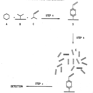
One embodiment of the present invention, termed the
"Direct Target Deposition Method", is illustrated in
Figure 1.
In Step a of this embodiment, the test sample
contain~ng the analyte (A) is first reacted with an
immobilized capture reagent ~B), such as an antibody,
and then wlth a reporter con~ugate comprising a target
nucleic acid sequence (C) to form an analyte dependent
reporter comp}ex (ADRC) (D) from which excess reagents
are removed by washing. In Step b, the ADRC is
contacted with a nucleic acid replication composition
and a replication process is performed to produce
replicated nucleic acids (E). In Step c, the replicated
nucleic acids are detected. In this embodiment, the
repor~er conjugate is a conjugate comprising a target
nucleic acid sequence and, for example, an antibody or
other analyte binding reagent.
WO93/15229 PCT/US93/01281
lI4 16
An ob~ious variation of the method, which is easily
practiced by one skilled in this art, is an adaption
wherein after step a, any excess, nonimmobilized
reporter conjugate remaining free in solution would be
separated from the immobilized capture reagent-analyte
complex. Ths amount of excess, nonimmobilized reporter
conjugate remaining free in the analyte sample would be
proportional to the amount of analyte initially present
in the sample. This nonimmobilized reporter conjugate,
after separation from the bound analyte complexes, could
then be replicated while free in solution, for example,
and the replicated nucleic acids are detected whereby
the presence of analyte in the sample is determined.
Another embodiment of the present invention, termed
the "Catalyzed Target Deposition Method", is illustrated
in Figure 2. In Step a of this embodiment, the test
sample containing the analyte (A) is first reacted with
an immobilized capture reagent (B), such as an antibody,
and then w~th a reporter conjugate (C) to form an
analyte dependent reporter complex (D) from which excess
reagents are removed by washing. In this embodiment the
reporter con~ugate (C) is comprised of an enzyme capable
of activating a moiety on the nucleic acid replication
substrate ~F) (such as horseradish peroxidase) and a
member of a bindlng pair (such as an antibody). In Step
b, the ADRC formed in Step a is reacted with a nucleic
acid repl~cation substrate (F), which contains the
target nucleic acid sequence, to form an activated
nucleic acid replication intermediate (G)/ which
-~0 deposits wherever receptor for the activated nucleic
acid replication intermediate is immobilized to produce
a deposited nucleic acid replication product (H).
Excess reagents are then washed off. In Step c, the
deposited nucleic acid _eplication product is contacted
with a nucleic acid replication composition to produce
WO93/15229 2 1 2 9 4 4 A PCT/US93/01281
17
replicated target sequence nucleic acids (E). In Step
d, the replicated nucleic acids are detected.
Another embodiment of the present invention, termed
the "Catalyzed Indirect Target ~eposition Method", is
illustrated in Figure 3. In Step a of this embodiment,
the test sample containing the analyte (A) iS first
reacted with an immobilized capture reagent (B) (such as
an antibody), and then with a reporter conjugate (C).to
form an analyte-dependent reporter complex (D) from
which excess reagents are removed by washing. In this
embodiment the reporter conjugate (C) is comprised of an
enzyme (such as horseradish peroxidase) which is oapable
of acti~ating a moiety on the binding substrate (I).
(I), the binding substrate, is a conjugate comprised of
~5 this substrate and a member of a binding pair. In Step
b, the ADRC is reacted with the-binding substrate ~I) to
form an activated bind;ng intermediate ~J) which
deposits wherever receptor for the activated binding
intermediate is immobilized, to produce a deposited
binding product (K). Excess reagents are then washed
off. In Step c, the deposited binding product is
reacted with a nucleic acid replication conjugate (L),
which conta~ns the target nucleic acid sequence and the
second member of the binding pair, to produce a nucleic'
acid replication binding pair complex (M). Excess
reagents are then washed off. ~n Step d, the nucleic
ac~d replication binding pair complex is contacted with
a nucleic acid replication composition to produce
replicated target sequence nucleic acids (E). In
Step e, these nucleic acids are detected.
Another embodiment is a variation of the "Direct
Target Deposition Method" of Figure l, and is
illustrated in Figure 4. The object of this embodiment
is to provide a method for detecting several different
WO93/15229 pcTluss3/o128l
212~44
18
analytes in a single sample, and may also be referred to
as the "mul~ianalyte method".
In Figure 4, at Step a, the test sample containing
different analytes (A and A') is first reacted with the
immobilized capture reagents (B and B'), and then with
the reporter conjugates (C and C') each comprising a
target nucleic acid sequence to form the analyte
dependent reporter complexes (D and D') from which
excess reagents are removed by washing. In Step b, the
A~RCs are contacted with a nucleic acid replication
composition and a replication process is performed to
produce replicated nucleic acids (E and E'). In Step c,
the replicated nucleic aclds are detected. In this
embodiment, there are more than one reporter conjugates,
each comprising an analyte specific antibody or other
analyte binding reagent linked to a target nucleic acid
sequence. The target nucleic acid sequence for each
type of reporter con~ugate has a specific length, which
is different in length from the target of any other
reporter con~ugate. Replication of the target nucleic
acid sequences thus gives amplification products of
dlfferent lengths and the presence of different analytes
msy be conveniently detected by analysis of the
smplification products on the basis of size, such as in
gel electrophoresis. In a particularly preferred
embodiment, the target nucleic acid sequences which
differ ~n length will be designed to comprise the same
5' and 3' primer binding regions, so that the same
primers can be used to replicate all of the various
targets present in the sample. In another preferred
embodiment of multianalyte detection which is useful for
detection of multiple sequences in sample nucleic acids
~the "multigene assay"), the reporter conjugates will be
comprised of target sequences which have been coupled to
other nucleic acid sequences which are complementary to
W093/15229 2 1 2 9 4 ~ ll PCT/US93/Ot2~1
19
specific nucleic acids which may be present in the
sample. The complementary sequences will hybridize to
sequences present in the sample nucleic acids, thereby
immobilizing the reporter conjugates. Target segments
of the reporter conjugates will then be replicated,
indicating presence of the specific sample sequences.
An add~tional embodiment of the present invention,
termed the "Competitive Binding Method",is illustrated
in Figure 6.
In Figure 6, at Step a of this embodiment, the test
sample containing the analyte ~A) is first reacted with
an immobilized capture reagent (B), such as an antibody,
and then with a ligand reporter conjugate (Q) comprising
a target nucleic acid sequence bound to a ligand wherein
the ligand is capable of competing with the analyte ~A)
for binding sites on the immobilized capture reagent
(B). The reaction results in the formation of an
immobilized analyte complex ~N) leaving the ligand
reporter conjugate (Q) unbound. The ligand reporter
con~ugate ~Q) and the immob~lized analyte complex ~N)
are then separated by washing. In Step b, either the
immobilized analyte complex (N) or the ligand reporter
con~ugate may be contacted with a nucleic acid
repllcatlon compo~ition. In the case where the ligand
2S reporter con~ugate~Q~ is contacted, nucleic acid
replication occurs and the presences of analyte is
detected. In the case where the immobilized analyte
complex ~N) is contacted, no replication occurs and no
replicated nucleic acids ~E) are produced. In this
embodiment, the ligand reporter conjugate is a conjugate
comprising a target nucleic acid sequence and, for
example, an antigen or other binding reagent capable of
competing with the analyte for binding sites on the
capture reagent.
WO93/15229 ~ 1? . v~ PCT/US93~01281
Additionally, one of ordinary skill will recognize
that the above several embodiments could be practiced
employing alternative immobilization points throughout
the assay. For example, in Figure 6, the ligand
S reporter conjugate co~ld be immobilized a~d the capture
reagent could be free in solution.
Thus, in all of the above embodiments the
production of replicated nucleic acids from a target
nucleic acid sequence is used to amplify detection of
the analyte.
The process of the present invention may be used to
detect the presence of a wide variety of analytes.
Generally, these include, but are not limited to,
plants, animals, nucleic acid segments, molecules,
cells, microorganisms and fragments and products
thereof, or any substance for which attachment sites,
binding members or receptors (such as antibodies) can be
developed. Of particular interest are pathogens,
viruses and bacteria. It is contemplated that the
sample material will be a liquid, a gas or a solid to be
dissolved in, extracted from or suspended in a test
fluid. The sample material will most likely be of
medical, veterinary, environmental, nutritional or
industrial significance. While not attempting to be
limiting, it is contemplated that specimens for human,
animal, or microbiological sources or habitats may be
tested by the present method, including body fluids such
as urine, blood, serum, plasma, milk, sputum, fecal
matter, lung aspirates, exudates; microbial culture
fluids; aerosols; crop materials; soils and ground
waters.
The immobi~ized capture reagent which binds the
test analyte will generally be comprised of, for
example, a binding protein, lectin, nucleic acid or an -
antibody, attached to an appropriate support. Any known
W093/15229 2 1 2 9 4 ~ 4 PCT/US93/01281
21
antibody could serve as the antibody of the immobilized
capture reagent. In addition, specific antibodies may
be prepared and utilized in this process. In certain
instances analyte may be captured directly by
nonspecific interaction with the support, as in, for
example, the hydrophobic interactions between proteins
and polystyrene.
Suitable immobilization supports used ln the ADRC,
recep~or supports and affinity supports ~to capture the
replicated nucleic acids) include synthetic polymer
supports, such as polystyrene, polypropylene,
polyglycidylmethacrylate, polystyrene, substituted
polystyrene ~e.g., aminated or carboxylated polystyrene;
polyacrylamides; polyamides; polyvinylchlorides, etc.);
glass beads; agarose; or nitrocellulose, etc. These
materials may be used as films, wells, beads, particles,
pins, pegs or membranes. Alternatively, the supports
could comprise magnetic and non-magnetic particles.
These supports can be used to prepare different
immobilized reagents. For exampie, depending on the
approach and reagent co~lfiguration, separate immobilized
support reagents could be prepared for binding the ADRC,
for binding o~ the activated conjugate receptor, or for
capture of the products of nucleic acid replication
during the detection step. Alternatively, under
circumstances in which the analyte, receptor and product
binding activities do not compete or interfere with the
other bind~ng functions, the analyte, conjugate and
product binding reagents could be co-immobilized on the
same support. In this way the ADRC, and the receptor
support could be prepared and used as separate supports,
or the bind~ng reagents could be combined on the same
support. Analyte binding molecules, and receptors can
be immobilized on the solid support using techniques
well known to those skilled in the art. H. Weetall,
WO93/15229 PCT/US93/01281
212!J~4 22
mmo~ L~I~LY~ 3CL3e~ ei~
(1975) Marcell Dekkerr Inc.~ New York.
Typically, the immobilized capture reagent can be
comprised of glycidyl methacrylate beads of about 30u in
diameter and an antibody such as goat anti-Rabbit IgG
antibody. Test beads and antibody are incubated at 4C
followed by a washing to remove excess antibody. The
beads are then treated with bovine serum albumin to bind
any unreacted epoxide groups and resuspended in buffer.
In practicing the present invention, two different
types of reporter conjugates are contemplated by
Applicants. The first type consists of a target nucleic
acid sequence coupled to an antibody or other binding
member which recognizes an analyte. These can be
prepared using variations of methods known to those
skilled in the art for linking proteins to amino-
oligcnucleotides. For example, this may be accomplished
using enzymatic tailing methods in which an amino-
modified dNTP is added onto the 3' end of the nucleic
acid. A. Xumar, Anal. Biochem,, 169, 376 (1988).
Alternatively, amino-modified bases can be synthetically
introduced into the nucleic acid base ~-~equence. P. Li,
et al-, ISe~ ch1~_L ~ , 5275 (1987). Antibodies
can then be attached to amino-modified nucleic acids by
sub-Qtituting an antlbody for an enzyme in the method of
Urdea. M. S. Urdea, ~ucleic ~ S Res., 16, 4937
~1988).
More specifically, preferred preparation of nucleic
acid/antibody conjugates involves the coupling of
heterobifunctional cross-linkers to the DNA
oligonucleotide targets which in turn are coupled to
antibodies using chemistry described by Tseng et. al. in
USSN 07/946247. A key advantage of this linking
chemistry over standard protocols in the art is that it
WO93/15229 2 1 2 ~ 'I 4 ~ PCT/US93/01281
23
reduces ~he occurrence of unwanted reactions such as
homo-DNA or homo-antibody polymers~
To facilitate the chemical attachment of the
oligonucleotides to the antibodies, the oligonucleotides
are amino-modified by introducing a primary amine group
at their 5' end during synthesis using cyanoethyl-
phosphoramidite chemistry. The amino-modified
oligonucleotides are further modified with a hetero-
bifunctional reagent that introduces sulfhydryl groups.
The reagent, N-succinimidyl S-acetylthioacetate (SATA)
is a heterobifunctional cross-linker agent that uses the
primary amine reactive group, N-hydroxyl-succinimide
~NHS) to couple to the amino-modified oligonucleotides
introducing an acetyl-protected sulfhydryl group. The
antibodies are modifled with another NHS cross-linking
agent, succinimidyl 4-(N-maleimidomethyl) cyclohexane-l-
carboxylate (SMCC). The SMCC reacts with primary amine
groups within the peptides (e.g., the e-groups on
lysine) of the antibody, introducing a maleimide group
(a free sulfhydryl reactive group) to the antibody. The
maleimide-modified an~ibodies are mixed with the SATA
modified antibodies. The acetyl-protected sulfhydryl
groups on the SATA-modified oligonucleotides are
activated with.the addition of hydroxylam~ne to produce
reactive, free sul~hydryl groups (USSN 07/946247). The
free sulfhydryl-containing oligonucleotides react
immediately with maleimide-modified antibodies forming
DNA to antibody conjugates.
The second con~ugate type comprises an enzyme
coupled to an antibody or other member of a binding
pair. These also may be prepared using methods well
known to those skilIed in the art. D. G. Williams,
J. Immun._Meth~, l9, 261 ~l984). Alternatively,
enzyme-binding conjugates can be generated using
recombinant DNA and genetic engineering techniques.
W093/1522g 212 3 ~ 4 4 PCT/US93/0128l
24
I. Pastan and D Fitzgerald, Science, ~, 1173 (1991).
Enzymes suitable for use in a reporter conjugate
include, but are not limited to, hydrolases, lyases,
oxido-reductases, transferases, isomerases and ligases.
Others are peroxidase, glucose oxidase, phosphatase,
esterase and glycosidase. Specific examples include
alkaline phosphatase, lipases, beta-galactosidase,
horseradish peroxidase and porcine liver esterase. The
choice of reporter con~ugate depends upon which
embodiment of the present invention is practiced.
Logarithmic nucleic acid replication technology
(for example, the polymerase chain reaction (PCR), or
the ligase chain reaction (LCR)) provides highly
Qensitive means for amplifying copies of a specific
nucle~c acid sequence. These technologies afford two
very important capabi~ s. One is the specificity of
the replication process. Information from a single
sequence can be specifically replicated in the presence
of Qamples containing complex mixtures of nucleic acids
and high concentrat~on of proteins. The second is the
hlgh sensitivity afforded by the process. Replications
of target DNA on the order of > 106 fold can be achieved~
by a temperature recycle proce8s. Currently, pathogens
can be detected in m~xtures of unknown samples by
Qequence probes; however, the sensitivity approaches
approximately only 103 cells/ml. Logarithmic sequence
replication of target DNA has now greatly extended probe
test sensitivity enabling as few as 1 to 5 cells /100 ml
to be detected, A. K. Bej et al., AD~. Environ.
~crobiol., 56, 307 (1990).
As noted above, the target nucleic acid is
replicated to produce amplified copies of the target
sequence nucleic acids. The design of the target
sequence is important because replication requires
WO93~15229 21 2 9 4 ~ PCT~US93/01281
suitable complementary ?rimer(s); and also because the
target can provide for different means of detection and
for flexibility in reaction conditions.
Specifically, the target nucleic acid sequence may
vary in length from 20 to 5000 bases. Preferably if the
target is to be used for PCR amplification it will range
between 30 and l000 bases. If the target is used for
LCR amplification the target length will range between
l00 and 500 bases. The target may be double-stranded
(ds), comprising a hybrid duplex of two complementary
nucleic acid strands, or may alternatively, be single-
stranded (ss). Double-stranded targets do not require ~
production of a complementary strand to participate in ~`
logarithmic chain polymerization. Either or both strands
can carry modified bases used for binding or detection.
When only one strand of the target is attached to the
support, the complem~ntary strand will be free to anneal
with primers in the solution phase. Once removed from
the ~upport matrix, strand replication is unhindered.
Double-stranded targets are thus particularly useful for
immobil~zed targets, since heat denaturation will free
one strand which is freed from the support for
replication.
Single-strands of double stranded targets however,
may also be used when preparing con~ugate reagents. For
ex~mple, as illustrated below, only one strand of the
target is used during the first ampiification cycle.
Annealing and extension of primer #l can then convert
the single-stranded target to a double-stranded duplex.
Acting in concert in subsequent cycles, primers #l and
~ will lead to logarithmic replication of the newly-
synthesized double-stranded target nucleic acid.Single-
stranded targets offer some reagent preparation
advantages in that they are l) cheaper, since only one
strand need be made, and 2) no prior annealing with the
W093/15229 PCT/US93/01281
2129~ 26
complementary strand is required. The same primers may
be used for amplification of appropriately designed ss
or ds targets.
In a preferred simplification of the Applicants'
invention, logarithmic replication can be achieved using
a single-stranded target and a single primer. This is
achieved by designing the target sequence to contain a
primer binding sequence at one end of the ss target and
a complement sequence of the primer binding site at the
opposite end of the target strand. Annealing and
extension of the primer will result in the formation of
a complementary target strand containing the identical ;
primer binding sites. In this way both the (+) and (-)
strands of the resulting ds target contain an identical
primer site at opposite ends of the target duplex, and
the same primer used in combination with the polymerase
and target nucleic acid promotes replication of both +
and - target strands.
The single primer approach affords advantages in
reduced assay complexity and increased reproducibility.
Simplification is achieved since only one primer must be
prepared and provided for detection. More importantly,
the 8ingle primer can enhance productivity of the
nucleic acid acid replication process since each primer
has exactly the same melting temperature ~Tm).
Temperature recycling constraints are thus more easily
controlled. Furthermore, the likelihood of nonspecific
nucleic acid formation resulting from primer-dimer
replication is reduced.
In other preferred embodiments, the base
composition and sequence of the target nucleic acid
sequence can be varied to accommodate different assay
requirements. For example, the target may contain
sequence segments which are not amplified during
replication, or variable regions used to alter the
W093/~5229 ~ 2~ PCT/US93/01281
27
length of the target sequence. It is contemplated, for
example, that a target nucleic acid sequence may be
designed to contain a coupling linkage for the
attachment of an antibody or ligand at the 5' end and a
primer binding site at the 3' end with a variable region
inserted between. The variable region could be of any
length or composition, limited only by the requirements
of the target amplification method. This is illustrated
in Figure 5, which provides an example of a reporter
con~ugate wherein the target nucleic acid has been
conjugated to an antibody through a chemical coupling
linkage at the 5' end. The target oligonucleotide may
contain a 5' binding region complementary to one of the
replication primers, and a 3' site for binding the other
replicated primer. Within the target sequence is a
variable region of nucleic acid bases, which could be
variable in length or in sequence, thereby providing
alternative means of detection of the replicated targets
based on s~ze or other factors, such as ability to bind,
or ability to emit distinctive signals (such as
fluorescence, radioactivity, etc.).
Another preferred emboidment for multi-analyte
detection is shown in Figure 4, wherein target nucleic
ac~ds wh~ch vary in length are u-qed to detect various
analytes. For example, a series of nucleic acid targets
having the same primer binding sites and same sequence,
but differing only in length of the inner target region
could be prepared, and coupled to different binding
pairs which are capable of binding to the different
analytes ~such as analyte-specific antibodies, lectins,
receptors, etc.). The replication products of each of
these receptor conjugates could then be readily
distinguished on the b&sis of size~ and conveniently
visualized, for example, by gel electrophoresis. In a
"multigene assay", where the analytes to be detected are
WO93~15229 PCT/US93/01281
212~`Q~ 28
.:
specific sequences of RNA or DNA contained within sample
nucleic acids, the reporter conjugates are comprised of
targets of varying lengths coupled to nucleic acid
sequences which are complementary to specific portions -
of the sample nucleic acid. In this way, multiple genes
or sequence sites within one sample can be conveniently
screened in one assay. The high resolution capability
of nucleic acid separation, wherein sequences which vary
in length by only one base can be resolved, renders this
method extremely attractive when large numbers of
samples containing multiple analytes are to be screened.
Additionally, the target sequence can be designed
to facilitate detection of the amplified nucleic acids
which are capable of emitting detectable signals. For
example, the target sequence may provide for
incorporation of labeled primers or labeled bases (e.g.,
fluorescence, radioactive, light emitting~ to produce
the correspondingly labeled signal generating nucleic ;
acids. The type, number of labeled bases and position
~0 within a chain, and between complementary chains, may be
designed to facilitate signal detection. In a preferred
embodiment, it is desired to position specifically
labeled bases in the sequence so as to enable energy
transfer between fluorophores, or to enable enzyme
channeling between proximally positioned coupled
enzymes.
Specifically, energy transfer between suitably
labeled bases can be achieved if the distance between
the excitation fluorophore (Fl) and the emission
fluorophore ~F2) are within 12 bases (ca. 50A) in the
helical duplex assemblage. R. A. Cardullo et al., ~Q~ -
~atl. Acad. Sci. USA, 85, 8790 (1988). A more preferred
distance is between 5 to 12 bases. This can be achieved
by designing the target and primer sequences so that one
of the labeled bases (Fl and F2) is alternately
WO93/15229 2 1 ~ 9 ~ l ~ PCT/US~3/01281
29
incorporated in the signal nucleic acid at each turn of
the helix. Alternatively, the base sequence of the
target or primers can ~e designed so that Fl and F2 are
incorporated into opposite strands of the signal nucleic
acid. The position of labeled bases is controlled so
that on strand hybridization, Fl and F2 are positioned
within the duplex at a distance of <50A. More
preferable, Fl and F2 will be positioned on the same
side of duplex one turn apart. Thus, within the signal
nucleic acid, both interchain and intrachain labeled
bases can position the fluorophores within a distance ;
suitable for energy transfer.
The requirements of fluorophores which participate
in energy transfer are well documented. L. E. Morrison,
Anal. Biochem., 174, lOl (1988). Generally, to achieve
energy transfer it is also important to select the
appropriate combination of fluorophores used for
labeling the excitation (Fl) and emission (F2) bases so
that the emission spectrum of the excitation fluorophore
~Fl) o~erlaps with the adsorption or excitation spectrum
of the exc~tation fluorophore (F2). For example, the
following fluorophore combinations include commonly
available suitable candidates for energy transfer:
F.~ on Fluoro~hore (F2)
Pyrenebutyrate ~-P~ycoerythrin
Fluore~ce~n Texaq Red
Luc~fer Yellow Rhodamine
Lucifer Yellow Texa~ Red
Fluore~cein Rhodamine
Fluore~camine Fluorescein
In another preferred embodiment, the sequences of
the target and primers can be designed to incorporate
bases labeled with the first member of a binding pair
(e.g. digoxigenin, biotin). The incorporated labeled
bases can be used to either immobilize the resulting
wo g3/l5229 2 1~ ~ ~ 4 4 PCT/US93/0l28l ~
nucleic acids, or to complex them with a second member
of the binding pair labeled with a reporter (e.g.
streptavidin-alkaline phosphatase, antidigoxigenin-
alkaline phosphatase)~ It is contemplated that the
target sequence may be designed to enahle the
incorporation of different bases or primers; one or more
labeled with binding members (e.g., biotin); and one or
more labeled with a reporter(s). It is desirable to
control the sequence so that the biotin-labeled bases
are incorporated predominantly at one end of a chain and
the reporter bases incorporated at the other end or some i~
distance from the binding members. Nevertheless, in
designing the base sequence it is important to avoid
both consecutive runs of C's and G's (3 or more) at the
3' ends, as well as with pzlindromic sequences. For
example, a target gene sequence could contain the
following sequence:
1 45
S' ATG CGT AGC AGC TTT ACC GCA GAG ATC ATG CCT ATG TAC CAT GCT 3'
3' TAC GCA TCG TCG AAA TGG CGT CTC TAG TAC GGA TAC ATG GT~ CGA 5'
46 75
5' ATC CTA CCT GTA AGT CAT AGC TGT TTC CTG 3' SEQ ID NO:l
3' TAG GAT GGA CAT TCA GTA TCG ACA AAG GAC 5' SEQ ID NO:2
The nucleic acid replication substrates are
comprised of a target nucleotide sequence, optionally a
spacer, and a moiety capable of being activated by an
enzyme. The~target nucleic acid sequence is prepared
according to the guidelines set out above. The moiety
capable of activation by an enzyme may be any moiety
which forms a reactive intermediate which can bind to a
receptor on the solid support of the immobilized capture
WO93/15229 PCT/US93/01281
212~4f~4
31
reagent. In preferred embodiments the enzyme reactive
moiety is tyramine.
Reporter conjugates, including the nucleic acid
replication substrate and nucleic acid replication
conjugate, may contain a molecular spacer segment
linking the two functional elements of the can jugate.
One purpose of the spacer is to extend the replication
segment of the target or binding functions away from the
surface of the solid phase support. Useful spacers are
well known in the affinity chromatography art. For
example, H. Schoot, Affinity Chromato~ra~h, ~l984),
Marcell Deckker, Inc., New York, describes different
~pacers and their use. Advantageously, the spacer
includes a chain of up to about 50 atoms, preferably 5
to 30 atoms. In composition, spacers may be a
polyfunctional segment including, but not limited to,
one or more of the groups: peptide, hydrocarbon,
polyalcohol, polyether, polyamine, polyimine and
carbohydrate e.g. -glycyl-glycyl-glycyl- or other
oligopeptide, carbonyl dipeptide, and omegaamino-
alkane-carbonyl radical such as -NH-~CH2)2-CO-, a
spermine or spermidlne radical, omega-alkanediamine
radical ~uch as -NH-~CH2)6-NH- or -HN-CH2-CH2-NH-. The
Qpacer ~egment may also be compri~ed of polymeric units
-~uch as polysaccharide, polyeth~lene oxide radicals,
glyceryl, pentaerythritol and like radicals. The spacer
Qegment may be linked directly or linked through a
divalent heterobifunctional or homobifunctional
couplers, for example SATA (N-succinimidyl S-acetylthio-
acetate), SMCC (succinimidyl 4-(N-maleimidomethyl)
cyclohexane-l-carboxylate)~, p-phenyl diisothiocyanate,
dithiobis succinimidyl propionate, l,4-butanediol
diglycidyl ether, a diisocyanate, carbodiimide, glyoxal,
glutaraldehyde or-sulfosuccinimidyl 6-(4'-azido-2'nitro-
phenylamino)-hexanoate.
WO 93/15229 PCr/US93/01281
2129 ~4 32
The length of the ~arget nucleic acid sequences may
be extended beyond the sites of primer attachment. The
extended length of the target thus can provide an
alternative spacers and thus reduce the length or
eliminate the need for a molecular spacer, and also
perhaps increase the efficiency of target replication.
For example, bases added at the target attachment site
will extend the target segment away from the point of
immobilization. In this way, spacer length can be
reduced or in some instances eliminated. Generally,
add~ng 5 to 30 bases to the target will be sufficient to
increase the efficiency of immobilized target
replication. The composition and length of molecular
spacers are designed to prevent interference during
amplification of nucleotide target sequence. Recent
findings indicate that ~arget sequences next to the
aminolink spacer are accessible for primer attachment
and logarithmic chain reaction. (S. Stamm and
J. Brosius, L~ oL~_ L~ S ~, 19 1350 (1991).
Once the target nucleic acid sequence is designed,
the nucleic acid replicat~on substrates may be prepared
using well-established procedures developed for
preparation of enzyme-labeled oligonucleotide probes.
-See e.g., G. H. Xeller and Manak, M. M., r~ a2:S,
(1989), pp. 136-148, Stockton Press, New York. More
specifically, during the synthesis of the target nucleic
ac~d sequence, a base modified with a spacer arm
containing a primary amine can be introduced at either
the 5' end or the 3' end of the target. Reagents for
introducing a base containing a S' amino group are
commercially availab}e (C8-aminohexyl-ATP and N6-amino-
hexyl ATP, Sigma Co)), and methods of accomplishing the
introduction into the sequence are known in the art.
The N-Monomethoxytrityl-C6-AminoModLfied cyanoethyl
phosphoramidite reagent~(Clontech Laboratories Inc.,
WO93/1522~ PCT~US93/01281
21~94~
9030 Fabian Way, Palo Alto, CA 99303) or AminolinkT~ 2
(Applied Biosystems, Inc.) provide an easy means of
introducing a 5' terminal primary aliphatic amine to an
oligonucleotide during synthesis of a target
oligonucleotide. Detailed procedures for the coupling
reaction are available from Clontech bulletin no.
PB022789-l, or from Applied Biosystems, Inc., Model 392
Manual. Once the amino-modified nucleic acid target has
been prepared, it can then be reacted with succinic
anhydride; which extends the length of the side chain
and also provides a terminal carboxylic acid which can
be activated using standard methods to form the N-
hydroxysuccinimide ~NHS) intermediate (l). This
intermediate can then be chemically coupled to tyramine,
for example, to form a nucleic acid replication
substrate ~2).
(l) NHS~C)n linker-5' target 3'
(2) Tyramine-~C)n linker- 5' target 3'
Generic reagents are also available from Cruachem
(460 Spring Park, Herndon, VA 22070) Clontech Laboratory
~4030 Fabian Way, Palo Alto, CA 44303) or Applied
Blosystems, Inc. (Foster City, CA) for introducing
$nternal single or multiple amino groups into the gene
sequence. However, end-labeled oligomers tend to be
more accessible for binding and reaction than internally
labeled nucleic acids.
In another preferred embodiment/ it is contemplated
that the sequences of the target nucleic acid can be
designed to incorporate a ligand. As used herein the
term ligand will encompass both ligands which are
structurally related to the analyte, and ligands which
mimic analyte binding, so long as the ligands have the
-t5 ability to compete with an analyte for receptor binding
WO93/1~229 PCT/US93/01281
21~9~ 3q
sites. Thus, ligand-target conjugates may function as a
first member of a binding pair and can mimic the binding
properties of an analyte, competing with the binding of
an analyte to a second member of a binding pair ~e.g.,
an antibody) For the purposes of the present invention
ligands of less than 3000 molecular weight are preferred
whereas ligands with molecular weights of less than 1500
are most preferred.
Variability in the positional orientation and
number of the incorporated ligands lends flexibility to
the target design, allowing for increased assay
sensitivity and optimized conjugate interaction with the
capture reagents. It is contemplated that ligands can
be incorporated into one or both strands of a duplex
lS target nucleic acid. ~ositionally, ligands can be
incorporated either at the 5' or 3' ends of the target
or incorporated on internal bases within the nucleic
acid sequence, where incorporation at the ends is
generally preferred. It is contemplated that any number
of ligands may be incorporated per target, however,
where the object is to achieve maximum sensitivity of
the assay, a relatively small number of ligands is
preferred, where a range of one to two is most
~preferred. In the situat~on where maximum rate of ~ ;
con~ugate capture is desired, a high number of ligands
per target is preferred.
The method of incorporation of the ligand into the
nucleic acid sequences may be accomplished either by
chemical or enzymatic means, or by direct incorporation
of ligand labeled bases into the target sequence.
Chemical incorporation would utilize chemistry similar
to that used for the synthesis of the tyramine
replication substrate, as previously discussed.
Typically, a base modified with a spacer arm containing
a primary amine can be introduced at either the 5' end
WO93/15229 21 2 4 ~ 4 PCT/US93/01281
or the 3' end of the target which can be further
modified to a N-hydroxysuccinimide (NHS) intermediate-
which may in turn be chemically coupled to the ligand.
In a preferred approach, ligand-incorporated
sequences are prepared using ligand-labeled bases or
primers during polymerase chain reaction. It is
contemplated that ligand labeling can be accomplished
either through the incorporation of primers modified
w~th ligand(s) or by using ligand-labeled dNTPs. Ligand
labeled primers can be prepared us~ng standard
oligonucleotide cyanoethyl phosphoramidite chemistry by
substituting selected bases with ligand-modified
phosphoramidite bases during primer synthesis.
Alternatively, if primers are prepared with modified
baQes containing a linkable molecular spacer, the
ligands can be chemically l~nked to the spacer after
primer synthesis. Another method would make use of
ligand-labeled dNTPs or amino-modified dNTPs which can
be incorporated into a target nucleic acid sequence
during the amplification procedure.
There are Qeveral advantages to synthesis of
ligand-incorporated nucleic acid sequences by PCR as
oppo8ed to chemical or enzymatic means. For example,
becau~e of failure of ~equences inherent in chemical
synthesis, targets of longer than lOO bases are more
easily constructed. Additionally, where labeled primers
are used, it is possible to control both the positioning
- and number of ligands within one or both strands of the
target sequence by the appropriate placement of the
ligand in the primers.
In contrast, although the use of labeled dNTPs
facilitates the preparation of multivalent ligand
reporter conjugates, precise control over ligand number
and labeling pattern is less reliable. This is because
dNTP-ligand incorporation is dependant on both
WO93/15229 PCT/US93/01281
~1~9~4
36
polymerase discrimination between dNTP and modified dNTP
analogs and the frequency of occurrence of a specific
base in the target nucleic acid sequence.
It should be appreciated that the above discussion
regarding preparation of target nucleic acid sequences
and replication conjugates is equally applicable to
preparation and design of the reference nucleic acid
sequences and reference replication conjugates of the
invention.
The binding substrate is comprised of a first
member of a binding pair species, optionally a spacer,
and a moiety capable of activation by an enzyme. The
spacer and moiety are the same as those described for
the nucleic acid replication substrate. Members of
specific binding pairs suitable for use in practicing
the invention can be of the immune or non-immune type. `-
Immune-specific binding pairs are exemplified by
antigen/antibody systems or hapten/anti-hapten systems.
The antibody member, whether polyclonal, monoclonal or
an ~mmunoreactive fragment thereof, of the binding pair
can bé produced by customary methods familiar to those
skilled in the art. The terms immunoreactive antibody
fragment or immunoreactive fragment re~er to fragments
which contain the binding region of the antibody. Such
fragments may be Fab-type fragments which are defined as
fragments devoid of the Fc portion, e.g., Fab, Fab! and
F(ab')2 fragments, or may be "half-molecule" fragments
obtained by reductive cieavage of the disulfide bonds
connecting the heavy chain components of the intact
antibody. If the antigen member of the specific binding
pair is not immunogenic, e.g., a hapten, it can be
covalently coupled to a carrier protein to render it
immunogenic.
Non-immune binding pairs include systems wherein
the two components share a natural affinity for each
W093/15229 21 ~ ? ~i ~ 1 PCT/US93/01281
37
other but are not antibodies. Exemplary non-immune
binding pairs are biotin-avidin or biotin-strepta~idin,
folic acid-folate b~nding protein, complementary probe
nucleic acids, Proteins A, G, and immunoglobulins, etc.
Also included are non-immune binding pairs which form a
covalent bond with each other. Exemplary covalent
binding pairs include sulfhydryl reactive groups such
as maleimides and haloacetyl derivatives and amine
reactive groups such as isothiocyanates, succinimidyl
esters and sulfonyl halides, etc. In preferred
embodiments, an exemplary binding substrate would be a
con~ugate of biotin coupled to tyramine via a N-
hydroxysuccinimido linker molecule. The binding
substrate can be synthesized using well known methods.
M. N. Bobrow, et. al., J. Immunol. Methods, l~, 279,
~1989)
The nucleic acid replication conjugate is comprised
of a second member of a binding pair species, optionally
a spacer, and a target nucleic acid sequence. The
target nucleic ac~d sequence and the spacer are designed
and prepared according to the principles set out above.
The second member of the binding pair species is chosen
80 ~s to be complementary to the first member of the
binding pair species utilized in the binding substrate.
A~ notèd above, in a preferred embodiment the binding
sub-qtrate is a conjugate comprised of biotin, a spacer
and tyramine. Thus, the choice of the second member of
the binding pair species used in the nucleic acid
replication conjugate will be avidin or streptavidin,
and thus the binding substrate will comprise avidin or
streptavidin as the second member of the binding pair
species. The nucleic acid replication conjugates can be
prepared using well established procedures. See, e.g.,
G. H. Xeller and Manak, M. M., DNA Probes, ~l989),
pp. 136-148, Stockton Press, New York. More
W093/15229 PCT/US93/01281
,~12 9 ~ L~l ~ 38
specifically during the synthesis of the target
oligonucleotide, a base modified with a spacer arm
containing a primary amine could be introduced at either
the 5' end or the 3' end. Reagents for introducing a 5'
amino group are commercially available (e.g.
AminolinkT~ 2; Applied Biosystems Inc., 800 Lincoln
Centre Drive, Foster City, CA 94404). AminolinkT~ 2 is
added as the last step in the synthesis of the oligomer.
The amino-modified nucleic acid target is then activated
by a bifunctional ester to both extend the length of the
side chain and to provide a terminal carboxylic acid
which can then be activated using standard methods to
form N-hydroxysuccinimide (NHS) intermediate ~l). This
intermediate can then be chemically coupled with avidin
lS to form the nuclei~ acid replicating conjugate (3):
(1) NHS~C)n linker-5' ~o~ococ- c~ -ooccc xx~xXXXXXX 3'
(3) Avidin -~C)~ linker- 5'~oao~ocCc~x~x~xx~o~Doo~x~x~ 3'
Generlc reagents are also available from Cruachem, and
Clontech for introducing internal single or multiple
amino groups into the target nucleic acid sequence.
However, end labeled oligomers tend to be more
.5 accessible for binding and reaction than in~ernally
l~beled nucleic acids.
Pr~mers:
In current practice, replication of the target
nucleic acid sequence requires "primer" oligonucleotides
which, as used herein, refers to all oligonucleotides
which anneal to target sequences to facilitate
replication of the target.
When target replication is performed by polymerase
chain reaction two specific primers are used. Each
primer specifically hybridizes with one of the two
WO93/15229 ~12 9 ~ 1 LI PCT/US93/01281
39
complementary strands of the target (or if the target is
single-stranded (ss) one of the primers is specific for
the second strand after synthesis). Replication of the
target requires that the 5' end of the primer which is
complementary to the ~-) sense target strand (primer
#2), corresponds to a region of the (~) sense strand
wh~ch is 5' to the 3~ end of the (+) sense strand
specific primer ~#l). Additionally, the primers should
not contain regions with sufficient complementarity to
form primer-dimers. Within these constraints, the total
length of the primers may range from shorter than, to
longer than, the target. In general, primers 10-30 ;
bases in length are most practical.
<------- s~ primer ~l
+ ~en-~e 5 ' ~ -3 '
- ~en~e 3 ' -Ym~YYYYY~YYYYYYY~Y-S '
pr~mer ~t2 5 ' ----------->
Primers may also contain sequences at their 5' ends that
have no complement in the target ~5' overhang or 5'
mismatch).
,. ~
<-----------5 ' pr~ner #1
+ ~en~e 5' -~_ -3'
S ' ~ -------> primer #2
- ~en~e 3 ' -m~Y~5 '
(X,Y represent complementary bases.)
~his 5' overhang or 5' mismatch can be used to
incorporate functionalized ~ases (e.g., signal-
generating or binding member-derivatized bases) on the
primers, or to extend the length of the replicated
WO93/15229 PCT/US93/01281
~1?~ 4~ 40
nucleic acid products by adding extra sequences. These
additions can be useful for capture of the resulting
nucleic acids and/or signal detection. In the primer a
3' segment complementary to the target can be joined to
a variety of different 5' segments. Thus, a series of
primers with a fixed hybridizing region linked to
different signal generating tails can be made. The
signal generated would depend on the 5' region of the
primer(s) used and could be tailored to the detection
method of interest. Hence, a single target sequence can
be used with different primers containing varying 5'
overhangs or mismatches to generate a number of
d~fferent sequence specific responses.
Typically, in PCR-type amplification techniques the
primers have different sequences and are not
complementary to each other. Depending on the desired
test conditions, the sequences of the primers should be
designed to pr~vide for both efficient and faithful
replication of the target nucleic acid. Some simple
rules are useful i~ selection and design of the primers.
Typically, primers should be lO to 35 base pairs in
length having a 50 to 60%, G+C composition. The
calculated Tm's for a given primer pair should be
balanced. For this purpoQe, a 2C for A or T and 4C
~5 for G or C can be added together to estimate the Tm of
the oligonucleotide. ~Thein and Wallace, "The use of
oli~onucleotide as specific hybridization probes in the
Diagnosis of Genetic Disordersn, in ~ y=~3 ~
Di~eases: A Practical A~pLQ~sh, K. E. Davis Ed., ~l986)
pp. 33-50 IRL Press, Herndon, Virginia). Depending on
the selected conditions, Tm's between 55C and 80C are
suitable. In addition to the Tm's, the complementarity
at the 3' end of the primers is an important
consideration. Generally, complementarity of primer
pairs should be avoided, especially at the 3' ends.
WO93/15229 ~1 Z ~ ~ ~ PCT/US93/01281
41
Also, consecutive runs of C's and G's (3 or more) at the
3' ends of the primers along with palindromic sequences
should be avoided. Consideration should also be given
to the concentration of primer molecules in the
replication milieu. Primer concentrations between 0.0l
and l.0 uM are generally suitable, with concentrations
of about 0.05 to l.0 uM being optimal.
When the ligase chain reaction ~LCR) is used for
replication of a target double-stranded nucleic acid,
two sets of target-specific primers will be required.
The members of one set of primers are complementary to
adjacent sequences found on a given strand of-the
target, while the members of the second set are
complementary to adjacent sequences on the opposite
strand. In this way a set of adjacent primers is
-Qpectfic for each target strand. During the replication
process the target nucleic acid is heated to denature
the two target strands. The four complementary
oligonucleottde primers comprising the two primer sets
are then hydridized near their melting temperature to
the separated target strands. A thermal-stable ligase
Wt 11 covalently attach the adjacent primers on each
~arget -qtrand. Only ad~acent primers that are perfectly~
complementary to the target will be ligated together.
In thts way, the products from the first stage of
ligation become targets for ~he next round of ligation.
The products thus increase exponentially with continued
cycles of target denaturation, primer hydridization and
ligation steps.
The requirements for non-complementarity between
primers, size, base composition and melting temperature
requirements of the primers tend to be similar to those
stated above for PCR replication. Generally, primers
for LCR replication should be sufficiently long so that
each will preferentially bind to its specific binding
WO93/15229 PCT/US93/01281
~1~9 ~44 42
site on the target nucleic acid. To insure specificity
of ligation, reactions can be carried out near the
melting temperature (Tm~ of the oligonucleotide primers.
At higher temperatures single-base mismatch at the
junction can form. This results not only in an
imperfect double helix but destabilizes hydridization of
the mismatched oligonucleotides.
In either PCR or LCR type replications, the primers
may contain bases labeled with reporter(s) or labeled
10 with one member of a specific binding pair. For ~;
example, biotin and fluorescein residues may be
incorporated into the primer during CE phosphoramidite
synthesis (NEN Products, Du Pont, Boston, MA; or
Clontech). Incorporation of the primers during
amplification will also result in nucleic acid products
containing biotin and fluorescein. In this way, primer
incorporation during replication process can be used as
a preferred means of introducing reporters and affinity
labels in the replicated nucleic acids.
~ UL~:Y~ 3n~
Practicing the analyte-dependent reporter system of
the present invention requires several steps. First, an
analyte-dependent reporter complex (ADRC) is formed.
Thl8 ~s accomplished us~ng well-known immunoassay
reagents and techniques. The reagents can be configured
for sequential, competitive, sandwich, and immunometric
mmunoassay approaches. Harlow, E. and Lane, D. L.,
dies - A Laboratory Manual, (1988), pp. 555-612,
Cold Spring Harbor Lab., Cold Spring Harbor, New York.
Once the ADRC complex has been formed excess free
reagents is removed. This is an important step since
any free reagent and non-specifically bound reporter can
contribute to the replication process. To aid in
reducing non-specific binding, stringent-wash conditions
which do not cause dissociation of the ADRC, such as
WO93/1~229 212 ~ 4 ~ 4 PCT/US93/01281
43
used in nucleic acid hybridization tests, can be
employed since target replication and readout are based
on nucleic acid chemistry. For example, heating, pH
changes, or (and) the addition of formamide, detergents
and salts can be used to increase the efficiency of the
wash step. Too stringent conditions can lead to
dissociation of the ADRC or destruction of the
immunoassay reporter. Tolerance to stringent wash
conditions will vary with the nature of the analyte,
binding member and specific reporter used. The
stringent conditions must, therefore, be experimentally
optimized for each assay. Howe~er, in washing to reduce
non-specific binding, if some of the ADRC is lost, this
can be compensated for by additional target replication
realized by increasing the number of temperature recycle
steps.
In cases where the ADRC is sensitive to stringent
wash conditions, a catalyzed reporter assay (Figure 2)
can be used. In this configuration reaction of the ADRC
with the substrate results in covalent coupling between
the target nucleic acid and the support. After
coupling, the solid support is washed leaving the
covalently bound target nucleic acid complex. ,
Speclf~cally, a.tyramine target substrate could be used
wlth an HRP enzyme reporter to covalently couple the
target to the support. In this way, enzyme
amplifications could be accomplished and stringent wash
conditions used before target replication.
In the next step, the target nucleic acid sequence
is replicated. If PCR is the replication method used,
the target sequence is mixed with the nucleic acid
replication composition ~lOOul) comprising two primers
~lO0 pmol/primer), a thermally stable DNA polymerase
such as Taq DNA polymerase ~2 units), required
nucleotides ~dNTP 200 uM/base) which may be signal
WO93/15229 PCT/US93/01281
~ 4 l 4 44 ;~
generating or ligand-containing nucleic acids, Tween 20
detergent (0.05~), 20 mM TRIS/HCl buffer (pH 8.3), MgCl2
(1.5 mM), KCl (25 mM), and nuclease-free gelatin
(lO0 ug~ml). `
Generally, excess Mg~+ in the replication reagent
composition can result in the non-specific amplification
whereas insufficient Mg+~ will reduce yields. It is
known tha~ deoxynucleotide triphosphates (dN~P) bind
Mg++, and the amount of binding depends upon the dNTP
concentration. In a reaction composition containing all
four bases (dNTP) this leaves a final free Mg+~
concentration of ca. 0.7 mM of the original l.5 mM Mg++.
If the dNTP concentrations are changed significantly, a
compensatory change in MgCl2 may be necessary.
Because of the diversity of applications in which
the present invention can be used, adjustments in the
concentrations and reagent compositions of the nucleic
acid replication reagent composition may be required.
Practical guidelines for optimizing and adjusting the -
replication milieu can be found R. K. Saiki and
Gelfand, D. H., in P~B Technology, ~l989), pp. 7-22,
Stockton Press, New York.
The reaction mixture is covered with mineral oil to~
prevent evaporation. The target nucleic acid is then
denatured to separate the duplex nucléic acid strands.
Generally this is carried out a high temperature
(90-95C) for 15 seconds; however, a longer initial time
can be required to assure complete denaturation.
Annealing of the oligonucleotide primers to the target
nucleic acid template is usually accomplished by
lowering the temperature to 37 to 60C for 30-60
seconds. Polymerase extension of the primers can then
be accomplished by equilibration at 72C for lO to 60
seconds, depending on the length of the signal-
generation products. The denaturation, primer annealing
W O 93/15229 212 9 g 4 4 PC~r/US93/01281
and primer extension steps are repetitively carried outin sequence to amplify the strand number of the signal-
generation product. The temperature cycling is
typically performed 10 to 40 times depending on the
desired degree of replication. The reaction can then be
stopped by addition of EDTA ~10 mM) and chilling to 4C.
In general, if LCR is the replication protocol
used, the target sequence of the ADRC is mixed with two
sets of adjacent oligonucleotides (40 fmol each); each
set will be complementary to one of the complementary
target nucleic acid strands; in 10 ul of buffer
containing 20 mM Tris.HCl buffer pH 7.6, 100 mM KCl,
1 mM EDTA, 10 mM NAD, 10 mM dithiothreitol, 4 ug salmon
sperm DNA and 15 nick-closing units of a thermostable
ligase, for example, T. aquatics ligase, (Tabor, S. and
Richardson, D. C. (1985) Proc. Natl. Acad. Sci. ~SA 82,
1074-1078).
The reaction mixture is then protected from
evaporation, for example, by covering with a drop of
mineral o~l, and then heated to 94C to insure
separation of the target strands. Annealing of the
oligonucleotide primers to the target is usually
accomplished by lowering the temperature to between 37
to 60 degrees. An optimum temperature close to melting
temperature of the primers is usually selected, but must
be determined dependent upon the primer length and
composition of the specific primers. The melting and
primer annealing cycle are then repeated 10 to 30 times.
The reaction is then chilled to 4C to stop the
reaction.
The next step involves detection or visualization
of the amplified nucleic acids. This can be
accomplished by several means including (a) direct
detection of the duplex nucleic acids using
intercalating dyes; (b) indirect or direct detection of
WO93/15229 PCT/US93/01~81
~ l~ 9 46
ligands, isotopes or reporters incorporated in the
nucleic acids; (c) hybridization of reporter probes to
the amplified nucleic acids; or (d) direct detection of
replicated product following separation of replicated
product from reaction milieu based on increased size of
replication product.
Specifically, amplified nucleic acids can be
detected in the reaction mixture by adding intercalating
dyes. Of particular use are those dyes of the ethidium,
phenazines, furocomarins, phenothiasines and quinoline
type which on intercalation with the duplex strands of
nucleic acids change dye detection properties. General
reviews and further information can be obtained in
Berman et al., Ann. Rev. BioDhys. Bioeng., 20, 87
~1981~. For example, a preferred dye is ethidium
bromide which on nucleic acid intercalation can be
detected by excitation of the reaction mixture with
short-wave uv light (259nm).
Incorporation of modified free bases or modified
primers during nucleic acid replication provides a means
- of introducing bases modified with ligands, isotopes, or
reporters. If ligase-type replication is used,
oligonucleotides with modified bases already
incorporated could be used to replicate the target
~equences. These techniques afford several detection
strategies. For example, the incorporation of
biotinylated or ligand modified bases provides means of
isolating the amplified nucleic acid products from
solution onto a solid support and discarding the
unincorporated bases. The addition of an avidin-signal-
generating conjugate then facilitates detection. The
amplified sequences may also contain signal-generating
labeled bases. These can be detected directly on the
solid phase support. Alternatively, methods of
collecting and detecting biotinylated DNA fragments on
W093/15229 21 2 3 ~ PCT/US93/01281
~7
magnetic beads containing immobilized avidin or
streptavidan are described by J. Wahlberg et al., ~nl~
Cell Probes, 4 285 (1990).
In another alternative, the sequence of the
amplified segment could be designed to position
fluorescent bases within the signal nucleic acids for
energy transfer or position the biotinylated bases so
that binding of avidin-labeled enzyme(s) reporters would
result in enzyme channeling. Using these approaches the
amplified target can be detected without the need for
separation from the unincorporated bases. According to
molecular modeling and recent reports, R. A. Cardullo
et. al., ~ , ~, 8790 (1988~,
energy transfer can be achieved at distances ~etween the
fluorophores of as much as 12 bases apart. However,
optimum distance appears to be somewhere between 5 to 12
bases. At one fluorophore base per helix turn, this
positions the donor and acceptor fluorophores in
appropriate proximity for energy transfer.
Analyte Quantitation :
The analyte-dependent reporter system (ADRS)
response depends upon the quantity of analyte present in
the sample, and also upon the efficiency of sequence
~mplification. While analyte concentrations in samples
can be interpolated from standard curvés by
experimentally relat~ng assay response under fixed
reaction conditions and known analyte concentrations,
the efficiency of sequence replication is difficult to
predict and control because of procedural and reaction
variables. Furthermore, amplification is highly
sensitive. Hence, if false negative test response can
be identified, the absence of analyte can be more
reliably determined and the useful range of the assay
extended.
WO93/15229 ~ ~2 ~ ~ 4 4 PCT/USg3/01281
48
Applicants contemplate that at least two types of
internal controls can be used to compensate for changes
in the efficiency of sequence replication and to provide
means of identifying false negative test response.
Applicants refer to these as ~amplification, or
replication control", and n capture control". An
"amplification control~ refers to a nucleic acid
sequence called the nreference sequence" (specifically,
a different sequence than the target sequence nucleic
acid) and its corresponding cognate primer(s). In use,
the reference sequence and cogna~e primers could be
included in the nucleic acid replication reagent
composition, and would serve to demonstrate that
react~on conditions are permissive for sequence
amplification. During testing if the amplification
control yields a signal, but the target sequence does
not, then the lack of target sequence amplification
cannot be the result of test conditions non-permissive
for ampllf~cation and the result indicates an analyte
concentration below the detectable level.
The ampl~fication control can also serve as the
internal reference for analyte quantification. For this
application, the reference sequence is added at a known
concentration approximating the analyte concentration to
be detected. During the replication réaction, the
reference sequence will theoretically be amplified with
the same efficiency as the target sequence. By
determin~ng the ratio of the signal responses from the
reference sequence and the target sequencel the
concentration of the analyte may be determined. In this
approach standard curves are determined by measuring the
ratio of the responses resulting from the target
sequence and the reference sequence in samples
containing a range of ~nown analyte concentrations and a
fixed concentration of reference sequence. In this way,
WO93/15229 ~1 2 ~9 4 fl 4 PCT/US93/01281
49
variations in efficiency of nucleic acid replication can
be compensated. Assay response can thus be more
accurately related to analyte concentration.
The reference sequence must be similar in molecular
weight to the amplification sequence, but must be
capable of producing a separate and distinct ~replicated
reference nucleic acid~ which is detectably
distinguishable from the replicated target sequence
nucleic acid. This can be achieved by designing target
and reference sequences to contain unique bases. In
this way during repli~ation, a unique base or primer
labeled with ligands, reporters, isotopes or reactive
groups can be incorporated respectively into the nucleic
acid products of both the reference sequence nucleic
acids and target sequence nucleic acids. The resulting
nucleic acids are thus labeled with separate reporters,
or can be isolated for detection via hybridization
reactions or binding with complementary members of a
specific ligand binding pair.
A "capture control" comprises an analyte reference
sequence conjugate (properly configured for each
embodiment - see below) and cognate primerls). The
capture control sequence con~ugate would be included
durlng the ana~yte capture step of the assay; and the
cognate primer pair would be included in the nucleic
acid replication composition. In this configuration, a
failure to detect the capture control replications could
result from either a lack of capture or conditions non-
permiss~ve for amplification. By using different
sequences for the capture and amplification controls,
both controls can be included in each reaction chamber
allowing differentiation between failed capture and
failed replication reaction conditions. Inclusion of
these controls is possible only because each of the
three amplification sites, (target sequence,
WO93/15229 212~ 4~4 PCT/USg3/0l~1
amplification control and capture control) could be
designed to generate a sequence-specific, differentially
detectable signal.
dNTP and primer levels should be adjusted so that
each of the three replication reactions can proceed to
completion. Targets and cognate primers should have
similar Tm, length and other characteristics that effect
amplification efficiency.
~ix~
The following examples are meant to illustrate key
embodiments of the invention but should not be constxued
to be limiting in any way.
Oligonucleotides to be used as the reporter or as
primers are prepared using standard cyanoethyl (CE)
phosphoramidite coupling chemist.ry on controlled pore
glass ~CPG) supports in an autom~ted DNA oligonucleotide
synthesizer ~Generator~, Du Pont Co., Wilmington, DE,
and Model 392, Applied Biosys~ems, Inc., Foster City,
CA) ~Beaucage and Caruthers, ~L~hedL~n 1~_, 22 (20),
1859 ~1981); Caruthers 8~ , Genetic En~in~gL1n~, vol.
4, ed., ~1982); Stelow and Hollaender, Plenum Publishing
Corp., New York). The amino-modifying phosphoramidite
reagent Aminolink 2~ is obtained from Applied
B~o-~ystems, Inc., Foster City, CA. Oligonucleotides are
radio-labeled with [~32p] cordycepin 5'-triphosphate
and scintillation fluid (Biofluor~) for scintillation
counting is obtained from NEN Products, Du Pont Co.,
Boston, MA and scintillation counting is accomplished
using a Beckman Model LS3801 scintillation counter
(Beckman Instruments, Inc., Palo Alto, CA ). Deoxy-
nucleotydyl transferase is obtained from Promega, Inc.,
Madison, WI. Poly-tergent SLF-18 is obtained from Olin
Corp., Stamford, CT. Kodak Xomat~AR 2 X-ray film for
autoradiography is obtained from Eastman Kodak Co.,
WO93/15229 2 1 2 ~ '~ 4 4 PCT/US93/01~1
Rochester, NY. The reagents, SATA (N-succinimidyl
S-acetylthioacetate~ and SMCC (succinimidyl
4-(N-maleimidomethyl) cyclohexane-l-carboxylate) are
obtained from Pierce, Rockford, IL. The SATA and SMCC
coupling chemistry is described by Tseng et al., in
commonly owned USSN 07/946247. ~eporter antibody, used
in the reporter conjugate (Affin1Pure Goat anti-Rabbit
IgG, H+L) is obtained from Jackson ImmunoResearch Labs.,
Inc.; Product No.~ 005-045. Antibody reaction
components are separated from the oligonucleotide
components by high-pressure liquid chromatography (HPLC)
using a Zorbax 250 Gel Exclusion column (9.4 x 250 mm,
with 0.2 M sodium phosphate buffer pH 7.0 and a column
flow rate of 1 ml/min.) (MacMod Analytical Inc. Chadds
Ford, PA) connected to a Waters 600 E System controller
and a Waters 991 Photodiode Array detector (Millipore
Corp., Milford, MA). Injections were made with a Waters
700 Satellite WISP - automated injection system. Test
beads of glycidyl methacrylate, (oxirane acrylic beads)
functioning as the immobilized capture are purchased
from Sigma (Product No. 0-9754, Sigma Chemical Co., St.
Louiæ, M0). Densitometer8 used in the following
examples were either a Densigraph 100~ (Graphic
Technology Inc., Cherry Hill, NJ) or the Model RD107R
Quanta L~g Densitometer (MacBeth Corp., Newbough, NY).
Bas~c polymerase chain reaction (PCR) protocols are
described in Saiki, R. S. Scharf, F. Faloona, K. Mullis,
G. Horn, H. A. Erlich, and N. Amheim, 1985. Science
~Q:1350 and the amplification reaction is done using
reagents obtained in the Perkin Elmer-Cetus GeneAmp~ kit
(N801-0055), the Perkin Elmer-Cetus 9600 GeneAmp~ PCR
System thermal-cycler (Perkin Elmer-Cetus, Norwalk, CT).
PC~ protocols used in the present invention were
modified to amplify short (<150 bases) single-stranded
DNA target sequences. In all of the examples provided
WO93/15229 PCT/US93/01~81
2129~
52
below, the ADRS assay is demonstrated using polymerase
chain reaction (PCR) replication techniques, however it
should be understood that any suitable method of nucleic
acid replication may be used including Ligase Chain
reaction, and isothermal or autocatalytic methods.
~L~
AmDli~i~d Analyte Detection In A Mouse IgG Assay
While not intending to be limiting, and for
illustration only, the sequences and primers used in
replication could be synthesized with the following
sequences.
Target Sequence for Single Primer Amplification:
l 4s
5' ATG CGT AGC AGC TTT ACC GCA GAG ATC ATG CCT ATG TAC CAT GCT 3'
3' TAC GCA TCG TCG AAA TGG CGT CTC TAG TAC GGA TAC ATG GTA CGA 5'
46 75
5' ATC CTA CCT GTA AGT AAA GCT GCT ACG CAT 3' SEQ ID NO:3
3' TAG GAT GGA CAT TCA TTT CGA CGA TGC GTA 5' SEQ ID NO:4
,. ~
Target/Primer Binding Sites for Single Prlmer
Amplification:
1 4S
5' ATG CGT AGC AGC TTT ACC GCA GAG ATC ATG CCT ATG TAC CAT GCT 3'
3' TAC GCA TCG TCG A~A TGG CGT CTC TAG TAC GGA TAC ATG GTA CGA 5'
5' ATG CGT AGC AGC TTT AC 3' Primer 1 SEQ iD NO:5
3' CA TTT CGA CGA TGC GTA 5' SEQ ID NO:5
46 75
5' ATC CTA CCT GTA AGT AA~ GC~ GC~_A~G CAT 3' SEQ ID NO:3
3' TAG GAT GGA CAT TCA TTT CGA CGA TGC GTA 5' SEQ ID NO:4
WO93/15229 21 2 .9 ~ PCT/US93/01281
53
Target Sequence for Double Primer Amplification:
5' ATG CGT AGC AGC TTT ACC GCA GAG ATC ATG CCT ATG TAC CAT GCT 3'
3' TAC GCA TCG TCG AAA TGG CGT CTC TAG TAC GGA TAC ATG GTA CGA 5'
5' ATC CTA CCT GTA AGT CAT AG~ TGT TTC CTG 3' SEQ I D NO:l
3' TAG GAT GGA CAT TCA GTA TCG ACA AAG GAC 5~ SEQ ID No:2
Primer 1:
5' A~G CGT AGC AGC TTT AC 3' SEQ ID NO:5
Primer 2
3' CAG TAT CGA CAA AGG AC 5' SEQ ID N~:6
Target Sequence/Primer Binding Sites for Double Primer
Amplification:
5' ATG CGT AGC AGC TTT ACC GCA GAG ATC ATG CCT ATG TAC CAT GCT 3'
3 ~ ~AC GCA TCG TCG AAA ~GG CGT CTC TAG TAC GGA TAC ATG GTA CGA 5'
5' ATG CGT AGC AGC TTT AC 3' Prlmer 1 SEQ ID NO:5
, .
3' CA GTA TCG ACA AAG GAC '5' Primer 2 SEQ ID NO:6
S~ ATC CTA CCT GTA ~ L~L~ L~G 3' SEQ ID NO:l
3~ TAG GAT GGA CAT TCA GTA TCG ACA AAG GAC 5' SEQ ID NO:2
Many alternative base sequences and chain lengths
can also be employed within the guidelines discussed
herein.
: The replication
composition useful in amplifying the sequence target in
polymerase-type amplifications may comprise a solution
WO93/15229 PCT/US93/01281
~2~1i 54
containin~ replication buffer (25 mM KCl, 20 mM TRIS
hydrochloride [pH 8.13], l.5 mM MgC12, 0.05% Tween 20
and O.l mg/ml autocla~ed gelatine [wt/vol]), 200 mM each
of the dNTPs, l.0 uM of each primer, 2.5 U of Taq DNA
polymerase made up in double-distilled water containing
(O.1% wt/vol.) diethylpyrocarbonate. When nucleic acid
replication is carried out using labeled deoxynucleotide
triphosphates, the above replication composition may be
modified so that one or more of the deoxynucleotide
triphosphates (dNTPs) is replaced with labeled bases.
Biotinylated dUTP ~bio-dUTP) (Enzo Biochem, New York,
NY) and fluorescein labeled dCTP (F-dCTP) (Boehringer
Mannheim Biochemicals, Indianapolis, IN) can be obtained
from Bethesda Research Laboratories, Inc., Maryland, and
may be ~ubsti~uted respectively for dTTP and dCTP in a
l:3 molar ratio of labeled to unlabeled base.
Generally, a 20 to 30% incorporation of labeled bases is
preferable in order to maintain efficient hybridization
V. T. Chan et al., ~g1glg~g~ U,, l~, 8083 (1985).
~ : Target and reference
sequence replication may be performed using a DNA
Thermal Cycler and Gene Amp Kit using native Taq
polymerase (Perkin-Elmer Cetus Corp., Norwalk Conn.).
For amplification, 50 to lO0 ul of the above
replication composition could be added to ADRC test well
containing the i~mobilized target sequence. The
replication composition is agitated to assure contact
with the support, and then overlaid with 75 ul of
mineral oil (BDH Paraffin oil). The nucleic acid
sequences are then denatured at 94C for l to 3 min. A
total of 25 to 40 temperature recycles could be
performed under the following conditions: denaturation
at 94C for 0.5 to l min., primer annealing at 37 to
60C for 0.5 to l min., DNA extension at 72C for l to 2
min.
WO93/1~229 2 1 2 g 1~ !x~ ~ PCT/US93/01~1
~5:~a_~ei~ e_N-Hydroxysuccinimide-Activated Taraet
~s~Qls_aGi~: Preparation of a 5' activated spacer arm
can be accomplished by substituting thymidine bases in
the target sequence with C5 thymidine analogs ;~
5 substituted with a C12 spacer linker arm terminating in :
an active esters as described by J. Ruth et al., E~
Proc., ~ (5), 1622 ~1985). The C5 amino-modified
thymidine analog can be synthetically incorporated into -
the oligonucleotide sequence using conventional
phosphoramidite activation chemistry (Applied
Biosystems, Foster City, CA, Model 392 DNA synthesizer)
and then derivatized with DSS ~disuccinimidyl suberate)
Pierce, Rockford, IL, as described by Ruth et al.,
(1985) to form an active N-hyroxysuccinimide (NHS)
ester.
Preparation of Anti~dy/Nucleic Acid Re~orter Conjugate: ;
A solution of the NHS oligonucleotide (0.5 umole
equivalents) may be coupled with 0.25 umoles of goat
anti-mouse IgG (Fab fragment specific) antibody (ICN) in
a lM NaHCO3 buffer at pH 9Ø The reaction mixture is
then incubated for 2 hr at room temperature in the dark.
The antibody reporter conjugate is purified away from
free nucleic acid and antibody using polyacrylamide (4
to 7~) gel electrophoresis under non-denaturing
conditlons (TBE) buffer). The conjugate is recovered by
cutting out the con~ugate band and placing it into a --~
stoppered Econo-column (Bio-Rad) containing phosphate
buffer saline solution (pH 7.4) as described by
G. H. Keller and Manak, M. M., in DNh_~okea, (1989),
pp. 129-142, Stockton Press New York. The conjugate is
then concentrated by centrifugation using a prewashed
Centricon lO (Amicon) micro-concentrator.
Polystyrene beads ~approximately 1-4 ~ microsphere
beads, PolySciences, Inc., Warrington, PA) are coated
with goat anti-mouse IgG (Fc fragment specific) antibody
WO93/15229 PCT/US93/01281
2 12 9 ~ 56
(sigma Chemical, St. Louis, MO) in O.l carbonate buffer
pH 9.6 to prepare the solid phase antibody supports.
After incubating overnight at 4C the antibody solution
is removed by centrifugation and aspiration. ~he beads
are blocked with a solution of 2% bovine serum albumin
(BSA) dissolved in the above carbonate buffer. The
beads are then washed free of excess reagents by rinsing
three times with a lO mM phosphate buffered (pH 7.4)
saline solution with 0.05% Tween ~PBSTi and th~n
resuspended in pssT. Dilutions of mouse IgG ~mIgG) are
made by dilution into PBST solution containing 1% BSA
~BSA-PBST) Aliquots of mouse IgG solution are added to
tubes containing the microsphere beads, to yield a range
of mou~e IgG concentrations from 0, O.OOl, O.Ol, O.l,
l.0 to lO ng/tube of mIgG. A solution (5 ul) containing
O.Ol ug/ml of the antibody/target reporter conjugate
described previously is then added, and the assay
mixture is incubated for l hour. Excess reagent is then
removed by waQhing the microbeads with three exchanges
of PBST wash fluid.
Target replication is then achieved by adding lO0
ul of the nucleic acid replication composition to test
micro8phere beads. The test solutions are mixed to
assure contact with the support and then overlaid with
50 ul of mineral oil (BDH Parrafin oil). The target
sequence is then denatured at 94C for l to 3 min. The
react~on mixtures are thermally cycled using a DNA
Thermal Cycler (Perkin-Elmer Cetus Corp., Norwalk,
Conn.) according to manufacturer instructions.
Measurement of test response in target sequence-
amplified wells could be achieved following
equilibration with the above sequence amplification
composition modified to contain biotinylated dATP and
fluorescein labeled dCTP as described above.
WO93/1522g 212~ Pcr/us93/ol28l
For detection of the amplified signal-generating
nucleic acids, the amplification reaction fluid in each
well is removed and transferred to microtiter plate
wells coated with streptavidin as follows: Polystyrene
S EIA Microtiter Strips (NUNC) are filled with
streptavidin (Sigma, St. Louis, M0) solution prepared in
0.1 M carbonate buffer pH 9.6. After incubating
overnight at room temperature (RT) the solution can be
removed and the strips blocked with a solution of 2
bovine serum albumin (BSA) dissolved in the above
carbonate buffer. The strips can then be washed free of
excess reagents by rinsing three times with PBST. The `
te8t fluids are then incubated in the avidin coated
wells for 30 min. and then washed free of unbound
target. The wells are then filled with 10 mM phosphate
buffer ~pH 7.4) saline (PBS) buffer, and the
fluorescence in each well is measured.
Assay response in this example would result from
binding of the antibody reporter conjugate to the
8urface of the test wells. The target sequence
contained on the reporter con~ugate is replicated using
replication composition containing biotinylated bases
and bases labeled with a fluorophore as previously
described. The incorporation of these bases during
sequence replication results in the formation of nucleic
acids containing both biotinylated and fluorescent
bases. For detection, the nucleic acids would be
isolated by capture onto an avidin solid-phase support,
and could then be detected by measuring the signal
nucleic acid fluorescence. Assay intensity would
increase in proportion to the concentration of mouse IgG
analyte in the sample.
WO93/15229 PCr~US93~01281
~1~9~ 5~
EXA~LE 2
Am~lifi~ Analyte Detection In A Mouse IqG Assay
Using The Indirect Target De~osition Method
Pre~aration of Tyramine Nu~leic Acid Substrate: A
solution of the NHS target oligonucleotide (0.5 umole
equivalents) can be coupled with 0.5 umoles tyramine
(recrystallized from water, Aldrich, Milwaukee, WI) in
2.5 ml of dimethylformamide by addition of l.0 ml of l M
triethylammonium bicarbonate, pH 7.5 and then heated at
50C for 3 hours. The solution can then be concentrated
to dryness on a rotary evaporator and purified by
recrystallization from water or by HPLC using a reverse-
phase column in a Perkin-Elmer high performance liquid
chromatograph.
pr~aL~lgn of Stre~tavidin Nucleic Acid Conju~ate: A
solution of the NHS ol gonucleotide (0.5 umole
equivalents) could be coupled with 0.25 mmoles of
streptavidin (Sigma) in a lM NaHCO3 buffer at pH 9Ø
The reaction m~xture can be incubated for 2 hr at room
temperature in the dark. The conjugate can be purified
away from free sequence and avidin using polyacrylamide
gel electrophoresis under non-denaturing conditions
using a TBE buffer as described by M. S. Ureda, ~~hQ~a
in ~zymol., l~, 22 (1987~ and M. S. Ureda et al.,
~Sl9i~ AC~ds ~,, 16, 4937 (1988).
Polystyrene microsphere beads (approximately 1-4 ~)
are coated with goat anti-mouse IgG (Fc fragment
specific) antibody in O.l M carbonate buffer pH 9.6.
After incubating overnight at room temperature the
antibody solution is removed by centrifugation and
aspiration. The microbeads are blocked with a solution
of 2% bovine serum albumin ~BSA) dissolved in the above
carbonate buffer. The beads axe then washed free of
excess reagents by rinsing three times with PBST.
Antigen dilutions of mouse IgG are prepared in PBST
WO93/15229 PCT/US93/012~1
2~ 9 4 1 A
containing 1% BSA (BSA-PBST) as described in Example 1,
and are then added to two sets of tubes containing the
beads. The beads are incubated at 37C for 1 hour,
followed by washing with PBST. Goat anti-mouse IgG-HRP
(Boehringer Mannheim) is diluted, as recommended by the
manufacturer, and incubated for 1 hour at 37C. Excess
reagent is then removed by washing and aspirating three
times with PBST.
A stock solution of biotin-tyramine con~ugate (1
mg/ml) in dimethyl sulfoxide is then prepared as
described above. Just before use, the stock solution is
diluted in 0.1 M borate buffer pH 8.5 containing 0.01%
H22 to prepare a substrate solution containing 10 ug/ml
biotin-tyramine. The substrate solution is then added
to both sets of the test beads and incubated for 15
minutes at room temperature. Unreacted substrate is
then removed and the test beads washed with PBST at
37C.
For a comparison of detection sensitivity,
streptavidin-alkaline phosphatase (Sigma Co., St. Louis,
MO) is diluted as recommended by the manufacturer and
added to one ~et of te8t beads as a reference control.
For target nucleic acid replication, streptavidin-target .
~equence con~ugate (1 ug/ml) in PBST is added to the
second set of beads. Both sets of test beads are then
incubated at room temperature for 30 minutes and then
washed 3 times with PBST to remove unreacted conjugate.
Measurement of response in reference control wells
is then achie~ed by addition of p-nitrophenyl phosphate
solution (1 mg/ml) in 10 mM diethanolamine (pH 9.5),
0.5 mM MgC12 buffer to both sets of test beads. After
15 minutes at 37C, color development is stopped by
addition of 50 ul of O.lM EDTA and optical densities are
read at 405 nm in a microtiter plate reader (Molecular
Devices Corp., CA).
WO93/15229 ~234~4 PCT~US93/01281
Measurement of test response in target nucleic acid
amplified wells is achieved by first equilibrating the
second set of beads with the replication composition as
described in Example l above. The test mixtures are
5 agitated to assure contact with the bead supports and
then overlaid with 75 ul of mineral oil (BDH Paraffin
oil). The target sequence is denatured at 95C for l to
3 min. The reaction mixtures are thermally recycled 30
times using a DNA Thermal Cycler according to
lO manufacturer instructions. For detection of the
amplified target nucleic acids, l ul of a stock solution
of lO0 ml of ethidum bromide (0.5 mg/ml) in a Tris
Acetate EDTA buffer (pH 8.1) containing 40 mM Tris base,
2 mM acetic acid, 0.2 mM EDTA is added to the reaction
15 supernatant. The solution is ~hen excited with short-
wave light at 254 nm and the fluorescence is detected.
Assay response in this example would result from
the ADRC-HRP catalyzed deposition of biotin/tyramine
reporter on the test bead surface followed by subsequent
20 binding of the streptavidin signal-generating sequence
target. The target is then amplified using nucleic acid
replication composition as described in Example l and
~he resulting nucleic acid products are detected by dye
intercalation. Assay intensity would increase in
25 proportion to the concentration of mouse IgG in samples,
and could be detected at mIgG concentration below that
which is detectable using an non-amplified ADRC-AP
reporter.
~a~.
30 Am~ s~ Analyte Detection In A Mouse IgG Assay Usin~
~atalyzed Direct Target DeDosition Method c
Polystyrene microspheres (1-4 ~) are coated with
goat anti-mouse IgG (Fc fragment specific) antibody in
O.l M carbonate buffer pH 9.6. After incubating
35 overnight at room temperature (RT) the antibody solution
W093/15229 PCT/US93/01281
2 1 ~ 4
61
is removed and the strips blocked with a solution of 2%
bovine serum albumin (BSA) dissolved in the above
carbonate buffer. The beads are then washed free of
excess reagents by rinsing three times with a 10 mM
phosphate buffered (pH 7.4) saline solution containing
O.05% Tween 20 (PBST). Antigen dilutions of mouse IgG
(mIgG) dissolved in a solution P8ST containing 1% BSA
~BSA-PBST) are then added to the beads as described in
Example 1. The beads are then incubated at 37C for
1 hour followed by washing 3 times with PBST, and
treated with goat anti-mouse IgG-HRP as described in
Example 2.
For catalyzed reporter deposition, a stock solution
of tyramine-gene target substrate (1 mg/ml) in dimethyl
sulfoxide is prepared. Just before use, the stock
solution is diluted in 0.1 M borate buffer pH 8.5
containing 0.01% H22 to prepare a substrate solution
containing tyramine-sequence target substrate
~10 ug/ml). The substrate so}utions is then added to
the test beads and incubated for 30 minutes at room
temperature. The reaction mixture is then removed and
the test wells washed with PBST at 37C.
Measurement of test response in sequence amplified
wells 1~ achleved following equilibration of each set of
test beads wlth the above sequence replication
composition modified to contain biotinylated and
'fluorescein labeled nucleotides as described in
Example 1 above. The test solutions are mixed to assure
contact with the support and then overlaid with 75 ul of
mineral oil (BDH Paraffin oil). The target sequence is
then denatured at 95C for 1 to 3 minutes, and then the-
reaction mixtures are thermally recycled 30 times using
a DNA Thermal Cycler according to the manufacturer
instructions.
W093/15229 PCT/US93/01~81
4 62
For detection of the amplified signal-generating
nucleic acids, the replication reaction fluid in each
set of test beads well is removed and transferred to
microtiter plate wells coated with streptavidin as
follows: Polystyren~ EIA microtiter plate wells are
filled with streptavidin (Sigma) solution prepared in
0.1 M carbonate buffer pH 9.6. After incubating
overnight at room temperature (RT) the streptavidin
solution is removed from each well and microtiter wells
blocked with a solu~ion of 2% bovine serum albumin (BSA)
dissolved in the above carbonate buffer. The wells are
then washed free of excess reagents by rinsing three
times with a 10 mM phosphate buffered (p~ 7.4) saline
solution containing 0.05% Tween 20 (PBST). Once
prepared the above test fluids are then transferred and
then incubated in the avidin-coated wells for 30 minutes
and then washed free of unbound target replication
reagents. The wells are then filled with PBS buffer,
and the fluorescence in each well is measured.
Assay response in this example would result from
the ADRC-HRP catalyzed deposition of tyramine-sequence
target reporter on the test bead surface. The
immobilized target nucleic acld is then amplified using
sequence replication composition containing biotinylated
ba8es and bases labeled with a fluorophore. The
lncorporation of these bases during sequence replication
would result in nucleic acids containing both biotin and
fluorescent-labeled bases. For detection, the nucleic
acids are isolated by capturing on an avidin solid-phase
support and then detected by measuring the nucleic acid
fluorescence. Fluorescent intensity increases with the
concentration of mouse IgG analyte in the sample.
WO g3/15229 2 1 2 ~ ; PCI~/US93/01281
63
EXAMPLE 4
Detection and Quantitation of Mouse IgG Using A
Reference Internal Control Assay
Pre~ara~ion Qf biotin, ~sa~4~_and fluorescence-
labeled ~
The target or reference sequences, and primer
sequences can be designed to enable incorporation of
biotin, isotope or ~and) fluorescence Iabeled bases or
primers. In this way, the sequence replication process
can produce nucleic acid strands providing means of both
capture and detection. For example, either the target
nucleic acid or a reference sequence could be prepared
with the following bases: .
Reference Nucleic Acid/Primer Binding Sites:
1 45
5' ATG CGT AGC AGC TTT ACC GCA GAG ATC ATG CCT ATG TAC CAT GCT 3'
3~ TAC Gca ~G T~G aaa ~GG CGT CTC TAG TAC GGA TAC ATG GTA CGA 5'
5' ATG CGT AGC AGC TTT AC 3' Primer 1 SEQ ID NO:5
3' AT CAT CTT TGT CGA CTG 5' Primer 5 SEQ ID NO:13
46 75
5' ATC CTA CCT GTA ATA GTA GAA ACA GCT GAC 3' SEQ ID NO:7
3~ TAG GAT GGA CAT TAT CAT CTT TGT CGA CTG 5' SEQ ID NO:8
Primer 1 ~capture~:
5' biotin - ATG CGT AGC AGC TTT AC 3' SEQ ID NO:5
. 30
Primer 5 (reporter):
3' AT CAT CTT TGT CGA CTG - Fluorophore (Flu) 5' SEQ ID NO:13
W093/15229 PCT/US93/01281
2 ~ 64
Target Sequence/Primer Binding Sites:
5' ATG CGT AGC AGC TTT ACC GCA GAG ATC ATG CCT ATG TAC CAT GCT 3'
3~ TAC GCA ~ TGG CGT CTC TAG TAC GGA TAC ATG GTA CGA S'
biotin-ATG CGT AGC AGC TTT AC 3' SEQ ID NO:5
3' AT CAT CTT TGT CGA CTG - Flu 5' SEQ ID NO:13
5' ATC CTA CCT G~A ~T~ G~A GAA ACA GCT GAC 3 ' SEQ ID NO: 7
3 ' TAG GAT GGA CAT TAT CA~ CTT TGT CGA CTG 5' SEQ ID NO: 8
Alternatively, sequences could be designed so that
the positions of fluorescent-labeled bases are in
appropriate spatial alignment for efficient energy
transfer between fluorophores.
Polystyrene microsphere beads (1-4 ~ diameter) are
coated with goat anti-mouse IgG (Fc fragment specific)
antibody (ICN) in O.l carbonate buffer pH 9.6. After
incubating overnight at room temperature the IgG
solution is removed and the beads blocked with a
Qolution of 2% bo~ine serum albumin ~BSA) dissolved in
the above carbonate buffer. The beads are then washed
free of excess reagents by rinsing three times with
lO mM PBST. For calibration, a standard curve is
established by determining the assay response from a
series of samples containing known concentrations of
mouse IgG (mIgG). To prepare the standard solutions,
dilutions of mIgG are made by dissolving mIgG in PBST
solution containing 1% BSA (BSA-PBST~. Each standard is
added to the same quantity of test beads. In this way,
samples containing a range of mouse IgG concentrations
from 0, O.OOl, O.Ol, O.l, l.0 to lO ng/tube of mIgG may
be prepared. A test sample containing an unknown
concentration of mIgG is also added to a separate set of
beads.
W093/15229 2 1 2 ~ PCT/USg3/01281
The tubes containing both the test and standard
beads are then incubated with 10 ul of a solution
(0.1 ug/ml) of the above anti-mIgG antibody/target
reporter conjugate. Excess reagents are then removed by
washing with PBST.
Amplification of the target nucleic acid on both
the antibody target reporter conjugate and a reference
~equence is achieved by adding to test wells 100 ul of a
replication composition which also contains replication
control at appropriate sequence and primer
concentrations. The test solutions are mixed to assure
contact with the support and then overlaid with 50 ul of
mineral oil ~BDH Parrafin oil). The target and
reference sequences can then be denatured at 94C for 1
to 3 minutes, and the reaction mixtures are thermally
recycled 30 times us~ng a DNA Thermal Cycler according
to manufacturer instructions as described in Example 1
above.
In this example, the capture primer $1 for both the
target and reference nucleic acid are identical, and
prepared ~o as to contain biotinylated dUTP at the 5'
end of the primer strand. The reporter primers (#2) for
the two different targets will comprise diffexent
~equences, one specific for the target nucleic acid, and
one Qpec~f~c for the reference nucleic acid. Each
pr~mer sequence i5 complementary for the antisense stand
and its respective target. Both the reporter primer and
reference primer can be amino-modified at their 5' end
with amino-modifying phosphoramidite reagent during ;
their synthesis on an automated DNA synthesizer. The
amino-modified reporter primer can be reacted with a
fluorescein NHS ester, labeling the 5' end of the primer
with fluorescein. The amino-modified reference primer
can be labeled with a rhodamine fluorophore NHS ester
using the~same chemistry.
WO93/15229 P~T/US93/01281
~ 66
For detection of the amplified nucleic acids, the
amplification reaction fluid in each test well is
removed and transferred to microtiter plate wells
previously coated with streptavidin as follows:
Polystyrene EIA Microtiter Strips (NUNC) are filled with
streptavidin (Sigma, St. Louis, MO) solution prepared in
O.l M carbonate buffer pH 9.6. After ~ncubating
overnight at room temperature the streptavidin solutions
are removed and strips blocked with a solution of 2%
bovine serum album~n (BSA) dissolved in the above
carbonate buffer. The strips can then be washed free of
excess reagents by rinsing three times with a lO mM
phosphate buffered (pH 7.4) saline solution containing
0.05% Tween 20 (PBST). The test fluids resulting from
the above nucleic acid replications are then incubated
in the streptavidin coated wells for 60 minutes and then
washed free of unbolnd signal nucleic acid products and
reagents. The rhodamine and fluorescein fluorescence in
each well is measured for both the standards and the
test sample, and the ratio of rhodamine to fluorescein
fluorescence in the standard samples and test samples is
computed. The fluorescein response in the test sample
would then be corrected based upon the average rhodamine
to fluore~cein re8ponse determined in the standard
samplos. The corrected fluorescein response would then
be used to determine the mIgG concentration by
interpolatlon of fluorescein response measured in the
mIgG standard wells. Fluorescein intensity in test
samples would increase in proportion to the
concentration of mouse IgG analyte in the sample.
W093/1~229 2 1 ~ 9 4 ~ 'I PCT~US93/01281
67
EXA~LE 5
PreDarat~ of a 75 base Oligonucleotide Re~orter-
Antibody Conjugate Using Heterobif~nctional
Crosslinkin~ Chemistry, for use in the
s.Di~ ~Lg~t De~osition Me~hod
li~onu~ sL5yn~hs~is or Pre~arati~ns:
The 75 base oligonucleotide used as a nucleic acid
reporter (target) was amino-modified at the 5' end, that
is, a primary amine group was introduced at the 5' end
of the oligonucleotide target. The primary amine group
was later used in the NHS-heterobifunctional chemistry
to couple the DNA target to the test antibody~b).
Amino-modification was accomplished using the CE
phosphoramidite chemistry during the synthesis of the
target on the automated DNA synthesizer (Smith, L. M.,
S. Fung, M. W. Hunkapiller and L. E. Hood ~1985) Nucleic
Acids Res. 1~:2399-2412; Sproat, B. S., B. Beijer and
P. Rider (1987) Nucleic Acids Res. 15:6181-6196).
Aminolink 2~ (Applied Biosystems) an amino-modifying
pho~phoramidite reagent was incorporated during the last
phosphoramidite coupling cycle of the oligonucleotide
synthesis.
The following target primer sequences were designed
and ~ynthesized to be used as the target sequence in the
con~ugate reporter.
Target Sequence ~75mer) for Dou~le Primer Conjugate
Reporter system:
5'X-GGC AGG AAG ACA AAC ACT GGC TGG TCT GTG GTG CTG TGC TTG TTC CCC TGT
..CCT AGT ATT GTT TTC TGG GTT GGT 3' SEQ ID NO:9
(X ~ Aminolink 2~ amino-modifier)
W093/15229 2 1 2 9 4 4 4 PCT/US93/01281
Primer 3 ~3l primer for the 75mer) ~17mer) sequence:
5'ACC AAC CCA GAA AAC AA 3' SEQ ID NO:10
The primer binding site for Primer 3 ~3' primer for the
75mer) is illustrated below:
5'X-GGC AGG AAG ACA AAC ACT GGC TGG TCT GTG GTG CTG TGC TTG TTC CCC TGT
..CCT AGT A~ G~ ~c ~Gr~ G~T GGT 3' SEQ ID NO:9
3'AA CAA AAG ACC CAA CCA 5' SEQ ID NO:10
The underlined sequence is the complementary sequence of
the Primér 3 or the "3' primer binding site".
The double stranded 75mer reporter produced from primer
extension (replication) is illustrated below:
5'X-GGC AGG AAG ACA AAC ACT GGC TGG TCT GTG GTG CTG TGC TTG TTC CCC TGT
3' CCG TCC TTC TGT TTG TGA CCG ACC AGA CAC CAC GAC ACG AAC AAG GGG ACA
..CCT AGT A,3__ 3_~L~ 9~ 3' SEQ ID NO:9
..GGA TCA TAA CAA AAG ACC CAA CCA 5' SEQ ID NO:ll
Primer 4 (5' primer of the 75mer)~16mer) primer binding
slte is illustrated below:
5~X-GGC AGG AAG ACA AAC ACT GGC TGG TCT GTG GTG CTG TGC TTG TTC CCC TGT
3' CCG TCC TTC ~GT T~G TGA CCG ACC AGA CAC CAC GAC ACG AAC AAG GGG ACA
5' GGC AGG AAG ACA AAC A 3' SEQ ID NO:12
..CCT AGT ATT GTT TTC TGG GTT GGT 3' SEQ ID NO:9
..GGA TCA TAA CAA AAG ACC CAA CCA 5' SEQ ID NO:ll
The underlined sequence is the complementary
sequence of Primer 4 or the "5' primer binding site".
WO93/lS229 2 1 2 ~9 ~ ~ 4 PCT/US93/01281
69
The crude target and primer oligonucleotides were
analyzed for full length products and failure sequences
by 8% polyacrylamide/8.3 M urea gel (denaturing)
electrophoresis ~Sanger, F and A. R. Coulson. 1978. FEBS
Lett. 87:107) and standard autoradiography. The
oligonucleotides were radio-labeled at the 3' end with
la32P] cordycepin 5'-triphosphate (5000 Ci/mmol) using
terminal deoxynucleotydyl transferase (TdT). This was
accomplished by adding lO0 ng of the oligonucleotide to
lO ul reaction solution containing lO0 mM cacodylate,
pH 6.8, 1 mM CoCl2 O.l mM DTT, lO0 ~g/ml BSA and lO
units of TdT. The reaction was incubated at 37C for 30
min. An aliquot, 2 ul, of the labeled oligonucleotides
was added to 6 ul of a loading solution ~90% formamide,
0.05% bromophenol blue and 0.05% xylene cyanol FF),
mixed, heated (90C for l min.) and loaded on an 8%
polyacrylamide/8.3 M urea gel (4~ x 30 x 0.04 cm) in lX
TBE buffer (8.9 mM Tris-borate, pH 8.2, 2 mM EDTA). The
electrophoresis was carried out by applying 55 Watts (or
l.35 W/cm) to the gel for 2.5 hrs or until the bromo-
phenol blue ran to the bottom of the gel. The gel was
transferred onto a piece of Whatman 3 MM paper ~Whatman
Internat~onal, Ltd., Maidstone, England) and dried on a
Model 583 gel dryer (Bio-Rad Laboratories, Richmond, CA)
at 80C for l hr. The dried gel and X-ray film (Kodak
Xomat~AR 2) were placed in an X-ray cassette
conta$ning an intensifying screen (Du Pont Cronex~
Lighting Plus~, Du Pont Co., Wilmington, DE), and the
film was exposed for an appropriate amount of time to
obtain an autoradiographic image.
Sy~ g55 ~e Re~orter Conj~g~Q,
The rational for the strategy used for synthesizing
the reporter conjugate is the same as outlined in the
specification. The protocol involves first coupling a
sulfhydryl group to the 75-base amino-modified oligo-
WO93~1~229 2 l ~ PCT/US93/01281
nucleotide using the SATA reagent followed by the
addition of a maleimide group to the goat antibody using
the SMCC reagent, and finally the linking of the
75-base, SATA-modified oligonucleotide to the maleimide-
modified goat antibody.Coupli~g a Su
modified Oligon~ otide:
The 75-~ase amino-modified oligonucleotide, 1.4 mg
(60 nmoles), was added to a 667 uL reaction mixture
containing 100 mM sodium bicarbonate buffer, pH 9.0,
13.3 mg SATA (N-succinimidyl S-acetylthioacetate) 50%
dimethyl formamide(DMF). The SATA reagent was prepared
by dissolving 20 mg in 500 uL of DMF. The reaction
mlxture was allowed to proceed for 30 min. at 25C, then
immediately applied to a Sephadex~ G-25 (Pharmacia LK8,
Uppsala, Sweden) column , 1 x 20 cm and eluted at room
temperature with 100 mM sodium phosphate buffer pH 6.5
at a flow rate of 1 ml/min. Fractions were monitored by
absorbance at 280 nm ~Pharmacia LK~ #2138 Unvicord S
Monitor) and collected on a Pharmacia Model Frac 100
fraction collector. The first peak fractions (1.0 ml),
containing the SATA-modified oligonucleotides were
pooled and concentrated to -1.0 ml using an Amicon
Centricon~ 3 concentrator ~Amicon, W. R. Grace & Co.,
Danvers, MA). The Centricon~ 3s were placed in a SM24
rotor (Du Pont Sorvall, Newtown, CT) and spun in a
Du Pont Sorvall~ RC5B Refrigerated Superspeed
centrifuge, e 7500 rpm (7000 x g) for 45 min. at 2~C.
The samples were pooled, placed in another set of
Centricon~ 3s and spun again for 45 min. using the same
centrifuge protocol. The SATA-modified oligonucleotide-
concentrate (-1.0 ml) was recovered using the protocol
recommended by the manufacturer (Amicon), and was saved
at 20C in the dark until needed for the final DNA-Ab
coupling protocol.
WO 93/15229 ~ PCI`/U593/01281
~Q~l1~9~ e Groups to the Goat Anti~ody:
The reporter antibody, used in the reporter
conjugate (AffiniPure Goat anti-Rabbit IgG, H~L,
1.5 mg/ml), 3 mg, was added to a 2.7 ml reaction mixture
S containing 100 mM sodium phosphate buffer, pH 7.0, 2 mg
SMCC, 1.5% dimethyl formamide~DMF). The SMCC was
prepared by dissolving 5 mg in 84 uL of DMF (60 mg/ml).
This reaction was started 75 min. after the 75 base
amino-modified oligonucleotide was reacted with the S~TA
reagen~. The reaction mixture was allowed to proceed
for 30 min. at 25C, then immediately applied to a
Sephadex~ G-25 column, 1 x 20 cm and eluted at room
temperature with 100 mM sodium phosphate buffer pH 6.5
at a flow rate of 1 ml/min. Fractions were monitored by
absorbance at 280 nm and collected on a Pharmacia Model
Frac 100 fraction collector. The first peak fractions
(1.0 ml), containing the SMCC-modified (maleimide-
modified) goat antibody were pooled (4 to 6 ml) into one
tube. The reaction product was ready for coupling to
the SATA-modified oligonucleotides.
The pooled maleimide-modifled goat antibody
fractions ~5 ml) were added to a 15 ml Falcon~ 2059 tube
~Becton and Dickinson and Co., Lincoln Park, NJ). The
concentrated 75 base SATA-modified oligonucleotide
~~l.0 ml) was added to the same reaction tube and mixed
well with the maleimide-modified goat antibody. The
coupling reaction was initiated by adding 75 ul of 1 M
hydroxylamine (HA)(Pierce, Rockford, IL), pH 7.0~ 50 mM -
EDTA and mixing well. The reaction was transferred to
an Amicon Model 3 MiniCell (6 ml stirred cell)
concentrator fitted with a YM5 filter (Amicon). The
MiniCell was connected to a helium source, adjusted to
60 psi, and placed on a magnetic stirrer. The reaction
W093/1s229 ~ PCT/US93~01281
was allowed to proceed while the reactions major
components (modified-Abs, modified-oligonucleotides and
newly formed DNA/Ab conjugate) were being concentrated
at room temperature covered with aluminum foil. The
reaction volume was reduced to approximately l.0 ml
(60 min.). The reaction was removed from the MiniCell
and transferred to a Wheaton 224812, amber 4.0 ml vial
(Wheaton, Millville, NJ) and incubated at room
temperature on a Lab Quake~ (Labindustries, Inc.,
Berkeley, CA), rotating until the total reaction time
reached 2 hrs. The reaction was terminated by the
addition of lO ul of lO mM N-ethylmaleimide in DMF.
Isolation of the Oli~onucleotide-Antibody Con~uaates
fro~ ~h~ Modifi~d Oli~onucleotide ComDonent:
lS The 75-base-oligonucleotide-goat-antibody conjugate
(the reporter conjuga~e or the oligonucleotide-antibody
conjugate) and the antibody reaction components were
separated from the oligonucleotide components by high-
pressure liquid chromatography(HPLC). This was achieved
using a Zorbax 250 Gel Exclusion column (9.4 x 250 mm,
w~th 0.2 M sodium phosphate buffer pH 7.0 and a column
flow rate of l ml/min.) connected to a Waters 600 E
System controller and a Waters 99l Photodiode Array
detector. In~ections (200 ul) were made with a Waters
700 Satellite WISP - automated in~ection system. The
first peak fractions (0.5 ml) resulted in a mixture of
the oligonucleotide-antibody conjugate and the maleimide
modified-antibody reaction component.
The fractions were further analyzed for the
oligonucleotide-antibody conjugate by gel electro-
phoresis and standard autoradiography. The conjugate- -
linked oligonucleotide was radio-labeled at the 3' end
with ta32p] cordycepin 5'-triphosphate (5000 Ci/mmol)
using TdT. This was accomplished by adding 2 ul of the
HPLC-isolated fraction to a lO ul reaction solution
WO93/15229 PCT/US93/01281
2129 ~'~4
73
containing 100 mM cacodylate, pH 6.8, 1 mM CoCl2 o.1 mM
DTT, 100 ~g/ml BSA, 10 units of TdT and 1 ~Ci of [a32p]
cordycepin 5'-triphosphate. The reaction was incubated
at 37C for 30 min. The samples were then analyzed on a
standard sodium dodecyl sulfate-polyacrylamide gel
electrophoresis (SDS-PAGE; Laemmli, U.K. 1970 Nàture
227:680-685) and standard DNA denaturing (urea)
polyacrylamide gels (Sanger, F and A. R. Coulson,1978,
FEBS Lett. 87:107).
A~ aliquot, 4 ul, of the labeled products was added
to 12 ul of protein gel loading buffer (62.5 mM Tris,
pH 6.8, 2% SDS, 10% glycerol, .01% bromophenol blue).
The samples were loaded on a SDS-PAGE protein gel ~8%
polyacrylamide separation gel (lS x 15 x 0.075 cm),
375 mM Tris, pH 8.8, 0.5% SDS; 4% polyacrylamide
stacking gel, 0.12 M Tris, pH 6.8, 1~ SDS; in Laemmli
running buffer, 0.25 mM Tris-~Cl,192 mM glycine, 0.1%
SDS, pH 8.3). The electrophoresis was started at 100 V
(6.7 V/cm.) and was increased to 225 V (15 V/cm.) after
the sample moved through the stacking gel. The
electrophoresis was continued until the dye front had
migrated approximately 13-14 cm.
A second aliquot of 2 ul was added to 6 ul of a
loading solution ~90% formamide, 0.05% bromophenol blue
and 0.05% xylene cyanol FF), mixed, and loaded on a 8%
polyacrylamide!8.3 M urea gel (40 x 30 x 0.04 cm) in lX
TBE buffer (8.9 mM Tris-borate, pH 8.2, 2 mM EDTA). The
gel electrophoresis was carried out by applying 5S Watts
(or 1.35 W/cm) to the gel until the bromophenol blue ran
to the bottom of the gel.
For autoradiography, both the SDS-PAGE protein gel
and the DNA denaturing PAGE gel were transferred onto a
piece of Whatmah 3MM paper and dried on a Bio-Rad Model
583 gel dryer at 80C for 1 hr. The dried gel and X-ray
film ~Kodak Xomat~AR 2) were placed in an X-ray
wo93/1522s PCT/US93/012X1
21294~4
- 79
cassette containing an intensifying screen (Du-Pont
Cronex~ Lighting Plus~), and the film was exposed for
an appropriate amount of time to obtain an
autoradio~raphic image.
S The spectrophotometric scans of the HPLC fraction
and data obtained from the autoradiography procedures
described above were used to determine which HPLC
fractions contained the oligonucleotide-antibody
conjugate free of the 75-base SATA-modified
oligonucleotide precursor. The peak fractions were
pooled and stored at 4C.
EXAMPLE 6
Direct
Ta~g~_~e~osition ~thod
m~unQassays:
The immobilized capture reagent (test beads) were
prepared by adding lO0 mg of glycidyl methacrylate beads
(oxlrane acrylic beads 30 ~), to a 500 ul solution of
the capture ant~body, goat anti-Rabbit IgG antibody
(0.5 mg) (AffiniPure Goat anti-Rabbit IgG, H~L,
l.5 mg/ml) in PBS buffer. The test beads were
incubated with rotation for 20 hrs. at~4C. The excess
goat antibody was removed by centrifuqation and
aspiration. The test beads were then pretreated with
bo~lne serum albumin (BSA) to pacify the unreacted
epoxide groups. The test beads were incubated with a
1 mg/ml solution of BSA in PBS (lO mM phosphate buffered
(pH 7.4) saline solution) buffer for 2 hr at room
temperature. The test beads were washed with water to
remove the BSA, then washed 4X with PBS buffer, and
finally resuspended in l.0 ml of PBS buffer (O.l mg/~L)
containing 0.02% azide.
WO93/15229 21 ~ 9 4 ~14 PCT/US93/01281
The Immobilized Capture Reagent's Immuno-Rea~tivity and
Immunoassay Procedure:
The test beads' immuno-reactivity is assayed in two
replicates by adding 0.5 mg of the test beads to 250 ul
Tris sample buffer (TSB) (50 mM Tris-HCl, 75 mM sodium
chloride, 0.1% poly-tergent SLF-18, 0.1% BSA and 0.02%
azide) in 500 ul ~eppendorf~ tubes (Eppendorf~,
Brinkmann Instruments Co., Westbury, NY). To assay
tubes receiving the test antigen, Rabbit IgG (Purified
Rabbit IgG), 20 ul of the stock solution (lO0 ~g/ml) was
added and the solutions were incubated a~ room
temperature for 30 min. To the control assay tubes
(test beads with no added antigen)~ 20 ul of TSB buffer
was added and the solutions were incubated at room
temperature for 30 min. The test beads were pelletted
by centrifugation and the supernatants were removed by
aspiration. Each test was washed 3X with TSB buffer.
Each test was then incubated at room temperature with
50 ul of the goat anti-R-IgG- alkaline phosphatase
con~ugate ~Sigma product No., A-8025) stock solution in
a final reaction ~olume of 250 ul for l hr. The
con~ugate reagent was removed by centrifugation and
a~piration and, then each test was washed 4X with TS~
buffer. The BCIP (bromochloroindoyl phosphate: Sigma
product No.710-3) reagent (20 ul) was then added to each
tube and incubated at room temperature for 30 min. The
color (blue-green) developed in the test was read to
determine the degree of immuno-activity.
Determination of the ~eporter Conjuaate's Immuno-
Activity and the Immunoassay's Sig~al to Noise Response:
The oligonucleotide-antibody conjugate (75 base
target oligonucleotide conjugated to the goat
anti-R-IgG) was radio-labeled at the 3' end with [a32P]
cordycepin 5'-triphosphate (5000 Ci/mmol) using TdT.
This was accomplished by adding lO ul of the pooled HPLC
WO93/15229 PCT/US93/01281
2129~ ~ ` 76
peak fractions of the olig~nucleotide-antibody conjugate
to 20 ul reaction solution containing lO0 mM cacodylate,
pH 6.8, 1 mM CoC12 0~1 mM DTT, lO0 ~g/ml BSA, 30 units
of TdT and 20 ~Ci of [a32p] cordycepin 5'-triphosphate.
The reaction was incubated at 37C for 30 min. Ten
microliters of the labeled oligonucleotide-antibody
con~ugate was added to 50 ul of "cold" conjugate.
Serial dilutions from 5 ul down to 0.005 ul of the
oligonucleotide-antibody conjugate were assayed for
immuno-reactivity. For each dilution to be tested, two
test replicates were set up by adding 0.5 m~ of the test
beads to 250 ul TSB buffer in 500 ul "eppendorf" tubes.
To a~say tubes receiving the test antigen, Rabbit IgG
(Pur~fied Rabbit IgG: Sigma, N I-5006), 20 ul of the
stock solution (lO0 ~g/ml were added and the solutions
were incubated at room temperature for 30 min. Two
control assay tests (test beads with no added antigen)
were made for each conjugate dilution. To each control
test, 20 ul of TSB buffer were added and the solutions
were incubated at room temperature for 30 min. The test
beads were pelletted by centrifugation and the
supernatants were removed by aspiration. Each test was
washed 3X with TSB buffer. Next, each test was
incubated at room temperature for l hr in 250 ul TSB
buffer and the appropriate dilution of the [32p] labeled
ol~gonucleotide-antibody conjugate. The non-reacted,
labeled reporter conjugate reagent was removed by
centrifugation and aspiration, and then each test was
washed 4X with TSB buffer. Each tube of assay test
beads or assay control beads was resuspended in lO ul of
TSB buffer and transferred to a scintillation vial
containing lO ml of scintillation fluid (Biofluor~).
The amount of radio-label in each test was counted in a
Beckman Model LS3801 scintillation counter (Beckman).
The amount of signal and non-specific label for each
2 l ~ 9 l~
WO93~15229 PCT/US93/01281
77
dilution was graphically plotted. These data were used
to determine a dilution of the 75 base oligonucleotide-
goat Ab conjugate at which the non-specific reactivity
was indistinguishable from background. This dilution
and the immediate dilutions below this dilution of the
reporter conjugate were used in immunoassays followed.
Aliquots made from serial dilutions of the test
analyte stock solution, rabbit IgG (lO0 ~g/ml) (Sigma,
N I-5006), l ug, l ng and l pg, were added to 500 ul
tubes containing 0.5 mg of test beads in 250 ul TSB.
There was also a control assay tube containing 250 ul of
TSB buffer, 0.5 mg test beads and no rabbit IgG. The
assay solutions were incubated at room temperature
(antigen capture step) for 30 min. The test beads were
pelletted by centrifugation and the supernatants were
removed by aspiration. Each test was washed 3X with TSB
buffer, and then incubated at room temperature with
250 ul equivalent dilution of the TSB containing 0.02 ul
of the oligonucleotide-antibody conjugate for 30 min.
The supernatants were removed by centrifugation and
a8piration. The test beads were washed 4X with TSB and
then lX with water The beads were resuspended in 50 ul
of water.
.
Amplification of the 75-base-oligonucleotide target
sequence conjugated to the reporter antibody (goat anti-
rabbit IgG, Jackson ImmunoResearch Labs.) was performed
using the polymerase chain reaction ~PCR). The protocol
required two separate additions of primers. The first-
addition was made before the initial cycle; the 3'
primer was added at a lOX excess over the 5' primer. A
second addition of primers was made after the fifteenth
replication cycle with both primers added at the same
093/15229 PCT/US93/01~1
t~
78
concentration. The amplification reaction was done
using reagents cited above and the following conditions.
~or each test sample, a final reaction volume of S0 ul
(containing 10 mM Tris-HCl, pH 8.3, 50 mM KCl, 1.5 mM
MgC12 0.001% gelatin, 100 ~M dATP, 100 ~M dCTP, 100 ~M
dGTP, 100 ~M dTTP and 0.25 units Taq DNA polymerase) was
prepared. For the initial reaction, 2.5 ul of the 3'
primer (400 nM stock sol.) and 2.5 ul of the 5' primer
(40 nM stock sol.) were added to a MicroAmp~ reaction
tube (Perkin Elmer-Cetus) containing 25 ul of distilled
water. Five microliters of the test beads resuspended
in water were added to the primers and the tube was
placed at 95C for five minutes. The lambda primers and
DNA, the ~kit~ control, were also run as an additional
sample for the PCR reagent control (using the
~anufacturer's recommended reactant concentrations for ~
primers and lambda DNA). A master reaction mix was `
prepared using the Perkin Elmer-Cetus kit reagents and
Taq DNA polymerase (Amplitaq~:Perkin Elmer-Cetus),
~[n+ll x lS ul, where n equals the number of test
samples) and was heated to 72C. The master reaction
mix was aliquoted (15 ul) to each sample tube, mixed and
transferred to the thermal-cycler block which was paused
on hold at 72C. After all samples were added to the
thermal-cycler, they were sub~ected to lS cycles of 90C
for lS sec. ~denaturing conditions) and then at 42C for
10 sec. (primer annealing conditions). Since the target
sequence was short (75 bases), an additional stage for
polymerization was not required. Polymerization was
accomplished during the ramp up to 90C for denaturation
~<1C/sec). After 20 cycles, the reaction was held at -
72C for the addition of 2.5 ul of the 3' primer (400 nM
stock sol.) and 2.5 ul of the S' primer (400 nM stock
sol.). The reactions were subjected to an additional 15
cycles using the same cycling program described above.
WO93/15229 2 1 2 9 4 4 4 PCT/US93/01~1
79
The reactions were then brought to 65C for 45 sec. and
then to 4C, holding for further analysis.
Amplification products were initially analyzed by
submarine gel electrophoresis. After the amplification
of the 75 base target (the reporter oligonucleotide
conjugated to the goat Ab), 15 ul of the amplified
sample was mixed with 3 ul of agarose gel loading buffer
(30% glycerol and 0.25% bromophenol blue) and analyzed
on a 3% agarose gel (8.5 x 6.0 x -0.5 cm:25 ml agarose
sol.) containing O.l ~g/ml ethidium bromide and 0.5X TBE
buffer. The gel electrophoresis was carried out by
applying 50 V (or 5.9 V/cm) to the gel for 40 min. The
results, ethidium bromide-stained DNA bands, were
visualized with a W transilluminator (310 nm
wavelength, Model TM-20, UVP, Inc., San Gabriel, CA) and
recorded on Polaroid type 57 black and white film
~Polaroid Corp., Cambridge, MA). The bands appeared on !'
the film as white or light gray bands on the dark gray
20 to black background (the gel). Further analysis of `~
amplification products was made by measuring the
reflection density of the ethidium bromide stained DNA
bands on the Polaroid Type 57 film using a densitometer
as cited above. The reflection densitometers were first
standardized to standard density plaques that gave
specific density reflections in density units. Next the
gel background was measured; then the ethidium bromide-
~tained DNA bands representing each of the amplification
product were measured. In this way, the amplification
response of each assay was measured, reflecting the
amount of antigen present in the sample when compared to
a standard curve (a dose-response cur~e).
Referring to Figure 7, lanes 3 through 6 show the
amplification response of the 75-~ase reporter sequence
using 0.02 ul of the conjugate in response to different
WO93/15229 PCT/US93/01281
~29J~ 80
amounts of antigen present in the immunoassay. The
samples tested in Lanes 3, 4, and 5 contain l ug, l ng
and l pg, respectively. Lane 6 is a control where no
analyte was added to test for the amount of non-specific
S binding ~n.s.b.). Lane l is Hae III digested ~ Xl74
molecular weigh~ markers, and lane 2 is ~ control primer
dimer.
In Table l below, the results represent the dose-
response of the immunoassay of Figure 7. Quantitation
of the 75-base product was done by measuring the
Polaroid 57 film for percent reflectance using the
Model RDl07R Quanta Log Densitometer. The data
expressed as the relative intensity of the ethidium
bromide stain (pexcent reflectance) indicating the assay
lS response as a function of the amount of analyte (Rabbit
IgG) present.
T~BT~E
Relative Band Density of Amplification Products
Produced in Response to Increasing R-IgG Analyte
Concentrations, in the Direct Tarqet Method
Lane Amt. of Analyte Band Density in
~R IaG)Percent Reflectance
6 0 9.S
S l.0 ~l pg) 10
4 l.0 (l ng) 18
3 l.0 ~l uq) 22
:
As can be seen by the data, assay amplification was
achieved when the analyte was present at l ng or above.
WO 93/15229 212 9 4 '~ 4 PCI/US93/01281
81
SEOUENCE LISTING
(1) GENERAL INFORMATION:
(i) APPLICANT: EBERSOLE, RICHARD C.
COLLIER, DA~ID N.
HENDRICKSON, EDWIN R.
HATFIE~D, TINA M.
MORAN, JOHN
(ii) TITLE OF INVENTION: AMPLIFICATION OF ASSAY
REPORTERS BY NUCLEIC
ACID REPLICATION
(iii) NUMBER OF SEQUENCES: 13
(iv) CORRESPONDENCE ADDRESS:
~A) ADDRESSEE: E. I. DU PONT DE NEMOURS ;
AND COMPANY
(B) STREET: 1007 MARKET STREET
(C) CITY: WILMINGTON
(D) STATE: DELAWARE
(E) COUNTRY: U.S.A.
(F) ZIP: 198g8
(v) COMPUTER READABLE FORM:
(A) MEDIUM TYPE: Diskette, 3.50 inch,
1.0 MB
(B) COMPUTER: MACINTOSH
(C) OPERATING SYSTEM: MACINTOSH 6.0
~D) SOFTWARE: MICROSOFT WORD, 4.0
(vi) CURRENT APPLICATION DATA:
(A) APPLICATION NUMBER: CR-8959-A
(B) FILING DATE:
~C) CLASSIFICATION:
(vii) PRIOR APPLICATION DATA:
~A) APPLICATION NUMBER: US 07/833,837 -
(B) FILING DATE: 04-FEB-1992
(viii) ATTORNEY/AGENT INFORMATION:
~A) NAME: GEIGER, KATHLEEN W.
(B) REGISTRATION NUMBER: 35,880
-.
.
WO93/15229 PCT/US93/01~1
212~
82
(C) REFERENCE/DOCKET NUMBER: CR-8959-A
~ix) TELECOMMUNICATION INFORMATION:
(A) TELEPHONE: 302-892-8112
(B) TELEFAX: 302-892-7949
(2) INFORMATI~N FOR SEQ ID NO:1:
(i) SEQUENCE CHARACTERISTICS:
~A) LENGTH: 75 base pairs
(B) TYPE: nucleic acid
(C) STRANDEDNESS: single
(D) TOPOLOGY: linear
(ii) MOLECULE TYPE: DNA (genomic)
(xi) SEQUENCE DESCRIPTION: SEQ ID NO:1:
ATGCGTAGCA GCTTTACCGC AGAGATCATG CCTATGTACC ATGCTATCCT ACCTGTAAGT 60
CATAGCTGTT TCCTG 75
(2) INFORMATION FOR SEQ ID NO:2:
~i) SEQUENCE CHARACTERISTICS:
(A) LENGTH: 75 base pairs
(B) TYPE: nucleic acid
(C) STRANDEDNESS: single
(D) TOPOLOGY: linear
(ii) MOLECULE TYPE: DNA (genomic)
(xi) SEQUENCE DESCRIPTION: SEQ ID NO:2:
CA66AAACAG CTATGACTTA CAGGTAGGAT AGCATGGTAC ATAGGCATGA TCTCTGCGGT 60
AAAGCTGCTA CGCAT 75
(2) INFORMATION FOR SEQ ID NO:3:
(i) SEQUENCE CHAR~CTERISTICS:
~A) LENGTH: 75 base pairs
(B) TYPE: nucleic acid
(C) STRANDEDNESS: single
(D) TOPOLOGY: linear
WO93/15229 PCT/US93/01281
212~4~4
83
(ii) MOLECULE TYPE: DNA (genomic)
(xi) SEQUENCE DESCRIPTION: SEQ ID NO:3:
ATGCGTAGCA GCTTTACCGC AGAGATCATG CCTATGTACC ATGCTATCCT ACCTGTAAGT 60
AAAGC~GCTA CGCAT 75
(2) INFORMATION FOR SEQ ID NO:4:
(i) SEQUENCE CHARACTERISTICS:
(A) LENGTH: 75 base pairs ~`~
(B) TYPE: nucleic acid
(C) STR~NnEDNESS: single
(D) TOPOLOGY: linear
(ii) MOLECULE TYPE: DNA (genomic)
(xi) SEQUENCE DESCRIPTION: SEQ ID NO:4:
A~GCGTAGCA GC~TTACTTA CAGGTAGGAT AGCATGGTAC ~TAGGCATGA TCTCTGCGGT 60
AAAGCTGCTA CGCAT /5
(2) INFORMATION FOR SEQ ID NO:5:
~i) SEQUENCE CHARACTERISTICS:
~A) LENGTH: 17 base pairs
~B) TYPE: nucleic acid
~C) STRANDEDNESS: single
~D). TOPOLOGY: linear
~ii) MOLECULE TYPE: DNA (genomic)
~xi) SEQUENCE DESCRIPTION: SEQ ID NO:5:
ATGCGTAGCA GCTTTAC 17
~2) INFORMATION FOR SEQ ID NO:6:
~i) SEQUENCE CHARACTERISTICS:
(A) LENGTH: 17 base pairs
(B) TYPE: nucleic acid
~C) STRANDEDNESS: single
(D) TOPOLOGY: linear
WO93/15229 PCT/US93/01281 .
~12g 44~ 84 :
(ii) MOLECULE TYPE: DNA (genomic)
(xi) SEQUENCE DESCRIPTION: SEQ ID NO:6:
CAGGAAACAG CTATGAC 17
(2) INFORM~TION FOR SEQ ID NO:7:
(i) SEQUEN(~E CHARACTERISTICS:
(A) LENGT~: 75 base pairs
(B~ TYPE: nucleic acid
(C) STRANDEDNESS: single
(D) TOPOLOGY: linear
(ii) MOLECULE TYPE: DNA (genomic)
(xi) SEQUENCE DESCRIPTION: SEQ ID NO:7:
ATGCGTAGCA GCTTTACCGC AGAGA~CATG CCTATGTACC ATGCTATCCT ACCTGTAATA 60
GTAGAAACAG CTGAC 75
(2) INFORMATION FOR SEQ ID NO:8:
(i) SEQUENCE CHARACTERISTICS:
(A~ LENGTH: 75 base pairs
(B) TYPE: nucleic acid
(C) STRANDEDNESS: single
(D) TOPOLOGY: linear
( ii ) MOLECULE TYPE: DNA (genomic)
(xi) SEQUENCE DESCRIPTION: SEQ ID NO:8:
GTCAGCTGTT TCTACTATTA CAGGTAGGAT AGCATGGTAC ATAGGCATGA TCTCTGCGGT 60
AAAGCTGCTA CGCAT 75
(2) INFORMATION FOR SEQ ID NO:9:
(i) SEQUENCE CHARACTERISTICS:
(A) LENGTH: 75 base pairs
(B) TYPE: nucleic acid
(C) STRANDEDNESS: single
(D) TOPOLOGY: linear
.
WO93/15229 ~ ~ PCT/US93/01281
212~4~
(ii) MOLECULE TYPE: DNA (genomic)
(xi) SEQUENCE DESCRIPTION: SEQ ID NO:9:
GGCAGGAAGA CAAACACTGG CTGGTCTGTG GTGCTGTGC~ TGTTCCCCTG TCCTAGTATT hO
GT~TTCTGGG TTGGT 75
(2) INFORMATION FOR SEQ ID NO:l0:
(i) SEQUENCE CHARACTERISTICS:
(A) LENGTH: 17 base pairs
(B) TYPE: nucleic acid
~C) STRANDEDNESS: single
(D) TOPOLOGY: linear
(ii) MOLECULE TYPE: DNA (genomic)
(xi) SEQUENCE DESCRIPTION: SEQ ID NO:l0:
ACCAACCCAG AAAACAA 17
(2) INFORMATION FOR SEQ ID NO:ll:
(i) SEQUENCE CHARACTERISTICS:
(A) LENGTH: 75 base pairs
(B) TYPE: nucleic acid
(C) STRANDEDNESS: single
(D) TOPOLOGY: linear
(li) MOLECULE TYPE: DNA (genomic)
(xi) SEQUENCE DESCRIPTION: SEQ ID NO:ll:
ACCAACCCAG AAAACAATAC TAGGACAGGG GAACAAGCAC AGCACCACAG ACCAGCCAGT 60
GTTTGTCTTC CTGCC 75
(2) INFORM~TION FOR SEQ ID NO:12:
(i) SEQUENCE CHARACTERISTICS:
(A) LENGTH: l6 base pairs
(B) TYPE: nucleic acid
(C) STRANDEDNESS: single
(D) TOPOLOGY: linear
WO93/15229 . PCT/US93/012~1 :
2129~4 86
(ii) MOLECULE TYPE: DNA (genomic)
(xi) SEQUENCE DESCRIPTION: SEQ ID NO:12:
GGCAGGAAGA CAAACA 16
(2) INFORMATION FOR SEQ ID NO:l3:
(i) SEQUENCE CHARACTERISTICS:
(A) LENGTH: 17 base pairs
(B) . TYPE: nucleic acid
(C) STRANDEDNESS: single
(D) TOPOLOGY: linear
(ii) MOLECULE TYPE: DNA (genomic)
(xi) SEQUENCE DESCRIPTION: SEQ ID NO:13:
GTCAGCTGTT TCTACTA 17