Note: Descriptions are shown in the official language in which they were submitted.
-~JVO 93~16652 i - PCI/GB93/00374
.. ... ~, .
., . ~ ~ .
; ~ ,~ '. ',.
~ ,.
APPARATUS FOR ULTRASONIC THERAPEUTIC TREATMENT
The present invention relates to an ultrascnic~therapeutic
treatment device.
The application of ultrasound in diagnostic scanning
techniques and therapeutic treatment of specific medical
conditions has been widely reported in the technical
literature over the last 20-30 years. However, we have
found no relevant reference to the use of frequencies in
the range 30-100 kHz. The reported applications of
therapeutic ultrasound relate almost exclusively to use of
frequencies in the MHz band. This stems from the concept
that therapeutic treatment using ultrasonic energy should
be directed accuratély to a well defined region of tissue
- ~and that this is best achieved with a finely focussed beam
which in turn diotates the use of high frequencies.
Similar arguménts apply to the selection of frequencies
for ultrasonic imaging ~applications. For example the
characteristic wavelength corresponding to a 3MHz
transmission through soft tissue is about 0.5mm; but at
OkHz the wavelength would be approximately 37.5mm.
Furthermore, it is~ known that the attenuation of
-ultrasonic waves increases with increasing frequency. The
general effect ~f high frequency transmission is to
produce relatively high energy absorption rates close to
the entry surface and for the effect to fall off with
increasing depth. It might therefore be concluded that
.
93/166~2 ~*~t~ ~ PCT/GB93/003
- 2 - ~
for a given power input there is a greater likelihood of
potentially harmful side effects in tissue near the
surface than for a corresponding low frequency input.
When treating deep tissue injuries this consideration
S becomes very important since in order to trans~it enough
energy to the required region, the risk of excessive
absorption in surface layers may become unreasonably high
when applying therapeutic ultrasound in the MHz band. For
this reason energy levels are limited by the requirement
that power input should not exceed 3 watts/cm2.
By selecting an operating frequency band in the range
30-130 kHz a good wave penetration through deep muscle
tissue is ensured and frequencies which are known to
result in high attenuation in bone tissue are avoided.
Experience of this form of therapy reveals a need to
effect treatment in regions of the body which naturally
inhibit access, particularly the hands and feet. In such
cases the use of a specially shaped therapy head may
- greatly facilitate the treatment of an injured joint. It
is recognised that under such circumstances repetitive
cyclic movement of the treatment head over the skin
surface may be difficult and that the need to avoid the
establishment of standing waves must therefore be
satisfied by an alternative technique.
It is an object of the present invention to address the
problem of probe access whilst at the same time choosing
an operating frequency which optimises the relationship
between wave penetration, treatment intensity and minimum
risk of tissue damage.
According to one aspect of the present invention there is
provided an apparatus to treat muscular injuries below a
body surface or to diagnose bone fractures characterised
W093/16652 3 _ ~ PCT/~is
in that it comprises piezo electric means to generate
ultrasonic energy at a frequency in the range 20 - 120
kHz, a head means adapted to be applied closely to the
body surface, and means to transfer said ultrasonic energy
to the head means and thereby into the body.
The frequency range employed permits significantly higher
energy dosage to deep seated injuries without causing
damage to surface tissue as might be the ca~e using more
conventional high frequency radiation.
According to another aspect of the invention, there are
provided methods by which low frequency vibrations are
introduced through a moulded plastic head permitting
effective coupling to irregularly shaped surfaces and
ensurinq even energy distribution throughout the targeted
volume of tissue.
The head may be machined or moulded from a range of dense
polymers including acetal, polypropylene and
polycarbonate. These and similar materials all permit the
transmission of low amplitude ultrasound in the frequency
range 30-lO0 kHz with very low energy absorption. The
head is machined from plastics material which is chosen
because its specific impedance closely matches that of
- human soft tissue. As an example, acetal may be used, in
which case figures are: Wa~etal = 1.86 x lO6kg m-2sec
Wsoft tissue = 1.65 x lO-kg m-2sec- . This allows good
coupling using virtually any fluid which excludes air from
the head/tissue interfàce. It must be noted that this
approach could not be used for radiation in the MHz band
since absorption in the head material would be very high
at these frequencies.
Clinical trials using this technique have so far proved
that the treatment is effective in rapidly reducing pain
Wo93/16652 ` ?~ `.S .~t ~'` PCI/GB93/003~,
;~2sæo
levels related to conditions such as; ankle sprains,
anterior knee pain, lower back pain, neck and wrist
sprains, and muscle spasm such as may be related to
spasticity.
According to another aspect of the invention there is
provided a method for the application of low frequency
vibrations to an injured limb in order to provide a simple
screening test for suspected fractures.
The presence of a hairline fracture would normally be
detected by radiography but if a high proportion of X-ray
examinations prove negative there is a strong argument for
conducting a simple preliminary examination to identify
those cases presenting definable symptons. When
ultrasound is transmitted through bone tissue containing a
fine crack the wave is partially reflected at the
interface between the two sections of bone, due to the
mechanical discontinuity in the transmission medium. Some
energy is however absorbed at the site of the injury
causing a local transient sensation of pain which provides
an initial indication of a fracture. It should be
emphasised that this preliminary indication may result
from a routine therapeutic treatment of an injury and not
a specific intention to test for a fracture.
.
Embodiments of the invention will now be described more
particularly, by way of example, and with reference to the
accompanying drawings, in which:
FIGURE 1 is a graphical representation of the
veiocity and stress distributed across the transducer and
head indicating the travelling wave amplitude in the head;
FIGURE 2 shows a schematic view of a piezo electric
transducer and head assembly;
FIGURES 3 to 7 show alternative head members for
~0 93/16652 Z~95Z PCI`/GB93/00374
5 _
. i i ,; ,~,. .,~.
use in an apparatus embodying the present invent~ o~
FIGURE 8 shows the electrical drive system for said
transducer and head;
FIGURE 9 shows an apparatus embodying the invention
and which incorporates an accelerometer; and
FIGURES 10 and 11 show an apparatus adapted for use
on an injured hand.
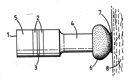
Referring now to the drawings; Figure 1 shows a vibrator
in the form of a PZT sandwich transducer incorporating a
backplate S, PZT ceramic rings 2 (piezo electric
transducer means), electrode 3 and stepped output section
4. This vibrator transmits waves at a predetermined
frequency through a shaped plastic head 6 into tissue 8
via coupling medium 7. Figure 1 shows the waveform in the
system. A standing wave is established in the transducer
with output amplitude, and this is transmitted through a
shaped therapy head 6 emerging as a travelling wave of
amplitude ~ LM. The velocity and pressure wave amplitudes
in the plastic head are seen to be relatively constant
under loaded conditions; they therefore represent the
travelling wave amplitude for energy transmitted into the
-patient. This condition is~established due to reflection
at the transducer/head interface and almost complete
transmission Aat the ~head/tissue interface. The shape of
the head~may be varied at least according to the examples
given in Figues 3~to 7. This characteristic reflects the
particular properties of the plastics chosen for the head
construction whioh allow accurate control of frequency and
amplitude. For given t'ransducer dimensions the shape and
size of the head can be varied between wide limits whilst
maintaining a controlled output power.
In operation the energy transmitted to the subject tissue
must not result in standing waves since this might cause
excessive local absorption. This would normally be
WO93/166~2 ` PCT/GB93/~3?~
- 6 -
avoided by moving the head over the tissue surface during
treatment; however when treating the hand or other
inaccessible area such movement may be inhibited by the
head shape and an alternative method must be used. The
S broad band transmission characteristic of the head permits
the use of frequency modulation derived from the system
shown in the drawings.
A further advantage which derives from the use of a
plastics, mouldable head is the ability to employ a shaped
head designed to give maximum contact in locations with
difficult access e.g. hand and feet. For example in
severe cases of rheumatoid arthritis the head could be
moulded to form a hand grip which when held by the patient
would permit qeneral treatment of the hand joints
lS simultaneously. This is shown in Figures l~ and ll. When
the power input is supplied by battery means or some other
transportable source, the transducer may be located within
the stem of a walking stick or cane, the grip of which
comprises the head.
This invention offers an improved method and means for the
therapeutic treatment of deep seated soft tissue injuriès
by ensuring that adequate power is safely transmitted to
the affected region. It offers a novel means of treating
irregularly shaped areas using moulding or machined heads
2~ that allow good transmission of energy without the need to
traverse the surface.
According to the invention, there is provided an apparatus
which offers a major benefit in the technique available to
monitor the treatment power delivered to a patient.
It is known that the intensity of transmitted ultrasound,
I, is related to displacement amplitude, ~ , by the
expression; I=l/2f c~2~o2, where f is the head material
~Z9~0
~093/16652 PCT/GB93/00374
~ 7 ~
density, c the phase velocity and~r, the angular frequency
defined as 2vf.
Since~ c is the wave impedance of the head material which
by design matches that of the treated tissue, then I ~ 2.
If we monitor the displacement amplitude within the
treatment head it is a relatively simple matter to obtain
a linear signal, proportional to displacement amplitude
and intensity, using a differentiating amplifier. This
method offers much greater reliability that the current
technique which depends on monitoring the electrical
signal to the transducer. Any variation in the transducer
performance would therefore cause a power neasuring error.
Figure 9 shows a head with a displacement sensor or
accelerometer incorporated. This enables the displacement
amplitude of vibrations transmitted to the patient to be
determined. The measurements may be transmitted to
indicating means for the user to control the power input.
The output of the displacement sensor may alternatively be
used to control directly the power input.
' I ' ~