Note: Descriptions are shown in the official language in which they were submitted.
CA 02131826 1999-12-23
-1-
MN GENE AND PROTEIN
FIELD OF THE INVENTION
The present invention is in the general area of medical
genetics and in t;he fields of biochemical engineering and
immunochemistry. More specifically, it relates to the
identification oi= a new gene--the MN gene--a cellular gene coding
for the MN protein. The inventors hereof found MN proteins to be
associated with t~umorigenicity. Identification of MN antigen as
well as antibodies specific therefor in patient samples provides
the basis for diagnostic/prognostic assays for cancer.
BACKGROUND OF THE INVENTION
MaTu is a novel quasi-viral agent with rather unusual
properties [Zavada, J., Arch. Virol, 50: 1-10 (1976)]. It is
presumably derived from a human mammary tumor. In some respects,
it resembles cla:~sical viruses whereas in other respects, it
resembles ~~slow° viruses (prions), and in still other respects it
is different from both classes of viruses.
MaTu was first detected by its capacity to complement
mutants of vesicular stomatitis virus (VSV) with heat-labile
surface G protein in HeLa cells (cell line derived from human
cervical adenocarcinoma), which had been cocultivated with human
breast carcinoma cells. The complementation resulted in the
formation of phenotypically mixed virions--the VSV(MaTu) pseudo-
types [Zavada et al., Nature New Biol, 240: 124-125 (1972)].
The virions contain the VSV genome, which is responsible for
their ability to produce plaques (as well as internal VSV
proteins), but the surface protein, corresponding to MaTu,
determines their host range and neutralization specificities.
One of the paradoxical features of the MaTu agent
is its host range:. VSV(MaTu) is infectious only for human
fibroblasts, but not for HeLa; however, the MaTu agent,
detected by its <:apacity to donate surface protein for the
VSV(MaTu) pseudot:ypes, is transmissible only to HeLa, but not
WO 93/18152 PCT/US93/02024
~~ X18 ~s = _ - 2 -
to fibroblasts [Zavada et al., J. Gen. Virol.. 24: 327-337
(1974)].
By its complementation of VSV mutants and by its
formation of pseudotypes, MaTu resembles known enveloped
viruses. However, MaTu is transmissible only by direct cell-
to-cell contact, and not by cell-free filtrates, thus
differing from both classical and "slow" viruses. Its only
permissive host appears to be HeLa cells. In those cells,
MaTu spreads extremely slowly, and does not form
morphologically distinct virions, thus resembling the "slow"
viruses. [Zavada et al., (1974); Zavada and Zavadova, Arch.
Virol. 118: 189-197 (1991)]. No known virus has HeLa cells
as an exclusive host.
Since the above-described properties suggest that
MaTu might be an entirely new type of molecular parasite of
living cells, and since it possibly originated from a human
tumor, there was a significant medical research interest to
characterize it in more detail. Herein elucidated is the
biological and molecular nature of MaTu. MaTu was found to be
2o a two-component system, having an exogenous transmissible
component, MX, and an endogenous cellular component, MN. The
MN gene was further found to be present i:n the chromosomal DNA
of all vertebrates tested, and its expression was~found to be
strongly correlated with tumorigenicity.
Described herein is the cloning and sequencing of
the MN gene and the production of a MN-encoded protein in a
bacterial vector. That genetically engineered MN protein as
well as other MN proteins/polypeptides, can be used in
serological assays according to this invention to detect MN-
specific antibodies. Further, such MN proteins/polypeptides
and antibodies reactive with MN antigen can be used in
immunoassays according to this invention to detect and/or
quantitate MN antigen. Such assays may be diagnostic and/or
prognostic for neoplastic and/or pre-neoplastic disease.
SUMMARY OF THE INVENTION
Herein disclosed is the MN gene, a cellular gene
which is the endogenous component of the MaTu agent.
WO 93/18152 PCT/US93/02024
21 318 2 6
Substantially the entire cDNA sequence for the apparently
intronless gene is shown in Figures lA-1B [SEQ ID NO.: i].
. This invention is directed to said MN gene,
fragments thereof and the related cDNA which are useful, for
example, as follows: 1) to produce MN proteins/ polypeptides
by biochemical engineering; 2) to prepare nucleic acid probes
to test for the presence of the MN gene in cells of a,subject:
3) to prepare appropriate polymerise chain reaction (PCR)
primers for use, for example, in PCR-based assays or to
Produce nucleic acid probes; 4) to identify MN proteins and
polypeptides as well as homologs or near homologs thereto; 5)
to identify various mRNAs transcribed from MN genes in various
tissues and cell lines, preferably human; and 6) to identify
mutations in MN genes. The invention further concerns
Purified and isolated DNA molecules comprising the MN gene or
fragments, thereof, or the related cDNA or fragments thereof.
The invention further concerns the discovery of a
hitherto unknown protein -- MN, encoded by the MN gene. The
expresssion of MN proteins is inducible by growing cells in
2p dense cultures, and such expression was discovered to be
associated with tumorigenic cells.
MN proteins were found to be produced by. some human
tumor cell lines in vitro, for example, by HeLa (cervical
carcinoma), T24 (bladder carcinoma) and T47D (mammary
carcinoma) and SK-Mel 1477 (melanoma) cell lines, by
tumorigenic hybrid cells and by cells of some human cancers _in
vivo, for example, by cells of uterine cervical, ovarian and
endometrial carcinomas as well as cells of some benign
neoplasias such as mammary papillomas. MN proteins were not
found in non-tumorigenic hybrid cells or in the cells of
normal tissues. Thus, MN proteins are considered to be tumor-
specif is .
In HeLa and in tumorigenic HeLa x fibroblast hybrid
(H/F/T) cells, MN protein is manifested as a "twin" protein
P54/58N; it is glycosylated and forms disulfide-linked
oligomers. As determined by electrophoresis upon reducing
gels, MN proteins have molecular weights in the range of from
about 40 kd to about 70 kd, preferably from about 45 kd to
II
WO 93/18152 PGT/US93/02024
21 31826
- 4 -
about 65 kd, more preferably from about 48 kd to about 58 kd.
Upon non-reducing gels, MN proteins in the form of oligomers
have a molecular weights in the range of from about 145 kd to
about 160 kd, preferably from about 150 to about 155 kd, still
more preferably from about 152 to about 154 kd. The predicted
amino acid sequence for a preferred MN protein of this
invention is shown in Figure lA-1B.
The discovery of the MN gene and protein and thus,
of substantially complementary MN genes and proteins encoded
thereby, led to the finding that the expression of MN proteins
was associated with tumorigenicity. That finding resulted in
the creation of methods that are diagnostic/ prognostic for
cancer and precancerous conditions. Methods and compositions
are provided for identifying the onset and presence of
neoplastic disease by detecting and/or quantitating MN antigen
in patient samples, including cell and tissue extracts from
vertebrates, preferably mammals and more preferably humans.
Such MN antigen may also be found in body fluids.
MN proteins and genes are of use in research
concerning the molecular mechanisms of oncogenesis, in cancer
diagnostics/prognostics, and may be of use in cancer
immunotherapy.
The present invention is useful for detecting a wide
variety of neoplastic and/or pre-neoplastic diseases.
Exemplary neoplastic diseases include carcinomas, such as
mammary, bladder, ovarian, uterine, cervical, endometrial,
squamous cell and adenosquamous carcinomas; and head and neck
cancers; mesodermal tumors, such as neuroblastomas and
retinoblastomas; sarcomas, such as osteosarcomas and Ewing's
sarcoma; and melanomas. Of particular interest are head and
neck cancers, gynecologic cancers including ovarian, cervical,
vaginal, endometrial and vulval cancers; gastrointestinal
cancer, such as, stomach, colon and esophageal cancers;
urinary tract cancer, such as, bladder and kidney cancers;
skin cancer; liver cancer; prostate cancer; lung cancer; and
breast cancer. Of still further particular interest are
gynecologic cancers; breast cancer; urinary tract cancers,
especially bladder cancer; lung cancer; gastrointestinal
WO 93/18152 PCT/US93/02024
- 21 31826
cancer, such as, stomach, colon and esophageal cancers; and
liver cancer. Even further of particular interest are
gynecologic cancers and breast cancer. Gynecologic cancers of
particular interest are carcinomas of the uterine cervix,
endometrium and ovaries; more particularly such gynecologic
cancers include cervical squamous cell carcinomas,
adenosquamous carcinomas, adenocarcinomas as well as
gynecologic precancerous conditions, such as metaplastic
cervical tissues and condylomas.
The invention further relates to the biochemical
engineering of the MN gene, fragments thereof or related cDNA.
For example, said gene or a fragment thereof or related cDNA
can be inserted into a suitable expression vector; host cells
can be transformed with such an expression vector; and an MN
protein/polypeptide, preferably an 1~1 protein, is expressed
therein. Such a recombinant protein or polypeptide can be
glycosylated or nonglycosylated, preferably glycosylated, and
can be purified to substantial purity. The invention further
concerns MN proteins/polypeptides which are synthetically or
otherwise biologically prepared.
Said MN proteins/polypeptides can be used in assays
to detect MN antigen in patient samples arid in serological
assays to test for MN-specific antibodies. MN
proteins/polypept~des of this invention are serologically
active, immunogenic and/or antigenic. They can further be
used as immunogens to produce MN-specific antibodies,
polyclonal and/or monoclonal, as well as an immune T-cell
response.
The invention further is directed to MN-specific
antibodies, which can be used diagnostically/prognostically
and may be used therapeutically. MN-specific antibodies can
be used, for example, in laboratory diagnostics, using
immunofluorescence microscopy or immunohistochemical staining;
as a component in immunoassays for detecting and/or
quantitating MN antigen in, for example, clinical samples; as
probes for immunoblotting to detect MN antigen; in
immunoelectron microscopy with colloid gold beads for
localization of MN proteins and/or polypeptides in cells; and
II
WO 93/18152 PCT/US93/02024
.~1 ~,~8 26 -
6 -
in genetic engineering for cloning the MN gene or fragments
thereof, or related cDNA. Such MN-specific antibodies can be
used as components of diagnostic/ prognostic kits, for
example, for in vitro use on histological sections; such
antibodies can also and used for in vivo diagnostics/
prognostics, for example, such antibodies can be labeled
appropriately, as with a suitable radioactive isotope, and
used in vivo to locate metastases by scintigraphy. Further
such antibodies may be used in vivo therapeutically to treat
cancer patients with or without toxic and/or cytostatic agents
attached thereto. Further, such antibodies can be used in
vivo to detect the presence of neoplastic and/or pre-
neoplastic disease. Still further, such antibodies can be
used to affinity purify MN proteins and polypeptides.
A hybridoma that produces a representative MN-
specific antibody, the monoclonal antibody M75, was deposited
at the American Type Culture Collection [ATCC; Rockville, MD
(USA)] on September 17, 1992, under ATCC Number HB 11128. The
M75 antibody was used to discover and identify the MN protein
and can be used to readily identify MN antigen in Western
blots, in radioimmunoassays and immunohistochemically, for
example, in tissue samples that have been'formalin fixed.
This invention also concerns recombinant DNA
molecules comprising a DNA sequence that encodes for an MN
protein or polypeptide, and also recombinant DNA molecules
that encode not only for an MN protein or polypeptide but also
for an amino acid sequence of a non-MN protein/polypeptide,
preferably which is not immunogenic to humans and which is not
typically reactive to antibodies in human body fluids.
Examples of such a DNA sequence is the alpha-peptide coding
region of beta-galactosidase and a sequence coding for
glutathione S-transferase or a fragment thereof. Further,
claimed herein are such recombinant fusion proteins/
polypeptides which are substantially pure and non-naturally
occurring. An exemplary fusion protein of this invention is
pGEX-3X-MN.
This invention also concerns methods ~f treating
neoplastic disease and/or pre-neoplastic disease comprising
WO 93/18152 PCT/US93/02024
- 21 31826
inhibiting the expression of MN genes by administering
antisense nucleic acid sequences that are substantially
complementary to mRNA transcribed from MN genes. Preferred
are antisense nucleic acid sequences that are substantially
complementary to sequences at the 5' end of the MN cDNA
sequence shown in Figure lA-iB. Preferably said antisense
nucleic acid sequences are oligonucleotides.
This invention also concerns vaccines comprising an
immunogenic amount of one or more substantially pure MN
l0 proteins and/or polypeptides dispersed in a physiologically
acceptable, nontoxic vehicle, which amount is effective to
immunize a vertebrate, preferably a mammal, more preferably a
human, against a neoplastic disease associated .pith the
expression of MN proteins. Said proteins can be
recombinantly, synthetically or otherwise biologically
produced. Recombinant 1~1 proteins includes fusion proteins,
as exemplified by pGEX-3X-1~T. A particular use of said
vaccine would be to prevent recidivism and/or metastasis. For
example, it could be ad~~inistered to a patient who has had an
MN-carrying tumor surgically removed, to prevent recurrence of
the tumor.
The invention still further concerns nucleic acid
probes that are substantially complementary to nucleic acid
sequences of the MN gene. Preferred nucleic acid probes of
this invention are those with sequences substantially
complementary to sequences from the MN cDNA shown in Figure
lA-1B. Test kits of this invention can comprise such probes
which are useful diagnostically/prognostically for neoplastic
and/or pre-neoplastic disease. Preferred test kits comprise
means for detecting or measuring the hybridization of said
probes to the MN gene or to the mRNA product of the MN gene,
such as a visualizing means.
The immunoassays of this invention can be embodied
in test kits which comprise MN proteins/polypeptides and/or
MN-specific antibodies. Such test kits can be in solid phase
formats, but are not limited thereto, and can also be in
liquid phase format, and can be based on ELISAS, particle
II
WO 93/18152 PCT/US93/02024
~~ ~~~ 2~ _
8_
assays, radiometric or fluorometric assays either unamplified
or amplified, using, for example, avidin/biotin technology.
Abbreviations
The following abbreviations are used herein:
AA - amino acid
ATCC - American Type Culture Collection
by - base pairs
BSA - bovine serum albumin
l0 Ci - curie
cm - centimeter
cpm - counts per minute
C-terminus - carboxyl-terminus
C - degrees centigrade
DMEM - Dulbecco modified Eagle medium
EDTA - ethylenediaminetetracetate
EIA - enzyme immunoassay
ELISA - enzyme-linked immunosorbent assay
F - fibroblasts
FCS - fetal calf serum
FIBR - fibroblasts
FITC - fluorescein isothiocyanate~
H - HeLa cells
HEF - human embryo fibroblasts
HeLa K - standard type of HeLa cells
HeLa S - Stanbridge's mutant HeLa D98/AH.2
H/F-T - hybrid HeLa fibroblast cells that are
tumorigenic; derived from HeLa D98/AH.2
H/F-N - hybrid HeLa fibroblast cells that are
nontumorigenic; derived from HeLa D98/AH.2
HGPRT- - hypoxanthine guanine phosphoribosyl
transferase-deficient
HRP - horseradish peroxidase
IPTG - isopropyl-Beta-D-thiogalacto-pyranoside
kb - kilobase
kd - kilodaltons
SUBSTITUTE SHEET
WO 93/18152 PCT/US93/02024
-9- 21 31826 V'
M - molar
- milliampere
~ - monoclonal antibody
ME - mercaptoethanol
MEM - minimal essential medium
mg - milligram
ml - milliliter
- millimolar
- mammary tumor virus
N - normal concentration
ng - nanogram
N-terminus amino-terminus
-
ODN - oligodeoxynucleotide
PAGE - polyacrylamide gel electrophoresis
PBS - phosphate buffered saline
PEST - combination of one-letter abbreviations for
proline, glutamic acid, serine, threonine
PI - isoelectric point
RIP - radioimmunoprecipitation
RIPA - radioimmunoprecipitation assay
SAC - protein A-BtaDhvlococcus aureus cells
SDS - sodium dodecyl sulfate
SDS-PAGE - sodium dodecyl sulfate-polyacrylamide gel
electrophoresis
SSPE - NaCl (0.18 M), sodium phosphate (0.01 M), EDTA
(0.001 M)
TCA - trichloroacetic acid
TC media - tissue culture media
~CCi - microcurie
~cg - microgram
ul - microliter
!~M - micromolar
VSV - vesicular stomatitis virus
X-MLV - xenotropic murine leukemia virus
Cell Lines
The following cell lines were used in the
experiments herein described:
SUBSTITUTE SHEET
CA 02131826 2000-O1-11
- 10 -
HeLa K -- standa.rd type of HeLa cells; aneuploid,
epithelial-like cell line isolated from a
.human cervical adenocarcinoma [Gey et al.,
;Cancer Res.. 12: 264 (1952); Jones et al.,
nbstet. Gynecol. 38: 945-949 (1971)]
~~btained from Professor B. Korych, [Institute
~of Medical Microbiology and Immunology,
Charles University; Prague, Czechoslovakia]
HeLa D98/AH.2 -- IKutant HeLa clone that is hypoxanthine
(also HeLa S) ~~uanine phosphoribosyl transferase-deficient
(HGPRT-) kindly provided by Eric J.
;~tanbridge [Department of Microbiology,
College of Medicine, University of
California, Irvine, CA (USA)] and reported in
:3tanbridge et al., Science, 215: 252-2'59 (15
Jan. 1'982); parent of hybrid cells H/F-N and
li/F-T, also obtained from E.J. Stanbridge.
NIH- 3T3 -- murine fibroblast cell line reported in
~~aronson, Science, 237: 178 (1987).
T47D -- c:ell lane derived from a human mammary
c:arcinnma [ICeydar et al. , Eur. J.- Cancer. - 15:
E~59-670 (1979)]; kindly provided by J. Keydar
[Haddaaah Medical School; Jerusalem, Israel]
T24 -- c;ell lane from urinary bladder carcinoma
(;Buben:ik et al., Int. J. Cancer. 11: 76'5-773
(;1973)] kindly provided by J. Bubenik
(;Instil~ute of Molecular Genetics,
Czechoslovak Academy of Sciences; Prague,
C:zecho:~lovakia ]
~2 -- cell line from melanoma [Svec et al.,
Nfeopla:ama. 35: 665-681 (1988) ]
_ _ _ -_- ' ~_ - _
CA 02131826 1999-12-23
-11-
HEF -- human embryo fibroblasts [Zavada et al., Nature
New Biology, 240: 124-125 (1972)]
SIRC -- cel:L line from rabbit cornea (control and X-MLV
infESCted) [Zavada et al., Virology, 82: 221-231
(19'77) ]
Vero cells-- African green monkey cell line [Zavada et al (1997)]
myeloma -- mye:Loma cell line used as a fusion parent
cell line in production of monoclonal antibodies [Galfre
NS-0 and Milstein, Methods Enzymol., 73: 3-46 (1981)]
SK-Mel -- human melanoma cell line kindly provided by K.E.
1477 Hel:Lstrom [Division of Tumor Immunology, Fred
Hut<:hins Cancer Research Center; Seattle,
Washington (USA)]
XC -- celJLs derived from a a rat rhabdomyosarcoma
induced with Rous sarcoma virus-induced rat sarcoma
[Svoboda, J., Natl. Cancer Center Institute
Monocrraph No. 17, IN: ~~International Conference
on Avian Tumor Viruses" (J. W. Beard ed.), pp. 277-
298 (1964)], kindly provided by Jan Svoboda
[Institute of Molecular Genetics, Czechoslovak
Academy of Sciences; Prague, Czechoslovakia]; and
Rat 2-Tk- -- a thymidine kinase deficient cell line, kindly
provided by L. Kutinova [Institute of Sera and
Vaccines; Prague, Czechoslovakia]
CGL1 -- H/F-N hybrid cells (HeLa D98/AH.2 derivative)
CGL2 -- H/F-T hybrid cells (HeLa D98/AH.2 derivative)
n
WO 93/18152 PCT/US93/02024
- _
12 -
CGL3 -- H/F-T hybrid cells (HeLa D98/AH.2 derivative)
CGL4 -- H/F-T hybrid cells (HeLa D98/Ah.2 derivative)
Nucleotide and Amino Acid Sequence Symbols
The following symbols are used to represent
nucleotides herein:
Base Symbol
adenine A
cytosine C
guanine G
thymine T
uracil U
There are twenty main amino acids, each of which is
specified by a different arrangement of three adjacent
nucleotides (triplet code or codon), and which are linked
together in a specific order to form a characteristic protein.
A three-letter convention is used herein to identify said
amino acids, as, for example, in Figure lA-1B, as follows:
Amino acid name Symbol
Alanine A-la
Arginine Arg
Asparagine Asn
Aspartic Acid Asp
Cysteine Cys
Glutamic Acid Glu
Glutamine Gln
Glycine Gly
Histidine His
Isoleucine Ile
Leucine Leu
Lysine Lys
Methionine Met
Phenylalanine Phe
Proline Pro
Serine Ser
Threonine Thr
SUBSTITUTE SHEET
~._.____ .______ _____.~. ~._ ~. _ _~___.__ ~._ ________ _
WO 93/18152 PCT/US93/02024
- 13 - 21 318 2 s
Tryptophan Trp
Tyrosine Tyr
Valine Val
BRIEF DESCRIPTION OF THE FIGURES
Figure lA-18 provides the nucleotide sequence for
the Mtl cDNA clone isolated as described herein and the
predicted amino acid sequence encoded by the cDNA [SEQ ID
NOS.: 1 and 2, respectively]. That sequence data has been
sent to the EMBL Data Library in Heidelberg, Germany and is
available under Accession No. X66839.
Figure 2A-2B graphically illustrates the expression
of MN- and MX-specific proteins in human fibroblasts (F), in
HeLa cells (H) and in H/F-N and H/F-T hybrid cells and
contrasts the expression in MX-infected and MX-uninfected
cells. Example 5 details the procedures and results.
Figure 3A-3B (discussed in Example 8) graphically
illustrates the results from radioimmunoprecipitation
experiments with 1251-pGEX-3X-MN protein and different
antibodies. The radioactive protein (15 x 103 cpm/tube) was
precipitated with ascitic fluid or sera and SAC as follows:
(A) ascites with MAb M75; (B) rabbit anti-MaTu serum; (C)
normal rabbit serum; (D) human serum L8; (E) human serum KH;
and (F) human serum M7.
Figure 4 (discussed in Example 8) shows the results
from radioimmunoassays for MN antigen. Ascitic fluid
(dilution precipitating 50% radioactivity) was allowed to
react for 2 hours with (A) "cold" (unlabeled) protein pGEX-3X-
MN, or with extracts from cells as follows: (B) HeLa + MX;
(C) Rat-2Tk-; (D) HeLa; (E) rat XC; (F) T24; and (G) HEF.
Subsequently 1251-labeled pGEX-3X-MN protein (25 x 103
cpm/tube) was added and incubated for an additional 2 hours.
Finally, the radioactivity to MAb M75 was adsorbed to SAC and
measured.
DETAILED DESCRIPTION
MaTu - MX and MN Components
As demonstrated herein MaTu is a two-component
system. One part of the complex, exogenous MX, is
SUBSTITUTE SHEET
II
WO 93/18152 PCT/US93/02024
~~ 3~8 26 = _.
- 14 -
transmissible, and is manifested by a protein, p58X, which is
a cytoplasmic antigen which reacts with some natural sera, of
humans and of various animals. The other component, MN, is
endogenous to human cells.
MN is a cellular gene, showing only very little
homology with known DNA sequences. It is rather conservative
and is present as a single copy gene in the chromosomal DNA of
various vertebrates. Described herein is the cloning and
sequencing of the MN cDNA, and the genetic engineering of a
l0 fusion protein, namely MN plus the carboxyl terminus of
glutathione S-transferase, that can be conveniently purified
by affinity chromatography.
MN is manifested in HeLa cells by a twin protein(s),
p54/58N, that is localized on the cell surface and in the
nucleus. Immunoblots using a monoclonal antibody reactive
with p54/58N (MAb M75) revealed two bands at 54 kd and 58 kd.
Those two bands may correspond to one type of protein that
differs by glycosylation pattern or by how it is processed.
(Both p54N and p58N are glycosylated with oligosaccharidic
residues containing mannose, but only p58N also contains
glucosamine.) Herein, the phrase "twin protein" indicates
p54/58N.
MN is absent in rapidly growing, sparse cultures of
HeLa, but is inducible either by keeping the cells in dense
cultures or, more efficiently, by infecting them with MX.
Those inducing factors are synergistic. Only p54/58N is
associated with virions of vesicular stomatitis virus (VSV),
reproduced in MaTu-infected HeLa. Whereas the twin protein
p54/58N is glycosylated and forms oligomers linked by
disulfidic bonds, p58X is not glycosylated and does not form
S-S-linked oligomers.
VSV assembles p54/58N into virions in HeLa cells,
indicating that the twin protein is responsible for
complementation of VSV G-protein mutants and for formation of
VSV(MaTu) pseudotypes. As only enveloped viruses provide
surface glycoproteins for the formation of infectious,
functioning pseudotypes, which can perform such specific
functions as adsorption and penetration of virions into cells
SUBSTITUTE SHEET
~. __ _._..W__~_ ~___.. ___~_ . __ .
WO 93/18152 PCT/US93/02024
-15- 21 31826
[Zavada, J., J. Gen. Virol. 63: 15-24 (1982)], that
observation implies that the MN gene behaves as a quasi-viral
sequence.
The surface proteins of enveloped viruses, which
participate in the formation of VSV pseudotypes, are
glycosylated as is the lid twin protein, p54/58N. I~1 proteins
also resemble viral glycoproteins in the formation of
oligomers (preferably tri- or tetramers); such
oligomerization, although not necessarily involving S-S bonds
(disulfidic bonds), is essential for the assembly of virions
[Kreis and Lodish, Cell. 46: 929-937 (1986)]. The disulfidic
bonds can be disrupted by reduction with 2-mercaptoethanol.
As reported in Pastorekova et al., Viroloctv. 187:
620-626 (1992), after reduction with mercaptoethanol, p54/58N
from cell extracts or from VSV looks very similar on
immunoblot. Without reduction, in cell extracts, it gives
several bands around 150 kd, suggesting that the cells may
contain several different oligomers (probably with a different
p54:p58 ratio), but VSV selectively assembles only one of
them, with a molecular weight of about 153 kd. That oligomer
might be a trimer, or rather a tetramer, consisting of 54 kd
and 58 kd proteins. The equimolar ratio of p54:p58 in VSV
virions is indicated by approximately the same strength of 54
kd and 58 kd bands in a VSV sample analyzed under reducing
conditions.
The expression of MN proteins appears to be
diagnostic/prognostic for neoplastic disease. The MN twin
protein, p54/58N, was found to be expressed in HeLa cells and
in Stanbridge~s tumorigenic (H/F-T) hybrid cells [Stanbridge
et al., Somatic Cell Genet 7: 699-712 (1981); and Stanbridge
et al., Science, 215: 252-259 (1982)] but not in fibroblasts
or in non-tumorigenic (H/F-N) hybrid cells [Stanbridge et al.,
~d_.] 1~1 proteins were found in immunoblots prepared from
human ovarian, endometrial and uterine cervical carcinomas,
and in some benign neoplasias (as mammary papilloma) but not
from normal ovarian, endometrial, uterine or placental
tissues. In HeLa cells infected with MX, observed were
conspicuous ultrastructural alterations, that is, the
SUBSTITUTE SHEET
I I
WO 93/18152 PCT/US93/02024
~~ 3828 ~ -
16 -
fonaation of abundant filaments on cell surfaces and the
amplification of mitochondria. Using an immunogold technique,
p54/58N was visualized on the surface filaments and in the
nucleus, particularly in the nucleoli. Thus MN proteins
appear to be tumor-specific as they do not appear to be
produced by normal non-tumor cells.
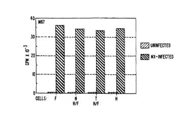
The examples herein show that MX and MN are two
different entities, that can exist independently of each
other. MX as an exogenous, transmissible agent can multiply
in fibroblasts and in H/F-N hybrid cells which are not
expressing MN-related proteins (Figure 2A-2B). In such cells,
MX does not induce the production of MN protein. MN protein
can be produced in HeLa and other tumor cells even in the
absence of MX as shown in Figure 2A-2B and in Examples 5 and
6. However, MX is a potent inducer of MN-related protein in
HeLa cells; it increases its production thirty times over the
concentration observed in uninfected cells (Examples 5 and 8,
and Table 1 in Example 8, below).
MN Gene--Cloning and Sequencing
Figure lA-1B provides the nucleotide sequence for
the MN cDNA clone isolated as described within this section.
It is understood that because of the degeneracy of. the genetic
code, that is, that more than one codon will code for one
amino acid [for example, the codons TTA, TTG, CTT, CTC, CTA
and CTG each code for the amino acid leucine (leu)], that
variations of the nucleotide sequence in, for example, Figure
lA-1B, wherein one codon is substituted for another, would
produce a substantially equivalent protein or polypeptide
according to this invention. All such variations in the
nucleotide sequence of the MN cDNA and complementary nucleic
acid sequences are included within the scope of this
invention.
It is further understood that the nucleotide
sequence herein described and shown in Figure lA-1B represents
only the precise structure of the cDNA nucleotide sequence
isolated and described herein. It is expected that slightly
modified nucleotide sequences will be found or can be modified
SUBSTITUTE SHEET
WO 93/18152 PCT/US93/02024
21 31826
- 17 -
by techniques known in the art to code for substantially
similar MN proteins and polypeptides, for example, those
having similar epitopes, and such nucleotide sequences and
proteins/polypeptides are considered to be equivalents for the
purpose of this invention. DNA or RNA having equivalent
codons is considered within the scope of the invention, as are
synthetic nucleic acid sequences that encode
proteins/polypeptides homologous or substantially homologous
to MN proteins/polypeptides, as well as those nucleic acid
sequences but for the degeneracy of the genetic code would
hybridize to said cDNA nucleotide sequence. Modifications and
variations of nucleic acid sequences as indicated herein are
considered to result in sequences that are substantially the
same as the MN sequence and fragments thereof.
To find the MN gene, a lambda gtli cDNA library from
MX-infected HeLa cells was prepared. Total RNA from MX-
infected HeLa cells was isolated by a guanidinium-thiocyanate-
CsCl method, and the mRNA was affinity separated on oligo dT-
cellulose. The synthesis of the cDNA and its cloning into
lambda gtll was carried out using kits from Amersham, except
that the EcoRI-NotI adaptor was from Stratagene [La Jolla, CA
(USA)]. The library was subjected to immunoscreening with
monoclonal antibody M75 in combination with goat anti-mouse
antibodies conjugated with alkaline phosphatase. That
immunoscreening method is described in Young and Davis, PNAS
(USA). 80: 1194-1198 (1983). One positive clone was picked
from 350,000 screened plaques (representing about one-half of
the whole library).
The positive clone was subcloned into the NotI site
of pBluescript KS [Stratagene] thereby creating pBluescript-
MN. Two oppositely oriented nested deletions were made using
Erase-a-BaseTM kit [Promega; Madison, WI (USA)] and sequenced
by dideoxy method with a T7 sequencing kit [Pharmacia;
Piscataway, NJ (USA)]. The sequencing showed a partial cDNA
clone, the insert being 1397 by long. That sequence is shown
in Figure lA-1B [SEQ ID NO.: 1]. The sequence comprises a
large 1290 by open reading frame and 107 by 3' untranslated
region containing a polyadenylation signal (AATAAA). Another
SUBSTITUTE SHEET
I I
WO 93/18152 PCT/US93/02024
21 318 26 _
- 18 -
interesting feature of the sequence is the presence of a
region contributing to instability of the mRNA (AUUUA at
position 1389) which is characteristic for mRNAs of some
oncogenes and lymphokines [Shaw and Kamen, Cell. 46: 659-667
(1986)]. As follows from a comparison of the size of the MN
clone with that of the corresponding mRNA in a Northern blot
(Example 12), the cDNA is missing about 100 by from the 5' end
of its sequence.
The open reading frame of MN cDNA clone encodes a
putative protein of about 48 kd (Figure lA-1B; SEQ ID NO.:
2). Analysis of the deduced translated amino acid (AA)
sequence failed to show any significant homology to published
protein sequences. The closest homology found was that of the
C-terminal part of the I~IN protein and different types of
carbonic anhydrase (about 30-35% in 170-200 AA overlap). The
active site as well as the Zn2+ binding domain of carbonic
anhydrase are well-conserved in the MN protein. However, the
MN gene is clearly a novel sequence derived from the human
genome.
Although as indicated, the MN gene shows some
homology with known carbonic anhydrases, it differs from them
in several repects. Seven carbonic anhydrases are known
[Dodgson et al. (eds.), The Carbonic Anhydrases, (Plenum
Press; New York/London (1991)]. Each of their genes contains
seven introns whereas the MN gene is apparently intronless.
Also, all the known carbonic anhydrases are proteins of about
kd, smaller than the p54/58N-related products of the MN
gene. Further, the carbonic anhydrases do not form oligomers
as do the MN-related proteins.
30 From the predicted amino acid sequence, it is
evident that the product of the MN gene is a basic protein (pI
9.08), with one potential N-glycosylation site located at the
amino acid positions 303-313. Those observations correspond
to the finding that p54/58N proteins from HeLa cells are
sensitive to Endo H and Endo F cleavage, which causes a loss
of about 3 kd each. The hydrophilicity profile reveals a
hydrophobic sequence of amino acids (at positions 371-395)
probably representing the region spanning the plasma membrane
SUBSTITUTE SHEE T
WO 93/18152 PCT/US93/02024
-19- 213182fi
and containing also a potential cleavage signal. The profile
fits well with the observation that p54/58N proteins are
localized on the cell membrane. There are no PEST regions in
the MN amino acid sequence, suggesting that the product of the
I~1 gene is a stable long-lived protein [Rogers et al.,
Science, 234: 364-368 (1986)]. Such a feature explains our
experience with inefficient metabolic labeling of p54/58N.
The deduced amino acid sequence displays also other features
namely, 10 potential phosphorylation and 7 myristylation
l0 sites, and 3 antigenic determinants.
To determine whether both p54/58N proteins were
encoded by one gene, antisense ODNs were used to inhibit
specifically 1~1 gene expression. [Such use of antisense ODNs
is reviewed in Stein and Cohen, Cancer Res.. 48: 2659-2668
(1988).] Those experiments are detailed in Example 11. The
findings indicated that cultivation of HeLa cells with ODNs
resulted in a considerable inhibition of p54/58N synthesis,
whereas the amount of different HeLa cell proteins produced
remained approximately the same. Further, and importantly on
immunoblotting, the specific inhibition by ODNs affected both
of the p54/58N proteins (Example 11). Thus, it was concluded
that the MN gene that was cloned codes for both of the p54/58N
proteins in HeLa cells.
To confirm whether the gene that was cloned codes
for the p54/58N-specific protein, it was subcloned into the
bacterial expression vector pGEX-3X [Pharmacia; Upsala,
Sweden], constructed to express a fusion protein containing
the C-terminus of glutathione S-transferase. That subcloning
is representative of one method to genetically engineer an MN-
related protein of this invention. The following description
is exemplary and not meant to limit the invention in any way.
Production of Fusion Protein pGEX-3X-MN
The cDNA insert from the above-described
pBluescript-MN was released by digesting the plasmid DNA by
NotI. It was then treated with S1 nuclease to obtain blunt
ends and then cloned into a dephosphorylated SmaI site of
SUBSTITUTE SHEET
- 20 -
pGEX-3X (Pharmacia). After transformation of XLl-Blue cells
and induction with iPTG, a fusion protein was obtained.
The fusion protein--MN glutathione S-transferase was
purified by affinity chromatography on Glutathione-S-Sepharose
4B (Pharmacia). Twenty micrograms of the purified recombinant
protein in each of two parallel samples were separated by SDS-
PAGE on a 10% gel. One of the samples (A) was stained with
Coomassie brilliant blue, whereas the other (B) was blotted
onto a Hybond C membrane (Amersham; Aylesbury, Bucks,
England). The blot was developed by autoradiography with 1~5I-
labeled MAb M75.
SDS-PAGE analysis provided an interesting result: a
number of protein bands with different molecular weights. A
similar SDS-PAGE pattern was obtained with another
representative fusion protein produced according to this
invention, beta-galactosidase-MN that was expressed from
lambda gtii lysogens. It appears that those patterns are due
to translation errors caused by the presence of 9 AGGAGG codon
tandems in the MN sequence. The use ofwthose codons is
strongly avoided in bacterial genes because of the shortage of
corresponding tRNAs. Thus, during the translation of AGGAGG
tandems from foreign mRNA, +1 ribosomal frameshifts arise with
a high frequency (about 50%) [Spanjaard et al., Nuc. Acid
Res.. 18: 5031-5036 (1990)]:
By immunoblotting, a similar pattern was obtained:
all the bands seen on stained SDS-PAGE gel reacted with the
MN-specific MAb M75, indicating that all the protein bands are
MN-specific. Also, that result indicates that the binding
site for MAb M75 is on the N-terminal part of the MN protein,
which is not affected by frameshifts.
As shown in Example 8 below, the fusion protein
pGEX-3X-MN was used in radioimmunoassays for MN-specific
antibodies and for MN antigen.
MN Proteins and/or Polypeptides
The phrase "MN proteins and/or polypeptides" (MN
proteins/polypeptides) is herein defined to mean proteins
and/or polypeptides encoded by an MN gene or fragments
* trade-mark
WO 93/18152 PCT/US93/02024
21 31826
- 21 -
thereof . An exemplary and preferred ICJ protein is that for
which the predicted amino acid sequence is shown in Figure lA-
iB (SEQ ID NO.: 2). Preferred MN proteins/polypeptides are
those proteins and/or polypeptides that have substantial
homology with that MN protein shown in Figure lA-1B.
A "polypeptide" is a chain of amino acids covalently
bound by peptide linkages and is herein considered to be
composed of 50 or less amino acids. A "protein" is herein
defined to be a polypeptide composed of more than 50 amino
acids.
It can be appreciated that a protein or polypeptide
produced by a neoplastic cell in vivo could be altered in
sequence from that produced by a tumor cell in cell culture.
Thus, MN proteins and/or polypeptides which have varying amino
acid sequences including without limitation, amino acid
substitutions, extensions, deletions, truncations and
combinations thereof, fall within the scope of this invention.
It can also be appreciated that a protein extant within body
fluids is subject to degradative processes, such as,
proteolytic processes; thus, 1~1 proteins that are
significantly truncated and MN polypeptides may be found in
body fluids, such as, sera. The phrase "MN antigen" is used
herein to encompass MN proteins and/or polypeptide~.
It will further be appreciated that the amino acid
sequence of MN proteins and polypeptides can be modified by
genetic techniques. One or more amino acids can be deleted or
substituted. Such amino acid changes may not cause any
measurable change in the biological activity of the protein or
polypeptide and result in proteins or polypeptides which are
within the scope of this invention.
The MN proteins and polypeptides of this invention
can be prepared in a variety of ways according to this
invention, for example, recombinantly, synthetically or
otherwise biologically, that is, by cleaving longer proteins
and polypeptides enzymatically and/or chemically. A preferred
method to prepare MN proteins is by a recombinant means. A
particularly preferred method of recombinantly producing a MN
protein is described above for the fusion protein pGEX-3X-MN.
SUBSTITUTE SHEET
II
WO 93/18152 PCT/US93/02024
- 22 -
Recombinant Production of MN Proteins and Polypeptides
A representative method to prepare the 1~T protein
shown in Figure lA-iB or fragments thereof would be to insert
the appropriate fragment of the NIrT cDNA into an appropriate
expression vector as exemplified above. A wide variety of
. host-cloning vector combinations may be usefully employed in
cloning the MN DNA isolated as described herein. For example,
useful cloning vehicles may include chromosomal,
nonchromosomal and synthetic DNA sequences such as various
known bacterial plasmids such as pBR322, other E. coli
plasmids and their derivatives and wider host range plasmids
such as RP4, phage DNA, such as, the numerous derivatives of
phage lambda, e.g., NB989 and vectors derived from
combinations of plasmids and phage DNAs such as plasmids which
have been modified to employ phage DNA expression control
sequences. The plasmid pGEX-3X is a preferred cloning
vehicle.
Useful hosts may be eukaryotic or prokaryotic and
include bacterial hosts such as E. coli and other bacterial
strains, yeasts and other fungi, animal or plant hosts such as
animal or plant cells in culture, insect cells and other
hosts. Of course, not all hosts may be equally efficient.
The particular selection of host-cloning vehicle combination
may be made by those of skill in the art after due
consideration of the principles set forth herein without
departing from the scope of this invention.
The particular site chosen for insertion of the
selected DNA fragment into the cloning vehicle to form a
recombinant DNA molecule is determined by a variety of
factors. These include size and structure of the protein or
polypeptide to be expressed, susceptibility of the desired
protein or polypeptide to endoenzymatic degradation by the
host cell components and contamination by its proteins,
expression characteristics such as the location of start and
stop codons, and other factors recognized by those of skill in
the art.
The recombinant nucleic acid molecule containing the
MN gene, fragment thereof, or cDNA therefrom, may be employed
SUBSTITUTE SHEET
_.._ .~~.~__ _ ._.______~._,_
WO 93/18152 PCT/US93/02024
-23- 2131826
to transform a host so as to permit that host (transformant)
to express the structural gene or fragment thereof and to
produce the protein or polypeptide for which the hybrid DNA
encodes. The recombinant nucleic acid molecule may also be
employed to transfona a host so as to permit that host on
replication to produce additional recombinant nucleic acid
molecules as a source of I~1 nucleic acid and fragments
thereof. The selection of an appropriate host for either of
those uses is controlled by a number of factors recognized in
the art. These include, for example, compatibility with the
chosen vector, toxicity of the co-products, ease of recovery
of the desired protein or polypeptide, expression
characteristics, biosafety and costs.
Where the host cell is a procaryote such as E. coli,
competent cells which are capable of DNA uptake are prepared
from cells harvested after exponential growth phase and
subsequently treated by the CaCl2 method by well known
procedures. Transformation can also be performed after
forming a protoplast of the host cell.
Where the host used is an eucaryote, transfection
methods such as the use of a calcium phosphate-precipitate,
electroporation, conventional mechanical procedures such as
microinjection, insertion of a plasmid encapsulated in red
blood cell hosts or in liposomes, treatment of cells with
agents such as lysophosphatidyl-choline or use of virus
vectors, or the like may be used.
The level of production of a protein or polypeptide
is governed by three major factors: (1) the number of copies
of the gene or DNA sequence encoding for it within the cell;
(2) the efficiency with which those gene and sequence copies
are transcribed and translated; and (3) the stability of the
mRNA. Efficiencies of transcription and translation (which
together comprise expression) are in turn dependent upon
nucleotide sequences, normally situated ahead of the desired
coding sequence. Those nucleotide sequences or expression
control sequences define, inter alia, the location at which an
RNA polymerase interacts to initiate transcription (the
promoter sequence) and at which ribosomes bind and interact
SUBSTITUTE SHEET
II
WO 93/18152 PCT/US93/02024
.~1 318 2 6 -
24 -
with the mRNA (the product of transcription) to initiate
translation. Not all such expression control sequences
function with equal efficiency. It is thus of advantage to
separate the specific coding sequences for the desired protein
from their adjacent nucleotide sequences and fuse them instead
to known expression control sequences so as to favor higher
levels of expression. This having been achieved, the newly
engineered DNA fragment may be inserted into a multicopy
plasmid or a bacteriophage derivative in order to increase the
n~er of gene or sequence copies within the cell and thereby
further improve the yield of expressed protein.
Several expression control sequences may be
employed. These include the operator, promoter and ribosome
binding and interaction sequences (including sequences such as
the Shine-Dalgarno sequences) of the lactose operon of E. coli
("the lac system"), the corresponding sequences of the
tryptophan synthetase system of E. coli ("the trp system"), a
fusion of the trp and lac promoter ("the tac system"), the
major operator and promoter regions of phage lambda (OLPL and
ORPR,), and the control region of the phage fd coat protein.
DNA fragments containing these sequences are excised by
cleavage with restriction enzymes from the DNA isolated from
transducing phages that carry the lac or trp operons, or from
the DNA of phage lambda or fd. Those fragments are then
manipulated in order to obtain a limited population of
molecules such that the essential controlling sequences can be
joined very close to, or in juxtaposition with, the initiation
codon of the coding sequence.
The fusion product is then inserted into a cloning
vehicle for transformation or transfection of the appropriate
hosts and the level of antigen production is measured. Cells
giving the most efficient expression may be thus selected.
Alternatively, cloning vechicles carrying the lac, trp or
lambda PL control system attached to an initiation codon may
be employed and fused to a fragment containing a sequence
coding for a MN protein or polypeptide such that the gene or
sequence is correctly translated from the initiation codon of
the cloning vehicle.
SUBSTITUTE SHEET
.__a_ .~ .. _ __._ .~__
WO 93/18152 PCT/US93/02024
21 31826
- 25 -
The phrase "recombinant nucleic acid molecule" is
herein defined to mean a hybrid nucleotide sequence comprising
at least two nucleotide sequences, the first sequence not
normally being found together in nature with the second.
The phrase "expression control sequence" is herein
defined to mean a sequence of nucleotides that controls and
regulates expression of structural genes when operatively
linked to those genes.
l0 Synthetic and Biologic Production of MN' Proteins and
Polypeptides
MN proteins and polypeptides of this invention may
be prepared not only by recombinant means but also by
synthetic and by other biologic means. Synthetic formation of
the polypeptide or protein requires chemically synthesizing
the desired chain of amino acids by methods well known in the
art. Exemplary of other biologic means to prepare the desired
polypeptide or protein is to subject to selective proteolysis
a longer MN polypeptide or protein containing the desired
amino acid sequence; for example, the longer polypeptide or
protein can be split with chemical reagents or with enzymes.
Chemical synthesis of a peptide is conventional in
the art and can be accomplished, for example, by the
Merrifield solid phase synthesis technique [Merrifield, J.,
Am. Chem. Soc.. 85: 2149-2154 (1963); Kent et al., Synthetic
Peptides in Biology and Med~~~~nP, 29 f.f. eds. Alitalo et al.,
(Elsevier Science Publishers 1985); and Haug, J.D., "Peptide
Synthesis and Protecting croup Strategy", American
Biotechnoloc~y Laboratory 5(1): 40-47 (Jan/Feb. 1987)].
Techniques of chemical peptide synthesis include
using automatic peptide synthesizers employing commercially
available protected amino acids, for example, Biosearch [San
Rafael, CA (USA)] Models 9500 and 9600; Applied Biosystems,
Inc. [Foster City, CA (USA)] Model 430; Milligen [a division
of Millipore Corp.; Bedford, MA (USA)] Model 9050; and Du
Pont's RAMP (Rapid Automated Multiple Peptide Synthesis) [Du
Pont Compass, Wilmington, DE (USA)].
SUBSTITUTE SHEET
II
WO 93/18152 PCT/US93/02024
2~ ~~~ 26 _
26 -
Nucleic Acid Probes and Test Kits
Nucleic acid probes of this invention are those
comprising sequences that are substantially complementary to
the MN cDNA sequence shown in Figure lA-iB or to MN gene
sequences. The phrase "substantially complementary" is
defined herein to have the meaning as it is well understood in
the art and, thus, used in the context of standard
hybridization conditions. The stringency of hybridization
conditions can be adjusted to control the precision of
complementarity.
Said probes can be used to detect MN DNA and/or RNA,
and thus can be used to test for the presence or absence of MN
genes, and amplification(s), mutations) or genetic
rearrangements of I~1 genes in the cells of a patient. For
example, overexpression of an 1~T gene may be detected by
Northern blotting using probes of this invention. Gene
alterations, as amplifications, translocations, inversions,
and deletions among others, can be detected by using probes of
this invention for in situ hybridization to chromosomes from a
patient's cells, whether in metaphase spreads or interphase
nuclei. Southern blotting could also be used with the probes
of this invention to detect amplifications or deletions of MN
genes. Restriction Fragment Length Polymorphism (RFLP)
analysis using said probes is a preferred method of detecting
gene alterations, mutations and deletions. Said probes can
also be used to identify MN proteins and/or polypeptides as
well as homologs or near homologs thereto by their
hybridization to various mRNAs transcribed from I~IN genes in
different tissues.
Said probes thus can be useful diagnostically/
prognostically. Said probes can be embodied in test kits,
preferably with appropriate means to enable said probes when
hybridized to an appropriate MN gene or MN mRNA target to be
visualized. Such samples include tissue specimens, body
fluids and tissue and cell extracts.
SUBSTITUTE SHEET
__ . _.___. _..~._~__ _.. . . ______.~
WO 93/18152 PCT/US93/02024
_ 2, _ 21 31826
s s
Assays according to this invention are provided to
detect and/or quantitate MN antigen or MN-specific antibodies
in vertebrate samples, preferably mammalian samples, more
preferably human samples. Such samples include tissue
specimens, body fluids, tissue extracts and cell extracts. MN
antigen may be detected by immunoassay, immunohistochemical
staining, immunoelectron and scanning microscopy using
immunogold among other techniques.
Preferred samples in which to assay MN antigen are
tissue and/or cell extracts. (Examples 7 and 8 below are
representative.) However, MN antigen may be detected in body
fluids, which can include among other fluids: blood, serum,
plasma, semen, breast exudate, saliva, tears, sputum, mucous,
urine, lymph, cytosols, ascites, pleural effusions, amniotic
fluid, bladder washes, bronchioalveolar lavages and
cerebrospinal fluid. It is preferred that the MN antigen be
concentrated from a larger volume of body fluid before
testing. Preferred body fluids to assay would depend on the
type of cancer for which one was testing, but in general
preferred body fluids would be breast exudate, pleural
effusions and ascites.
MN-specific antibodies can be bound by serologically
active MN proteins/polypeptides in samples of such body fluids
a'' blood, plasma, serum, lymph, mucous, tears, urine, spinal
fluid and saliva; however, such antibodies are found most
usually in blood, plasma and serum, preferably in serum. A
representative assay to detect MN-specific antibodies is shown
in Example 8 below wherein the fusion protein pGEX-3X-MN is
used. Correlation of the results from the assays to detect
and/or quantitate MN antigen and MN-specific antibodies
reactive therewith, provides a preferred profile of the
disease condition of a patient.
The assays of this invention are both diagnostic
and/or prognostic, i.e., diagnostic/prognostic. The term
"diagnostic/ prognostic" is herein defined to encompass the
following processes either individually or cumulatively
depending upon the clinical context: determining the presence
SUBSTITUTE SHEET
II
WO 93/18152 PCT/US93/02024
,~q31826
- 28 -
of disease, determining the nature of a disease,
distinguishing one disease from another, forecasting as to the
probable outcome of a disease state, determining the prospect
as to recovery from a disease as indicated by the nature and
symptoms of a case, monitoring the disease status of a
patient, monitoring a patient for recurrence of disease,
and/or determining the preferred therapeutic regimen for a
patient. The diagnostic/prognostic methods of this invention
are useful, for example, for screening populations for the
presence of neoplastic or pre-neoplastic disease, determining
the risk of developing neoplastic disease, diagnosing the
presence of neoplastic and/or pre-neoplastic disease,
monitoring the disease status of patients with neoplastic
disease, and/or determining the prognosis for the course of
neoplastic disease.
The present invention is useful for screening for
the presence of a wide variety of neoplastic diseases
including carcinomas, such as, mammary, urinary tract,
ovarian, uterine, cervical, endometrial, squamous cell and
adenosquamous carcinomas; head and neck cancers; mesodermal
tumors, such as, neuroblastomas and retinoblastomas; sarcomas,
such as osteosarcomas and Ewing's sarcoma; and melanomas. Of
particular interest are gynecological cancers including
ovarian, uterine, cervical, vaginal, vulval and endometrial
cancers, particularly ovarian, uterine cervical and
endometrial cancers. Also of particular interest are cancers
of the breast, of the stomach including esophagus, of the
colon, of the kidney, of the prostate, of the liver, of the
urinary tract including bladder, of the lung, and of the head
and neck.
The invention provides methods and compositions for
evaluating the probability of the presence of malignant or
pre-malignant cells, for example, in a group of cells freshly
removed from a host. Such an assay can be used to detect
tumors, quantitate their growth, and help in the diagnosis and
prognosis of disease. The assays can also be used to detect
the presence of cancer metastasis, as well as confirm the
absence or removal of all tumor tissue following surgery,
SUBSTITUTE SHEET
r.
WO 93/18152 PCT/US93i02024
21 31826
- 29 -
cancer chemotherapy and/or radiation therapy. It can further
be used to monitor cancer chemotherapy and tumor reappearance.
The presence of MN antigen or antibodies can be
detected and/or quantitated using a number of well-defined
diagnostic assays. Those in the art can adapt any of the
conventional immunoassay formats to detect and/or quantitate
MN antigen and/or antibodies. Example 8 details the format of
a preferred diagnostic method of this invention--a
radioimmunoassay. Many other formats for detection of MN
antigen and MN-specific antibodies are, of course available.
Those can be Western blots, ELISAs (enzyme-linked
immunosorbent assays), RIAs (radioimmunoassay), competitive
EIA or dual antibody sandwich assays, among other assays all
commonly used in the diagnostic industry. In such
i~unoassays, the interpretation of the results is based on
the assumption that the antibody or antibody combination will
not cross-react with other proteins and protein fragments
present in the sample that are unrelated to MN.
Representative of one type of ELISA test for MN
antigen is a format wherein a microtiter plate is coated with
antibodies made to MN proteins/polypeptides or antibodies made
to whole cells expressing MN proteins, anQ to this is added a
patient sample, for example, a tissue or cell extract. After
a period of incubation permitting any antigen to bind to the
antibodies, the plate is washed and another set of anti-MN
antibodies which are linked to an enzyme is added, incubated
to allow reaction to take place, and the plate is then
rewashed. Thereafter, enzyme substrate is added to the
microtiter plate and incubated for a period of time to allow
the enzyme to work on the substrate, and the adsorbance of the
final preparation is measured. A large change in absorbance
indicates a positive result.
It is also apparent to one skilled in the art of
immunoassays that MN proteins and/or polypeptides can be used
to detect and/or quantitate the presence of MN antigen in the
body fluids, tissues and/or cells of patients. In one such
embodiment, a competition immunoassay is used, ~,~herein the MN
protein/polypeptide is labeled and a body fluid is added to
SUBSTITUTE SHEET
II
WO 93/18152 PCT/US93/02024
~r~3~826 -30-
compete the binding of the labeled MN protein/polypeptide to
antibodies specific to MN protein/polypeptide. Such an assay
can be used to detect and/or quantitate l~IN antigen as
described in Example 8.
In another embodiment, an immunometric assay may be
used wherein a labeled antibody made to a MN protein or
polypeptide is used. In such an assay, the amount of labeled
antibody which complexes with the antigen-bound antibody is
directly proportional to the amount of MN antigen in the
l0 sample .
A representative assay to detect I~IN-specif is
antibodies is a competition assay in which labeled NIr1
protein/polypeptide is precipitated by antibodies in a sample,
for example, in combination with monoclonal antibodies
recognizing MN proteins/polypeptides. One skilled in the art
could adapt any of the conventional immunoassay formats to
detect and/or quantitate MN-specific antibodies. Detection of
the binding of said antibodies to said MN protein/polypeptide
could be by many ways known to those in the art, e.g., in
humans with the use of anti-human labeled IgG.
An exemplary immunoassay method of this invention to
detect and/or quantitate MN antigen in a vertebrate sample
comprises the steps of:
a) incubating said vertebrate sample with one or
more sets of antibodies (an antibody or antibodies) that bind
to MN antigen wherein one set is labeled or otherwise
detectable;
b) examining the incubated sample for the presence
of immune complexes comprising MN antigen and said antibodies.
Another exemplary immunoassay method according to
this invention is that wherein a competition immunoassay is
used to detect and/or quantitate MN antigen in a vertebrate
sample and wherein said method comprises the steps of:
a) incubating a vertebrate sample with one or more
sets of MN-specific antibodies and a certain amount of a
labeled or otherwise detectable MN protein/polypeptide wherein
said MN protein/ polypeptide competes for binding to said
antibodies with MN antigen present in the sample;
SUBSTITUTE SNS~
____~ ____..._~._._ ~.~.~..~..~._.___ _
WO 93/18152 PCT/US93/02024
-31- 2131826
b) examining the incubated sample to determine the
amount of labeled/detectable MN protein/polypeptide bound to
said antibodies; and
c) determining from the results of the examination
in step b) whether MN antigen is present in said sample and/or
the amount of MN antigen present in said sample.
Once antibodies (including biologically active
antibody fragments) having suitable specificity have been
prepared, a wide variety of immunological assay methods are
to available for determining the formation of specific
antibody-antigen complexes. Numerous competitive and
non-competitive protein binding assays have been described in
the scientific and patent literature, and a large number of
such assays are commercially available. Exemplary
immunoassays which are suitable for detecting a serum antigen
include those described in U.S. Patent Nos. 3,791,932;
3,817,837; 3,839,153; 3,850,752; 3,850,578; 3,853,987;
3,867,517; 3,879,262; 3,901,654; 3,935,074; 3,984,533;
3,996,345; 4,034,074; and 4,098,876.
Antibodies employed in assays may be labeled or
unlabeled. Unlabeled antibodies may be employed in
agglutination; labeled antibodies may be~employed in a wide
variety of assays, employing a wide variety of labels.
Suitable detection means include the use of labels
such as radionuclides, enzymes, coenzymes, fluorescers,
chemiluminescers, chromogens, enzyme substrates or co-factors,
enzyme inhibitors, free radicals, particles, dyes and the
like. Such labeled reagents may be used in a variety of well
known assays, such as radioimmunoassays, enzyme immunoassays,
e.g., ELISA, fluorescent immunoassays, and the like. ee for
example, U.S. Patent Nos. 3,766,162; 3,791,932; 3,817,837; and
4,233,402.
Methods to prepare antibodies useful in the assays
of the invention are described below. The examples below
detai~ representative assays according to this invention.
SUBSTITUTE SHEET'
WO 93/18152 PCT/US93/02024
~~ ~~~2s ~ _
32 -
Immunoassay Test Kits
The above outlined assays can be embodied in test
kits to detect and/or quantitate MN antigen and/or MN-specific
antibodies (including biologically active antibody fragments).
Kits to detect and/or quantitate MN antigen can comprise MN
protein(s)/polypeptides(s) and/or MN-specific antibodies,
polyclonal and/or monoclonal. Such diagnostic/prognostic test
kits can comprise one or more sets of antibodies, polyclonal
and/or monoclonal, for a sandwich format wherein antibodies
recognize epitopes on the MN antigen, and one set is
appropriately labeled or is otherwise detectable.
Test kits for an assay format wherein there is
competition between a labeled (or otherwise detectable) MN
protein/polypeptide and MN antigen in the sample, for binding
to an antibody, can comprise the combination of the labeled
protein/polypeptide and the antibody in amounts which provide
for optimum sensitivity and accuracy.
Test kits for MN-specific antibodies preferably
comprise labeled/detectable MN proteins(s) and/or
polypeptides(s), and may comprise other components as
necessary, for example, to perform a preferred assay as
outlined in Example 8 below. Such test kits can have other
appropriate formats for conventional assays.
Preparation of MN-Specific Antibodies
The term "antibodies" is defined herein to include
not only whole antibodies but also biologically active
fragments of antibodies, preferably fragments containing the
antigen binding regions. Such antibodies may be prepared by
conventional methodology and/or by genetic engineering.
Antibody fragments may be genetically engineered, preferably
from the variable regions of the light and/or heavy chains (VH
and VL), including the hypervariable regions, and still more
preferably from both the VH and VL regions. ~'or example, the
term "antibodies" as used herein comprehends polyclonal and
monoclonal antibodies and biologically active fragments
thereof including among other possibilities "univalent"
antibodies [Glennie et al., Nature, 295: 712 (1982)]; Fab
SUBSTITUTE SHEET
T.
WO 93/18152 PCT/US93/02024
21 31s2s
- 33 -
proteins including Fab' and F(ab')2 fragments whether
covalently or non-covalently aggregated; light or heavy chains
alone, preferably variable heavy and light chain regions (VH
and VL regions), and more preferably including the
hypervariable regions [otherwise known as the complementarity
determining regions (CDRs) of said VH and VL regions]; F~
proteins; "hybrid" antibodies capable of binding more than one
antigen; constant-variable region chimeras; "composite"
immunoglobulins with heavy and light chains of different
origins; "altered" antibodies with improved specificity and
other characteristics as prepared by standard recombinant
techniques and also by oligonucleotide-directed mutagenesis
techniques [Dalbadie-McFarland et al., PNAS (USA), 7~: 6409
(1982)].
It may be preferred for therapeutic and/or imaging
uses that the antibodies be biologically active antibody
fragments, preferably genetically engineered fragments, more
preferably genetically engineered fragments from the VH and/or
VL regions, and still more preferably comprising the
hypervariable regions thereof.
There are conventional techniques for making
polyclonal and monoclonal antibodies well=known in the
immunoassay art. Immunogens to prepare MN-specific~antibodies
include MN proteins and/or polypeptides, preferably purified,
and MX-infected tumor line cells, for example, MX-infected
HeLa cells, among other immunogens.
Anti-peptide antibodies are also made by
conventional methods in the art as described in European
Patent Publication No. 44,710 (published Jan. 27, 1982).
Briefly, such anti-peptide antibodies are prepared by
selecting a peptide from an MN amino acid sequence as from
Figure lA-iB, chemically synthesizing it, conjugating it to an
appropriate immunogenic protein and injecting it into an
appropriate animal, usually a rabbit or a mouse; then, either
polyclonal or monoclonal antibodies are made, the latter by
the Kohler-Milstein procedure.
Besides conventional hybridoma technology, newer
technologies can be used to produce antibodies according to
SUBSTITUTE SHEET
WO 93/18152 PCT/US93/02024
~~13~82s _
34 -
this invention. For example, the use of the polymerase chain
reaction (PCR) to clone and express antibody V-genes and phage
display technology to select antibody genes encoding fragments
with binding activities has resulted in the isolation of
antibody fragments from repertoires of PCR amplified V-genes
using immunized mice or humans. [Marks et al., BioTechnology,
~0_: 779 (July 1992) for references; Chiang et al.,
BioTechniques. 7(4): 360 (1989); Ward et al., Nature. 341:
544 (Oct. 12, 1989); Marks et al., J. Mol. Biol.. 222: 581
(1991); Clackson et al., Nature, 352: 624-628 (15 August
1991); and Mullinax et al., PNAS (USA), 87: 8095 (Oct. 1990).
Descriptions of preparing antibodies, which term is
herein defined to include biologically active antibody
fragments, by recombinant techniques can be found in U.S.
Patent No. 4,816,567 (issued March 28, 1989); European Patent
Application Publication Number (EP) 338,745 (published Oct.
25, 1989); EP 368,684 (published June 16, 1990); EP 239,400
(published September 30, 1987); WO 90/14424 (published Nov.
29, 1990); WO 90/14430 (published May 16, 1990); Huse et al.,
Science. 246: 1275 (Dec. 8, 1989); Marks et al.,
BioTechnoloay. 10: 779 (July 1992); La Sastry et al., PNAS
(USA), 86: 5728 (August 1989); Chiang et al., BioTechniques,
7(40): 360 (1989); Orlandi et al., PNAS (USA), 86I:~ 3833 (May
1989); Ward et al. Nature. 341: 544 (October 12, 1989);
Marks et al., J. Mol. Biol. 222: 581 (1991); and Hoogenboom
et al., Nucleic Acids Res.. 19(15): 4133 (1991).
Prebaration of Monoclonal Antibodies
Monoclonal antibodies for use in the assays of this
invention may be obtained by methods well known in the art for
example, Galfre and Milstein, "Preparation of Monoclonal
Antibodies: Strategies and Procedures," in Methods in
Enzvmoloav: Immunochemical Techniaues 73: 1-46 [Langone and
Vanatis (eds); Academic Press (1981)]; and in the classic
reference, Milstein and Kohler, Nature, 256: 495-497 (1975).
Although the representative hybridoma of this
invention is formed by the fusion of murine cell lines,
human/human hybridomas [Olsson et al., PNAS (USA), 77: 5429
SUBS .TITUTE SHEET
WO 93/18152 PCT/US93/02024
-35- 2131826
(1980)] and human/murine hybridomas [Schlom et al., PNAS
(USA), 77: 6841 (1980); Shearman et al. J. Immunol. 146:
928-935 (1991); and Gorman et al., PNAS (USA), ,~: 4181-4185
(1991)] can also be prepared among other possiblities. Such
humanized monoclonal antibodies would be preferred monoclonal
antibodies for therapeutic and imaging uses.
Monoclonal antibodies specific for this invention
can be prepared by immunizing appropriate mammals, preferably
rodents, more preferably rabbits or mice, with an appropriate
immunogen, for example, MaTu-infected HeLa cells or MN
proteins/polypeptides attached to a carrier protein if
necessary. The production of hybridoma W-M75 which secretes
MAb M75 is exemplary and described below. MAb M75 serves to
identify MN proteins/polypeptides in various laboratory
diagnostic tests, for example, in tumor cell cultures or in
clinical samples. Also produced by the method described for
producing MAb M75 (isotype IgG2B) were MAbs M16 (isotype
IgG2A) and M67 (isotype IgGl).
MAb M75
Monoclonal antibody M75 (MAb M75) is produced by
mouse lymphocytic hybridoma W-M75, whichYwas initially
deposited in the Collection of Hybridomas at the Institute of
Virology, Slovak Academy of Sciences (Bratislava,
Czechoslovakia) and was deposited under ATCC Designation HB
11128 on September 17, 1992 at the American Type Culture
Collection (ATCC) in Rockville, MD (USA).
Hybridoma W-M75 was produced according to the
procedure described in Gerhard, W., "Fusion of cells in
suspension and outgrowth of hybrids in conditioned medium,"
In: Monoclonal Antibodies Hvbridomas~ A New D ~nension in
Biological Analysis, page 370 [Kennet et al. (eds
.); Plenum NY
(USA)]. BALB/C mice were immunized with MaTu-infected HeLa
cells, and their spleen cells were fused with myeloma cell
line NS-0. Tissue culture media from the hybridomas were
screened for monoclonal antibodies, using as antigen the p58
immunoprecipitated from cell extracts of MaTu-infected HeLa
with rabbit anti-MaTu serum and protein A-Staphylococcus
SUBSTITUTE SHEEP
I I
WO 93/18152 PCT/US93/02024
2~ 31s 2s f
- 36 -
aureus cells (SAC) [Zavada and Zavadova, Arch. Virol.. 118
189-197 (1991)], and eluted from SDS-PAGE gels. Monoclonal
antibodies were purified from TC media by affinity
chromatography on protein A-Sepharose [Harlow and Lane,
"Antibodies: A Laboratory Manual," Cold Spring Harbor, Cold
Spring Harbor, NY (USA); 1988].
The monoclonal antibodies useful according to this
invention to identify MN proteins/polypeptides can be labeled
in any conventional manner, for example, with enzymes such as
horseradish peroxidase (HRP), fluorescent compounds, or with
radioactive isotopes such as, l2sI, among other labels. A
preferred label, according to this invention is 1251, and a
preferred method of labeling the antibodies is by using
chloramine-T [Hunter, W.M., "Radioimmunoassay," In: Handbook
of Extierimental Immunologv, pp. 14.1-14.40 (D. W. Weir ed.;
Blackwell, Oxford/London/Edinburgh/Melbourne; 1978)].
MAb H460
Monoclonal antibody H460 (MAb H460) was prepared in
a manner similar to that for MAb M75 except that the mice were
immunized with HeLa cells uninfected with MaTu, and
lymphocytes of the mice rather than spleen cells were fused
with cells from myeloma cell line NS-0. MAb H460 reacts about
equally with any human cells.
~'heraoeutic Use of MN-Specific Antibodies
The MN-specific antibodies of this invention,
monoclonal and/or polyclonal, preferably monoclonal, more
preferably MAb M75, may be used therapeutically in the
treatment of neoplastic and/or pre-neoplastic disease, either
alone or in combination with chemotherapeutic drugs or toxic
agents, such as ricin A. Further preferred for therapeutic
use would be biologically active antibody fragments as
described herein. Also preferred MN-specific antibodies for
such therapeutic uses would be humanized monoclonal
antibodies.
SUBSTITUTE SHEET
WO 93/18152 PCT/US93/02024
21 318 26
- 37 -
The MN-specific antibodies can be administered in a
therapeutically effective amount, preferably dispersed in a
physiologically acceptable, nontoxic liquid vehicle.
Imavina Use of Antibodies
Further, the MN-specific antibodies of this
invention when linked to an imaging agent, such as a
radionuclide, can be used for imaging. Biologically active
antibody fragments or humanized monoclonal antibodies, may be
preferred for imaging use.
A patient's neoplastic tissue can be identified as,
for example, sites of tumors and locations of any metastases.
Antibodies, appropriately labeled or linked to an imaging
agent, can be injected in a physiologically acceptable carrier
into a patient, and the binding the antibodies can be
detected by a method appropriate to the label or imaging
agent, for example, by scintigraphy.
Antisense MN Nucleic Acid Seauences
MN genes are herein considered putative oncogenes
and the proteins encoded thereby are considered to be putative
oncoproteins. Antisense nucleic acid sequences substantially
complementary to mRNA transcribed from MN genes, as~
represented by the antisense oligodeoxynucleotides (ODNs) of
Example 11, 'nfra, can be used to reduce or prevent expression
of the MN gene. [Zamecnick, P.C., "Introduction:
Oligonucleotide Base Hybridization as a Modulator of Genetic
Message Readout," pp. 1-6, Prospects for Antisense Nucleic
Acid Therapy of Cancer and AIDS, (Wiley-Liss, Inc., New York,
NY, USA; 1991); Wickstrom, E., "Antisense DNA Treatment of HL-
60 Promyelocytic Leukemia Cells: Terminal Differentiation and
Dependence on Target Sequence," pp, 7-24, id.; Leserman et
al., "Targeting and Intracellular Delivery of Antisense
Oligonucleotides Interfering with Oncogene Expression," pp.
25-34, id.; Yokoyama, K., "Transcriptional Regulation of c-mvc
Proto-oncogene by Antisense RNA," pp. 35-52, id.; van den Berg
et al., "Antisense fos Oligodeoxyribonucleotides Suppress the
Generation of Chromosomal Aberrations," pp, 63-70, ~d.;
SUBSTITUTE SHEET
I I
WO 93/18152 PCT/US93/02024
~q ~1a 2s
- 38 -
Mercola, D., "Antisense fos and un RNA," pp. 83-114, id.;
Inouye, Gene, 72: 25-34 (1988); Miller and Ts'o, inn. Reports
Med. Chem.. 23: 295-304 (1988); Stein and Cohen, Cancer Res..
48: 2659-2668 (1988); Stevenson and Inversen, J. Gen. Virol..
70: ?673-2682 (1989); Goodchild, "Inhibition of Gene
Expression by Oligonucleotides," pp. 53-77,
Oliaodeoxynucleotides: Antisense Inhibitors of Gene
Expression (Cohen, J.S., ed; CRC Press, Boca Raton, Florida,
USA; 1989); Dervan et al., "Oligonucleotide Recognition of
Double-helical DNA by Triple-helix Formation," pp. 197-210,
'~d.; Neckers, L.M., "Antisense Oligodeoxynucleotides as a Tool
for Studying Cell Regulation: Mechanisms of Uptake and
Application to the Study of Oncogene Function," pp. 211-232,
id.; Leitner et al., PNAS (USA), $7: 3430-3434 (1990);
Bevilacqua et al., PNAS (USA), 85: 831-835 (1988); Loke et
al. Curr. Toy. Microbiol. Immunol.. 141: 282-288 (1988);
Sarin et al., PNAS (USA), 85: 7448-7451 (1988); Agrawal et
al., "Antisense Oligonucleotides: A Possible Approach for
Chemotherapy and AIDS," International Union of Biochemistry
Conference on Nucleic Acid Therapeutics (Jan. 13-17, 1991;
Clearwater Beach, Florida, USA); Armstrong, L., Ber. Week, pp.
88-89 (March 5, 1990); and Weintraub et al., Trends. 1: 22-25
(1985).] Such antisense nucleic acid sequences, preferably
oligonucleotides, by hybridizing to the MN mRNA, particularly
in the vicinity of the ribosome binding site and translation
initiation point, inhibits translation of the mRNA. Thus, the
use of such antisense nucleic acid sequences may be considered
to be a form of cancer therapy.
Preferred antisense oligonucleotides according to
this invention are gene-specific ODNs or oligonucleotides
complementary to the 5' end of MN mRNA. Particularly
preferred are the 29-mer ODN1 and 19-mer ODN2 for which the
sequences are provided in Example 11, infra. Those antisense
ODNs are representative of the many antisense nucleic acid
sequences that can function to inhibit MN gene expression.
Ones of ordinary skill in the art could determine appropriate
antisense nucleic acid sequences, preferably antisense
SUBSTITUTE SHEET
WO 93/18152 PCT/US93/02024
- 21 31826 ~y
oligonucleotides, from the nucleic acid sequence of Figure lA-
1B.
Vaccines
It will be readily appreciated that 1~1 proteins and
polypeptides of this invention can be incorporated into
vaccines capable of inducing protective immunity against
neoplastic disease and a dampening effect upon tumorigenic
activity. MN proteins and/or polypeptides may be synthesized
l0 or prepared recombinantly or otherwise biologically, to
comprise one or more amino acid sequences corresponding to one
or more epitopes of the MN proteins either in monomeric or
multimeric form. Those proteins and/or polypeptides may then
be incorporated into vaccines capable of inducing protective
immunity. Techniques for enhancing the antigenicity of such
polypeptides include incorporation into a multimeric
structure, binding to a highly immunogenic protein carrier,
for example, keyhole limpet hemocyanin (KLH), or diptheria
toxoid, and administration in combination with adjuvants or
any other enhancers of immune response.
Preferred MN proteins/polypeptides to be used in a
vaccine according to this invention would be genetically
engineered MN proteins. A preferred recombinant I~ protein is
the fusion protein pGEX-3X-MN, produced according to this
invention.
A preferred exemplary use of such a vaccine of this
invention would be its administration to patients whose MN-
carrying primary cancer had been surgically removed. The
vaccine may induce active immunity in the patients and prevent
recidivism or metastasis.
It will further be appreciated that anti-idiotype
antibodies to antibodies to MN proteins/polypeptides are also
useful as vaccines and can be similarly formulated.
An amino acid sequence corresponding to an epitope
of an MN protein/polypeptide either in monomeric or multimeric
form may also be obtained by chemical synthetic means or by
purification from biological sources including genetically
modified microorganisms or their culture media [See Lerner,
SUBSTITUTE SHEET
WO 93/18152 PCT/US93/02024
~~1 318 2 6 _
40 -
"Synthetic Vaccines", Sci. Am. 248(2): 66-74 (1983)]. The
protein/polypeptide may be combined in an amino acid sequence
with other proteins/polypeptides including fragments of other
proteins, as for example, when synthesized as a fusion
protein, or linked to other antigenic or non-antigeneic
polypeptides of synthetic or biological origin.
The term "corresponding to an epitope of an I~1
protein/polypeptide" will be understood to include the
practical possibility that, in some instances, amino acid
sequence variations of a naturally occurring protein or
polypeptide may be antigenic and confer protective immunity
against neoplastic disease and/or anti-tumorigenic effects.
Possible sequence variations include, without limitation,
amino acid substitutions, extensions, deletions, truncations,
interpolations and combinations thereof. Such variations fall
within the contemplated scope of the invention provided the
protein or polypeptide containing them is immunogenic and
antibodies elicited by such a polypeptide or protein cross-
react with naturally occurring MN proteins and polypeptides to
a sufficient extent to provide protective immunity and/or
anti-tumorigenic activity when administered as a vaccine.
Such vaccine compositions will be combined with a
physiologically acceptable medium, including immunologically
acceptable diluents and carriers as well as commonly employed
adjuvants such as Freund's Complete Adjuvant, saponin, alum,
and the like. Administration would be in immunologically
effective amounts of the MN proteins or polypeptides,
preferably in quantities providing unit doses of from 0.01 to
10.0 micrograms of immunologically active MN protein and/or
polypeptide per kilogram of the recipient's body weight.
Total protective doses may range from 0.1 to about 100
micrograms of antigen.
Routes of administration, antigen dose, number and
frequency of injections are all matters of optimization within
the scope of the ordinary skill in the art.
The following examples are for purposes of
illustration only and not meant to limit the invention in any
way.
SUBSTITUTE SHEET
_..~._ ... ..._... __.~ _.~.__. _ _._.. T.._. __._
WO 93/18152 PCT/US93/02024
-41- 2131826
Materials and Methods
The following materials and methods were used in the
Examples below.
MaTu-Infected and Uninfected HeLa Cel7~
MaTu agent [Zavada et al., Nature New Biol.. 240:
124-125 (1972); Zavada et al., J. Gen. Viro 4: 327-337
(1974)] was from original "MaTu" cells [Widmaier et al., arch.
Geschwu~stforsch 44: 1-10 (1974)] transferred into our stock
of HeLa by cocultivation with MaTu cells treated with
mitomycin C, to ensure that control and MaTu-infected cells
were comparable. MaTu cells were incubated for 3 hours at
37°C in media with 5 ~cg/ml of mitomycin C [Calbiochem,
LaJolla, CA (USA)]. Mixed cultures were set to 2 x 10⁵ of
mitomycin C-treated cells and 4 x 10⁵ of fresh recipient cells
in 5 ml of medium. After 3 days they were first subcultured
and further passaged 1-2 times weekly.
Control HeLa cells were the same as those described
in Zavada et al., Nature New Biol 240: 124-125 (1972).
Sera
Human sera from cancer patients, from patients
suffering with various non-tumor complaints and from healthy
women were obtained from the Clinics of Obstetrics and
Gynaecology at the Postgraduate Medical School, Bratislava,
Czechoslovakia.
Human sera KH was from a fifty year old mammary
carcinoma patient, fourteen months after resection. That
serum was one of two sera out of 401 serum samples that
contained neutralizing antibodies to the VSV(MaTU) pseudotype
as described in Zavada et al., Nature New Biology 240: 124-
125 (1972). Serum L8 was from a patient with Paget's disease.
Serum M7 was from a healthy donor.
Rabbit anti-MaTu serum was prepared by immunizing a
rabbit three times at intervals of 30 days with 10-5 x 10~
viable MaTu infected HeLa cells.
SUBSTITUTE SHEET
42 -
RIP and PAGE
RIP and PAGE were performed essentially as described
in Zavada and Zavadova, Arch. Virol.. 118: 189-197 (1991),
except that in the experiments described herein [35S]methionine
(NEN), 10 ~Ci/ml of methionine-free MEM medium, supplemented
with 2% FCS and 3% complete MEM were used. Confluent petri
dish cultures of cells were incubated overnight in that media.
For RIP, the SAC procedure [Kessler, J. Immunol..
115: 1617-1624 (1975)] was used. All incubations and
centrifugations were performed at 0-4°C. Cell monolayers were
extracted with RIPA buffer (0.14 M NaCl, 7.5 mM phosphate
buffer, pH 7.2, 1% Triton X-100, 0.1% sodium deoxycholate, 1
mM phenylmethylsulfonyl fluoride and Trasylolj. To reduce
non-specific reactions, antisera were preabsorbed with foetal
calf serum [Barbacid et al., PNAS (USA), 77: 1617-1621
(1980)] and antigenic extracts with SAC.
For PAGE (under reducing conditions) we used 10%
gels with SDS [Laemmli, ~Jature, 227: 680-685 (1970)]. As
reference marker proteins served the Sigma kit (product MW-
SDS-200). For fluorography we used salicylate [Heegaard et
al., Electrophoresis. 5: 263-269 (1984)].
lmmunoblots
Immunoblotting used as described herein follows the
method of Towbin et al., PNAS (USA), 76: 4350-43'54 (1979).
The proteins were transferred from the gels onto
nitrocellulose [Schleicher and Schuell; Dassel Germany; 0.45
~cm porosity] in Laemmli electrode buffer diluted 1:10 with
distilled water, with no methanol or SDS. The transfer was
for 2 1/2 hours at 1.75 mA/cm2. The blots were developed with
l2sl_labeled MAbs and autoradiography was performed using
intensifying screens, with X-ray films exposed at -70°C.
In extracts from cell cultures containing only small
amounts of MN antigen, we concentrated the antigen from 0.5 or
1 ml of an extract by adding 50 ~1 of a 10% SAC suspension,
pre-loaded with MAb M75. This method allowed the
concentration of MN antigen even from clinical specimens,
containing human IgG; preliminary control experiments showed
* trade-mark
WO 93/18152 PCT/US93/02024
21 318 26 -
- 43 -
that such a method did not interfere with the binding of the
MN antigen to SAC-adsorbed M75. Tissue extracts were made by
grinding the tissue with a mortar and pestle and sand
(analytical grade). To the homogenates was added RIPA buffer,
10:1 (volume to weight) of original tissue. The extracts were
clarified for 3 minutes on an Eppendorf centrifuge.
Example 1
Immunofluorescence of MaTu Specific Anif ig~Pns
Immunofluorescence experiments were performed on
control and MaTu-infected HeLa cells with monoclonal
antibodies, prepared as described above, which are specific
for MaTu-related antigens. FITC-conjugated anti-mouse IgG was
used to detect the presence of the monoclonal antibodies.
Staining of the cells with Giemsa revealed no clear
differences between control and MaTu-infected HeLa cel'_.s.
MAbs, which in preliminary tests proved to be
specific for MaTu-related antigens, showed two different
reactivities in immunofluorescence. A representative of the
first group, MAb M67, gave a granular cytoplasmic fluorescence
in MaTu-infected HeLa, which was only seen in cells fixed with
acetone; living cells showed no fluorescence. MAb M16 gave
the same type of fluorescence. With either M67 or M16, only
extremely weak "background" fluorescence was seen in control
HeLa cells.
Another MAb, M75, showed a granular membrane
fluorescence on living MaTu-infected cells and a granular
nuclear fluorescence in acetone-fixed cells. However, M75
sometimes showed a similar, although much weaker, fluorescence
on uninfected HeLa cells. A relationship was observed based
upon the conditions of growth: in HeLa cells uninfected with
MaTu, both types of fluorescence with MAb M75 were observed
only if the cells were grown for several passages in dense
cultures, but not in sparse ones.
The amount of M75-reactive cell surface antigen was
analyzed cytofluorometrically and was dependent on the density
of the cell cultures and on infection with MaTu. Control and
MaTu infected HeLa cells were grown for 12 days in dense or
SUBSTITUTE SHEET
a
WO 93/18152 PCT/US93/02024
231826 -44-
sparse cultures. The cells were released with Versene (EDTA),
and incubated with MAb M75 or with no MAb, and subsequently
incubated with FITC-conjugated anti-mouse IgG. The intensity
of fluorescence was measured.
It appeared that the antigen binding MAb M75 is
inducible: it was found to be absent in control HeLa grown in
sparse culture, and to be induced either by the growth of HeLa
in dense culture or by infection with MaTu. Those two factors
were found to have an additive or synergistic effect. Those
observations indicated along with other results described
herein that there were two different agents involved:
exogenous, transmissible MX, reactive with M67, and
endogenous, inducible MN, detected by MAb M75.
Example 2
Immunoblot Analvsis of Proteins) Reactive with MAb M75
To determine whether MAb M75 reacts with the same
protein in both uninfected and MaTu-infected HeLa, and to
determine the molecular weight of the protein, extracts of
those cells were analyzed by PAGE and immunoblotting (as
described above). HeLa cells uninfected or MaTu-infected,
that had been grown for 12 days in dense or sparse cultures,
were seeded in 5-cm petri dishes, all variants at 5~x 105
cells per dish. Two days later, the cells were extracted with
RIPA buffer (above described), 200 ~1/dish. The extracts were
mixed with 2x concentrated Laemmli sample buffer containing 6~
mercaptoethanol and boiled for five minutes. Proteins were
separated by SDS-PAGE and blotted on nitrocellulose. The
blots were developed with 125I_labeled MAb M75 and
autoradiography.
MAb M75 reacted with two MN-specific protein bands
of 54 kd and 58 kd, which were the same in uninfected HeLa
grown at high density and in MaTu-infected HeLa, evidencing
that M75 recognizes the same proteins) in both uninfected and
MaTu-infected HeLa cells. Consistent with the
cytofluorometric results, the amount of the antigen depended
both on cell density and on infection with MaTu, the latter
being a much more potent inducer of p54/58N.
SUBSTITUTE SHEET
______ _... _.___....~_~_.. ..
WO 93/18152 PCf/US93/02024
-45- 213182fi
Example 3
Had~o~m_munoassay of MaTu-Specific Antigens In Situ
In contrast to the results with M75, the other MAb,
M67, appeared to be specific for the exogenous, transmissible
agent MX. With M67 we observed no immunofluorescence in
control HeLa, regardless of whether the cells were grown in
dense or in sparse culture. That difference was clearly
evidenced in radioimmunoassay experiments wherein 1251-labeled
MAbs M67 and M75 were used.
For such experiments, parallel cultures of
uninfected and MaTu-infected cells were grown in dense or
sparse cultures. The cultures were either live (without
fixation), or fixed (with methanol for five minutes and air-
dried). The cultures were incubated for two hours in petri
dishes with the 1251-labeled MAbs, 6 x 10⁴ cpm/dish.
Afterward, the cultures were rinsed four times with PBS and
solubilized with 1 ml/dish of 2 N NaOH, and the radioactivity
was determined on a gamma counter.
The simple radioimmunoassay procedure of this
example was performed directly in petri dish cultures.
Sixteen variants of the radioimmunoassay enabled us to
determine whether the MX and MN antigens are located on the
surface or in the interior of the cells and how the~expression
of those two antigens depends on infection with MaTu and on
the density, in which the cells had been grown before the
petri dishes were seeded. In live, unfixed cells only cell
surface antigens can bind the MAbs. In those cells, M67
showed no reaction with any variant of the cultures, whereas
M75 reacted in accord with the results of Examples 1 and 2
above .
Fixation of the cells with methanol made the cell
membrane permeable to the MAbs: M67 reacted with HeLa
infected with MaTu, independently of previous cell density,
and it did not bind to control HeLa. MAb M75 in methanol-
fixed cells confirmed the absence of corresponding antigen in
uninfected HeLa from sparse cultures and its induction both by
growth in dense cultures and by infection with MaTu.
SUBS T ITUTE SHEET
46 _ 2~ 3826
Example 4
Identification of MaTu Components Reactive with
Animal Sera or Associated with VSV Virions
Immunoblot analyses of MaTu-specific proteins from RIPA
extracts from uninfected or MaTu-infected HeLa and from
purified VSV reproduced in control or in MaTu-infected
HeLa, identified~which of the antigens, p58X or p54/58N,
were radioimmunoprecipitated with animal sera, and which of
them was responsible for complementation of VSV mutants and
for the formation of pseudotype virions. Details concerning
the procedures can be found in Pastorekova et al.,
Virology, 187: 620-626 (1992).
The serum of a rabbit immunized with MaTu-infected HeLa
immunoprecipitated both MAb M67- and MAb M75-reactive
proteins (both p58X and p54/58N), whereas the
"spontaneously" immune sera of normal rabbit, sheep or
leukemic cow immunoprecipitated only the M67-reactive
protein (p58X). On the other hand, in VSV reproduced in
MaTu-infected HeLa cells and subsequently purified, only
the M75-reactive bands of p54/58N were present. Thus, it
was concluded that_MX and MN are independent components of
MaTu, and that it was p54/58N that complemented VSV mutants
and was assembled into pseudotype virioas.
As shows is Figure 2A discussed below is Example 5, MX
antigen was found to be present is MaTu-infected
fibroblasts. In Zavada and Zavadova (1991), su ra, it was
reported that a p58 band from MX-infected fibroblasts could
not be detected by RIP with rabbit anti-MaTu serum. That
serum contains more antibodies to MX than to MN antigen.
The discrepancy can be explained by the extremely slow
spread of MX in infected cultures. The results reported in
Zavada sad Zavadova (1991) supra were from fibroblasts
tested 6 weeks after infection, whereas the later testing
was 4 months after infection. We have found by immunoblots
that MX can be first detected in both H/F-N and H/F-T
hybrids after 4 weeks, in HeLa cells after six weeks and is
fibroblasts only 10 weeks after infection.
r
WO 93/18152 PCT/US93/02024
-4,_ 2131826
Exam 1~ 5
Expression of MN- and MX- Specific Proteins
Figure 2A-2B graphically illustrates the expression
of MN- and MX- specific proteins in human fibroblasts, in HeLa
cells and in H/F-N and H/F-T hybrid cells, and contrasts the
expression in MX-infected and uninfected cells. Cells were
infected with MX by co-cultivation with mitomycin C-treated
MX-infected HeLa. The infected and uninfected cells were
grown for three passages in dense cultures. About four months
after infection, the infected cells concurrently with
uninfected cells were grown in petri dishes to produce dense
monolayers.
A radimmunoassay was performed directly in confluent
petri dish (5 cm) culture of cells, fixed with methanol
essentially as described in Example 3, supra. The monolayers
were fixed with methanol and treated with 1251-labeled MAbs M67
(specific for exogenous MX antigen) or M75 (specific for
endogenous MN antigen) at 6 x 10⁴ cpm/dish. The bound
radioactivity was measured; the results are shown in Figure
2A-2B.
Figure 2A shows that MX was transmitted to all four
cell lines tested, that is, to human embryo fibroblasts, to
HeLa and to both H/F-N and H/F-T hybrids; at the same time,
all four uninfected counterpart cell lines were MX-negative.
Figure 2B shows that MN antigens are present in both MX-
infected and uninfected HeLa and H/F-T cells, but not in the
fibroblasts. No MN antigen was found in the control H/F-N,
and only a minimum increase over background of MN antigen was
found in MaTu infected H/F-N. Thus, it was found that in the
hybrids, expression of MN antigen very strongly correlates
with tumorigenicity.
Those results were consistent with the results
obtained by immunoblotting. The MN-specific twin protein
p54/58N was detected by immunoblotting in HeLa cell lines
(both our standard type, that is, HeLa K, and in the
Stanbridge mutant HeLa, that is, D98/AH.2 or HeLa S) and in
tumorigenic H/F-T; however, p54/58N was not detected in the
fibroblasts nor in the non-tumorigenic H/F-N even upon
SUBSTITUTE SHEET
WO 93/18152 PCT/US93/02024
.13186 _48_
t
deliberately long exposure of the film used to detect
radioactivity. Infection of the HeLa cells with MX resulted
in a strong increase in the concentration of the p54/58N
protein(s).
The hybrid cells H/F-N and H/F-T were constructed by
Eric J. Stanbridge [Stanbridge et al., Somatic Cell Genetics,
7: 699-712 (1981); and Stanbridge et al., Science. 215: 252-
259 (1982)]. His original hybrid, produced by the fusion of a
HeLa cell and a human fibroblast was not tumorigenic in nude
mice, although it retained some properties of transformed
cells, for example, its growth on soft agar. Rare segregants
from the hybrid which have lost chromosome 11 are tumorigenic.
The most likely explanation for the tumorigenicity of those
segregants is that chromosome 11 contains a suppressor gene
(an antioncogene), which blocks the expression of a as yet
unknown oncogene. The oncoprotein encoded by that oncogene is
critical for the capacity of the H/F hybrids to produce tumors
in nude mice. Since the p54/58N protein shows a correlation
with the tumorigenicity of H/F hybrids, it is a candidate for
that putative oncoprotein.
Example 6
Immunoblots of MN Antigen from Human Tumor Cell
Cultures and from Clinical Specimens of Human Tissues
The association of MN antigen with tumorigenicity in
the H/F hybrid cells as illustrated by Example 5 prompted
testing for the presence of MN antigen in other human tumor
cell cultures and in clinical specimens. Preliminary
experiments indicated that the concentration of MN antigen in
the extracts from other human tumor cell cultures was lower
than in HeLa; thus, it was realized that long exposure of the
autoradiographs would be required. Therefore, the sensitivity
of the method was increased by the method indicated under
Materials and Methods: Immunoblottina, su ra, wherein the MN
antigen was concentrated by precipitation with MAb M75-loaded
SAC.
Immunoblots of MN proteins in cell culture extracts
were prepared from the following: (A) MX-infected HeLa cells;
SUBSTITUTE SHEET
WO 93/18152 PCT/US93/02024
21 31826
- 49 -
(B) human fibroblasts; (C) T24; (D) T47D; (E) SK-Me1 1477; and
(F) HeLa.cells uninfected with MX. The proteins were
separated by PAGE after heating in a sample buffer, either
with or without 3% mercaptoethanol (+ ME or O ME,
repectively). Thus, one run was done for each extract + ME
and then O ME. In lane (A) (the cell culture extract from MX-
infected HeLa cells) was analysed directly (10 ~,1 per lane),
whereas the antigens from the other extracts (lanes 8-E) were
each concentrated from a 500 ~1 extract by precipitation with
MAb M75 and SAC. The immunoblots indicated that two other
human carcinoma cell lines contain MN-related proteins -- T24
(bladder carcinoma; lane C) and T47D (mammary carcinoma; lane
D). Those cells contain proteins which react with MAb M75
that under reducing conditions have molecular weights of 54 kd
and 56 kd, and under non-reducing conditions have a molecular
weight of about 153 kd. The intensity of those bands was at
least ten times lower than that for the p54/58N twin protein
from HeLa cells.
An extremely weak band at approximately 52 kd could
be seen under reducing conditions from extracts from human
melanoma cells (SK-Mel 1477; lane E), but no bands for human
fibroblast extracts (lane B) could be seen either on the
reducing or non-reducing gels.
Immunoblots of human tissue extracts including those
of surgical specimens were compared to that of a cell extract
from MX-infected HeLa (lane A). The tissue extracts in the
other lanes were prepared from the following: (B) full-term
placenta; (C) corpus uteri; (D, M) adenocarcinoma endometrii;
(E, N) carcinoma ovari; (F, G) trophoblasts; (H) normal ovary;
(I) myoma uteri; (J) mammary papilloma; (K) normal mammary
gland; (L) hyperplastic endometrium; (O) cervical carcinoma;
and (P) melanoma. The MN-related antigen from all the
extracts but for lane A (analysed directly at 10 ~,1 per lane)
was first concentrated from a 1 ml extract as explained above.
MN proteins were found in endometrial (lanes D and M), ovarian
(lanes E and N) and in uterine cervical (lane 0) carcinomas.
In those extracts MN-related proteins were found in three
bands having molecular weights between about 48 kd and about
SUBSTITUTE SHEET
II
WO 93/18152 PCT/US93/02024
8 2 6 ,, - 5 0 _
t2 ~ 3 ~
58 kd. Another MN-related protein was present in the tissue
extract from a mammary papilloma; that protein was seen as a
single band at about 48 kd (lane J).
Clearly negative were the extracts from full-term
placenta (lane B), normal mammary gland (lane K), hyperplastic
endometrium (lane L), normal ovaries (lane H), and from
uterine myoma (lane I). Only extremely slightly MN-related
bands were seen in extracts from trophoblasts (lanes F and G)
and from melanoma (lane P).
The observations that antigen related to p54/58N was
expressed in clinical specimens of several types of human
carcinomas but not in normal tissues of corresponding organs
further strengthens the association of MN antigen with
tumorigenesis. However, it should be noted that for human
tumor, a normal tissue is never really an adequate control in
that tumors are believed not to arise from mature,
differentiated cells, but rather from some stem cells, capable
of division and of differentiation. In body organs, such
cells may be quite rare.
Example 7
MN Antigen in Animal Cell Lines ,
Since the MN gene is present in the chromosomal DNA
of all vertebrate species that were tested, MN-related antigen
was searched for also in cell lines derived from normal
tissues and from tumors of several animal species. MN-related
protein was found in two rat cell lines: one of them was the
XC cell line derived from rat rhabdomyosarcoma induced with
Rous sarcoma virus; the other was the Rat2-Tk- cell line. In
extracts from both of those rat cell lines, a single protein
band was found on the blots . its molecular weight on blots
produced from a reducing gel and from a non-reducing gel was
respectively 53.5 kd and 153 kd.
Immunoblots of MN proteins were prepared from (A)
MX-infected HeLa cells and from (B) Rat-TK-cells [wherein, as
indicated above, the proteins were separated by PAGE after
heating in a sample buffer with 3% mercaptoethanol (+ ME) or
without 3% mercaptoethanol (O ME)]. The concentration of MN
SUBSTITUTE SHEET
~ __. _._.__
51 -
- 21 31826
antigen in those two cell lines was found to be similar both
in the + ME and O ME runs. The extracts were analysed
directly (40 ul per lane).
MN-related protein from XC cells showed the same
pattern as for Rat2-Tk- cells both under reducing and non-
reducing conditions, except that its concentration was about
30x lower. The finding of a MN-related protein--p53.5N--in
two rat cell lines (the just discussed immunoblots and Figure
4) provides the basis for a model system.
None of the other animal cell lines tested contained
detectable amounts of MN antigen, even when the highly
sensitive immunoblot technique in which the MN antigens are
concentrated was used. The MN-negative cells were: vero
cells (African green monkey); mouse L cells; mouse NIH-3T3
cells normal, infected with Moloney leukemia virus, or
transformed with Harvey sarcoma virus; GR cells (mouse mammary
tumor cells induced with MTV), and NMG cells (normal mouse
mammary gland).
Example 8
Radioimmunoassays in Liquid Phase
Usinct Recombinant MN Protein for
MN-Specific Antibodies and for MN Anti en
The genetically engineered MN protein fused with
glutathione S-transferase--pGEX-3X-MN--prepared and purified
as described above was labeled with 1251 by the chloramine T
method [Hunter (1978)]. The purified protein enabled the
development of a quantitative RIA for MN-specific antibodies
as well as for MN antigens. All dilutions of antibodies and
of antigens were prepared in RIPA buffer (1% TRITON X-100*and
0.1% sodium deoxycholate in PBS--phosphate buffered saline, pH
7.2), to which was added 1% of foetal calf serum (FCS).
Tissue and cell extracts were prepared in RIPA buffer
containing 1 mM phenylmethylsulfonylfluoride and 200 trypsin
inhibiting units of Trasylol (aprotinin) per ml, with no FCS.
1251-labeled pGEX-3X-MN protein (2.27 ~CCi/~,g of TCA-
precipitable protein) was before use diluted with RIPA + 1%
FCS, and non-specifically binding radioactivity was adsorbed
* trade-mark
A~
II
WO 93/18152 PCT/US93/02024
. 52
with a suspension of fixed protein A-Staphylococcus aureus
cells (SAC).
In an RIA for MN-specific antibodies, MAb-containing
ascites fluids or test sera were mixed with l2sl_labeled
protein and allowed to react in a total volume of 1 ml for 2
hours at room temperature. Subsequently, 50 ~1 of a 10%
suspension of SAC [Kessler, supra] was added and the mixture
was incubated for 30 minutes. Finally, the SAC was pelleted,
3x washed with RIPA, and the bound radioactivity was
l0 determined on a gamma counter.
Titration of antibodies to MN antigen is shown in
Figure 3A-3B. Ascitic fluid from a mouse carrying M75
hybridoma cells (A) is shown to have a 50% end-point at
dilution 1:1.4 x 10⁻⁶. At the same time, ascitic fluids with
~s specific for MX protein (M16 and M67) showed no
precipitation of 125I-labeled pGEX-3X-MN even at dilution 1:200
(result not shown). Normal rabbit serum (C) did not
significantly precipitate the MN antigen; rabbit anti-MaTu
serum (B), obtained after immunization with live MX-infected
HeLa cells, precipitated 7% of radioactive MN protein, when
diluted 1:200. The rabbit anti-MaTu serum was shown by
immunoblot as indicated in Example 4 (above) to precipitate
both MX and MN proteins.
Only one out of 180 human sera tested (90 control
and 90 sera of patients with breast, ovarian or uterine
cervical cancer) showed a significant precipitation of the
radioactively labeled MN recombinant protein. That serum--L8-
-(D) was retested on immunoblot (as in Example 4), but it did
not precipitate any p54/58N from MX-infected HeLa cells.
Also, six other human sera, including KH (E), were negative on
immunoblot. Thus, the only positive human serum in the RIA,
L8, was reactive only with the genetically engineered product,
but not with native p54/58N expressed by HeLa cells.
In an RIA for MN antigen, the dilution of MAb M75,
which in the previous test precipitated 50% of maximum
precipitable radioactivity (= dilution 1:1.4 x 10⁻⁶) was mixed
with dilutions of cell extracts and allowed to react for 2
hours. Then, 1251-labeled pGEX-3X-MN (25 x 103 cpm/tube) was
SUBSTITUTE SHEET
_.___ ~_._..__.~._ ___
WO 93/18152 PCT/US93/02024
21 31826
- 53 -
added for another 2 hours. Finally, the radioactivity bound
to MAb M75 was precipitated with SAC and washed as above. One
hundred percent precipitation (= 0 inhibition) was considered
the maximum radioactivity bound by the dilution of MAb used.
The concentration of the MN antigen in the tested cell
extracts was calculated from an inhibition curve obtained with
"cold" pGEX-3X-MN, used as the standard (A in Figure 4).
The reaction of radioactively labeled pGEX-3X-MN
protein with MAb M75 enabled us to quantitate MN antigen
directly in cell extracts. Figure 4 shows that 3 ng of "cold"
pGEX-3X-MN (A) caused a 50% inhibition of precipitation of
"hot" pGEX-3X-MN; an equivalent amount of MN antigen is
present in 3 x 10³ ng of proteins extracted from MaTu-infected
HeLa (B) or from Rat2-Tk- cells (C). Concentrations of MN
protein in cell extracts, determined by this RIA, are
presented in Table 1 below. It must be understood that the
calculated values are not absolute, since MN antigens in cell
extracts are of somewhat different sizes, and also since the
genetically engineered MN protein is a product containing
molecules of varying size.
SUBST( T UTE SHEET
n
WO 93/18152 PCT/US93/02024
2~3~a26 -54-
TABLE 1
Concentration of MN Protein in Cell Extracts
Cells ng MN/mg total protein
HeLa + MX 939.00
Rat2-Tk- 1065.00
HeLa 27.50
XC 16.40
T24 1.18
HEF 0.00
The data were calculated from the results shown in Figure 4.
Example 9
RIP of MX Antigen
An approximate concentration of p58X protein can be
obtained by RIP from extracts of MaTu-infected HeLa cells that
have been metabolically labeled with [35S]-methionine or with a
mixture of [14C]-amino acids. The results are shown in Table
2.
SUBSTITU T E SHEET
_.____~__.__ _ _ _ ~_
WO 93/18152 . PCT/US93/02024
-55- 2131826
TABLE 2
Radioimmunoprecipitation of metabolically
labeled p58X protein
R a d i o a c t i v i t y
Precipit. with
MAb M16 + SAC % cpm
Inter- Total cpm x 10-3 in p58X
Label val Cells cpm x 10-6 Prec.l Prec.2 (prec.l+2)
l0
A HeLa 7.850 11.455 7.631 0
35S HeLa+MX 9.337 93.797 12.117 0.891
methi-
onine B HeLa 6.270 7.299 5.947 0
HeLa+MX 6.469 67.099 7.346 0.935
A HeLa 4.223 6.423 4.168 0
14C HeLa+MX 3.577 29.280 4.936 0.705
amino
acids
B HeLa 3.266 4.915 3.805 . 0
HeLa+MX 2.627 24.323 4.346 ~ 0.824
Radioactivity counts are cpm of total or immunoprecipitated
radioactivity per dish. Intervals: A - cells labeled
overnight; B - parallel cultures after 24 hours' chase.
From the results shown in Table 2 it follows that p58X
represents approximately 0.8% of the proteins in the cell
extracts.
Very similar values were obtained in cultures after
overnight incubation with labeled amino acids and in parallel
cultures, which in addition were incubated for another 24
hours in "cold" media with a full complement of amino acids.
Those results indicate that the values of radioactivity
obtained reflect already an equilibrium state, rather than the
velocity of incorporation; therefore, the values cannot be
SUBSTfTUTE SHEET
II
WO 93/18152 PCT/US93/02024
3 ~ $ 2 6 - 56 -
very different from the actual contents of p58X in the
extracts. Extracts from cells labeled with [35S]-methionine
gave values of p58X similar to those of cells from extracts of
cultures labeled with a mixture of [14C]-amino acids.
Example 10
Immunoelectron and Scanning Microscopy of
Control and of MX-infected HeLa Cells
As indicated above in Example 1, MN antigen,
detected by indirect immunofluorescence with MAb M75, is
located on the surface membranes and in the nuclei of MX-
infected HeLa cells or in HeLa cells grown in dense cultures.
To elucidate more clearly the location of the MN antigen,
immunoelectron microscopy was used wherein MAb M75 bound to MN
antigen was visualized with immunogold beads. [Herzog et al.,
"Colloidal gold labeling for determining cell surface area,"
IN: Colloidal Gold, Vol. 3 (Hayat, M.A., ed.), pp. 139-149
(Academic Press Inc.; San Diego, CA).]
Ultrathin sections of MX-uninfected (control) and of
MX-infected HeLa cells were stained with MAb M75 with and
without immunogold. [Some cells were fixed and treated with
M75 and immunogold before they were embedded and sectioned.
That procedure allows for immunogold decoration only of cell
surface antigens. Some of the cells were treated with M75 and
immunogold only once they had been embedded and sectioned, and
thus antigens inside the cells could also be decorated. Some
of the cells that were not treated with immunogold were in a
terminal phase of cell division.]
Immunoelectron and scanning micrographs of the
stained cells demonstrated the location of MN antigen in the
cells, and in addition, the striking ultrastructural
differences between control and MX-infected HeLa. A control
HeLa cell was shown to have on its surface very little MN
antigen, as visualized with gold beads. The cell surface was
rather smooth, with only two little protrusions. No
mitochondria could be seen in the cytoplasm. In contrast, MX-
infected HeLa cells were shown to have abundant, dense
filamentous protrusions from their surfaces. Most of the MN
SUBSTITUTE SHEET
WO 93/18152 PCT/US93/02024
-57- 2131826
antigen is located on those filaments, which were decorated
with immunogold when immunogold was used to stain them. The
cytoplasm of MX-infected HeLa was shown to contain numerous
mitochondria. MN antigen was found to be in the nucleus of
MX-infected cells: some of the MN antigen was in the
nucleoplasm (possibly linked to chromatin), but a higher
concentration of the MN antigen was in the nucleoli. Again,
the surface of normal HeLa cells was rather smooth whereas MX-
infected HeLa cells have on their surface, numerous filaments
and "blebs". Some of the filaments appear to form bridges
connecting them to adjacent cells.
It has been noted that in some instances of in vitro
transformed cells compared to their normal parent cells that
one of the differences is that the surface of normal cells was
smooth whereas on the transformed cells were numerous hair-
like protrusions [Darnell et al. "Molecular Cell Biology,"
(2nd edition) Sci. Am. Books; W.H. Freeman and Co., New York
(1990)]. Under that criteria MX-infected HeLa cells, as seen
in the micrographs discussed herein, have a supertransformed
appearance.
Further in some tumors, amplification of
mitochondria has been described [Bernhard; W., "Handbook of
Molecular Cytology," pp. 687-715, Lima de Faria (ed:), North
Holland Publishing Co.; Amsterdam-London (1972)]. Such
amplification was noted for MX-infected HeLa cells which
stained very intensely with Janus' green, specific for
mitochondria whereas control HeLa were only weakly stained.
It should be noted that electron microscopists were
unable to find any structural characteristics specific for
tumor cells.
Example 11
Antisense ODNs Inhibit MN Gene Expression
To determine whether both of the p54/58N proteins
were encoded by one gene, the following experiments with
antisense ODNs were performed. Previously sparse-growing HeLa
cells were seeded to obtain an overcrowded culture and
incubated for 130 hours either in the absence or in the
SUBSTITUTE SHEET
i~
WO 93/18152 PCT/US93/02024
2131826.x -58-
presence of two gene-specific ODNs complementary to the 5' end
of MN mRNA. HeLa cells were subcultured at 8 x 10⁵ cells per
ml of DMEM with 10% FCS. Simultaneously, ODNs were added to
the media as follows: (A) 29-mer ODN1 [5'
CGCCCAGTGGGTCATCTTCCCCAGAAGAG 3' (SEQ. ID. NO.. 3),
complementary to positions 44-72] in 4 ~M final concentration,
(B) 19-mer ODN2 [5' GGAATCCTCCTGCATCCGG 3' (SEQ. ID. NO.. 4),
complementary to positions 12-30] in 4 ~M final concentration
and (C) both ODN1 and ODN2 in 2 ~,M final concentration each.
(D) Cells treated in the same way, but incubated without ODNs,
served as a control. After 130 hours, extracts from the cells
were prepared and analyzed by immunoblotting using 1251-labeled
MAb M75. Protein extracts from the cells were analyzed by
immunoblotting and RIA using MAb M75.
It was found that cultivation of HeLa cells with the
ODNs resulted in considerable inhibition of p54/58N synthesis.
The 19-mer ODN2 in 4 ~,M final concentration was very
effective; as determined by RIA, it caused 40% inhibition,
whereas the 29-mer ODN1 (4 ~tM) and a combination of the two
ODNs, each in 2 ~,M final concentration, were less effective in
RIA showing a 25-35% increase of the MN-related proteins. At
the same time, the amount of different HeLa cell protein
determined by RIA using specific MAb H460 was in all cell
variants approximately the same. Most importantly was that on
immunoblot it could be seen that specific inhibition by the
ODNs affected both of the p54/58N proteins. Thus, we
concluded that the MN gene we cloned coded for both p54/58N
proteins in HeLa cells.
Example 12
Northern Blotting of MN mRNA in Tumorigenic
and Non-Tumorictenic Cell Lines
Northern blotting of MN mRNA in human cell lines was
performed. Total RNA was prepared from the following cell
lines by the guanidinium thiocyanate-CsCl method: HeLa cells
growing in a dense (A) and sparse (B) culture; CGL1 (H/F-N)
hybrid cells (C); CGL3 (D) and CGL4 (E) segregants (both H/F-
T); and human embryo fibroblasts (F). Fifteen ~,g of itNA were
SUBSTITUTE SHEET
T_
-59 - 21 31826=
separated on a 1.2% formaldehyde gel and blotted onto a Hybond
C Super membrane (Amersham). MN cDNA NotI probe was labeled
by random priming (Multiprime DNA labelling system; Amersham).
Hybridization was carried out in the presence of 50% formamide
at 42°C, and the final wash was in 0.1% SSPE and 0.1% SDS at
65°C. An RNA ladder (0.24-9.5 kb) [Bethesda Research
Laboratories (BRL); Bethesda, MD (USA)] was used as a size
standard.
Detected was a 1.5 kb MN-specific mRNA only in two
tumorigenic segregant clones--CGL3 and CGL4 (H/F-T), but not '
in the non-tumorigenic hybrid clone CGL1 (H/F-N) or in normal
human fibroblasts. Further, the 1.5 kb mRNA was found in the
HeLa cells growing in dense but not in sparse culture.
Thus, the results of the Northern blotting were
consistent with those of the above example in regard to MN-
related proteins being associated with tumorigenicity.
Example 13
Southern Blottinct of Genomic DNAs from Different
Vertebrate Species to Detect MN Gene
MN genes in the genomic DNAs of various vertebrates
were detected by Southern blotting. Chromosomal DNA digested
by SstI was as follows: (A) chicken; (B) bovine; (C) feline;
(D) MX-infected HeLa cells; (E) mouse NIH 3T3 cells; (F) human
placental cells; (G) HeLa cells; (H) sheep; (I) human melanoma
cells; and (J) monkey vero cells. Restriction fragments were
separated on a 0.7% agarose gel and alkali blotted onto a
Hybond N membrane (Amersham). The~MN cDNA probe labelling and
hybridization procedures were the same as for the Northern
blotting analyses described in Example 12.
The Southern blots made with SstI restriction
fragments of chromosomal DNA showed in every species only one
discrete band of about 1.5 kb. Further, such hybridizations
with restriction fragments resulting from cleavage with XhoI
and SalI gave in each chromosomal DNA sample only one band of
4.5 kb and 4.7 kb, respectively. Those results indicate that
the MN gene is present as a single copy in vertebrate genomes.
* trade-mark
CA 02131826 2000-O1-11
- 60 -
The results further indicate that the MN gene together with
its flanking sequences is rather conservative.
Since th~~ cleavage sites of SstI create natural
boundaries in the ZdN gene, and since the size of MN mRNA is
the same as the si:.e of 'the MN gene on Southern blot (compare
results of Example 12), it was inferred that there are no
introns in the MN gene. That conclusion was also supported by
the fact that restriction patterns of MN cDNA and of MN-
specific genomic S:~tI fragments are the same.
The material listed below was deposited with the -
American Type Culture Collection (ATCC) at 12301 Parklawn
Drive, Rockville, t4aryland 20852 (USA). The deposits were
made under the provisions of the Budapest Treaty on the
International Recognition of Deposited Microorganisms for the
Purposes of Patent Procedure and Regulations thereunder
(Budapest Treaty). Maintenance of a viable culture is assured
for thirty years from the date of deposit. The organism will
be made available by the ATCC under the terms of the Budapest
Treaty, and subject: to an agreement between the Applicants and
the ATCC which assures unrestricted availability upon issuance
of the pertinent U.,S. Patent. Availability of the deposited
strain is not to be: consitrued as a license to practice the
invention in contravention of the rights granted under the
authority of any Government in accordance with its patent
laws .
Hybridoma Deposit Date ATCC
W-M75 September 17, 1992 HB 11128
The description of the foregoing embodiments of the
invention have been presE~nted for purposes of illustration and
description. They are not intented to be exhaustive or to
limit the invention to the precise form disclosed, and
obviously many modifications and variations are possible in
light of the above teachings. The embodiments were chosen and
described in order to explain the principles of the invention
and its practical application to enable thereby others skilled
in the art to utilize the invention in various embodiments and
with various modifications as are suited to the particular use
-61- 2131826
contemplated. It is intended that the scope of the
invention be defined by the claims appended hereto.
A