Note: Descriptions are shown in the official language in which they were submitted.
- 2 - 2~3331~
The invention relates to a process for diagnosing
circulation (cerebrovascular) disorders, in order to recogni-
ze vascular diseases at the possible earliest time and makes
the screening of cerebrovascular affectedness and threaten-
edness possible. The invention relates also to the equipment
for effectuating the process.
Among the cases of death, the diseases of heart, circu-
lation and vascular system take worldwide a leading place.
Thus, their healing has a prominent importance and the
medicine makes strong efforts for the improvement of treat-
ment and effective prevention. The precondition of an effici-
ent healing and well-timed intervention is the comprehensive
and good diagnostic work.
According to the present practice of medical diagnos-
tics various electrodiagnostical examinations are succes-
sively carried out for exploration of cerebrovascular disor-
ders on the basis of complaints of the patient. Such exami~
nations are e.g.: ECG (electrocardiography), EEG (electro-
encephalography) as well as the measurement of pulse wave.
From the analogue signals obtained from these examinations,
the neurologist physician states a diagnosis or affords an
,information of diagnostical value.
On examination of the vascular system diseases diagnos-
tical curves are taken up (plotted), usually by the means of
an instrument working on the basis of Doppler effect.
The greatest drawback of the present investigating
methods is that the separate examinations are carried out
' '; ~ ` ' e~
r ~ :
~ 3 - 2133314
separately in space and in time usually by not the same
neurologist, physlcian since internists, cardiologists,
specialists of the heart and vascular system, angiologists
and the like work separately and the data taken up succes-
sively - and not simultaneously - do not provide an infor-
mation of satisfying accuracy from the status of the person
examined. The data recorded in time points differing from
another do not permit to obtain finer, more graded informa-
tion, which can be concluded from connections between the
separated parameters; or to accomplish the most effective
preventive and complex measures to be done on the basis
thereof. Of course, the present methods of examination have
also the drawback that the patient has to visit the various
diagnostic sections (departments) for the informations
needed to the differential diagnostics which can be received
in several cases only in a troublesome and complicated
manner. A further disadvantage of the present diagnostical
examination methods is that they (including the Doppler
effect) are unuseful to state (detect) the arteriosclerosis
in its quite advanced phase.
The task of the invention is ~o provide a process for
diagnosing disorders, in other words: for establishing
(ascertaining) human arteriosclerosis, which process makes
possible to recognize the circulation system of the person
examined in a complex and highest fineness (in the fullest
detail) in a simple manner, within a short time; and which,
- 4 - 213331~ :
thereby affords the possibility to select not only the best
(optimum) method(s) or treatment of healing, respectively
but also to intervene prophylactically e.g. in the processes
of sclerosis so early that it has not been possible by the
examination-diagnostic methods known at present. A further
task of the invention is to provide a modern equipment for
accomplishing (effectuating) the process.
The invention is based on the following recognitions.
From the viewpoint of examination of the status of whole
circulation system, the investigation of cerebral blood
flow and within this, the investigation of decrease in the
function of cerebral vessels bears a prominent importance.
This function decrease may be caused by an illness or aging
and can quantitatively be stated. An other one of our recog-
nitions is that it is indispensable to weigh also the peri-
pheral circulation and phychophysiological status together
with determination of the cerebrovascular orientation to
assure a complex result with a highest fineness~ Furthermore,
it has been recognized that the precondition for an~ optimum
diagnosis is the combined use of filling out an informative
(information) questionary (interrogating of the person
examined) and instrumental examination. Based on the infor-
mations arising from these sources of two kinds, a diagnosis
can be established, which is more accurate and fine than any
of the earlier ones and provides the possibility of an early
prophylactic intervention, too.
~ 5 ~ 2133314
Based on the above recognitions, the set task has been
solved according to the invention by the means of a process,
in the course of which arterial blood flow parameters are
measured and recorded, and a diagnosis is stated on the
basis of the results obtained. It is characteristic of this
process that:
a) questions are posed to the patient in relation to
his neurological status and the responses are
recorded;
b) - the brain electrical activity,
- rheographic pulse waves on the head and
extremities and
- electrical activity of the heart of the patient
are instrumentally measured and recorded;
c) the data of verbal and instrumental examinations
are evaluated together and the diagnosis is
established on the basis of this common evaluation. By the
means of this process the subjective and objective data of
human arteriosclerosis can be collected and (numerically)
measured partly by weighing the neurological symptoms
indicating a disturbance in the brain circulation, and
partly by informations containing the accentuated risk
factors /stress, smoking, elevated (high) blood pressure,
diabetes, heart disease, alcohol consumption and the like/,
too. In the process, the measurement and data processing of
the physiological parameters occur parallelly or
substantially parallellY during a short period. In the
. ~,.
, ~.
~` 21333~4
- 6
~ .
course of this, in addition to the traditional
(conservative) processings, an important viewpoint is to
clear up the relations (connections) and informations
between the separate (individual) parameters and to draw a
diagnostical conclusion therefrom (e.g. ECG + pulse wave
pulse delay) as well as to perform simultaneous measurements
practically extended to the whole body (head, hands, feet)
in at least three modalities. These latter ones are EEG,
rheogram and rheoencephalogram parallelly with ECG.
The process provides simultaneous examination of data
and physiological indices accomplishing a data processing of
novel conception, which is not common in the present medical
practice. This is achieved by using the GRAL language,
which can be described by the principle of intermodal
information treatment and mathematically approximated
through the time series analysis.
Within the complexity mentioned above, the system
pronouncedly builds on the measurement of the pathological
phenomenon called as decrease in the wind box function of -~
the vessel wall, which means the decrease in the elasticity
of vascular wall because of aging or arteriosclerosis,
respectively.
: :
on the basis of these, the process is useful for the
numerical measurement (quantitative measurement) of deterio-
ration (disease) occurring in the status of circulation of
the brain and extremities, detecting and following it in the
earliest phase; large-scale (mass) screening examinations;
'
` ` :; ' . ' ~
: .~ ' `.'
~ 7 - 21333~4
following the drug action; intensive patient-monitoring as
well as visualisation and archivation of the recorded
phys~ological indices (parameters) in the form of analogue
curves. It is suitable to solve tasks of preparing decisions
(diagnostics) and to be transformed to an expert system in
the case of built in limit establishments.
According to a preferred way of implementation of the
process, the verbal and instrumental examinations are
successively carried out one after each other. According to
an other criterion of the process, first the major part of
verbal examination, then the instrumental examination and
subsequently, the remaining part of the verbal examination
are carried out; it is suitable to perform in the first
verbal examination a first stress examination comprising
(including) the measurement of blood pressure and heart rate
(pulse frequency) in order to establish the vegetative
balance and to perform a similar second stress examination
in the residual part of the verbal examination.
According to an other preferred way of implementation of
the process, the risk factors of arteriosclerosis, in particu-
lar smoke, systematic alcohol consumption, diabetes, heart dis- ~ `
ease and high blood pressure are stated in the verbal examination. ~ ;
The equipment serving for the implementation of the
process includes a computer. It is characteristic of this
equipment that the computer is connected through a data-
transfer channel with a patient-inserting unit containing
signal receiver-transformer channels for recording- and
'' ' ' ' t~ ' ~ ? -:~
2~333~
transfer of human physiological parameters, data-collecting
unit and supply unit; whereas the computer contains an
inserting card relating to the patient. The data are stored
in a magic database-treating system; the analogue signal
processing is realized by the means of the GRAL programme
and displayed by own developed visualizing program (Redirec)
The operation suitably occurs by using a patient-inserting
unit conveniently connected before the IBM-compatible AT
computer, which unit contains therefore the analogue measur-
ing unit (receiving, amplifying and coding the physiological
signals of the patient), the measurement data-collecting
(processing) unit as well as the supply unit (corresponding
to the IEC 601 standard) providing the operation thereof.
The computer contains the inserting card of the patient.
In the following, the invention is described on the
basis of the enclosed drawings, which contain the devices
useful for implementation of the invention as well as
illustrate block schemes and results of determinations. In
the drawings:
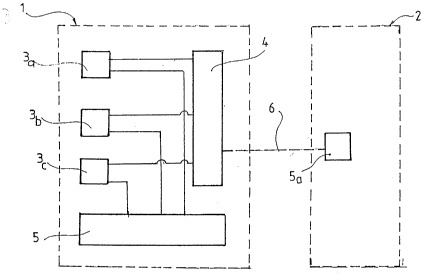
` in Figure 1, the schematic building-up of a preferred
example of effectuating the equipment use-
ful for the process can be seen;
Figure 2 is the block scheme of the examination;
Figure 3 is the conceptual drawing of measurement
and data processing of the physiological
functions;
in Figure 4, the result of a frequency analysis -(EEG
- 213331~
g
examination) carried out on the right and
left side of head of a patient can be seen;
in Figure 5, the result of pulse wave measurement carri-
ed out on the right side and left side of
head of the same patient is illustrated
whom Figure 4 relates to.
Computer 2 (which may be e.g. an IBM-AT computer) of -
the equipment visible in Figure 1 is connected through the
data transfer channel 6 with the patient-inserting unit -
signed by reference number 1 as a whole. This latter one
possesses the signal receiver-transformer channels 3a-3c,
data-collecting unit 4, supply unit 5 and inserting card 5a.
The signal receiver-transformer channels 3a-3c serve for
receiving, amplifying and transmission of the physiological
signals of the patient. The data-collecting unit 4 transmits -
the measurement results. Computer 2 stores and evaluates the
measurement results and informations obtained by interrogat-
ing. The supply unit 5 connected with the signal receiver-
transformer channels 3a-3c is preferably a type
corresponding to the prescriptions of IEC-601. The data trans- ;
fer channel 6 is a highspeed, galvanic separated two-waY
data transfer channel.
In Figure 2, the individual blocks represent the most
important phases of examination, i.e. of the process
according to the invention. Block 7 contains the identifying
questions and responses directed to the subject of the patient
(name, data of birth, sex, body weight, height, time
. !~
- 213331~ :
- 1 0 - " '
of examination and the like). The questions and responses
relating to the common diseases and risk factors (smoking,
systematic consumption of alcohol, elevated blood pressure,
heart disease, diabetes) are incorporated to block 8.
The result of the first verbal stress examination
(investigation of the psychophysiological status) supplement- :
ed with blood pressure and heart rate measurements arrive at
block 9. In the case of an examination by questionary, the
respective part of questionary is assembled as follows.
''.~
Table 1 . :~.
PSYCHOPHYSIOLOGICAL STATUS ~ ;.
(at the start of examination - "just now")
___ ____ _ _ ___________ __________________ __ ________ ______ ..
Blood pressure Hgmm Heart rate /min : ~
__________ ------------ .~
I feel myself to be care-free (1,2,3,4) ~ ;~
I am nervous (1,2,3,4)
I am free of any tension (1,2,3,4)
I am satisfied (1,2,3,4)
I am anxious (1,2,3,4)
_________________________________________ _____--__----_----_---------------- ..
1 = not at all; -~
2 = in some measure;
¦ 3 = fairly; .::
¦ 4 = very/fully -
- 213331~
:`:
Neurological ~uestions and responses (related to cerebro- ~
vascular spasms; TIA /Transient Ischemic attack/) belong to :
block 10, which can be included ~o a questionary e.g. in the
manner according to Table 2
Table 2
Neurology I. :
Sign it by X when any of the complaints listed has earlier
occurred
_______________ ~:,
Temporary weakness ~
on one extremity ...... X on extremities of one side .. X ; .
on all the four on more extremities or in : :~
extremities ........... X other form .................. X
Temporary numbness, sensory decrease or defect
on one extremity ...X on extremities of one side ...X
on all the four on more extremities or in
extremities ...X other form ...X
on the face or trunk...X
Temporary disturbance in the speaking (difficulty in the
phonetics and apperception of words)
Temporary disturbance in the vision
temporary disturbance or defect of vision of one eye ... X
disturbances in the vision of both eyes ...X Diplopia ... X
-~- 2~33314
- 12 -
Neurology II.
Temporary dysphagia
Dizziness
rotary vertigo .... X
feeling of uncertainty
(decisively in one direction, e.g. to the left) ..... X
feeling of uncertainty (without directedness) ....... X
Temporary apraxia of one extremity or extremities ..... X
Loss of memory or temporary defect of memory .... X
Temporary disturbance of reading, writing, `~
counting or spatial orientation .... X ~
Did you have any temporary disturbance being of ;
neurological origin in your opinion? .... X
___________ __ __________------------------------------------ -- ------ :~.~:
`.';'~
Block 11 is the block of physiological examinations.
Within the framework of physiological examinations: the
brain electric activity (EEG, i.e. electroencephalogram) is
measured through the signal receiver-transformer channels
3a-3c visible in Figure 1; electric heart examination (ECG,
i.e. electrocardiogram) is carried out; and the pulses both
~on the head (REG, i.e. rheoncephalogram) as well as on the
extremities (rheographic or impedance pulse) are recorded
The measurement data recorded are summarized in a table (see
later).
Block 12 corresponds to a second stress examination
(investigation of the psychophysiological status), which is
- 13 - ~ ~333~ ~
carried out precisely according to the first such examina-
tion (Table 1). ~:
Block 13 relates to the evaluation summarizing the
result of verbal and instrumental examination as described ~ :
above and supplementing those with the results of other -- -
examination(s) (absent from the questionary) arising from `.
block 16; such other examinations are chiefly directed to
the determination of blood cholesterol and blood sugar level
as well as they comprise an ophthalmologic (fundus) examina~
tion.
Block 14 represents the findings, i.e. the guiding
diagnosis, which can be assembled as a part of a questionary
according to the following Table 3.
Table 3
Opinion
(sign the opinion selected by X)
1. Neither cerebral circulation disturbance
nor data indicating it are present ..... X i~
2. The uncertainty of complaints and/or
findings further on require control
(medical observation) ..... X
-- 2133314
- 14 -
:::
3. Check-up is suggested because of suspicion -
of cerebral circulation disturbance ..... X
4. Check-up is suggested because of suspicion
of peripheral circulation disturbance ..... X
5. Internal or neurological check-up is
suggested ..... X
___ ___ _____ .~ ~
Based on the findings, i.e. diagnosis, the physician
may propose the patient to carry out additional examination.
Block 15 represents the carrying out of blood circulation
examination (Doppler control) whereas block 17 corresponds
to other examinations possibly required on the basis of
findings (block 14).
In Figure 3, the block 11 indicated in Figure 2
relating to the instrumental physiological examination is
illustrated in detail. Figure 3 actually demonstrates a
process organization. Block 18 is the block of EEG
(electroencephalogram examination and instrument): the arrow
31 corresponds to the informations obtained from the right
side of head whereas arrow 32 indicates the informations
measured on the left side of head. Block 19 represents the
REG (rheoencephalogram) examination the meanings of arrows
-1 and 32 are the same as given for the block 18.
Block 20 relates to the instrumental examination
-- 213331~ ~
- 15 -
(measurement) of the pulse (heart rate); arrows _1 and _2 ~`
show the pulse signals of the right or left hand,
respectively, whereas arrows l1 and 12 demonstrate the pulse
signals taken from the right or left foot, respectively. Block
21 is the block of ECG lead II; and the arrow m shows the
data transfer channel. The signal receive-transformer channels
3a-3c of the equipment according to Figure 1 are provided for
carrying out the operations corresponding to blocks 18-21.
Block 23 corresponds to a computer related activity, to
a rapid Fourier analysis known in se; whereas block 22
represents a computer-aided averaging separately occurring in
each case. An averagins operation takes place during about
seconds. Block 22a is needed to the procedures demon-
strated by blocks 22 and 23 for computer-related reasons: it
demonstrates the formation of the trigger pulse arising from
the ECG signal (the reference signal is the "R" wave of
ECG).
Block 24 is the block of peak-search; block 25 is that
of E~G-related spectrum peak-search; these are maximum-mini-
mum searching calculating operations.
Finally, block 26 corresponds to a part of questionary,
wherein the calculated values of instrumental examination
(blocks 24 and 25) are inscribed to the questionary (see
Table 4 later) The values arriving at the block 26 are
usually computer-calculated asymmetry values, the values
obtained from the right-side body parts are considered as
basal values and values calculated from the left side are
':
` 21333~4 :
- 16 -
.
formed in relation to these.
In Figure 4, the graphically illustrated final result
of procedures described in relation to blocks 18, 23 and 25
according to Figure 3, can be seen, i.e. the Figure is the -
representation of the examination of E~G spectrum of both -
brain hemispheres in a specific case. The results of
instrumental measurements obtained from the left hemisphere
are visible below, those obtained from the right hemisphere -~
can be seen above, which are the average of about 40 measure- ~-
ment values. Figure 4 proves that, in the given specific --
case, no difference exists between the electric activity
(background) of the right and left brain hemispheres of the
patient. It should be noted that, according to the worldwide
accepted standpoint, a difference of about 30 % in the EEG
amplitudes is not pathologic but allowed in the practice. -
In the case according to Figure 4, the percentage ~ ;~
difference between the two brain hemispheres is negligible
since this is not a "native" curve but it can be considered
to be magnified several hundred times since it was prepared
by the computer.
In Figure 5, the graphically illustrated final results
of procedures described in relation to blocks 19, 22 and 24
according to Figure 3 can be seen: the pulse waves (REG
examination) arising from the right and left brain
hemispheres of the same patient are shown whom Figure 4
relates to. Also in this case, the pulse wave starting from
- 17 - 213331~
the left brain hemisphere is illustrated below, that
starting from the right hemisphere is shown above (average
of about 40 measurements). It appears unequivocally from
Figure 5, that there exists an essential difference between
the two pulse waves, namely the distance e (demonstrating
a time interval) signed at the pulse wave of the right brain
hemisphere shows a length indicating a pathologic alternation
(the internationally used limit value is 180 msec, which is
highly surpassed by the value corresponding to the distance.
The invention is hereinafter described in detail by an
Example.
ExamPle
The data of the patient examined (block 7 in Figure 2)
were as follows:
name: Y.X.
data of birth: 18. 05. 1936, Budapest
body weight: 79 kg
height: 174 cm
time of the examination: January 5, 1992
sex of the patient: woman.
:.
After taking up the above data, the following questions
were posed to the person examined and answered negatively
(block 8 in Figure 2):
~ 213331~
- 18 - ~ .
Do you smoke?
Do you systematically consume alcohol?
Do you suffer from high blood pressure?
Do you suffer from heart disease? ~
Do you suffer from diabetes? ~: :
Immediately after answering the questions, the first
stress exa~ination was carried out /block 9 in Figure 2 :
(Table 1)/, within the framework of which -
a) the blood pressure and pulse frequency (heart rate)
of the patient were measured. The result of measure- -~
ment was as follows:
120/85; 74/minute
b) questions were posed for determination of the
temporary anxiety (brief variant of the Spielberger
test) and the responses were recorded. -~
In the next step, our questions relating to the
neurological symptoms indicating cerebral vascular spasm
(TIA) were posed (block 10 in Figure 2, i.e. the neurologi- .
cal block). These were targeted essentially to the following
syndromes:
- temporary weakness of extremities, numbness, sensory
decrease;
- temporary numbness, sensory disturbance on the face :
or trunk;
- transitory disturbance of speaking, vision, swallowing
- dizziness, temporary apraxia of extremities and loss
of consciousness;
3 ~i~
- 19 -
- loss of memory, transitory disturbance of reading,
writing, counting or spatial orientation;
- other disturbances considered to be of nervous
system origin.
On the questionary cited above the questions can be
summarized according to Table 2. The patient responded to
the questions relating to the above syndromes in several
cases in such a manner that he has disturbances on his left
side (temporary weakness on the left foot, temporary
numbness on the left foot, left face and left part of the
trunk); feeling of uncertainty at the left, apraxia of the
left extremities). In addition, he indicated disturbance of
speaking and disturbances in the vision of both eyes.
The above questions and responses are fixed (recorded)
by filling out the questionary several times mentioned
previously. After filling out the questionary and after
inscribing the informations obtained into a database
treating program, they are charged to a computer (Figure 1)
including also the responses given to the first stress
examination.
By answering and entering the above questions into a
computer, the major part of verbal examination has been
closed. Thereafter, the electrodes required to EEG and REG
examinations were fastened to the patient seated, then she
lay backwards onto the examinating bed. Then, the electrodes
required for examining ECG and extremity pulses were placed
onto her body (above the wrist and ankle both on the right
:,
~ ~33~
- 20 -
'~ ' -'
and left sides). The electrodes join to signal receiver-
transformer channels forming a part of the patient-inserting ~;
unit connected with the computer mentioned above (Figure 1)
namely, they are the inputs thereof. By starting the
computer program, the measurement and data processing of the
analogue physiological signals, namely EEG, REG and ECG
signals are initiated (Figure 3).
The following data were obtained as a result of this
operation (process).
Table 4
Measurement data of recordings
Left side Riqht side
10.2/14.6 dominant frequency of
EEG [Hz]amplitude10.2/9.77 `
on the head, amplitude/time [msec]
-20.3/131 1st minimum -11.7/140 `~
30.5/368 maximum 17.7/362
On the neck, amplitude/time [msec]
-11.7/149 1st min. 3.91/419
61.7/476 max. 66.4/707
On the arm, amplitude/time [msec]
-16.4/179 1st min. -16.4/176
9.37/254 max. 14.8/254
2nd min. -9.37/1656
On the foot, amplitude/time [msec]
-7.03/-70 1st min. -1.56/-70
26.6/53 max. 23.4/53
" '
- 21 - 2~3~4
The graphical illustration of data of the first line
(EEG frequency spectrum) was also carried out (Figure ~).
Similarly, the REG displayed in the 2nd line (pulse wave on
the head) was graphically demonstrated. However, the
graphical illustration is not indispensably required; at
most in the case when it can be perceived in this phase of
examination that further examination of circulation will be
performed.
Immediately after disassembling the electrodes, the
second stress examination (block 12 in Figure 2, Table 1)
was carried out, within the framework of which the blood
pressure and pulse frequency (heart rate) were again
measured and gave the following results:
130/80 and 60/minute
and responses were asked to the same questions as in the
first stress examination; these responses as a continuation
of the former (previous) database were also entered into the
computer.
From the results of examination recorded by the
computer, the findings were formed namely, the following
decision was made.
a) Based on the questionary, the following status can
be concluded, which can be judged to be pathologic:
based on the symptoms of the left side, the -
circulation disturbance of the right brain hemisphere can be
supposed. No other sign indicating any pathological altera-
tion was found. No response given to other questions (remain-
- 2133~1~
- 22 -
ing as empty in the questionary) (other symptoms, block 16,
Figure 2) indicated that any disorder of circulation could be
supposed in addition to that defined above.
b) Subsequently, the results of physiological data
processing were evaluated, according to which:
- no evaluable or pathological, respectively
difference between the right and left sides (Figure 4) `~
exists in EEG and pulse waves of the exlremities (blocks 18
and 20 as well as 24 in Figure 3);
- in the REG, also graphically represented in
Figure 5, a difference appears between the right and left brain
hemispheres namely, the right-side pulse wave represents
a pathological alteration indicating arteriosclerosis.
Based on these examinations of two kinds, our decision,
i.e. the part "Opinion" of the findings (block 13 in Figure ~
2) was as follows. `
Check-up is suggested because of suspicion of dis-
turbance in the brain circulation (blocks 14 and 15 in
Figure 2).
In relation to the Example it should be noted that the
district physician would not have posed the groups of
questions posed by us (namely, those are substantially neuro-
logical-professional asks), when the patient had turned to
him and, on the other hand, he would have considered the
complaints to be of psychic origin. However, if this
physician had made to carry out the Doppler control on the ~ ~
patient, he would have obtained a negative result. It is ~ ;
- 2~3331~
- 23 -
noted that 28 persons belonging to our own patients were
controlled by using the Doppler test and the résult was in
each case negative. Subsequently, the same group of patients
were examined by using the process according to the
invention and on about 80 % (22 persons) of them,
quantifiable differences were found in the pulse waves of
head, hands and feet. Based on this, further examination of
the affected persons was proposed, but at least a medical
observation was suggested.
The advantageous effects connected with the invention
may be summarized as follows.
The greatest advantage of the invention is that it
provides to recognize in the possibly earliest phase the
arteriosclerosis and preceding circulation disturbances
(decrease in the wind box function of the vessel wall),
which cannot yet be considered to be pathologic. By using
the process according to the invention, the difference from ~-
the normal status can already be detected when an actual
sclerosis of the vessel wall has not yet developed but its
elasticity has been diminished; thus, the invention is an
ideal tool for the prevention. (No process is known at :
present, which could be useful to early recognize the -
alterations (sclerosis of the vascular system; e.g. by using
the Doppler method considered to be most up-to-date, the
consequence of a sclerotic vessel wall inducing flow dis-
turbance can only be measured). The process has (involves)
no invasive element, it is patient-saving, pain-free and ~-~
~ ~.
---` 213331~
- 24 -
can be carried out during 30 minutes. It is considerablY
economic.
The process is equally useful both for screening
examinations and performing tasks of patient-care. At the
end of examination the participant receives the examination
protocol containing the results and, when required, he can ~ ~
utilize it for further examinations. -
The invention is of course not limited to the solvings
described above but it can be accomplished in a number of
ways within the scope of protection defined in the claims.