Note: Descriptions are shown in the official language in which they were submitted.
-1- '2133a5s
BACKGROUND OF THE INVENTION
I. Field of the Invention
This invention is directed generally to the permanent
augmentation of soft tissue and, more particularly, to the
treatment of urological disorders, e. g., incontinence, vesi-
coureteral reflux, gastric fluid reflux, etc., by endoscopic
injection of compatible micro particle implants into the
submucosal tissue. Since the invention is closely related to
the treatment of incontinence, it will be described in detail
by reference thereto.
With the exception of urinary incontinence secondary
to neurogenic disorders, incontinence occurs when the resistance
to urine flow has decreased excessively, i. e., urethral
resistance to urine outflow, from whatever cause, has been
lowered to the point when it can no longer resist increased
intra-abdominal pressure. While this may seem to be an over-
simplification of the problem, in general nearly all procedures
developed to restore continence are designed on this basis to
restore the lost resistance to urine outflow. Similarly, the
present invention allows for the control of gastric fluid reflux
when submucosal injections of the micro implants are made to the
esophageal-gastric junction and to the gastric-pyloric junction.
To these ends, several surgical procedures and devices
have heretofore been developed and tried with varying degrees of
success, e. g., suspension procedures, plications, constrictive
procedures and various combinations of these. Devices which
have been developed primarily operate as plugs and cannot be
used on a permanent basis. Electrical stimulation and biofeed-
back
77466-1
WO 93/19702 PCT/US93/029f'~
,~ 13 3 7'~ 6
-2-
techniques have so far been demonstrated to have limited
success in treatment of incontinence and gastric reflux.
II. Discussion of the Related Art
As examples of such treatments and procedures
heretofore known in the art, mention may be made of a .
variety of prosthetic devices based on the compression of
the urethra at a given point. ~"''(See, for example,
"Treatment of urinary incontinence by implantable
prosthetic sphincter," by Bradley and Timm, Uroloctv, 1:252
(1973); "Treatment of post-prostatectomy urinary
incontinence using a gel prostheses", by Kaufman, Brit. J.
U o ., 45:646 (1973) and "Treatment of post-prostatectomy
urinary incontinence using a silicon gel prostheses", Brit.
J. Urol., 48:646 (1973).
In the practice of plastic and reconstructive surgery,
inert materials have frequently been implanted to fill in
defects or augment weakened tissue. These have been
fabricated of a variety of materials and have been
implanted using several techniques.
Certain very small particle species compounded in a
lubricious material have been implanted by subcutaneous
injection for both soft and hard tissue augmentation.
Heretofore success has been limited. Undesirable
subsequent particle migration and serious granulomatous
reactions have commonly resulted. This is well documented
with such materials as polytetrafluoroethylene (PTFE)
particles of very small diameter (>90% of a diameter <30
microns) in glycerine. One such product includes PTFE
particles, suspended in glycerine with a minor amount of
polysorbate is available under the name Polytef~ (trademark
of Mentor Corp. of California). This is discussed, for
example, in Malizia, et al., JAMA, Volume 251, No. 24, pp.
3277-3281 (1984).
U.S. Patent No. 4 773 393 issued September 27, 1988
to Haber and Malizia and assigned to C.R. Bard, Inc.
relates to an apparatus for hypodermically implanting a
genitourinary prosthesis comprising an extensible,
CVO 93/19702 213 3 7 5 6 P~/US93/02986
-3-
inflatable tissue expanding containment membrane to be
located in the proximal periurethral tissues to add bulk to
these tissues and thereby overcome urinary incontinence by
means of localized, controlled tissue volume increase. In
~ 5 column 1, reference is made to the aforementioned
article co-authored by the co-patentee Anthony A. Malizia
with respect to the widespread migration of polytef
particles along with granulomas. Accordingly, the patented
invention is said to obviate these problems by providing a
prosthesis comprising an elastomerical biocompatible
containment membrane into which a biomeric fluid or
suspended particulate matter such as TEFLON particles is
percutaneously injected to inflate the membrane.
The use of very small diameter particulate spheres
(approximately 1-20 microns) or small diameter elongated
fibrils, (generally 1-20 microns in diameter) of various
materials such as cross-linked collagen or synthetic
polymers suspended in an aqueous medium to which a
biocompatible fluid lubricant has been added as injectable
implant composition is disclosed in Wallace et al., U.S.
Patent 4 803 075. While these materials create immediate
augmentation, this result is generally short-lived as the
material also has a tendency to migrate and/or be
reabsorbed from the injection site by the host tissue.
Most recently, three companies have indicated in
published reports their intent to enter the market for
treatment of urinary incontinence with an injectable
material. Mentor Corporation has received limited approval
from the FDA for use of their injectable material,
"Urethrin", in treating incontinent male post-prostatectomy
patients. Previous published reports stated that C.R.
Bard, Inc. and Collagen Corporation were developing an
incontinence treatment called "Contigen Bard Collagen
' . Implant," understood to be Collagen Corporation's
"contigen" injectable bovine collagen material.
Subsequently, it was reported that C.R. Bard is also
evaluating for urinary incontinence treatment a product
--- ~ 2133756
-4-
called "Hylagel-Muscle" which is said to be based upon
Biomatrix's patented technology on modifying naturally occurring
hyaluronan "to form three-dimensional sponge-like matrixes in
the form of high molecular mass fluids, gels and solids that can
separate tissue, cells and molecules".
From the foregoing survey of the current state of the
art, it will thus be seen that of recent date many approaches
and treatments have been proposed to cure or relieve conditions
of urinary incontinence by injection. While some of these
approaches have enjoyed modest success, relief has been, for the
most part, only temporary in those patients where success is
noted. This generally is due to granuloma reactions and/or
migration of injected particulate material and reabsorption of
gellular materials. Thus, there remains a very important need
for a treatment that will provide a lasting remedy for success-
fully treating such urological disorders.
SUMMARY OF THE INVENTION
The invention provides a composition for injecting
submucosally or peri-urethrally into tissue at at least one
injection site to provide long-term treatment of urological and
gastric disorders, comprising: an effective amount of
relatively soft, malleable, elastic, biologically compatible
prosthetic micro particles dispersed in a non-retentive
compatible physiological vehicle, the micro particles of the
composition further being of a designed average particle size
distribution and characterized by a rough surface having a
plurality of surface irregularities generally randomly formed
therein, such that the effects of average particle size and
77466-1
i 21 33758
-4a-
average particle surface roughness cooperate in combination in
an autogenous manner to essentially prevent loss of the micro
particles from any injection site, the particles remaining to
be incorporated as long-term tissue augmentation.
The invention also provides a composition for inject-
ing submucosally or peri-urethrally into tissue at at least one
injection site to treat urological and gastric disorders,
comprising: (a) an effective amount of relatively soft,
resilient, malleable, biologically compatible micro particles
consisting essentially of a polysiloxane material, the micro
particles having an average unidimensional particle size above
60 microns and having a highly irregular particle surface
configuration including indentations, cavities and pores
generally randomly formed therein such that the effects average
particle size and irregular particle surface cooperate in an
autogenous manner to essentially prevent loss of the micro
particles from an injection site; and (b) a compatible physio-
logical vehicle that will promote injection of the micro
particles but once injected is non-retentive of the particles.
The present invention is useful for treating urologic-
al disorders such as stress incontinence and gastric fluid
reflux disorders. The micro-implant particles are characterized
as being biocompatible, and immunologically non-reactive, and
will take advantage of the body's own mechanism to encapsulate
the micro-implanted particles to prevent migration from the
injection site.
The textured micro particles preferably have a nominal
unidimensional measurement of between about 30 and 3,000 microns
77466-1
2~33~5s
-4b-
(.003 to 3.0 mm), and a preferred range for most applications
is between about 80 and 600 microns (0.008 to 0.6 mm). The
textured micro particles present generally
77466-1
PCT/US93/02986
,~--~WO 93/ 19702 2 I 3 3 7 5 S
-5-
amorphous surfaces, and normally possess surface
irregularities including indentations ranging in size from,
for example, l0A (angstroms) to 500 microns, with the
indentations themselves having irregular configurations and
surfaces. A minimal inter-indentation distance is
maintained that enables the particles to be injected
through an hypodermic needle of the appropriate preselected
size, and with or without a physiologic vehicle.
Examples of appropriate physiologic vehicles are
saline, solutions of sodium hyaluronate, various starches,
hydrogels, polyvinylpyrrolidones, other polymeric
materials, polysaccharides, organic oils or fluids, all of
which are well known and utilized in the art. Vehicles
that are biologically compatible, i.e., cause minimal
tissue reaction and are removed or metabolized without
cytotoxicity, are, of course, preferred. Biologically
compatible saccharides such as glucose have been found
useful, aqueous solutions of starch or sodium hyaluronate
may be employed and certain fats may also be found useful.
In certain instances, it may be desirable to employ a
totally inert vehicle. The patient's own plasma may be
derived from blood withdrawn from the patient, centrifuged
to remove cells (or not) and mixed with appropriate
aliquots of particles and the mixture injected in the
desired locations.
In this connection, highly compatible vehicles include
esters of hyaluronic acids such as ethyl hyaluronate and
polyvinylpyrrolidone (PVP). PVP normally has the general
empirical formula [ (CHCH2)ZN(CHz)3C0]~ wherein n equals 25-
500, a form of which is otherwise known and marketed as
Plasdone"' (trademark of GAF Corporation, New York, New
' York). Additionally, polyvinylpyrrolidone (Plasdones),
hyaluronate, collagen and other biocompatible substances
' may be incorporated into the elastomer or combined with its
surface.
In certain instances, it has been found desirable to
utilize a surface modifier in combination with the micro
WO 93/19702 PCT/US93/029~'"'
2133756
-6-
particles, with materials such as polyvinylpyrrolidone,
polytetrafluoroethylene, collagen, or hyaluronates having
been found suitable. In this connection, the surface
modifiers may be mixed into the substance of or with the
micro particles, and furthermore may.thereafter be coated
with a layer of a hyaluronate ;~or hyaluronic acid.
Specifically, certain mb.tl'ifiers such as
polytetrafluoroethylene may be ~2~mixed with, for example,
a poly di-substituted siloxane particle material prior to
cure to impart an average surface modification to the cured
particle. A material such as hyaluronic acid may be
attached to the micro particle surface either through
physical or chemical bonding. Surface modifiers also can
be used to typically assist in detoxification and promote
the desired tissue ingrowth encapsulation. Other bioactive
substances that can be included in the carrier or attached
to the surface of the beads to promote encapsulation
include f ibronectin, n, transforming growth factor beta,
and various other cytokines such as interleukin-1.
BRIEF DESCRIPTION OF THE DRAWINGS
Figure 1 is a perspective view of a micro particle
useful in accordance with the present invention, and
illustrating surface irregularities typically present in
the particle;
Figure 2 is a vertical sectional view taken along the
line and in the direction of the arrows 2-2 of Figure 1;
Figure 3 is a schematic illustration of a fragmentary
portion of human skin organ, and illustrating a hypodermic
needle of appropriate size being utilized to introduce
materials in accordance with the present invention into the
subcutaneous zone beneath a depressed scar;
Figure 4 is a view similar to Figure 3, and
illustrating the same location following subcutaneous
injection of the textured micro particles in accordance
with the present invention;
~"'"WO 93/19702 ' ~ ~ PGT/US93/02986
FIGURE 5 is a perspective view of a modified form of
a useful wherein the surface irregularities project
outwardly from a body member in pillar form;
FIGURE 6 is a cross-sectional view of the device of
. 5 Figure 5;
FIGURE 7 is a fragmentary schematic view which
illustrates the submucosal injection of the microparticles
in the vicinity of a bladder neck; and
FIGURE 8 is an actual photomicrograph of particles
useful in accordance with the invention.
DETAINED DESCRIPTION
As heretofore mentioned, the present invention is
directed to the treatment of urological and gastric fluid
reflux disorders, particularly stress incontinence, by
endoscopic injection of specified micro particles. The
above-referenced copending application relates to an
improved micro-implantation method and composition for
filling depressed scars, unsymmetrical orbital floors,
muscle, lip, and other tissue defects in reconstructive
surgery procedures. The tissues to be augmented exhibit
varying degrees of softness.
As disclosed, textured micro particles having an
outside diameter between about 30 microns and 3000 microns
are employed with an appropriate physiologic vehicle, as
will be detailed hereinafter. A more preferred range is
above about 80 microns and depending on the precise
application between about 80 to 100 and 600 microns.
Equivalent smooth particles should be somewhat larger.
In accordance with the invention, the particles are
preferably injected through a hypodermic needle of an
appropriate preselected size, preferably with an
appropriate lubricious physiologic vehicle which is
biocompatible, i.e. causes minimal tissue reaction and is
removed or metabolized without cytotoxicity. As indicated
above, and by way of illustration, possible suitable useful
disclosed physiologic vehicles include, saline, various
starches, hydrogels, polyvinylpyrrolidones (Plasdones),
WO 93/19702 PCT/US93/029f""'~
2133756
polysaccharides, fats, organic oils or fluids and other
polymeric materials, all of which are well known and
utilized in the art. In this connection, highly compatible v
vehicles also include esters of hyaluronic acids such as
ethyl hyaluronate and polyvinylpy~z~olidone (PVP). PVP
normally has the general-:..' empirical formula
[ (CHCHZ) ZN (CHZ) 3C0] ~ wherein n is in the range of about 25-
500, a form of which is otherwise known and marketed as
Plasdone'"', or the patient s own plasma.
Additionally, polyvinylpyrrolidone (Plasdones),
hyaluronate, collagen and other biocompatible substances
may be incorporated into the elastomer or combined with its
surface. As used herein, a "surface modifier" connotes a
material combined into the formed particle, applied to the
surface of the particle or added to the carrier vehicle to
alter inter-particle or prosthesis-host interaction and/or
particle identifiability. These surface modifiers may
alter the coefficient of friction of the particles, as by
making them more lubricious, render the particles more
radiopaque, assist in detoxification, and/or render the
surface of the particles more susceptible to tissue
ingrowth or facilitate tissue encapsulation of individual
particles. Useful surface modifiers include PVP, collagen,
hyaluronates, polytetrafluoroethylene, and others.
The surface modifiers such as polyvinylpyrrolidone or
polytetrafluoroethylene may be mixed into the substance of
or with the micro particles, which furthermore may
thereafter be coated with a layer of a hyaluronate or
hyaluronic acid. Specifically, certain modifiers such as
polytetrafluoroethylene may be admixed with, for example,
a poly di-substituted siloxane particle material prior to
cure to impart an average surface modification to the cured
particle. A material such as hyaluronic acid may be
attached to the micro particle surface either thorough
physical or chemical bonding. Surface modifiers also
typically are selected to assist in detoxification and
promote the desired tissue encapsulation. As mentioned
21 3375fi
-9-
above, other bioactive substances that can be included in the
carrier or attached to the surface of the micro implants to
promote encapsulation include fibronectin, n, transforming
growth factor beta, and various other cytokines such as
interleukin-1.
Once implanted, the body will form a thin scar tissue
around each of the implants so as to provide initial encapsula-
tion. Polyvinylpyrrolidone, hyaluronate or collagen or other
biocompatible substances may be chemically or physically combined
with the particle substance or its surface to enhance the
acceptance of the implant by the host. While in most situations
the particles are of random size and configuration, but within
the constraints of size indicated, it is generally desirable
that the particles be of generally uniform configuration for use
in a given procedure. With respect to relative resilience of
the augmentation mass, it is preferably designed to closely
simulate the tissue of the implant or injection site.
For soft tissue, a soft elastomer such as silicone
rubber is a desirable material for the textured particles. This
is preferably a poly(dimethylsiloxane) but may have substitute
alkyl or aromatic groups. When a firm area is being treated,
such as connective tissue or the like, polytetrafluoroethylene
sold under the Trade-mark "Teflon" or polyethylene may be
satisfactorily utilized. In those instances wherein the
requirement is for hard substances, biocompatible materials such
as certain calcium salts including hydroxyapatite or other such
crystalline materials, biocompatible ceramics, biocompatible
metals such as certain stainless steel particles or glass may
be utilized.
77466-1
r1
21 3375fi
-9a-
By way of further background, the average diameter of
a capillary is approximately 16 microns, or roughly two times
the diameter of a red cell. Therefore, since the size of the
textured micro particles is in the area of at least approximate-
ly 30 microns, they will not be absorbed into the capillaries,
but will on the other hand, remain
77466-1
WO 93/19702 PCT/US93/029~
2133756
-10-
generally captive and fixed in place. Smaller particles,
including some in the sub-micron range, have been
implicated in causing chronic inflamruation and may be
ingested by host cells. Thus, particles in the range of
between about 30 and 3000 microns are, employed.
The fibroblast cell is the scar-forming cell of the
human body, and these cells range 'in size from between
about 20 microns up to about 100 microns, and because of
contact guidance and reduced micromotion, they will form an
efficient scar tissue or collagen-based coating around an
inert foreign body. Furthermore, such scar tissue will
conform to the irregularities in the surface of the foreign
body, particularly if they are of sufficient size to
accommodate tissue ingrowth. Our previous studies
(American Society of Artificial Internal Organs; U. S.
Patent Nos. 3,638,649; 3,657,744; 4,239,492; and 4,240,794)
have shown that foreign substances can be substantially
firmly anchored in a predetermined location in the body.
Because of the inherent ability of fibroblasts to form scar
tissue in and around irregularities of the surface, such
anchoring occurs in many locations, including locations
within the blood stream.
Figure 1 illustrates a micro-implant particle
generally designated 10 which has an inner-core having
various randomly distributed indentations or pores 11-11
throughout its surface. These openings or pores are spaced
apart by connective pillar members 12. As indicated above,
the indentations, interstices or pores preferably have a
minimum indentation depth or open dimension of about 10
Angstroms, along with a maximum dimension of about 500
microns. The interconnective or pillar zones 12-12 which
separate or otherwise define solid material between
openings or indentations 11-il have a dimension or breadth
sufficient so that the majority or greater portion of the
surface is defined by indentations, openings or pores.
Actual particles are shown in the photomicrograph of
Figure 8. As can be seen from the scale of the Figure, the
~O 93/19702
213 3 7 5 6 P~'/US93/02986
-11-
size range of the illustrated particles ranges from about
100 to 600 microns. The irregular particle shapes and
surface configurations including indentations, openings and
pores is dramatically illustrated.
With continued attention being directed to Figures 1
and 2 of the drawings, connective elements 12 are available
on the surface of the micro-implant particles and provide
for mechanical stability of the individual particle. This
arrangement is illustrated in particular in Figure 2 and is
apparent from the photomicrograph of Figure 8.
As further disclosed in the cross-referenced
application, it has been found that inert foreign tissue
augmentation particulate matter having a mean diameter less
than about 30 microns will generally become subject to
significant migratory loss from the site of injection,
regardless of surface configuration absent extraordinary
protection. The textured or irregular nature of the
surface of the microspheres of the invention, however,
imparts to them an apparent size equivalency which, in the
case of at least the relatively smaller sized particles
(particularly in the range of 30-60 and up to 80 microns),
makes them behave, once injected, as much larger smoother
particles might behave with respect to host implant or
prosthesis migration tendencies and benign assimilation in
scar tissue. Particulate matter of the class of 'the
present invention which is of a size ranging from about 30
microns to about 3000 microns and having a textured surface
in which the surface irregularities vary in size over a
range of about 10 Angstroms to 500 microns.
The irregularities, pores and interstices are designed
to have widths ranging from those having a diameter or
opening size which will just accommodate the infiltration
of a typical connective tissue fibril or protein molecule
at the lower end to those large enough to accommodate
ingrowth of much larger cross-linked protein, possibly
collagen protein, fibrillar structures or actual
fibroblasts at the high end. In this regard, it is well
II
WO 93/19702 PCT/US93/0298'~
~ 133 5 6 _12_
known that the collagen fiber is composed of fibrils and
filaments. The basic poly-peptide chain is arranged into
micro-filaments of tropocollagen having a diameter of
approximately 20 Angstroms. It has been found that surface
irregularities as small as 10 An~~~~oms will interdigitate
with the filaments on the surface of the fibers and serve
to resist host-prosthesis interface motion.
Further, with respect to particle size, it will be
appreciated that particle size, particularly of those
l0 species contained in preparations utilized in prior
injectable compositions, tends to vary over a range within
any group of particles so that there will be a percentage
of the group larger and a percentage of the group smaller
than at target size of the indentations, pores or
interstices associated with a give group of particles will
also describe a range. It will further be appreciated that
one must take into account the normal variation in patient-
to-patient acceptance and reaction to tissue augmentation
injection of micro particles. With this in mind, certain
observations have been made regarding optimum particle
size, particularly with regard to the severe problems of
unwanted migration and formation of granulomatous
reactions.
Observations in a variety of clinical situations
indicate that particles less than about 60 microns in
diameter can be engulfed by macrophages and transported to
regional lymph nodes. Submicron-sized particles may be the
most easily transported and may remain intracellular
indefinitely. However, larger particles, particles that
approach the size of a macrophage, i.e., from about 20 to
about 60 microns, may cause the death of a cell when
engulfed. This begins a progression in which the dead cell
releases its intercellular enzymes (cytokines), and those
attract other phagocytes which, again, encounter and engulf
the particle with the debris of the first encounter. In
this manner, a vicious cycle continues on a larger scale as
"'~"'"WO 93/19702 213 3 7 5 6 P~/US93/02986
-13-
a chronic inflammatory response. Of course, such a
response is highly undesirable.
Particles greater than about 60 microns, however, have
not been observed within a cell or within lymph nodes; and,
certainly, particles. greater than 80 microns appear safe
from initiating such foreign body reactions. Further, as
in the example below, particles of an average diameter of
100 to 600 microns with textured surfaces having an average
indentation cavity or pore size from about 10 microns to
about 200 microns have been observed to work quite well.
Theoretically, there is no upper limit to the size of the
textured particles, and this is borne out by the success of
sintered-surface hip implants, textured breast implants and
others. However, the useful upper limit of micro implant
dimensions is probably somewhere in the vicinity of 1 to 3
mm in defects just beneath the skin surface because
particles of a size greater than this may be perceived as
surface irregularities when palpitated. Large textured
implants have also been employed in breast reconstruction,
for example.
It will be appreciated that textured spheroids of the
class contemplated for use in the present invention may be
molded, for example, by any gravity-free technique wherein
the spheroids are formed with centrifugal force equal to
that of gravity in cases where the spheroids are formed of
rather malleable synthetic material. Spheroids can be
fabricated from a variety of inert substances such as
polytetrafluoroethylene, poly(methylmethacrylate), poly
substituted siloxanes (silicones) and a variety of other
synthetic polymeric materials, ceramics and others and
different fabrication processes may be applicable to each
material for the augmentation of soft tissue. Of course,
fabrication of the spheroids from a malleable polymer
material such as a silicone rubber is preferred as it will
more closely imitate the texture of the natural tissue it
replaces. With respect to malleable polymers such as
silicone rubber, the following fabrication techniques are
II
WO 93/19702 PCT/US93/029F
2.~33~56
-14-
exemplary of those that will readily enable manufacture by
those skilled in the art. It will be appreciated that a
technique that might be preferred for one material may not
work equally well for all.
In one process, a malleable~~ stock of unvulcanized
polydimethylsiloxane is rolled!:' into spheroids of
approximately 100 microns or other desired size diameter.
-.
The surface is then textured Hy impacting each spheroid
with an appropriate force. The textured spheroids are then
vulcanized and mixed with the appropriate vehicle for
injection.
In another successful method, generally preferred for
forming beads of silicone rubbers, poly(di-substituted
siloxane) may be dispersed in an appropriate volatile
solvent and then partially cured by droplets being forced
through a specific distance of air from an orifice having
a specific diameter. This is a very familiar process
technique generally known with respect to the operation of
a shot tower in making lead shot. The size of the
particles or spheroids is easily regulated by varying the
viscosity of the mixture and/or the orifice of origin. As
the particle travels a known distance through air, it is
partially cured as the volatile vehicle evaporates. The
specifically formed spheroid or bead is then separated by
a suitable fluid medium. The spheroids may then be pressed
against an appropriate surface or impacted by an
appropriate force to impart the desired texture, the
surface having an appropriate mold release. Partially
cured spheroids are then vulcanized by heat irradiation.
The particles are then sized and graded by physical means.
Spheroids are then mixed with the appropriate vehicle in
appropriate ratios, placed in containers and finally
sterilized within the container.
Texture can be imparted to the beads or spheroids in
a number of ways. In addition to the molding method, other
techniques include ion-beam microtexturing which makes it
possible to produce controlled microtextured surfaces,
~WO 93/19702
~ 13 3 7 5 fi PCT/US93/02986
-15-
chemical and plasma etching and impacting the beads with
solid particles. Of course, it is contemplated that other
methods could also occur to those skilled in the art.
If desired, surface modifiers, as explained above, can
be incorporated in the material prior to formation of the
spheroids or beads or may be thereafter be added as a
coating on the deformed surfaces. In this manner, certain
materials such as hyaluronic acid, for example, may be
attached to the micro particle surface either through
physical or chemical bonding in a well-known manner after
formation and texturing.
EXAMPLE I
Amounts of particles with average diameters of 100,
150 and 600 micrometers were fabricated with.a textured
surface from fully polymerized and vulcanized
poly(dimethylsiloxane). The polymer was mixed to form a
biocompatible solution with an organic polymer hydrogel.
The hydrogel was a polyvinylpyrrolidone gel having an
average molecular weight of approximately 13,700 and one of
a family of such material known as Plasdones. These
Plasdones in a molecular weight range of interest are
freely transported through tissue fluids and excreted
unchanged by the kidneys. The mixture utilized was
approximately 38% by weight of the polymer particles and
62% of the gel material. The polymer/gel mixture was mixed
until the inert particles were evenly dispersed and then
placed in syringes with small pistons placed in the
proximal ends. The distal end of each cylinder would have
a Luer taper to which an appropriate needle or cannula
could be attached. A highly leveraged injection ratchet
mechanism was utilized to accept the syringe cartridges and
deliver precise amounts of the gel mixture through a
cannula into the subcutaneous plane of the ear tissue of 20
large, adult white rabbits. Controls using commercially
available collagen derivatives were injected in the
subcutaneous plane in adjacent sites in the rabbits ears
I I
WO 93/19702 213 3 7 5 6 PCT/US93/029F~'
-16-
using small gauge needles provided by the manufacturers of
the collagen derivatives.
With respect to the injected collagen control sites,
subsequent histologic sections indicated that after three
weeks, no residual collagen could be found at the site of
the injection. In dramatic cori~rast, the histologic
sections of the micro particles evidenced a dramatic
transition in which the gelwwphase of the material was
replaced by a fibrin and protocollagen matrix surrounding
each of the micro particles. In three days, the fibrin
matrix was complete, with all the gel having been removed
by the host. Connective-tissue cells had developed and had
begun to replace the matrix with host collagen fibrils. By
the sixth week, this fibrosis was complete, and each
individual textured particle appeared to be encased in its
own individual. inner connected covering of fibrous tissue.
The thickness of the implanted area and the degree of
fibrosis as measured by transillumination, micrometer and
light and electron beam microscopy remained constant for
more than a year.
Subsequent histologic examination of the regional
lymph nodes at the base of the rabbit ears revealed no
migration of particles. Cross-sections of the ear below
the injected area showed no particles. Through
transillumination, the size and density of the areas of
injection were easily and atraumatically monitored for each
rabbit. No textured micro implants were found at the base
of the ears or in the regional lymph nodes of any of the
rabbits under study.
The dimensions of the subcutaneous deposits of
textured micro implants remained approximately the same
throughout the period of study, as was evidenced by
transillumination photographic record and micrometer
measurement. Opacity was noted to decrease over the last
few weeks as the transillumination became brighter but then
appeared to stabilize between the end of the first and the
sixth months.
~~WO 93/19702 ~ ~ ~ ~ PCT/US93/02986
-17-
The results obtained with the experimental particles
of Example 1 illustrate the dramatic contrast between this
material and the injection of collagen-containing
materials. Although the collagen-containing materials
created immediate soft tissue augmentation, these
substances - which are only about 3.5 to 6.5% solid
collagen material - soon became invaded by host capillaries
and were absorbed. No absorption or migration of the 100,
150 or 600 micron silicone rubber particles was observed,
even after 382 days.
In other experiments, particles having an average
diameter of 80 microns and incorporating tracer material in
the form of gamma radiation-emitting material were injected
into the ears of other rabbits. These particles showed no
migration from the injection site during a subsequent six-
month monitoring period.
While prior work by the inventors and others have
shown that surface irregularities preferably are in the 20
to 200 micron range in order to achieve adequate contact
guidance of the fibroblasts so as to create or develop a
scar tissue pattern that is a mirror image of the substrate
surface, it is also appreciated that the particle size in
relation to the relative size of the surface irregularities
is a factor to be considered. In this connection, if the
openings, indentations or pores are too shallow in their
depth dimension, or in the event their diameter is not
sufficiently great, the fibroblasts will tend to bridge
across the defect so as to provide a substantially smooth
surface.
In the preferred embodiment of the present invention,
the particles indicated or selected for a specific
procedure to assist in correcting a given defect are
previously loaded into a hypodermic syringe with a needle
having an adequately sized interior bore so that upon
injection of the needle into the area of the depression
being corrected, the particles together with the
appropriate physiologic vehicle enables the spheroids to be
I I
WO 93/19702 PCT/US93/0298
2133756
-18-
injected directly into the area of the depression.
Appropriate vehicles, as previously indicated, include
physiologic saline or polysaccharide lubricants, each of
these enabling the spheroids to be injected as set forth.
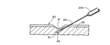
With attention being directed to Figure 3 of the
a,
drawings, it will be noted that surface tissue as shown at
20 includes a depression area;~a~l, with the depression area
extending into the subcutaneous tissue as at 22. For
utilization of the concept of the present invention, the
needle 23 is shown as it is injected into tissue.
Particles 30, of the type illustrated in Figures 1 and 2,
along with vehicle 31 are injected into the predetermined
site, with the result being filling of the depression area,
particularly as illustrated in Figure 4. Upon withdrawal
of the needle 23, the injected material is left in situ at
the selected site. The supply of particles 30 is retained
in syringe body zone 23A for passage through hollow needle
23.
As further illustrated in Figure 7, the needle 23 may
be provided with a marker as at 50, which may be any
desired color, to indicate~the depth of tissue penetration
so that the precise relative location of the needle bevel
relative to a bladder neck 51, for example, may be gauged
without fluoroscopy.
Syringes of this type are, of course, commercially
available, and suitable for particles in the low to
mid-size range, while larger particles within the size
range may require an inwardly tapered out-flow tract. For
certain applications, it has been found desirable to
utilize a syringe-needle combination which tapers
continuously, thereby providing an elongated syringe-needle
combination with a inwardly tapered out-flow tract.
Generally, upon completion of the inflammatory phase
of wound healing, or after approximately one week,
formation of scar tissue commences with this becoming
complete after about three weeks. Following completion of
the deposition and formation of scar tissue, a remodeling
~"~~VO 93/ 19702
213 3 7 5 6 PCT/US93/02986
-19-
phase or operation may be undertaken. In view of the
specific irregularities and indentations of the surfaces of
the individual particles, contact guidance will normally
allow for the resulting scar tissue to firmly anchor and
- 5 attach the implanted particles 30 wherever deposited. As
borne out by the example, although various biological
substances have been used for similar purposes, such as
collagen and fibril, these other previously utilized
substances are normally broken down by the body over a
period of time and digested autogenously.
It is anticipated that the micro particles fabricated
of silicone rubber, polytetrafluoroethylene (Teflon),
ceramic or other appropriate inert substances will mimic
the durometer hardness of the host tissue being filled,
with the softer materials, such as silicone rubber being
utilized for normal subcutaneous fat tissue, and with
ceramic materials being utilized for bone tissue.
Polytetrafluoroethylene (Teflon) is deemed suitable for
cartilage, and silicone elastomer with variations in
firmness for subcutaneous fat in various regions of the
body. In the event the procedure involves an
over-correction, the use of lipoplasty techniques of
suction lipectomy with a cannula of appropriate diameter
will allow for fine tuning, even after several months or
years. Removal of an appropriate quantity of filler
material may be accomplished ix~ that fashion.
Specif is attention is now directed to the modification
of particle configuration illustrated in Figures 5 and 6.
Specifically, the textured micro particle generally
designated 40 comprises a central body portion 41 of
generally spheroidal form, together with a number of
outwardly projecting pillar members 42-42 thereon.
Inter-pillar indentations of generally arcuate form are
shown at 43-43. Textured micro particles of the type
illustrated in Figures 5 and 6 may also be found useful in
connection with the various aspects of the present
invention. In actual use, these micro particles will be
-2~- : 21 33 756
combined with an appropriate vehicle, of the type previously
referred to, such as physiologic saline, PVP or polysaccharide
lubricant, so as to enable these textured micro particles to be
injected into the body. Also, textured micro particles of the
type illustrated in Figures 5 and 6 may be formed of the same
material as indicated in connection with the embodiment of
Figures 1-4, such as for example, silicone rubber, polytetra-
fluoroethylene (Teflon Trade-mark), biocompatible solids such
as, for example, hydroxyapatite or other biocompatible solids
of the type listed hereinabove.
Radiopaque substances such as, for example, barium
compounds, may be utilized to make the particles more visible.
Radioactive materials may also be incorporated for certain
applications. In most instances, however, utilization of such
radiographic tagging will not be required.
The foregoing detailed description has been provided
directed to the micro particles contemplated in the practice of
the present invention to render the instant specification
complete in and of itself.
As was previously stated, the essence of the present
invention is to provide novel procedures for treating urological
disorders, particularly stress incontinence and ureteral reflux,
wherein textured micro particles of the foregoing description in
a biocompatible liquid vehicle are injected endoscopically into
submucosal tissue in order to add bulk.
In accordance with the present invention, stress
incontinence may be treated by a plurality of spaced injections
77466-1
A
-20a- 2 1 3 3 7 5 fi
of the aforementioned micro particles into the submucosal space
of the urethra in order to provide the necessary bulk. The
amount of the micro particles to be injected will depend at
least in part upon the amount of bulk desired for the particular
procedure. Accordingly, it
77466-1
~''"~VNO 93/ 19702
~ 13 3 7 5 G PCT/US93/02986
-21-
is not capable of precise quantification. For this reason,
the amount to be injected may be referred to as an
"effective amount", meaning an amount effective to provide
the desired result. By way of illustration, an "effective
amount" in the treatment of stress incontinence is the
amount needed to provide the necessary bulk to elevate the
mucosa a predetermined desired distance, e.g., on the order
of about 2.0 cm.
The procedure, which may be performed under local,
regional or general anesthesia, is performed so as to
provide a series of mounds which usually include the
urethral lumen. The micro particles to be implanted are
combined with a biocompatible polymer liquid carrier or
vehicle in order to permit the contemplated micro-implant
surgery to be accomplished by endoscopic injection.
Thus, according to the present invention, soft tissue
augmentation may be obtained by direct cannula injection
surgery. Following implantation in the desired submucosa
site(s), the micro particle/liquid vehicle combination will
undergo a transformation whereby the liquid vehicle
component is rapidly scavenged by the host inflammatory
cells and then replaced by host fibrin. In this manner,
all of the liquid vehicle carrier phase is dispersed by the
mammalian host and then completely excreted by the kidneys
within a few days. In vivo studies of both animals and
humans reported in the literature have shown that massive
amounts of the liquid carrier injected intravenously or
subcutaneously are promptly excreted from the body
chemically unaltered. Examples of these are as
follows: Rhodes, J.E.: "Various plasma expanders in man."
ANNUAL. NEW YORK ACADEMY OF SCIENCE, 55:522-525, 1952;
Harwicke, J.: Advances in Nephroloctv. 2:61-74, 1972;
Kojima, M., Takahashi, K. & Honda, K.: "Morphological
study on the effect of polyvinylpyrrolidone infusion upon
the reticuloendothelial system." TOKYO J. EXP MED.,
92:27-54, 1967."
I I
WO 93/19702 PCT/US93/029P
2133756
-22-
The transformation of the injected substances into
specific individual micro-implant particles, each encased
in a host collagen lattice occurs in an orderly step-by-
step fashion over a relatively short period of time, e.g.,
over on the order of several weeks: First, as previously
stated, the liquid vehicle is replaced by fibrin. Then,
the host fibrin is replaced; by connective tissue cells
which deposit collagen betwein and through the textured
particles.
The following example shows by way of illustration and
not by way of limitation procedures steps for treating
stress incontinence in accordance with this invention.
EXAMPLE 2
The micro particles/liquid vehicle composition to be
injected comprised textured poly(dimethylsiloxane) micro
particles ranging generally from about 100-600 micrometers
mixed with a PVP gel to provide a biocompatible biphasic
solution as described in Example 1. In the following
procedure, this solution was contained in a syringe mounted
in a pressure injection gun.
1. As desired, local, regional or general
anesthesia is administered.
2. The patient is positioned in the
lithotomy position.
3. A cystoscope equipped with a
panendoscopic lens is inserted into the urethra
and the urethra then examined for the suitability
of submucosal injection.
4. Assuming suitability, the patient s
bladder is then filled with sterile water from on
the order of one-fourth to one-half full.
5. A long 16-gauge needle with a cuff one
centimeter from the end is passed into the
cystoscope or it may be inserted outside the
urethra, through the peritoneum, into the region
of the bladder neck. The needle is guided by
palpitation and visual control through the scope.
''"~'WO 93/19702 PCT/US93/02986
2133756
-23-
The syringe mounted in the pressure injection gun
and containing the micro particle solution to be
injected is attached to the proximal end of the
needle.
6. The needle is advanced to the six
o'clock position and inserted (bevel up) into the
submucosal space, approximately 1-3 cm caudad to
the bladder neck, as illustrated in Figure 7.
7. The position of the needle is checked
l0 by inserting a small amount of the micro particle
solution. If the needle is properly placed, a
. bump will appear immediately in the submucosa.
8. If the injection site is correct,
approximately 1.0 to 5.0 cc will generally be
required per injection site. The injection
should elevate the mucosa for a distance of about
2.0 cm. In making the injection, the needle
should be held in place for about 30 seconds.
The needle is then backed away from the injected
material approximately 0.5 cm for 20-20 seconds
after the injection is completed in order to seal
the injection site:
9. The injection is then repeated at each
of the 3 o'clock and 9 o'clock positions and, if
necessary, at the 12 o'clock position.
As heretofore mentioned, the final result should be a
series of mounds which visually occlude the urethral lumen.
This allows the patient to gain needed closure control.
While the invention is particularly directed to the
treatment of stress incontinence, it is expressly to be
understood that it may also be employed for treatment of
other urological disorders by injection of the
aforementioned texture micro particle solution. 8y way of
further illustration, it may for example be employed in the
correction of vesicoureteral reflux which has heretofore
been treated by endoscope injection of
polytetrafluoroethylene paste under the intravesical
II
WO 93/19702 PCT/US93/029'
~133~56
-24-
portion of the affected ureter. This is described in, for
example, "TECHNICAL REFINEMENTS IN ENDOSCOPIC CORRECTION OF
VESICOURETERAL REFLUX", by O'Donnel and Puri, The Journal
of Uroloav, vol. 140, November, 1988, pp. 1101-1102.
In accordance with the present invention, endoscopic w
injection may be made in thWsame manner as that described
in the above-mentioned Urology Journal, substituting
applicants' novel textured micro particle solution for the
polytetrafluovopthylene parts hereto employed. For
example, with the patient positioned with the thighs
extended and fully abducted to flatten the case of the
bladder,
Insert the needle bevel upwards into about 6 to
10 mm, of the submucosa (lamina propria) at
exactly the 6 o'clock position and 5 mm. should
be under the ureter itself. after the needle is
in place but before injection lift the needle
gently under the ureter so that one can outline
exactly the position of the point of the needle.
It is important not to inject the paste into the
muscle of the bladder and not to perforate the
ureter. Injection should be done slowly and the
effect of each increment should be visualized.
The paste is injected until a nipple is created
by the paste on top of which sits the now
flattened ureteral orifice like an inverted
crescent. The volume of paste required varies
with the condition of the orifice and the age of
the patient. The needle is kept in position for
abut 30 seconds after injection to avoid
extrusion . . . .
As further described in this article, the needle hole may
then be irrigated to remove any loose particles of paste.
In general it can be said that the present invention
is applicable to the correction of the various urological
disorders heretofore treated by endoscopic injection of
particles to fill defects and/or provide bulk. Treatment
PCT/US93/02986
~WO 93/ 19702 213 3 7 5 6
-25-
of other urological disorders are also contemplated by the
present invention. For example,l the treatment of post-
prostatectomy incontinence and incontinence of females
associated with cystourethroceles by intraurethral
- 5 injection of polytetrafluoroethylene particles is known.
(See, for example, "PERIURETHRAL POLYTETRAFLUOROETHYLENE
INJECTION FOR URINARY INCONTINENCE", by Politano, The
Journal of Urologv, vol. 127, March, 1982, pp. 439-442.)
This invention has been described herein in
considerable detail in order to comply with the Patent
Statutes and to provide those skilled in the art with the
information needed to apply the novel principles and to
construct and use such specialized components as are
required. However, it is to be understood that the
invention can be carried out by specifically different
equipment and devices, and that various modifications, both
as to the equipment details and operating procedures, can
be accomplished without departing from the scope of the
invention itself.