Note: Descriptions are shown in the official language in which they were submitted.
2133765 ~'' ~~~'''~ vyloy~.z ~
CYCLIC-SUBSTITUTED UNSYMMETRICAL CYANINE DYES
FIELD OF THE INVENTION
The invention relates to fluorescent dyes for nucleic acids. In particular,
the invention relates to dyes
derived from unsymmetrical cyarune dyes having a saturated or unsaturated
cyclic substituent that stain
nucleic acids in a variety of media.
BACKGROUND INFORMATION
In many fields of life sciences research, including biological, biomedical,
genetic, fermentation,
aquaculture, agricultural, forensic and environmental research, there is a
need to identify nucleic acids both
isolated and within cells as a routine component of standard experimental
methods. Such applications require
a fast, sensitive, and selective methodology that can detect nucleic acids,
even when bounded (or surrounded)
by cellular membranes, such as living cells. Additionally, analysis of cells
from mixed populations of cells or
microorganisms for both viability and/or Gram sign is a routine component of
standard experimental methods.
Although ce°n unsymmetrical cyanine dyes were first described before
the genetic role of nucleic
acids was established (Brooker, et al., J. AM. CHEM. SOC. 64, 199 (1942)), a
variety of unsymmetrical
cyanine dyes have now been found to be very effective in the fluorescent
staining of DNA and RNA. The
compound sold as Thiazole Orange has particular advantages in the quantitative
analysis of immature blood
cells or reticulocytes (U.S. Patent No. 4,883,867 to Lee, et al. (1989)) or in
preferentially staining the nucleic
acids of bloodborne parasites with little staining of nucleated blood cells
(U. S. Patent No. 4, 937,198 to Lee, et
al. (1990). Thiazole Orange and similar thioflavin dyes are permeant to many
mammalian cells, yet are
impermeant to some eukaryotic cells.
The inventors have discovered that attachment of various cyclic structures to
a parent unsymmetrical
cyanine produces a family of superior nucleic acid dyes. Surprisingly,
although bulkier, the new dyes more
quickly penetrate the cell membranes of a wider variety of cell types,
including both gram-positive and
gram-negative bacteria, yeasts, and eukaryotic cells as well as prokaryotic
cells. The subject dyes also more
rapidly stain electrophoretic gels used for the separation of nucleic acids.
Direct comparison of the rate of
uptake in bacteria with known dyes such as Thiazole Orange and its homologs,
shows enhanced uptake of the
new compounds (Table 1). Even in applications where cell permeability is not a
factor, the quantum yield of
most of these dyes is unexpectedly, and significantly, better than that of
Thiazole Orange (Table 2).
Furthermore, by simple synthetic modification, a family of dyes having
absorption and emission
spectral properties that cover most of the visible and near-infrared spectrum
can be prepared. The improved
fluorescent properties of the dyes of the present invention present
significant advantages for the detection of
2133755
cellular or non-cellular nucleic acids in all areas of nucleic acid research.
These dyes are particularly useful in
combination with other dyes, for example to differentiate cells and/or
determine viability.
Table 1: Loading Time
To ak sec To E uilibrium
(sec)
DYE S. aureus E. coli S. aureus E. coli
T T T T
61 3.4 18.2 66.9 270.9
63 7.9 ND 172.2 ND
613 9.1 11.3 149.0 163.1
619 7.3 15.5 34.3 243.3
624 7.6 24.3 27.6 89.4
628 19.6 36.8 47.2 89.9
591 6.3 25.3 116.3 73.3
634 14.5 12.5 86.3 154.2
73 10.0 23.3 145.1 58.6
720 6.8 21.6 216.4 221.6
Thiazole87,2 39.2 242.0 125.9
Oran
a
Loading time is expressed as: time required to reach half of the maximal
fluorescence (To.s) and to reach 95%
of the fluorescence measured at equilibrium (To.95).
Table 2: Properties of Representative Dyes
DYE Ex Pro RNA
/Em erties ~
nm on
DNA
DNA' RNA' lCpz QY' P.B." F.E.S F.E.S
61 500/527510/5301.OE07 0.46 1.10 353 502
63 514/531515/5373.9E06 0.24 1.08 582 696
613 506/523508/5295.3E06 0.33 1.14 225 1614
619 488/517492/5299.7E06 0.62 0.89 301 518
624 480/501485/505S.OE06 0.58 1.17 661 1435
628 488/506490/5107.OE06 0.40 1.13 771 166
591 509/532517/5364.8E06 0.09 1.11 169 653
634 510/530511/5332.OE06 0.18 1.10 176 122
73 508/525510/5314.4E06 0.31 1.12 700 371
720 487/507490/5231.2E07 0.52 1.09 1330 107
Thiazole810/530509/5354.8E06 0.18 1.01 143 811
Oran
a
1. Obtained using a standard ratio of 50 ~M by of DNA (bases of RNA) to 1 ~M
dye (standard solution) in
Tris buffered saline ( 10 mM Tris base, 1 mM EDTA and 50 mM NaCI), pH 7.4, in
a spectrophotometer
(absorbance), or in a fluorometer (emission) using 10-fold less dye and
nucleic acid.
2. Partition coefficient (I~ determined by linear fitting of plots of
reciprocal fluorescence enhancement
versus reciprocal DNA concentration, as measured using a CytoFluor microtiter
plate fluorescence reader.
3. Quantum yield (QY) of dye on DNA (standard solution in Tris buffered saline
adjusted to pH 10) in
comparison with fluorescein (fluorescein assumed to have quantum yield of 0.92
under test conditions).
4. Photobleaching (P.B.), expressed as the residual fluorescence from the new
dye relative to that of
fluorescein under identical conditions. A 0.05 OD standard solution in Tris
buffered saline is illuminated
at 485 nm (ex. bandwidth of 20 nm), fluorescence is measured at time 0 and 30
min. Fraction of new dye
fluorescence after 30 minutes is divided by fraction of fluorescein
fluorescence under identical conditions.
5. Fluorescence enhancement (F.E.) is the fluorescence of the standard
solution divided by the fluorescence of
the same dye in the absence of nucleic acids.
2133765
This invention provides a compound of the formula
R2
+~
N
(Rl}~ ~(CH=CHI CHQ
~X
to
wherein
each R1 is independently H; or an alkyl group having from
1-6 carbons; or a trifluoromethyl; or a halogen; or -ORB,
-SRB or - (NRBR9) where RB and R9, which can be the same or
different, are independently H; or alkyl groups having
1-6 carbons; or 1-2 alicyclic or aromatic rings; or 1-2
heteroalicyclic or heteroaromatic rings containing 1-4
heteroatoms wherein the heteroatoms are O, N or S; or RB
and R9 taken in combination are - (CHZ) 2-L- (CHz) 2- where L =
a single bond, -O-, -CH2-, or -NRl°-, where Rl° is H or an
alkyl group having 1-6 carbons; and t = 1-4;
RZ is an alkyl group having 1-6 carbons;
X is O or S;
n=O, 1 or 2;
Z- is a biologically compatible counterion;
2a
B
21 337 fi5
Q has the formula Q1 or Q2
RS
RS Ym N R11
Ym N
Rg y ~ R12
Y
to R7 R14 R13
(Q 1 ) (Q2)
wherein
Y is -CR3=CR4-;
p and m = 0 or 1, such that p + m = 1;
RS is an alkyl group having 1-6 carbons; or RS is an
OMEGA;
R3, R4, R6 and R', which may be the same or different,
are independently H; or an alkyl group having 1-6
carbons; or a halogen; or -OSO2R19 where R19 is alkyl
having 1-6 carbons, or perfluoroalkyl having 1-6
carbons, or aryl; or an OMEGA; or -OH, -ORB, -SRe,
- (NRBR9) ;
or R6 and R', taken in combination are - (CHZ) ~- where
v = 3 or 4, or R6 and R' form a fused aromatic ring
according to formula Q2;
R11, R12, R13, and R14, which may be the same or
different, are independently H; or an alkyl group
having 1-6 carbons; or a halogen; or an OMEGA; or
2b
2133765
-OH, -ORe, -SRe, or - (NRBR9) ;
OMEGA is cyclohexyl, cyclohexenyl, morpholino,
piperidinyl, naphthyl, phenyl, thienyl,
benzothiazolyl, furanyl, oxazolyl, benzoxazolyl or
pyridinyl that is unsubstituted or optionally
substituted one or more times, independently, by
halogen, alkyl, perfluoroalkyl, amino, alkylamino,
dialkylamino, alkoxy or carboxyalkyl, having 1-6
carbons, and that is attached as R3, R4, R5, R6, R',
Rll~ R12, R13, or R14 by a single bond;
such that at least one of R3, R4, R5, R6, R', Rll, R12,
R13, and R14 is an OMEGA, and, where more than one of
R3 , R4 , RS , R6 , R' , Rl1, Rlz , R13 , and R14 i s an OMEGA,
each OMEGA is optionally the same or different, and
where Q has the formula Q1, n = 0.
This invention also provides a cyclic-substituted
unsymmetrical cyanine dye, comprising a first
heterocyclic ring system that is a substituted
benzothiazolium, benzoxazolium, benzoselenazolium,
benzimidazolium, or dialkylindolinium; that is linked by
a monomethine, trimethine, or pentamethine bridging
moiety attached at the 2-position of said first ring
system to the 2- or 4- position of a second heterocyclic
ring system that is a substituted quinolinium, or that is
linked by a monomethine bridging moiety attached to the
2-position of said first system to the 2- or 4-position
of a second heterocyclic ring system that is a
substituted pyridinium; wherein one or more substituents
of said second ring system is an OMEGA, where OMEGA is a
cyclohexyl, cyclohexenyl, morpholino, piperidinyl,
naphthyl, phenyl, thienyl, benzothiazolyl, furanyl,
oxazolyl, benzoxazolyl or pyridinyl that is unsubstituted
or optionally substituted one or more times,
independently, by halogen, alkyl, perfluoroalkyl, amino,
2c
B
21 3 37 6 5
alkylamino, dialkylamino, alkoxy or carboxyalkyl, having
1-6 carbons, and where there is more than one OMEGA, each
OMEGA is the same or different.
This invention also provides a compound of the
formula
R14 B R14 R4
to R1 R3 R1 R3
or
R1 R4 R1 B
R11 R5
Z Z
wherein RS is an OMEGA where OMEGA is a cyclohexyl,
cyclohexenyl, morpholino, piperidinyl, naphthyl, phenyl,
thienyl, benzothiazolyl, furanyl, oxazolyl, benzoxazolyl
or pyridinyl that is unsubstituted or optionally
substituted one or more times, independently, by halogen,
alkyl, perfluoroalkyl, amino, alkylamino, dialkylamino,
alkoxy or carboxyalkyl, having 1-6 carbons;
B is methyl;
R3 , Rll , Rlz , R13 , and R14 are independent ly H or al kyl
having 1-6 carbons;
R4 is F, C1, Br, I, or -OSOZR19 where R19 is alkyl having
1-6 carbons, or perfluoroalkyl having 1-6 carbons, or
aryl; and
Z- is a biologically compatible counerion.
This invention further provides a compound of the
formula
2d
B
21 337 65
B R4
R7 \ R3 R7 \ R3
or
R6 N+ R4 R6 N+ B
IS
R RS
Z Z
to
or of the formula:
R14 B R14 R4
R13, ~ ~ _ R3 R1
or
R12~ ~ 'N+ 'R4 Rl
2 o Rl 1 RS R11 R5
Z Z
wherein RS is an OMEGA where OMEGA is a cyclohexyl,
cyclohexenyl, morpholino, piperidinyl, naphthyl, phenyl,
thienyl, benzothiazolyl, furanyl, oxazolyl, benzoxazolyl
or pyridinyl that is unsubstituted or optionally
substituted one or more times, independently, by halogen,
alkyl, perfluoroalkyl, amino, alkylamino, dialkylamino,
alkoxy or carboxyalkyl, having 1-6 carbons;
B is methyl;
R6 and R' are H;
R3 , Rll , R12 , R13 , and R1' are independent ly H or al kyl
having 1-6 carbons;
2e
B
2'~ 337 65
R4 is F, C1, Br, I, or -OSO2R19 where R19 is alkyl having
1-6 carbons, or perfluoroalkyl having 1-6 carbons, or
aryl; and,
Z- is a biologically compatible counterion.
2f
v
.w ( 21 337 65
DESCRIPTION OF DRAWINGS
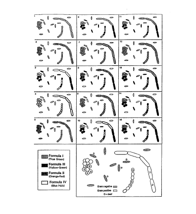
Figure 1: Each numbered panel (1-15) corresponds directly to the combination
of stains shown in Table 5.
SUMIvIARY OF THE INVENTION AND DESCRIPTION OF PREFERRED EMBODIMENTS
The cyclic-substituted unsymmetrical cyanine dyes of the invention are
virtually non-fluorescent when
diluted in aqueous solution. When bound to nucleic acid polymers such as DNA
and RNA, however, the
resultant dye-nucleic acid complex becomes extremely fluorescent upon
illumination. The dyes of the present
invention are highly permeant and label nucleic acids in a wide variety of
solid or liquid samples, particularly
in cells and gels. These dyes are optionally used in combination with other
detection reagents to differentiate
various properties of cells such as viability-, Gram sign, or antibody
staining.
The dyes of the invention comprise three parts: 1) a first heterocyclic ring
system that is a substituted
benzazolium ring system, 2) a linking methine bridge and 3) a second
heterocyclic ring system that is a
pyridinium or quinolinium ring system, one or more positions of which is
substituted by a saturated or
unsaturated, substituted or unsubstituted, cyclic substituent. The two ring
systems are optionally further
substituted independently by lower alkyl, ether, thioether, substituted or
unsubstituted amine, sulfonate ester,
halo, or cyclic substituents. Preferably the ring nitrogen of the second
heterocyclic ring system contains a
cyclic substituent, adjacent to which is a second non-hydrogen substituent.
The non-hydrogen substituent is
preferably another cyclic substituent, or a halo, an ether, a thioether, a
substituted or unsubstituted amine, or a
sulfonate ester substituent.
Specific examples of the dyes of the present invention are described by the
formula:
R2 5
Z_ + ~ /R
N Ym N
(R1 )t ~~--(CH=CH)n CH~ ~ R6
YP
R~
where the substituted benzazolium ring system on the left is linked by a
methine bride to the righthand
pyridinium or quinolinium ring system, one or more substituents of which must
be an OMEGA.
An OMEGA is a saturated or unsaturated, substituted or unsubstituted, cyclic
substituent that has a
total of 2-16 ring carbon atoms in 1-2 alicyclic, aromatic, or hctcroalicyclic
or heteroaromatic rings containing
3
213375
1-4 heteroatoms (wherein the hetero atoms are O, N or S) that is directly
bonded to the pyridinium or
quinoliruum ring system by a single bond. Examples of OMEGA are substituted or
unsubstituted cyclohexyls,
cyclohexenyls, morpholinos, and piperidinyls. Examples of OMEGA that are
aromatic include substituted or
unsubstituted naphthyls, phenyls, thienyls, benzothiazolyls, furanyls,
oxazolyls, benzoxazolyls, and pyridinyls.
Substituents on OMEGA are independently hydrogen, halogen, alkyl,
perfluoroalkyl, amino, alkylamino,
dialkylamino, alkoxy or carboxyalkyl, each alkyl having 1-6 carbons. Preferred
embodiments of OMEGA are
substituted or unsubstituted naphthyl, phenyl, thienyl, morpholino, and
cyclohexyl, more preferably substituted
or unsubstituted phenyl.
Although R' on the benzazolium ring system is usually H, incorporation of one
or more non-hydrogen
substituents R' can be used to fine tune the absorption and emission spectrum
of the resulting dye. For
instance when R' is a methoxy (compound 770 ) its absorption spectrum shifts ~
12 nm and its emission
spectrum shifts ~ 18 nm (Table 5) relative to the comparable compound where R'
is H (compound 63). The
benzazole may contain more than one substituent R', which may be the same or
different (t = 1-4). Each R' is
optionally an alkyl group having from 1-6 carbons; or a trifluoromethyl; or a
halogen; or -ORB, -SR8 or
-(NRBR~ where RB and R9, which can be the same or different, are independently
H or alkyl groups having 1-6
carbons; or 1-2 alicyclic, aromatic, or heteroalicyclic or heteroaromatic
rings having a total of 3-16 ring atoms
(wherein the hetero atoms are O, N or S); or RB and R9 taken in combination
are -(CHZ)Z-L-(CHz)z- where L =
-0-, -NR'°, -CH2- or a single bond where R'° is H or an alkyl
group having 1-6 carbons. Typically, the
compound contains no more than one R' that is not H.
The substituent Rz is an alkyl group having 1-6 carbons, preferably methyl or
ethyl, more preferably
methyl.
The counterion Z- is a biologically compatible ion that is stable and
synthetically accessible.
Examples of Z- include, among others, chloride, bromide, iodide, sulfate,
alkanesulfonate, arylsulfonate,
phosphate, perchlorate, tetrafluoroborate, tetraarylboride, nitrate and anions
of aromatic or aliphatic carboxylic
acids. Preferred Z- counterions are chloride, iodide, perchlorate and various
sulfonates.
X is one of O, S, Se or NR'S, where R'S is H or an alkyl group having 1-6
carbons. Alternatively, X is
CR'6R", where R'6 and R", which may be the same or different, are
independently H or alkyl groups having
1-6 carbons, or the carbons of R'6 and R" taken in combination complete a five
or six membered saturated
ring. Generally, R'6 and R" are methyls.
The two heterocyclic ring systems are linked by 1, 3 or 5 methine (-CH=)
groups in such a way as to
permit extensive electronic delocalization. When n = 0 the dyes are
unsymmetrical monomethine dyes; when
n = 1 the dyes are trimethine dyes; when n = 2, the dyes are pentamethine
dyes. As with similar compounds
21337fi5
(Griffiths, COLOUR AND CONSTITUTION OF ORGANIC MOLECULES, pp. 241 (1976)), the
number of
methine groups between the heteroaromatic rings influences the spectral
properties of the dye (Table 3).
The N-bound substituent RS is an alkyl, alkenyl, polyalkenyl, alkynyl or
polyalkynyl group having 1-6
carbons; or RS is an OMEGA. Most commonly RS is an OMEGA.
The second ring system contains a ring fragment Y that is -CR'=CR'-, with
subscripts p and m equal
to 0 or 1, such that p + m = 1. For all embodiments, the ring contains a 6
membered pyridinium-based
heterocycle according to one of these formulations
R2 R5 R6
+N
(R1 )t ~~(CH=CH)n C ~ R7
~X
R3 R4
or
Z R2 R3 R4
+N
(R1 )t ~>---(CH=CH)n C ~- R5
~X
R~ R6
In preferred embodiments of the invention, m = 1 and p = 0 (4-pyridinium).
The substituents on the second heterocyclic ring system, R3, R", R6 and R',
may be the same or
different and are independently H; or an alkyl, alkenyl, polyalkenyl, alkynyl
or polyalkynyl group having 1-6
carbons; or a halogen; or -0H, -0R8, -SRB, -(NR8R9), as defined previously; or
-0SOzRl9 where R'9 is alkyl
having 1-6 carbons, or perfluoroalkyl having 1-6 carbons, or aryl; or an OMEGA
(defined above); or R6 and
R' taken in combination are -(CH2)~ where v = 3 or 4, forming a fused 5 or 6
membered ring, or R6 and R',
taken in combination form a fused 6 membered aromatic ring.
Where R6 and R' taken in combination form a fused 6 membered aromatic ring,
embodiments of this
invention are quinolinium derivatives according to the formula
2133765
R2 R5
R11
N Ym
(R1 )t ~(CH=CH)n CH~
Y ~~ R12
P
R14 R13
where ring substituents R", R'z, R", and R'° may be the same or
different, and are independently H; or an
alkyl, alkenyl, polyalkenyl, alkynyl or polyalkynyl group having 1-6 carbons;
or a halogen; or -0H, -ORB, -SRB,
-(NRBR9), where R8 and R9 are as defined previously; or -0SOZR'9 where R'9 is
alkyl having 1-6 carbons, or
perfluoroalkyl having 1-6 carbons, or aryl; or an OMEGA. A preferred
embodiment of the invention is a
quinolinium wherein m = 1 and p = 0 (4-quinoliruum).
For all embodiments of the invention, one or more of the substituents of the
pyridinium or
quinolinium ring system is an OMEGA. Preferably, one or two substituents are
OMEGAs. When more than
one OMEGA is bound to a compound of the present invention, the two or more
OMEGAs may be the same or
different. For embodiments of the invention that contain pyridinium ring
systems, OMEGA is preferably R5,
or R6 or both. For embodiments of the invention that contain a 4-quinolinium
ring system, OMEGA is
preferably R4 or R5, or both. For embodiments of the invention that contain a
2-quinolinium ring system,
OMEGA is preferably R5, R" or both. For all embodiments of the invention,
preferably RS is an OMEGA.
One embodiment of the invention contains exactly two non-hydrogen substituents
on the second
heterocyclic ring, one of which is an OMEGA. In one preferred embodiment, RS
is an OMEGA and the
substituent adjacent to RS (R6 for pyridiruums, R4 for 4-quinoliniums, and Rl'
for 2-quinoliniums) is a non-
hydrogen substituent. In one aspect, the substituent adjacent to RS is halogen
or -0SOZR'9, more preferably
halogen. In another aspect, the substituent adjacent to RS is an OMEGA. In
another preferred embodiment,
one non-hydrogen substituent is -0RB, -SRB, or -NRBR9, preferably -NRBR9.
6
2133765
Table 3
DYE EXmaa QY QY Kp
EM maa NA A
Thiazole Oran510/530 0.18 0.15 4.8 E6
a
61 500/527 0.46 0.34 1.0 E7
63 514/531 0.24 3.9 E6
64 450/523
71 508/526 0.31
72 515/535 0.026 1.2 E6
73 508/525 0.31 4.4 E6
200 739/759
542 510/527
578 470/504 4.1 ES
582 516/533
591 509/532 0.09 0.13 4.8 E6
613 506/523 0.33 5.3 E6
616 471/510 3.8 ES
619 488/517 0.62 0.22 9.7 E6
621 635/656
624 480/501 0.58 0.57 5.0 E6
628 488/506 0.40 7.0 E6
630 517/544 0.19
633 489/508 0.12 7.4 ES
634 510/530 0.18 2.0 E6
637 601/622 0.28
639 513/548 0.20 8.0 E6
640 471/516
641 503/526 0.35 2.0 E7
672 586/611
720 487/507 0.52 1.2 E7
742 570/611
752 494/518 0.51
758 504/524 0.44 8.5 E6
760 483/510 0.68
?64 486/508 0.58 0.46 1.1 E7
765 506/524 0.50 1.1 E7
770 526/549 1.7 E6
774 517/533 7.9 E6
776 0.65
780 (CI 513/536 0.09 3.4 E6
780 S 0.31
830 517/533
834 486/507
835 495/518
853 516/555
854 483/520
856 502/523 0.43
5103 511/530 0.18 5.4 E6
6104 505/523 0.52 1.3 E7
2133765
Table 4
DYE X heter cle Rl RZ R4 Rs R" Rl2 n
# I
125 S 2- 'diniumH Me H hen 1 - - 0
578 S 4- 'diniumH Me Cl hen 1 - - 0
616 S 4- 'diniumH Me CI o-Me0- - - 0
hen I
640 S 4- 'diruumH Me H hen 1 - - 0
742 S 4- idiruumH Me n-bu 1 hen 1 - - 1
64 S 2 uinoliruumH Me H hen 1 H H 0
61 S 4 uinoliniumH Me n-bu 1 hen 1 H H 0
63 S 4 uinoliniumH Me H hen 1 H H 0
71 S 4 uinoliniumH Me n-bu 1 thien H H 0
1
72 S 4 uinoliniumH Me H Me hen H 0
1
73 S 4 uinoliniumH Me H clohe H H 0
1
130 S 4 uinoliniumH Me -NH- hen hen 1 H H 0
1
100 S 4 uinoliniumH Me n-bu 1 hen 1 H H 2
200 S 4 uinoliniumH Et Cl hen 1 H H 0
542 S 4 uinoliniumH Me H clohexen H H 0
1
582 S 4 uinoliniumH Me CI -Me0- H H 0
hen 1
591 S 4 uinoliniumH Me CI hen I H H 0
613 S 4 uinoliniumH Me Me hen 1 H H 0
619 S 4 uinoliniumH Me -NE hen I H H 0
621 S 4 uinoliruumH Me n-bu 1 hen 1 H H 1
624 O 4 uinoliruumH Me n-bu 1 hen 1 H H 0
628 S 4 uinoliniumH Me -0Me hen 1 H H 0
630 S 4 uinoliniumH Me hen 1 hen 1 H H 0
633 O 4 uinoliniumH Me Cl hen 1 H H 0
634 S 4 uinoliniumH Me H n-he 1 H H 0
637 O 4 uinoliniumH Me n-bu 1 hen I H H 1
639 S 4 uinoliniumH Me hen 1 Me H H 0
641 S 4 uinoliniumH Me -SMe hen I H H 0
672 O 4 uinoliruumH Me -0Me hen 1 H H 1
720 S 4 uinoliniumH Me -0Et hen 1 H H 0
752 S 4 uinoliniumH Me mo holin Me H H 0
1
758 S 4 uinoliniumCl Me n-bu 1 hen 1 H H 0
760 S 4 uinoliniumH Me -NE hen 1 H -0Me0
764 S 4 uinoliniumH Me -0-iPr hen 1 H H 0
765 S 4 uinoliniumH Me clohe 1 hen 1 H H 0
770 S 4 uinolinium-0MeMe H hen 1 H H 0
774 S 4 uinoliniumH Me Br hen 1 H H 0
776 S 4 uinoliniumH Me -N-nPr hen 1 H H 0
780 S 4 uinoliniumH Me CI clohe H H 0
Cl 1
780 S 4 uinoliniumH Me -SMe clohe H H 0
S 1
823 S 4 uinoliniumH Me Cl hen 1 H H 1
830 S 4 uinoliniumH Me Cl thien H H 0
1
834 S 4 uinoliniumH Me F hen 1 H H 0
835 S 4 uinoliniumH Me -0- hen hen 1 H H 0
1
853 S 4 uinoliniumH Me -S-2- 'd hen 1 H H 0
1
854 S 4 uinoliruumH Me -0SO CF hen 1 H H 0
856 S 4 uinoliniumH Me N-Me- i hen 1 H H 0
r 1
5103 S 4 uinoliniumH Me CI hen 1 H -OMe0
6104 S 4 uinoliniumH Me clohe 1 Me H H 0
2133765
synthesis
In general, synthesis of these dyes requires three precursors: a benzazolium
salt, a pyridinium (or
quinoliruum) salt (both of which have the appropriate chemical subsdtuents),
and (where n = 1 or 2) a source
for the methine spacer. Although the combination that enables these compounds
to be useful stains for nucleic
acids has not been described previously, the chemistry that is required to
prepare and combine these precursors
so as to yield any of the subject derivatives is generally well-understood by
one skilled in the art.
The benzazolium moiety.
A wide variety of derivatives of this type have been described (Brooker, et
al., J. AM. CHEM. SOC.,
64, 199 (1942)) and Hamer, "The Cyanine Dyes and Related Compounds", THE
CHEMISTRY OF
HETEROCYCLIC COMPOUNDS, Vol. 18, A. Weissberger, Ed., Interscience, New York
(1964). These
precursors have the common structure:
R2
Z
+N
(R1 )t ~-A
X may be O (benzoxazolium), S (benzothiazolium), Se (benzoselenazolium), N or
an alkyl-substituted
N (benzimidazolium) or a carbon atom substituted by two alkyl groups R'6R"
(indolium) (where R'6 and R"
are independently alkyl groups having 1-6 carbons, or R' 6 and R" taken in
combination complete a five or six
membered saturated ring).
R' is usually incorporated in the parent benzazole molecule prior to
quaternization with an alkylating
agent. Rz is usually obtained by alkylation of the parent heterocycle with Rz-
Z, where Rz is an alkyl group
having 1-6 carbons and Z is an electronegative group that frequently becomes
the counterion on the resultant
dye. Z- is a biologically compatible counterion that additionally is stable
and synthetically accessible. The
counterion may be exchanged for another counterion by methods known in the
art, such as the use of ion
exchange resins or by precipitation. Preferred RZ-Z are compounds that yield
RZ = methyl, such as methyl
iodide.
A is a substituent whose nature is determined by the synthetic method utilized
to couple the
benzazolium precursor with the pyridinium or quinolinium precursor. When n =
0, A is usually alkylthio,
commonly methylthio, or A is chloro, bromo or iodo. When n = 1 or 2, A is
methyl.
9
21 337 65
The pyridinium or quinolinium moiety.
The second heterocyclic precursor is a pyridinium or quinolinium salt. These
can sometimes be
generated from the corresponding pyridine or quinoline by alkylation at
nitrogen using a suitable alkylating
agent RS-Z. However, 2- and 4-pyridones and 2- and 4-quinolones are much more
versatile chemical
intermediates, with the added advantage of being easily prepared (for examples
see HETEROCYCLIC
COMPOUNDS, VOL. 4, R. C. Eldcrfield cd., John Wiley and Sons Inc., (1952) pp 1-
331 or Wawzonek et al.,
J. HETEROCYCLIC CHEM., 25, 381 (1988)).
Typically the required pyridinium salt precursor has the structure
B R4
R~ R3 R~ R3
\ \
or
R6 N+ R4 R6 N+ B
R5 Z_ R5 Z_
and the quinolinium salt precursor has the structure
R14 B R14 R4
R13 R3 R13 R3
/ \ / \
R12 \ ~ N, 4 or 12 \
+ R R ~ ~N+ B
1 'R5 R11 ~ 5
Z R Z
At all times, the ring is a cationic f-membcred pyridinium- or quinolinium-
based heterocycle.
When n = 0, B is methyl, or B is chloro, bromo or iodo. When n = 1 or 2, B is
methyl. Only when n
= 1 or n = 2 is any part of B incorporated in the final compound.
When RS is an OMEGA or alkyl, the 2-pyridone or 4-pyridone or 2-quinolone or 4-
quinolone can be
treated with a powerful nucleophile such as a Grignard or an alkyl lithium
reagent (Example 11) or with a
metal hydride (Example 12)) to generate the pyridinium or quinolinium salt
after acid-catalyzed
dehydroaylation.
l0
CA 02133765 1999-08-10
The pyridone or quinolone can also be converted to a pyridinium or quinolinium
salt
by using an agent such as phosphorous oxychloride, phosphorous tribromide,
diethylaminosulfur trifluoride (Example 5) or trifluoromethanesulfonic
anhydride. The
resulting activated intermediate can be condensed with the appropriate
benzazolium salt to
form the dye directly (Example 6) or the activated intermediate can be treated
with alcohols,
phenols, or alkoxides to yield ether derivatives (Example 10), thiols or
thiophenols to yield
thioether derivatives (Example 8) or ammonia or amines to yield substituted or
unsubstituted
amino derivatives (Example 7).
l0 The methine bridge.
The methine bridge consists of 1, 3 or 5 methine (-CH = ) groups that bridge
the
benzazolium rings and the pyridinium or quinolinium rings) in such a way as to
permit
extensive electronic conjugation.
Synthesis of monomethine dyes (n = 0) commonly uses a combination of reagents
where the methine carbon atom results from either A on the 2-position of the
benzazolium salt
or B on the 2- or 4-position of the pyridinium or quinolinium salt being
methyl and the other
of A or B being a reactive "leaving group" that is typically methylthio or
chloro
2 0 (Brooker et al. , supra).
To synthesize trimethine dyes (n = 1 ) or pentamethine dyes (n = 2) both A and
B are
methyl. In these cases the additional methine carbon of is provided by a
reagent such as
N-methylformanilide or ethyl
30
11
2133765
orthoformate (HOUBEN-WEYL METHODON DER ORGANISCHEN
CHEMIE, Band V/ld 231-299 (1972)) or the additional
trimethine fragment is provided by a malonaldehyde
equivalent such as 1, 1, 3, 3-tetramethoxypropane; 1, 1,
3-trimethoxypropane, 3-(N-methylanilino) propenal or 1-
anilino-3-phenylimino-1-propene (Sprague, su ra).
Subsequent modification of dyes
As described earlier, the reactivity of the 2-
halogenated pyridinium or quinolinium intermediate offers
a variety of synthetic methods for attachment of various
substituents at the 2-position. However, the reactivity
of the 2-halo derivatives is preserved even after
conjugation with the benzazolium precursor, enabling
conversion of the resulting dye in which R4 is halogen
into the appropriate ether, amine and thioether analogs,
as described above for the pyridinium and quinolinium
precursors (Examples 7, 8 and 10).
Method of Use
The use of the invention comprises combining a dye
of the present invention with a sample that contains or
is thought to contain a nucleic acid, incubating the
sample for a time sufficient to obtain a detectable
fluorescent response, and observing the fluorescent
response. The sample is optionally combined
lla
B
2133765
with one or more additional dyes (preferably fluorescent dyes) having a
response detectably different from that
of the subject dyes.
Typically, the subject dye is present as a staining solution, which is
prepared by addition of the dye to
an aqueous solution that is biologically compatible with the sample. The
staining solution is made by
dissolving the dye directly in an aqueous solvent such as water, a buffer
solution, such as buffered saline
(preferably non-phosphate), or an organic water-miscible solvent such as
dimethylsulfoxide (DMSO),
dimethylformamide (DMF), or a lower alcohol such as methanol or ethanol, or
acetonitrile. Typically the dye
is preliminarily dissolved in an organic solvent (preferably 100% DMSO) at a
concentration of greater than
about 100-times that used in the staining solution, then diluted one or more
times with an aqueous solvent
such as water or buffer, such that the dye is present in an effective amount.
An effective amount of dye is the
amount su~cient to give a detectable fluorescent response when in the presence
of nucleic acids. Typically
staining solutions for cellular samples have a dye concentration greater than
about 0.1 nM, and less than about
100 N.M, more typically greater than about 1 nM. Staining solutions for
electrophoretic gels typically have a
dye concentration of greater than about 1 pNi and less than about 10 N.M, more
typically about 4-5 N.M.
Staining solution for detection of free nucleic acids in solution typically
have a concentration 10 nM-1 liM.
The specific concentration of the staining solution is determined by the
physical nature of the sample, and the
nature of the analysis being performed, and can be optimized according to
standard procedures such as
described for cell samples in Example 16.
The dye is combined with a sample that contains a nucleic acid. The nucleic
acid in the sample may
be RNA or DNA, or a mixture thereof. Any DNA is optionally single-, double-,
triple-, or quadruple-stranded
DNA. The nucleic acid may be natural (biological in origin) or synthetic
(prepared artificially). The nucleic
acid may be present as nucleic acid fragments, oligonucleotides, or nucleic
acid polymers, and may contain
unnatural bases. The nucleic acid may be present in a condensed phase, such as
a chromosome. The presence
of the nucleic acid in the sample may be due to a successful or unsuccessful
experimental methodology,
undesirable contamination, or a disease state. Nucleic acid may be present in
all, or only part, of a sample, and
the presence of nucleic acids may be used to distinguish between individual
samples, or to differentiate a
portion or region within a single sample.
The nucleic acid may be enclosed in a biological structure, for example
contained within a viral
particle, an organelle, or within a cell. Cell types include, but are not
limited to, eukaryotes, such as nucleated
plant and animal cells, and prokaryotes, such as bacteria (including both Gram-
negative and Gram-positive
bacteria such as Bacillus cereus, Bacillus subtilus) Clostridium sporogenes,
Corynebacterium xerosis,
Micrococcus luteus, Mycobacterium phlei, Propionibacterium freunderreichii)
Staphylococcus aureus,
Streptococcus pyogenes, Lactobacillus acidophilus, Cytophaga psychrophila)
Enterobacter aerogenes,
Escherichia coli, Flavobacterium meningosepticum) Klebsiella pneumonia,
Neisseria sub'lava) Pseudomonas
aeruginosa, Rhizobium trifolii, Salmonella oranienburg, Shigella sonnei,
vibrio parahaemolyticus or
12
zi337s~
combinations thereofj, as well as yeast and other fungi, mycobacteria and
mycoplasma. The nucleic acids
enclosed in biological structures may be obtained from a wide variety of
sources, including unfiltered or
separated biological fluids (such as urine, cerebrospinal fluid, blood, lymph
fluids, tissue homogenate, mucous,
saliva, stool, or physiological secretions or other similar fluids);
environmental samples such as soil, water and
air; a fermentation medium such as from a biological reactor or food
fermentation process such as brewing; or
surface washes of materials, (e.g. food) or small amounts of solids such as
retentates, scrapes, and smears; or
liquid growth medium in which cells have been introduced for culturing. The
cells are optionally discrete or
individual cells, including microorganisms, or multiple cells associated with
other cells in two or three
dimensional layers, including multicellular organisms, embryos, tissues,
biopsies, filaments, biofllms, etc. The
nucleic acid may be endogenous or introduced as foreign material, such as by
infection or by transfection. The
cells may be viable or dead cells or a mixture thereof. The nearly universal
permeability of the instant dyes,
their accelerated rate of uptake and the low toxicity of the dyes to living
systems enable the examination of
nucleic acids in living samples with little or no perturbation caused by the
dye itself. The dyes can also be
used for staining nucleic acids in a cell or cells fixed and treated with
routine histochemical or cytochemical
procedures.
Alternatively, the nucleic acid, in any of the forms described previously, is
not enclosed within a
biological structure, but is present as a sample solution. The sample solution
can vary from one of purified
oligonucleotides or nucleic acids to crude mixtures such as cell extracts,
biological fluids and environmental
samples from the sources listed above. In some cases it is desirable to
separate the nucleic acids from a
mixture of biomolecules or fluids in the solution prior to combination with
the dye. Numerous techniques exist
for separation and purification of nucleic acids from generally crude mixtures
with other proteins or other
biological molecules. These include such means as electrophoretic techniques
and chromatographic
techniques using a variety of supports. When used for poststaining
electrophoresis gels, the high sensitivity of
the dyes of the present invention allow the detection of previously
unmeasureable amounts of nucleic acids
without requiring destaining. One embodiment of the invention, when used in
conjunction with an ultraviolet
transilluminator, allows detection of as little as 20 picograms of double-
stranded DNA per band.
The sample may be combined with the staining solution by any means that
facilitates contact between
the dye and the nucleic acid. The contact can occur through simple mixing, as
in the case where the sample is
a solution. The dye may be added to the nucleic acid solution directly or may
contact the solution on an inert
matrix such as a blot or gel, a testing strip, or any other solid or semi-
solid surface, for example where only a
simple and visible demonstration of the presence of nucleic acids is desired.
Any inert matrix used to separate
the sample can be used to detect the presence of nucleic acids by observing
the fluorescent response on the
inert matrix. While the subject dyes have shown an ability to permeate
cellular membranes rapidly and
completely upon addition of the dye solution, any other technique that is
suitable for transporting the dye
across cell membranes with minimal disruption of the viability of the cell and
integrity of cell membranes is
also a valid method of combining the sample with the subject dye. Examples of
suitable processes include
13
21337s~
action of chemical agents such as detergents, enzymes or adenosine
triphosphate; receptor- or transport
protein-mediated uptake; pore-forming proteins; microinjection;
electroporation; hypoosmotic shock; or
minimal physical disruption such as scrape loading or bombardment with solid
particles coated with or in the
presence of the dyes.
The sample is incubated in the presence of the dye for a time sufficient to
form the fluorescent nucleic
acid-dye complex. Detectable fluorescence in a solution of nucleic acids is
essentially instantaneous.
Detectable fluorescence within cell membranes requires the permeation of the
dye into the cell. Preferably, the
dye is added at a temperature optimal for normal activity of the cells within
the operating parameters of the
dyes (between about 5 °C and about 50 °C); typically this is
room temperature (23 °C). At temperatures
between 5-45 °C, visibly detectable fluorescence is obtained within
about 15-20 minutes of combination with
the sample, commonly within about 5 minutes. Preferred embodiments give
detectable fluorescence inside
cells in less than about 2 minutes. Lymphocytes loaded with 5 pM dye solutions
give a fluorescent response in
less than 5 seconds, too fast to measure by conventional fluorometry. This
property is useful for observing
nuclear structure and rearrangement, for example such as occurs during mitosis
or apoptosis. While
permeation and fluorescence is rapid for all embodiments, optimal permeation
of the dye or formation of the
nucleic acid complex is dependent upon the physical and chemical nature of the
individual sample and the
sample medium, and can be determined according to standard procedures such as
described in Example 17.
The subject dyes bind non-covalently with nucleic acids to yield enhanced
fluorescence, the level of
enhancement being generally about 100-1000 fold, typically greater than about
300-fold (Table 2). These dyes
generally exhibit improved quantum yields upon binding to nucleic acids,
relative to Thiazole Orange, which
translate directly into improved sensitivity in nucleic acid detection. While
not every dye shows an improved
quantum yield, other attributes of the subject dyes represent significant
improvement, including enhanced
permeation, enhanced rate of permeation, and/or the selectivity of excitation
and emission bands to suit
specific instrumentation.
To facilitate the detection of the nucleic acid-dye complex, the excitation or
emission properties of the
fluorescent complex are utilized. For example, the sample is excited by a
light source capable of producing
light at or near the wavelength of maximum absorption of the fluorescent
complex, such as an ultraviolet or
visible lamp, an arc lamp, a laser, or even sunlight. Preferably the
fluorescent complex is excited at a
wavelength equal to or greater than about 300 nm, more preferably equal to or
greater than about 340 nm.
The equipment commonly available for excitation of samples near 254 nm,
between 300 and 310 nm, and near
365 nm can be used to excite any of the dyes of the present invention.
Excitation by a source more appropriate
to the maximum absorption band of the nucleic acid-dye complex, such as the
488 nm band of the argon laser,
results in even higher sensitivity. Some examples permit excitation beyond 600
nm.
14
zi337s5
The fluorescence of the complex is detected qualitatively or quantitatively by
detection of the resultant
light emission at a wavelength of greater than about 400 nm, preferably
greater than about 480 nm, more
preferably at greater than about 500 nm. The emission is detected by means
that include visible inspection,
photographic film, or the use of current instrumentation such as fluorometers,
quantum counters, plate readers,
epifluorescence microscopes, and flow cytometers, or by means for amplifying
the signal such as a
photomultiplier. The nucleic acid concentration in a sample can also be
quantified, as the fluorescence of the
nucleic acid-dye complex is linearly dependent on concentration (Examples 22-
23).
The wavelengths of the excitation and emission bands of the dyes vary with dye
composition to
encompass a wide range of illumination and detection bands (e.g. Table 3).
This allows the selection of
individual dyes for use with a specific excitation source or detection filter.
In particular, dyes can be selected
that match their excitation band with the commonly used argon laser, or
emission bands that match
preexisting filters such as a typical fluorescein long-pass set or multi-band
set with fluorescein excitation and
emission bands.
In addition, the dye can be selected to give a detection response that is
different from that of other
dyes desired to be used in combination with the subject dyes. Preferably the
additional dye or dyes are
fluorescent, for which the response to illumination that is detectably
different from that of the subject cyclic-
substituted unsymmetrical cyanine dyes. Any fluorescence detection system can
be used to detect the
difference in spectral characteristics between dyes. Preferably the dyes have
the same or overlapping
excitation spectra, but possess visibly different emission spectra, generally
having emission maxima separated
by >10 nm, preferably >20 nm, more preferably >50 nm.
The additional dyes are optionally used to differentiate cells or cell-free
samples containing nucleic
acids according to size, shape, metabolic state, physiological condition,
genotype, or other biological
parameters or combinations thereof. In one aspect of the invention, the
additional dye or dyes are metabolized
intracellularly to give a fluorescent product inside certain cells but not
inside other cells, so that the fluorescent
response of the cyclic-substituted unsymmetrical cyanine dye predominates only
where such metabolic process
is not taking place. Alternatively, the additional dye or dyes are specific
for some external component of the
cell such as cell surface proteins or receptors. In yet another aspect of the
invention, the additional dye or dyes
actively or passively cross the cell membrane and are used to indicate the
integrity or functioning of the cell
membrane.
The additional dyes are added to the sample being analyzed to be present in an
effective amount, with
the optimal concentration of dye determined according to the cell density as
above. Typically the
concentration of each dye is between about 0.01 N.M and about 100 1,~M, more
typically between 0.1 N.M and 10
pNI. Each dye is optionally prepared in a separate solution or combined in one
solution. Generally the dyes
are present in the staining solution within about a five-fold molar range, but
the molar ratio one to the other in
CA 02133765 1999-08-10
the sample can vary from out 1:1 to about 1:100, and may vary depending on
whether the
dyes are added to the sample simultaneously or sequentially. After
illumination of the dyed
cells at a suitable wavelength, as above, the cells are analyzed according to
their fluorescent
response to the illumination. In addition, the differential fluorescent
response can be used as
a basis for sorting the cells or nucleic acids for further analysis or
experimentation. For
example, all cells that "survive" a certain procedure are sorted, or all cells
of a certain type in
a sample are sorted. The cells can be sorted manually or using an automated
technique such
as flow cytometry according to the procedures known in the art such as in U.S.
patent 4,655,024 to Mansour, et al. (1987).
In one embodiment of the invention, the subject dyes are used in combination
with a
second fluorescent dye (Dye II) to distinguish viable cells from dead cells,
where Dye II is
selective either for viable or for dead cells. In one aspect of the invention,
Dye II gives a
detectable fluorescent response only in viable cells. Such as fluorescent
enzyme substrates and
reagents described in Haugland, HANDBOOK OF FLUORESCENT PROBES AND
RESEARCH CHEMICALS (1992-94) to selectively stain viable cells, including
haloalkyl
esterase substrates and calcein AM. Alternatively, Dye II gives a detectable
fluorescent
response only in dead cells, such as an impermeant dye that only becomes
fluorescent upon
passing through the cell membrane to bind to some intracellular component,
such as an
2 o intracellular protein or nucleic acid. While there is not an exact
equivalence between an
intact cell membrane and the term "viability" (technically defined as the
ability of a cell to
maintain its existence), it is common to refer to cells where the cell
membrane has been
irreversibly disrupted as "dead" cells or "non-viable" cells. Suitable dyes
include
impermeant phenanthridium or benzazolium derivatives, including monomers or
dimers
2 5 thereof, such as ethidium homodimer, ethidium bromide, propidium iodide,
TOTO*,
BOBO*, POPO*, YOYO*, TO-PRO*, BO-PRO* PO-PRO* and YO-PRO* (Molecular
Probes) that give an enhanced fluorescence when complexed to intracellular
nucleic acids.
Loading times for impermeant Dye II dyes such as phenanthridium or banzazolium
dyes, is
generally the same as previously discussed above. Cell permeant Dye II dyes
selective for
3 o viable cells generally require longer loading times, particularly if such
dyes require
intracellular activity to generate fluorescence.
*trade-marks
16
CA 02133765 1999-08-10
In cells for which Dye II is selective, both dyes are present because the
subject dyes
stain all cells, including those for which Dye II is selective. In the cells
for which Dye II is
selective (and both dyes are present) the intracellular fluorescent response
of Dye II is
optionally the same as the fluorescent response of Dye II alone (e.g. where
Dye II effectively
competes for nucleic acid binding relative to the subject dye) or is a
response indicative of the
presence of both dyes (as is the case where the competitive binding is less
effective or where
Dye II is not a nucleic acid stain). The fluorescent response of the subject
dye alone is
indicative of cells for which Dye II is not selective, either viable or non-
viable cells as the
case may be. The cells for which Dye II is selective are optionally sorted or
counted, as
1 o above.
20
30
16a
2133765
In another embodiment of the invention, the sample is combined with multiple
fluorescent dyes to
determine identification, and optionally viability. The additional fluorescent
dyes) binds selectively to cell
surface components or is selectively pcrmeant to certain cell types, and can
be used in combination with Dye II
to also indicate viability. The surface label that only stains externally is
distinguishable from the dyes that
stain intracellularly. When a surface dye is used in combination with one or
more intracellular stains such as
nucleic acid stains, a "bullseye" pattern of Staltllng is seen -- i.e. a
brightly stained interior within an exterior
ringstain. Preferably, the surface label also has an emission spectrum that is
detectably different from that of
the other dyes used. Preferably the excitation spectnun of each dye or dye-
nucleic acid complex overlaps the
excitation spectrum of the other dye(s). More preferably, each dye complexed
with nucleic acids has an
excitation maximum between about 480 nm and 510 nm. Most preferably, each dye
or dye-complex also
excites in the UV between about 300 nm and 365 nm.
In one aspect of the invention, the appearance of the stained bacteria
indicates the Gram reaction of
the bacteria in the sample, and optionally whether or not the G+ or G-
bacteria present in the sample are
viable. Gram positive (G+) bacteria are those that give a positive Gram stain,
including but not limited to
Bacillus, Lactobacillus, A~licrococcrr.v, .Sfreptococcrr.s, Clo.slridicnu,
.Stapl?vlococcus, and Mvcobacteriuru,
among others. Gram negative (G-) bacteria are those that are negative for the
Gram stain, including but not
limited to Escherichia, EnterobacTer, .Salmonella, I'.seudmuonas, .Slrigella,
Klebsiella, Haernophilus,
Neisseria, Proteus, I~ibrio, Carrrpvlobacter, and 1'ersinia, among others.
Preferably a cyclic-substituted unsynunctrical cyanine dye that (in
combination with intracellular
nucleic acids) gives a green or yellow-green fluorescence is used in
combination with one or more of the
following dyes: a) a C4 C8 alkyl substituted phenanthridium nucleic acid stain
(preferably hexidium or C6
substituted phenanthridium, Watkins, J. CHEM. SOC. 3059 ( 1952)) that
selectively stains live G+ bacteria and
all dead bacteria with an orange red fluorescent signal that partially or
completely replaces the signal of the
cyclic-substituted unsymmetrical cyanine dye; and/or b) a protein that is
covalently bound to a fluorophore
with a fluorescent response different from that of the phcnanthridium dye in
a) and from that of the cyclic-
substituted unsymmetrical cyanine dyc, preferably a lcctin such as wheat gene
agglutinin labeled with AMCA
or Cascade Blue dye (Molecular Probes) that is selective for the cell surface
of G+ bacteria, live or dead; and
optionally c) a membrane impermeant bcnzazolium nucleic acid stain according
to Dye I1 above, that has a
fluorescent response different from that of the other dyes used, preferably
dyes sold under the names TOTO,
YOYO, BOBO, POPO, TO-PRO, YO-PRO, BO-PRO, PO-PRO (Molecular Probes).
Table 5 sununarizes the spectral response, where the cyclic-substituted
unsymmetrical cyanine dye (I)
has an emission maximum between 500 nm and 535 nm (e.g. dye 624); the
phenanthridium dye (II) has an
emission maximum between 580 nm and 650 nm (c.g. hcxidium); the membrane
impermeant benzazolium
(III) nucleic acid complex has an emission maximum between 530 nm and 590 nm
(e.g. TOTO, YOYO, TO-
PRO or YO-PRO); and the labeled protein (IV) has an emission maximum between
410 nm and 480 nm (e.g.
17
,, ,
Th.
21337fi5
AMCA- or Cascade Blue-labeled wheat germ agglutinin). Careful matching of
other fluorescent stains with
equivalent selective permeability, excitation/emission spectra, and
preferential binding affinity for nucleic
acids allows substitution of the preferred combination of nucleic acid stains
to discriminate between many
different organisms, whether live or dead.
Table 5
Panel Dyest Live Gram Live Gram Dead Gram Dead Gram
# in (+) (-) (+) (-)
Fi a Bacteria Bacteria Bacteria Bacteria
1
1 I G G G G
2 II O - O O
3 III - - Y Y
4 IV B - B -
5 I, II O G O O
6 I, III G G Y Y
7 I, IV G with G G with B G
B
8 II, III O - Y Y
9 II, IV O with - O with B O
B
III, IV B - Y with B Y
11 I, II, O G Y Y
III
12 I, II, O with G O with B O
IV B
13 I, III, G with G Y with B Y
IV B
14 II, III, O with - Y with B Y
IV B
I, II, O with G Y with B Y
III, B
IV
Color B = Blue
Ke : Halo -=
G = Unstained
True-
een
Y =
Yellow-
reen
O =
Oran
a
The examples below are given so as to illustrate the practice of this
invention. They are not intended
to limit or define the entire scope of this invention. In the structural
formulae below, the substituent phenyl is
10 represented by the symbol Ql, as is generally used and understood in the
art.
Example 1: Preparation of 1.2-dihvdro-4-methyl-1-phenyl-2-quinolone (1)
The following compound is prepared:
The synthetic precursor (1) is prepared either by an Ullmann coupling
according to a literature procedure
(Wawzonek, et al., supra.) or via the reaction of the corresponding
diarylamine with diketene followed by acid
cyclization ( Elderfield, su ra . Thus 10.0 g (62.9 mmoles) of 2-hydroxy-4-
methylquinoline is heated at reflux
with 24.0 g (377 mmoles) of copper powder, 8.68 g (62.9 mmoles) of potassium
carbonate and 19.2 g (94
18
21337fi5
mmoles) of iodobenzene for 48 hours. The reaction is cooled to room
temperature, partitioned between water
and ethyl acetate, filtered, and the organic layer is dried over magnesium
sulfate. The crude product is
purified on a silica gel column, eluting with 1:1 ethyl acetate/hexanes to
yield 8.1 g of the desired product.
Example 2: Preparation of 1,2-dihvdro-4-methyl-1-phenyl-2-pyridone (2)
The following compound is prepared:
~3
~O
Synthetic precursor 2 is prepared as in Example 1 with a 40% yield, except
that the starting material is 1,2-
dihydro-4-methyl-2-pyridone.
Example 3: Preparation of 1,2-dihvdro-1,4-dimethvl-2-guinolone (3)
The following compound is prepared:
Synthetic precursor 3 is prepared by first conjugating N-methylaniline with
diketene, followed by an acid
cyclization of the amide intermediate. Thus 10.0 g (0.12 moles) of diketene is
added dropwise to 10.7 g (0.1
moles) of N-methylaruline and the reaction is heated at 100 °C for an
additional 30 minutes. To the resulting
mixture is added 30 mL of acetic acid and 30 mL of sulfuric acid, and the
mixture is heated at 50 °C
overnight. The reaction is worked up with water and ethyl acetate and purified
on a silica gel column to yield
9.5 g of the desired product.
If the synthesis is performed using N-methyl-2-phenylaniline (generated by
methylation of 2-phenylaniline
using ICzC03 and CH3I) the resulting product is 1,2-dihydro-1,4-dimethyl-8-
phenyl-2-quinolone.
Example 4: Preuaration of 1,2-dihydro-7-methoxy-4-methyl-1-phenyl-2-quinolone
The following compound is prepared:
19
2133765
CH30
N-(3-hydroxyphenyl)-N-phenylamine is O-methylated with potassium carbonate and
methyl iodide in acetone
in 39% yield. The resulting N-(3-methoxyphenyl)-N-phenylamine is then reacted
with diketene to generate
the corresponding acetoacetamide which, without purification, is cyclized in
acetic acid/sulfuric acid as in
Example 3 to generate the desired quinolone in 41% yield.
Example 5: Preparation of 2-chloro-4-methyl-1-phenylquinolinium chloride (4)
The following compound is prepared:
CI _
CI
H3C ~ 'N+ Q~
To 2.8 g (11.9 mmoles) of 1 in 20 mL of methylene chloride is added 1.85 g of
POC13 and a catalytic amount
of dimethylformamide (Marson, TETRAHEDRON., 48, 3659 (1992)). The resulting
mixture is heated to
reflux for 24 hours. ABer cooling, the product is purified using column
chromatography.
The corresponding bromide is prepared using PBr3 rather than POC13.
The corresponding fluoride is prepared using diethylaminosulfur trifluoride,
rather than POC13.
Example 6: Preparation of 2-chloro-4-f2,3-dihvdro-3-methyl-(benzo-1,3-thiazol-
2 girl)-methvlidenel-1-
phenvlc~uinolinium iodide (dye 591)
The following compound is prepared:
20
21337~~'
A room temperature solution of 4 ( 11.9 mmoles) is prepared, and 3. 5 g (9.6
mmoles) of N-methyl-2-
methylthiobenzothiazolium tosylate (5) (Rye, et al., NUCLEIC ACIDS RES., 20,
2803 (1992)) is added
followed by 1.3 mL (9.4 mmoles) of triethylamine. The mixture is stirred for
an additional 6 hours. The crude
product is purified on silica gel using ethyl acetate:chloroform:methanol,
3:3:1 as eluant. The product is then
recrystallized from methanol/chloroform/ethyl acetate.
The corresponding bromide (Dye 774) is prepared analogously using 2-bromo-4-
methyl-1-phenylquinolinium
bromide in place of 4.
The corresponding fluoride (Dye 834) is prepared analogously using 2-fluoro-4-
methyl-1-phenylquinolinium
fluoride in place of 4.
The methoxyquinolinium analog (Dye 5103) is prepared in the same way, except
using 1,2-dihydro-7-
methoxy-3-methyl-1-phenyl-2-quinolone.
The pyridiruum analog (Dye 678) is prepared in the same way, except using the
pyridinium analog of 4.
The trimethine dye analog (Dye 823) is prepared similarly, except using 2-(2-
anilinovinyl)-3-
methylbenzothiazolium tosylate in place of 5.
An additional synthetic route to Dye 591 utilizes 4-[2,3-dihydro-3-methyl-
(benzo-1,3-thiazol-2-yl)-
methylidene]-1,2-dihydro-1-phenyl-2-quinolone (6), which in turn is prepared
from 1 and 5. Thus the lithium
enolate of 1 ( prepared from treating the quinolone with 2.7 equivalent of
lithium diisopropyl amide) or the
silyl enolate of 1 (from (1) and trimethylsilyl trifluoromethanesulfonate and
diisopropylethylamine) is stirred
with 5. The desired intermediate (4) is isolated by column chromatography. The
quinolone (6) is then treated
with POCl3 to generate Dye 591.
Example 7: Preparation of 2-diethylamino-4-f2,3-dihvdro-3-methyl-(benzo-1,3-
thiazol-2~1)-methvlidenej-1-
yhenylquinolinium iodide lDve 619)
The following compound is prepared:
21
21 3 37 6 5
I +CH3 NEt2
w
Dye 619 is prepared by heating Dye 591 (26 mg) at 55 °C with 0.5 mL of
diethylamine in 1.5 mL of DMF
overnight. The desired product is isolated by a simple filtration.
Dye 752 is prepared similarly, except using morpholine in place of
diethylamine in DMF at 50 °C.
Dye 856 is prepared similarly, except using N-methylpiperazine in place of
dicthylamine.
Dye 130 is prepared similarly, except using aniline in place of dicthylamine.
2-(N-3-dimethylaminopropyl)-N-propyl a m i no-.1-12, 3-di hydro-3-methyl-
(benzo-1, 3-thiazol-2-yl)-methylidene]-
1-phenylquinolinium iodide (Dye 1037) is prepared similarly, except using N-(3-
dimethylaminopropyl)-N-
propylamine in place of diethylaminc.
Example 8: Preparation of 4-12.3-dihvdro--t-mcih~~l-(benzo-1,3-thiazol-2-vl)-
methvlidenej-1-phenyl-2-(2-
pyridylthio)-quinolinium iodide (dye 853)
The following compound is prepared:
i
I CH3 S ~
+N
w I v / - N_~
s
\ /
2-Mercaptopyridine (6.3 mg) is added to 25 mg of Dye 591 in 2 mL of methylene
chloride, followed by 13 pL,
of triethylamine, and the resulting mixture is stirred at room temperature for
1.5 hours. The volume of solvent
is reduced to about 0.5 mI. under reduced pressure and the product is isolated
by filtration.
2-(2-Dimethylaminoethylthio)-4-12.3-dihydro-3-methyl-(benzo-1,3-thiazol-2-yl)-
methylidene]-1-
phenylquinolinium iodide (Dye 1()()=1) is prepared analogously, using Dye 633
in place of Dye 591, and 2-
dimethylaminoethanethiol in place of 2-mercaptopyridinc.
22
z1~37s~
Example 9: Preparation of 2-chloro-4-f2,3-dihydro-3-methyl-(benzo-1.3-thiazol-
2-yl)-methvlidenej-1-
c~clohexvlQUinolinium tosvlate (Dpe 780 (Cl))
The following compound is prepared:
S 03-
CH3
1-Cyclohexyl-1,2-dihydro-4-methyl-2-quinolone is prepared using N-
cyclohexylaniline as starting material.
The quinolone (0.482 g, 2 mmol) is transformed to the 2-chloro-1-
cyclohexylquinolinium chloride with a
procedure similar to Example 5, and is then reacted with 5 (0.74 g, 2 mmol)
and triethylamine (0.28 mL, 2
mmol) to yield the product.
Example 10: Preparation of 2-methoxv-4-f2,3-dihydro-3-methyl-(benzo-1,3-
thiazol-2-vl)-meth~rlidenel-1-
phenylquinolinium iodide (dye 628)
The following compound is prepared:
Dye 591 (4.3 mmoles) and methanol (10 mL) are heated to reflux for 2 hours.
The methanol is removed under
reduced pressure, and 10 mL of methylene chloride is added, followed by 1.56 g
(4.3 mmoles) of 5 and 1.5 mL
of triethylamine. The resulting mixture is stirred at room temperature for 3
days. The crude material is
purified on a silica gel column by eluting with 5:5:1 ethyl acetate:
chloroform: methanol.
The corresponding ethoxide (Dye 715) is prepared analogously, using ethanol
rather than methanol.
Example 11: Preparation of 2~utv1-4-f2,3-dihvdro-3-methyl-(benzo-1.3-thiazol-2-
vl)-methylidenel-1-
phenylquinolinium iodide (dye 61)
The following compound is prepared:
23
_.. 2I3376~
To 0.235 g (1 mmole) of 1 in 10 mL of THF at -78 °C under nitrogen, 1.2
equivalents of n-butyl lithium is
introduced. The reaction is stirred at -78 °C for 15 minutes, and then
the temperature is raised to 0°C for
another 30 minutes, then the reaction is quenched with acetic acid and the
solvent is evaporated. The residue
is dissolved in 5 mL of methylene chloride and 0.367 (1 mmole) of 5 is added
followed by 0.28 mL (2 mmoles)
of triethylamine. The reaction mixture is stirred for 20 minutes at room
temperature and the crude product is
isolated as the iodide salt after a salt exchange. The crude iodide is
recrystallized from methanol.
Dye 624 is prepared similarly, except that 3-methyl-2-methylthiobenzoxazolium
tosylate (7) (Rye, et al.,
supra) is used instead of 5 in the synthesis.
Dye 6104 is prepared similarly, except that cyclohexyl magnesium bromide is
used instead of butyl lithium.
The corresponding trimethine dye (Dye 621) is prepared similarly, except that
2-(2-anilinovinyl)-3-
methylbenzothiazolium tosylate is used in place of 5.
The corresponding pentamethine dye (Dye 100) is prepared similarly, except
that 2-(4-anilino)-1,3-
butadienyl)-benzothiazolium iodide is used in place of 5. 2-(4-Anilino)-1,3-
butadienyl)-benzothiazolium
iodide is prepared using methods known in the art (CT. S. Patent No. 2,269,234
to Sprague ( 1942); and
HOUBEN-WEYL METHODON DER ORGANISCHEN CHEMIE, Band V/ld, 231-299 (1972 )) from
1,3-
dimethylbenzothiazolium iodide and 1-anilino-3-phenylimino-1-propene
hydrochloride.
Example 12: Preuaration of 4-f(2,3-dihydro-3-methyl-(benzo-1,3-thiazol-2-yl)-
methvlidenel-1-
phenvlpvridinium iodide (dye 640)
The following compound is prepared:
I_ +NH3
' , -~s
s
24
2133755
To 0.37 g (2 mmoles) of 1,2-dihydro-1-phenyl-2-pyridone in 10 mL of methylene
chloride at 0 °C is added 2.2
mL of 1.0 M DIBAL (in cyclohexane) and the resulting mixture is stirred at a
low temperature for 2 hours.
Acetic acid (0.3 mL) is added, and the volatile components are evaporated. The
residue is dried, then is
redissolved in 15 mL of methylene chloride. 0.74 g (2 mmoles) of 5 is added
followed by 0.28 mL (2 mmoles)
of triethylamine. The reaction mixture is stirred at room temperature for 3
hours and the crude product is
loaded on a silica gel column and eluted with 3:3:1 ethyl
acetate/chloroform/methanol. The fractions
containing the product are pooled and evaporated, redissolved in 5 mL of DMF
and added to 5 g of sodium
iodide in 75 mL of water. The precipitate is filtered and recrystallized from
methanol.
If 1,2-dihydro-1,4-dimethyl-8-phenyl-2-quinolone is used in place of the
pyridone, the reaction produces Dye
72.
Example 13: Preparation of 4-f(2,3-dihvdro-3-methyl-(benzo-1,3-thiazol-2-vl)-
methvlidenel-1-
cyclohexylquinolinium iodide (due
The following compound is prepared:
A mixture of 1.43 g ( 10 mmoles) of lepidine and 2.1 g ( 10 mmoles) of
cyclohexyl iodide is heated at
130°C for 2 hours. Ethyl acetate (20 mL) is added and 1.36 g of solid
is obtained after filtration. The solid is
stirred in 50 mL of methylene chloride with 1.41 g of 5 and 1.12 mL of
triethylamine for several hours. The
crude product is converted to the iodide salt and recrystallized from methanol
to yield the pure product.
Example 14: Preparation of 2-f(2,3-dihvdro-3-methyl-(benzo-1,3-thiazol-2-vl)-
methylidenel-1-
phenylquinolinium iodide (dye 64)
The following compound is prepared:
/ \
\I s / N /
21337fi5
The intermediate N-phenyl-2-chloroquinolinium chloride is prepared according
to Marson (TETRAHEDRON,
48, 3659 (1992)). Thus 1.06 g (5 mmoles) of N,N-diphenylacetamide is heated
with 1.69 g (11 mmoles) of
POC13 and 0.44 g (6 mmoles) of DMF at 120 °C for 2 hours. The reaction
mixture is cooled to room
temperature and 15 mL of methylene chloride is added to dissolve the residue.
To the solution is added 1.68 g
(5 mmoles) of 2,3-dimethylbenzothiazolium tosylate and 1.46 g (12 mmoles) of 4-
dimethylaminopyridine, and
the reaction is stirred overnight (Elderfield, su ra . The crude product is
first purified on a silica gel column
eluting with 2:2:1 ethyl acetate/chloroform/methanol and then metathesized to
the iodide salt and
reerystallized from methanol to obtain the pure product.
Example 15: Preparation of 4-f2,3-dihvdro-4-methyl-(benzo-1,3-thiazol-2-vl)-
methylidenel
-1-phenyl-2-trifluoromethanesulfonvlox~quinolinium iodide (dye 854)
The following compound is prepared:
+CHs OS02CF3
W
Trifluoromethanesulfonic acid anhydride (66 ~I,) is added to 0.1 g of 6 in 5
mL of 1,2-dichloroethane, and the
solution is heated at 80 °C for 3 hours. The reaction is worked up with
water and chloroform, and the
resulting product is purified by chromatography on silica gel.
Example 16: Optimization of Dve Loading
Cell density is determined by counting or by the following extrapolation. A
cell culture is washed by
centrifugation and resuspended in water to its original volume. Using flat-
bottom 96-well microtiter plates,
150 wI, volumes of suspension are loaded per well. A single well of sterile
water is the well background
standard. Using a Dynatech MR600 microplate reader equipped with a 410 nm
filter, absorbance is
determined for the initial volumes of suspension. The suspension is diluted
seven times by serial ten-fold
dilutions in water, 150 wI, of suspension per well, and the absorbance
measured for each dilution. Following
the absorbance measurements, each dilution loaded into wells is further
diluted 1:10 and plated in duplicate on
nutrient growth agar. The colonies are counted and expressed as colony forming
units per milliliter (cfu/mL).
Using the turbidity of the dilution in the microtiter plate, the suspension is
diluted to a density of about 1x10⁹
cfu/mL.
The cell suspension, adjusted to a known density, is diluted seven times by
serial ten-fold dilutions in
water; 150 ~L, of suspension per well. Three-fold serial dilutions of dye are
used (30-0.04 pM); 50 pI, of dye
26
2I337fi5
at 4x final concentration. Using 96-well flat-bottom plates, a matrix is set
up whereby the cell concentration
decreases across the plate and the dye concentration decreases down the plate,
final volume per well is 200 wl,.
The top row and first column are reserved for the control, sterile water. The
plate is incubated at 37 °C for 30
minutes, then read in a CytoFluorTM 2350 fluorescence microplate reader at a
fixed excitation of 485 +/-10 nm
and each of three emission wavelengths, 530 +/-12, 620 +/-20, or 645 +/-20 nm.
The results determine the
best dye range (30-1 ~ and the best cell concentrations (concentrated through
first three ten-fold dilutions)
for optimal dye loading. These results lead to the next staining optimization
assay. Using the four dye
dilutions and the four cell dilutions, many cultures and dyes can be assayed
quickly. The data collected allow
the determination of optimal dye and cell concentration required for maximal
fluorescence intensity per cell.
Example 17: Rate of Dve Loading
Minimum times for dye loading are obtained as follows: Cells are grown in
nutrient broth to log
phase, washed by centrifugation, and resuspended in water to a density
previously shown to allow dye loading
to maximal fluorescence/cell. Fluorescence cuvettes containing the cell
suspension are placed in a
fluorescence spectrophotometer equipped with a temperature regulated cuvette
holder and magnetic stirrer.
The suspensions are brought to the appropriate temperature prior to dye
addition. Millimolar dye stock
solutions in DMSO are added at the appropriate concentrations to produce
maximum attainable
fluorescence/cell at the peak emission wavelength of each dye. Fluorescence
intensity of the suspensions is
measured at or near the peak excitation and emission wavelengths for the dye
(see e.g. Table 3). Sampling of
fluorescence is carried out until the fluorescence signal stabilizes.
Comparison of loading times at 5 °C, 23 °
C, and 37 ° C shows a marked enhancement of rate of loading as the
temperature increases, after equilibrating
the suspensions at the appropriate temperatures and adding the dye as
described above.
Example 18: Staining Motile Cells
A frozen suspension of goat sperm is thawed and held at 32 °C. Enough
of a 10 mM dye stock
solution (dye 628, 624, 835 or 591) is added to the sperm suspension to obtain
a final concentration of 0.5 pNi
dye. The sperm are labeled by incubation in the dye solution for 10 min. Sperm
cells stain with all of the
dyes, and the order of brightness is 628 > 624 » 835 > 591. Motility is
retained at 0.5 p,M, but is lost in some
sperm at 5 pM dye.
Example 19: Stairun Tg issue
A leaf ofAucuba spp. is cross-sectioned with a razor blade and immersed in 0.5
mL of a 10 wM
solution of dye 624 in E-pure water in a 35 mm glass dish. The tissue is
stained for 30 min at room
temperature in the dark. The tissue preparation is mounted in the presence of
dye between coverglass and
slide. The leaf epidermal layer is demarcated by a large amount of yellow
autofluorescence, however both the
vascular bundle and cell nuclei stain bright green in the dye 624-loaded
cells.
Example 20: Staining Compartmentalized Nucleic Acids
27
2I337fi5
A 10 mM stock solution of dye 613 is added to a suspension of Infectious
Hepatic Necrosis Virus in
135 mM NaCI, 5 mM KCI, 1 mM MgCl2, 1.8 mM CaCl2, 20 mM Na-HEPES, at pH 7.4
(HBSS+) to give a 40
EaM dye solution. After incubation for 10 minutes at 15 °C, the viruses
are observed in an epifluorescence
microscope using a 100x objective lens. The virus particles (~30 x 160 nm) are
below the resolution limit of
the microscope using visible light. Incorporation of dye 613 into the viral
RNA results in a sufficient
concentration of the dye in the particle to render it visible as a bright
point of green light when observed using
a standard fluorescein long-pass filter set.
Example 21: Stairun~Organellar Nucleic Acids
3T3 mouse fibroblast cells are grown on coverslips in calf serum-supplemented
Dulbecco's Modified
Eagle medium. Coverslips of cells are washed using HBSS+, then incubated for
30 min at room temperature
in solutions of dye 835 with final concentrations of either 2 ~M or 0.2 pM
prepared in HBSS+. Cells are then
washed in HBSS+ and viewed by epifluorescence microscopy using a long-pass
fluorescein filter set. After 30
minutes all of the cells are stained green in both the nucleus and cytoplasm,
although to different intensities,
when viewed through the long-pass fluorescein filter. Cells loaded with 0.2 wM
dye show distinct
mitochondrial staining whereas cytoplasmic fluorescence appears to be less
punctate in cells incubated with 2
1.~M dye. Nuclear staining is fairly uniform and is not concentrated in the
nucleolar regions. Cell viability, as
determined using an ethidium homodimer counterstain, is maintained throughout.
Example 22: Staining Cell-free Nucleic Acids
To quantify the amount of DNA or RNA in solution, dye 61 is prepared as 10 mM
stock solution in
DMSO, then diluted to 2 NM in THE buffer (2 M NaCI, 10 mM Tris, 1 mM EDTA,
adjusted to pH 7.4). Calf
thymus DNA or yeast ribosomal RNA solutions between 1-40 ~g/mL are prepared in
THE buffer and mixed
1:1 with diluted dye. Fluorescence of 100 pI, samples is measured in a
CytoFluor fluorescence microplate
reader. A linear increase in fluorescence is obtained with increasing DNA or
RNA concentration.
Example 23: Quantitative Analysis Using a Fluorometer
The density of a suspension of E. coli is indicated by adding enough cells to
cuvettes containing
~IvI dye 624 to effect final densities of 105 -108 bacteria/mL and incubating
for 5 minutes. The suspensions
30 are excited at 480 nm and the fluorescence emission spectra of the
suspensions are measured in a fluorometer.
The green fluorescence of the bacterial suspensions increases with decade
changes in bacterial cell density.
Example 24: Viability Analysis Using Visual Observation
Peripheral blood lymphocytes are isolated from whole goat blood using the
standard ficoll density
gradient protocol. The cells are incubated in saline buffer with coverslips
coated with a cell adhesive. After
attachment to the coverslip, the cells are incubated with either
28
_ ~1 337 65
a) 1 pNI dye 637 for 30 minutes followed by washing, and subsequently
incubating with 1 pM calcein AM for
30 minutes, or
b) as above but labeling first with calcein AM, and subsequently staining with
dye 637.
After washing with saline, the stained cells are viewed through a long-pass
fluorescein filter to view
calcein fluorescence and a long-pass Texas Red~~ filter to view the emission
of dye 637. The majority of cells
are visible using both the red and green filters. Celts that are dead,
however, are stained only with dye 637
and do not exhibit green fluorescence, regardless of order of staining.
Example 25: Viability Analysis Using Flow Cyometry:
Cultures of either E. coli or .Staph. acre rr.s are grown to late log phase in
30 mL of nutrient broth. A
25 mL suspension of the culture is concentrated by centrifugation at 10,000
rpm for 10-15 minutes. The
supernate is discarded and the pellet is rcsuspendcd by triturating in 2 mL
sterile, filtered water. Two 30-40
mL centrifuge tubes are prepared containing. respectively. 20 rnL sterile
water (for the live bacteria standard)
and 20 mL 70% isopropyl alcohol (for the dead bacteria standard). To each of
the centrifuge tubes is added 1
mL of the resuspended bacterial sample. Both tubes arc then incubated at room
temperature for 1 hour,
mixing every 15 minutes. Both samples are then centrifuged as above and
washed. The pellets are then
resuspended in separate tubes using 10 mL of sterile water in each tube. The
optical density of each
suspension is then determined at 670 nm. The optical density of the
suspensions is then adjusted to 1 x 10⁸
bacteria/mL (0.03 OD6») for E. coli or 1 x 10' bacteria/mL (0.149 OD6~;~) for
Staph. aureus. The 1 x 10⁸
bacteria/mL (0.03 ODb~r~) suspensions are then diluted 1:100 in sterile water
to give a final bacterial density of
1 x 10⁶ bacteria/mL for both bacterial samples. Eleven different proportions
of E. coli are prepared to yield
live:dead ratios between 0 and 100'%, in 10'%~ increments. The volume of each
bacterial sample is 2 mL.
A staining solution is prepared that is 1.67 mM in dye 624 and 10 mM in
propidium iodide. Each of
the 11 samples is stained with 6 ErL of the staining solution and mixed
thoroughly. The samples are incubated
in the dark for 15 minutes.
The bacterial samples are analysed using a flow cytomcter (Coulter
Electronics, Hialeah, Florida)
equipped with an argon laser (488 nm excitation), two photomultipliers (PMT),
and a 76 pm flow tip. The
emission light path contains a ~ 1 i nm blocking filter, X90 nm dichroic
filter before the Green PMT, and a 610
nm absorbance filter before the Red PMT. The fluorescence acquisition is gated
on the log integrated green
fluorescence (LIGFL) and discriminated at the 15'%. level on LIGFL since both
live and dead bacteria have a
measurable green signal. The populations oCbactcria are discriminated by the
ratio of LIRFL to LIGFL and
the numbers of bacteria found within these regions are used to determine the
percentage of viable organisms in
the population.
Example 26: Viability Analysis Using a Microplatc Reader:
Suspensions of live and dead E. cnli or .ftaph. arrrecr.c are prepared as in
Example 25, except that the
Staph. aureus suspension is adjusted to an optical density of s x 106
bacteria/mL ((1.074 OD6~~), and
29
A
2I337~5
incremental mixtures of live and dead bacteria are likewise prepared. Sterile
filtered water serves as a reagent
blank.
A staining solution is prepared that is 1.67 mM in dye 624 and 10 mM in
propidium iodide. To 6.6
mL of sterile water is added 40 pI, of the staining solution, and the new
solution is mixed thoroughly.
Into each test well of a 96-well flat-bottom microplate is pipetted 100 wI, of
the mixed live/dead
bacterial suspensions. Using a new pipet tip for each row, 100 ~I, of the
diluted staining solution is pipetted
into each appropriate well in the row. The plate is then incubated in the dark
for 15 minutes. The appropriate
gain setting and filters are set on the specific fluorescence microplate
reader. The excitation filter is set to 485
f 20 nm (blue) and the emission filter is set to 530 f 25 nm (1). The
fluorescence emission intensity of the
entire plate is measured, and the data saved. The emission filter is set to
620 t 40 nm (2), retaining the blue
excitation. The fluorescence emission intensity of the entire plate is
measured, and the data saved. The
fluorescence data are analyzed by subtracting the fluorescence of the reagent
solution in water from the
fluorescence of the stained cell suspensions with each filter combination and
dividing the corrected
fluorescence emission 1 by the fluorescence emission 2. The corrected ratio
versus percent live bacteria
suspension is plotted and used as a calibration curve for determining
live/dead ratios in bacterial samples.
Example 27: Cell Differentiation Using Flow Cvtometry
Blood is collected aseptically in a K3EDTA-containing tube and maintained at
room temperature. 5
wL, of whole blood is added to 1 mL of a 30-90 nM solution of dye 628 or 591
in 135 mM NaCI, 5 mM KCI,
and 20 mM Na-HEPES, at pH 7.4 (HBSS-). The suspension is incubated at room
temperature for between 10
min and 3 hr. The cells are analyzed in a flow cytometer by gating around the
erythrocyte population.
Fluorescence is excited at 488 nm and emission is measured between 520 and 550
nm. Cells with fluorescence
above the autofluorescence of the erythrocyte population without dye are
counted as reticulocytes. Reticulocyte
staining of patient blood samples is compared with staining of reticulocyte
standards (Retic Chex, Streck).
Dyes 591 and 628 are effective stains for reticulocytes, both by the
measurement of commercial reticulocyte
standards and with populations of reticulocytes in normal blood and in blood
from patients with hemolytic
anemia.
Example 28: Cell Differentiation Using Multiple es
The Gram reaction and viability of the mixed bacterial suspension of Staph.
aureus (5 x 10⁵/mL) and
E. coli (1 x 10⁶/mL) in water is determined by automated fluorescence
microscopy by loading the bacteria with
1 p,M of TOTO-1 dye (Molecular Probes) in combination with 5 NM of dye 624 and
1 NM of hexidium
bromide dye (C6 alkyl substituted phenanthridium, Watkins, J.CHEM. SOC. 3059
(1952) ). All dyes are
prepared by dilution of 1 mM DMSO stock solutions in water. All dead bacteria
appear very brightly
fluorescent yellow-green, while live S. aureus bacteria appear orange-red and
live E. coli appear green.
(Figure 1& Table 5). Cell fragments that have no associated nucleic acids are
not stained. Cells that are
stained with a 2-fold lower concentration of the same dyes are analyzed using
a flow cytometer equipped with
213375
a 488 nm Argon laser. The cells are sorted or counted based on red/green ratio
and spectral intensity. Three
populations are discerned.
Example 29: Detection of Bacterial Contamination
Whole goat blood smears are prepared with 30 pI, blood diluted 50:50 with HBSS-
. Blood with or
without 5 wI, of Mycobacterium phlei (in 1% TX-100) per 100 pI, of blood is
used for the smears. Smears are
air dried and heat fixed at 50 °C for 2 hours. 15 pI, of 5 p,M dye 628
in water are added to the smears. A
coverslip is placed over the dye droplet and sealed. Bacteria are visible
after < 30 sec. Numerous extremely
bright bacteria can be seen in blood to which Mycobacteria have been added.
Low background fluorescence is
observed in blood without Mycobacteria, aside from a few tiny bright dots,
which are much smaller than
bacteria and not nearly as bright when observed by epifluorescence microscopy
using a 40x or 100x objective
lens.
It is to be understood that, while the foregoing invention has been described
in detail by way of
illustration and example, numerous modifications, substitutions, and
alterations are possible without departing
from the spirit and scope of the invention as described in the following
claims.
31