Note: Descriptions are shown in the official language in which they were submitted.
~13~~.~:~
WO 93/21981 PCT/US93/03802
1
REBBIR~ITORY 8U8pORT 8Y8TEM
T'ECFIT1ICAL FIEIaD
1. F7 e~ d of i~~e Inver~fi 1 on
This invention relates generally to apparatus used in
conjunction with a respiratory support system. More
specifically, the present invention relates to a method and
appara~:us for using a suction catheter device as part of a
respiratory support system. Even more specifically, the
present invention relates to the attachment and detachment
of a suction catheter device~from a suction control valve
and a ventilator manifold used with a respiratory support
system without interruption or loss of continuous
respiratory support of a patient.
2. ~ior Art
Respiratory support systems used for the ventilation of
critically ill patients are now commonly used in medical
facilities. Typically, a prior art respiratory support
system includes a tracheal tube positioned either directly,
or through the nose or mouth, into the trachea of a patient,
a manifold connected to the tracheal tube at one port
position thereof, and a source of breathable gas connected
at a second port thereof. The purpose of the respiratory
support system is to assist the patsent in maintaining
adequate blood oxygenation levels without overtaxing the
patient's heart and lungs.
While a patient is attached to the respiratory support
system, it is periodically necessary to aspirate fluid from
the.patient's trachea or lungs. In the past, in order to
accomplish aspiration, it has been necessary to disassemble
part of the respirator~r support system, either by removing
the ventilator manifold therefrom or by opening a port of
the manifold and inserting a~small diameter suction tube
down the tracheal tube and into the patient's trachea and
lungs. The fluid was then suctioned from the patient and
~13412~ . ,
WO 93/21981 PCT/US93/03802
2
the suction catheter was removed and the respiratory support
system reassembled. However, due to the interruption of
respiratory support during this procedure; a patient's blood
oxygen often dropped to an unacceptably low level, even when
other previously known breathing assistance efforts were
simultaneously provided.
One solution to the above problem, which is generally
exemplary of the prior art, is shown in U.S. Patent No.
5,073,'164 to 8ollister et al., which includes a ventilator
aanitold having an access port therethrough which is adapted
to receive~a connector o! the suction catheter device. The
suction catheter device positions a catheter within the
ventilator manifold without substantial manifold pressure
.loss: The suction catheter device includes an envelope
which is positioned around the catheter portion thereof in
order to prevent contamination of catheter surfaces intended
to bg inserted into the patient's trachea and lungs.
Although this type of ventilator manifold and suction
catheter device connection allows continuous respiratory
2o support-of the patient during suctioning of fluid from the
patient's trachea and lungs, it nevertheless has several
drawbacks associated with ita use. For example, removal of
the suction catheter device from'the manifold, such as for
the purpose of replacing the suction catheter device, or for
attachinganother accessory to the manifold (e. g., a manual
resuscitation bag or a metered doss inhaler) cannot be
accomplished without loss of internal manifold pressure and
thereby a compromise of the integrity of the respiratory
system. Further, separation of the 8ollister et al. suction
catheter device from their suction control valve cannot be
accomplished without opening the manifold to atmospheric
pressure through the catheter. Therefore, replacement of
either the suction catheter device or the suction control
valve s not possible without loss of internal manifold
pressure. Instead, respiratory support of the patient is
compromised whenever the suction catheter device or the
WO 93/21981 PCT/US93/03802
3
suction control valve is removed from the system for any
reason: Since the suction catheter device tends to become
contaminated relatively quickly with respect to the suction
control valve and the ventilator manifold, it must be
changed out of the system and replaced on a relatively
frequent basis. However, because of the problems caused by
loss of respiratory support during replacement, the
ventilator manifold and/or the suction control valve are
often prematurely discarded along with the suction catheter
device in order to limit replacement time and the number of~
replacement procedures required.
U.S. Patent No. 4,351,328 to Bodai attempts to solve
one of the above problems by forming an opening in the
ventilator manifold which is blocked by a pre-punctured
resilient seal through which a catheter can be passed
without substantially affecting the integrity of the system,
i.e., without substantial gas exchange or pressure loss
between the interior of the manifold and the atmosphere.
The Bodai device, although allowing entry and removal of a
suction catheter through the ventilator manifold during
continuous respiratory support of a patient, nevertheless
fails to completely resolve the existing problems in the
prior art. Specifically, the pre-punctured resilient
material in Bodai's manifold opening allows only for the
insertion of a catheter therethrough, and fails to
accommodate a suction catheter device which includes a
collapsible envelope which surrounds and seals the catheter
against exterior surface contamination. Further, there is
no design consideration for the attachment of other
accessory devices to the manifold, such as a manual
resuscitation bag or a metered dose inhaler, which are often
necessary for use in the care of a patient.
Also, the system described by Hodai tends to cause
mucus and other fluids from the patient's lungs and trachea
to collect in the manifold as the catheter is pulled past
the pre-punctured resilient seal when being withdrawn.
~~3~~~:~
WO g3l21981 PCT/US93/03802
4
Because of this contamination problem, it is often necessary
to replace the manifold on a more frequent basis than would
otherwise be necessary, which necessitates a pressure breach
in the support system..
There therefore exists a need in the art for a
respiratory support system which includes a ventilator
manifo~.d which allows simple attachment and detachment of a
suction catheter device therefrom during continuous patient
respiratory support, without substantial pressure loss from
the manifold and without substantial collection of body
.fluids in the manifold. There also exists a need in the art
for a suction catheter device and a suction control valve
which can be disassembled and reassembled, individually or .
collectively, from the respiratory support system during use
thereof, and reassembled or replaced thereafter, without
causing interior pressure loss from the ventilator manifold.
DISCIASURE OF INVENTION
A principle object of the present invention is to
provide a respiratory support system which allows attachment
thereto and detachment therefrom of a suction catheter
device without interruption of continuous patient
respiratory support.
A further object of the present invention is to provide
a suction catheter device which is designed to be capable of
interchangeably engaging and disengaging a normally closed
valve of a manifold port of a respiratory support system at
one end thereof, and a suction control valve at the other
end thereof, without comprising internal manifold pressure
integrity. '
Another object of the present invention is to provide a
suction catheter device which is capable of being
disassembled from the respiratory support system to allow
replacement of the suction catheter device or a component
part of the respiratory support system, such as the suction
control valve thereof, during respiratory support of a
O 93 PCT/US93/03802
W /21981
patient without compromising the integrity of the ventilator
manifold.
A further object of the present invention is to provide
a suction catheter device which is designed to be capable of
5 engaging a ventilator manifold at one end.thereof and
allowing engagement and disengagement of a suction control
valve at an opposite end thereof without compromising
internal pressure integrity of the ventilator manifold.
it is also an object of the present invention to
l0 provide a respiratory support system having a manifold and a
suction control valve, and a suction catheter device usable
therewith, which may include "time-in-use" indicators on one
or more of the component.parts of the respiratory support
system or the suction catheter device which indicate the
amount of time each component has been a part of the overall
respiratory support system and which may also indicate
preferred or recommended time periods for replacement of
each individual component.
A further object of the present invention is to provide
a respiratory system having a ventilator manifold which ,
includes an access port with a normally closed valve
therein, which can accomanodate an adaptor formed as part of
the suction catheter and designed o seal against and open
the port, the normally closed valve allowing interchangeable
use of suction catheters with the manifold while maintaining
manifold pressure integrity..
Another object of the present invention is to provide a
manifold for a respiratory system which includes an access
port which is adapted to allow cleaning fluid to be injected
therein in order to clean the adaptor and suction catheter
while positioned within the access port.
A further object of the present invention is to provide
a suction control valve which is designed to provide the
user with an auditory signal~corresponding to the
availability of suction pressure from a vacuum source.
~~.3~~2~ _
WO 93/21981 PCT/US93/03802
6
Another principal object of the present invention is to
provide a suction control valve designed with a locking and
unlocking valve actuator which includes an auditory
signaling means which informs the user of the locked or
unlocked status of the valve.
It is further an object of the present invention to
provide a suction control valve which allows attachment of
ancillary devices such as a Yankauer device thereto for
accessing the vacuum source without the necessity of
to removing the suction catheter from the valve. ,
These and other objects~of the present invention are
realized in a presently preferred embodiment thereof,
described by way of example and not necessarily by way of
limitation, which provides for interchangeable use of
components of a respiratory support system and a suction
catheter device during respiratory support of a patient,
without comprising the integrity of the respiratory support
system through loss of internal pressure in the manifold
thereof. The invention includes a ventilator manifold
2o formed with an access port which includes a normally closed
valve therein. The valve maintains the pressure
differential between the atmosphere and the interior of the
manifold regardless of manifold pressure fluctuations. The
access port also includes a sleeve member positioned within
the port, so as to line the port interior surface, which
assists in sealing against an adaptor inserted into the
port. The sleeve member also passes through a side opening
in the port and attaches to a pigtail type fluid in3ection
tube which is adapted for allowing injection of fluid
the=ethrough into the access port and through the sleeve
member into the adaptor. The pigtail.may also include a
one-way valve therein for preventing retrograde movement of
fluid therethrough. The invention also includes a suction '
catheter device which includes a manifold-end connector
having an adaptor formed to fit within the access port of
the manifold and to sealingly engage therewith. Positioning
WO 93/21981 ~ ~ ~ ~ ~ ~ ~ PCT/US93/03802
7
_ the adaptor into the access port of the manifold forces a
normally closed valve therein to an open position. The
access port and adaptor may include a detent and stop-type
locking arrangement for locking the adaptor within the port
against inadvertent withdrawal thereof during use, and for
orienting the adaptor in a single unique position relative
to the access port to align the side opening through the
side of the access port with an opening through the side of
the adaptor which allows cleaning and/or lavage fluid to be
injected into~the interior of the adaptor and/or the
interior of the manifold if desired.
The manifold-end connector allows the catheter to pass
freely therethrough and includes a window having a
magnifying lens therein which allows a user to view a
portion of the catheter within the adaptor in a magnified
size. The catheter itself may also include positioning
marks thereon which, when viewed through the lens of the
connector, inform the user of the position of the distal tip
of the catheter relative to the connector so that the user
2o can readily determine how far the catheter has been inserted
into the patient's trachea or lungs, or conversely, how far
the catheter has been withdrawn through the connector.
.The suction catheter device also includes a valve-end
connector which is designed to allow snap-in connection of
an insert within the connector housing which will properly
position both the end of the suction catheter and the
catheter sleeve within the connector. The connector
includes a septum which closes the end of the catheter
against fluid flow therethrough until the suction control
valve is properly attached to the connector to force the
septum open and allow fluid flow between the catheter and
the suction control valve.
The suction control valve includes a main body forming
a fluid flow channel therethrough and includes an actuator
for opening and closing the fluid flow passage of the main
body. The actuator is normally biased to a position in
213:12
WO 93/21981 ~ PCT/US93/03802
8
which the fluid flow passage is closed to prevent fluid
passage therethrough and can be actuated by the user against
the biasing thereof in order to open the fluid flow passage.
The actuator may also be rotated relative to the valve body '
to a locked position in which the actuator can no longer be
actuated to cause fluid flow through the valve. A tubular
extension for attachment of the valve to a suction source
and to a primary device such as the suction catheter is
included on each end of the fluid flow charnel through the
10. valve body.
The valve body also includes an ancillary device
connection port positioned opposite the valve actuator which
is~normally closed by a flip top cap and can be opened to
expose a connection port which is designed to receive an
ancillary device such as a Yankauer suctioning wand therein.
Ths port is placed in fluid flow connection with the fluid
flow passage through the valve body when the'actuator is
rotated to the locked position, which in turn is in fluid
flow connection with the suction source to which the valve
2o is attached.
alternatively, a second embodiment of the suction
oath~tsr device may include a dual-lumen catheter for
suction-and irrigation purposes, and the valve-end connector
~erefore can include a one-way saline injection port
attached in fluid flow connection with the fluid injection
lumen of the dual-lumen catheter.
HRIEF DESCRIPTION OF DRAWINGS
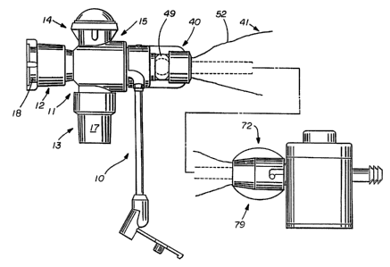
Figure i shows a suction control valve and a manifold
o! a respiratory support system attached for use to a
suction catheter device formed in accordance with the
principles of the present invention:
Figure Z is a plan view of a portion of the suction
catheter device which includes the manifold-end connector
thereof formed in accordance with the principles of the
present invention:
~~.~~~.~f
WO 93/21981 PGT/US93103802
9
Figure 3 is a cross-sectional view of the manifold-end
connector of the suction catheter device shown in Figure 2:
Figure 4 is a side view of the ventilator manifold;
Figure 5 is a cross-sectional view of the ventilator
manifold shown in Figure 4:
Figure 6 is a cross-sectional view of the ventilator
manifold with the manifold end connector of the suction
catheter device attached to the access port thereof;
Figure 7 is a cross-sectional view of the valve-end
connector of the suction catheter device formed in
accordance with the principles of the present invention; .
Figure 8 is a cross-sectional view of the housing
portion of the valve-end connector;
Figure 9 is a cross-sectional view of, the insert
portion of the valve-end connector;
Figure 10 is a plan view of an alternative embodiment
of the suction catheter device formed in accordance with the
principles of the present invention:
Figure li is a cross-sectional view of the valve-end
connector of the alternative embodiment of the suction
catheter device shown in Figure lo:
,Figure 12 is a perspective view of a suction control
valve formed in accordance with the principles of the
present invention;
Figure 13 is a cross-sectional view of the fluid flow
valuing device of Figure 12;
Figure 14 is a cross-sectional view of the valve
housing of the fluid flow valuing device of the present
invention;
. Figure i5 is a cross-sectional view of the valve body
of the fluid flow valuing device of the present invention;
Figure 16 is a top view of the rotatable core of the
fluid tlow valuing device formed in accordance with the
principles of the present invention; and
2~3~12
WO 93/21981 PCT/US93/03802
Figure 17 is a cross-sectional view of the fluid flow
valuing device as shown in Figure 13, showing the actuator
and core rotated to the locked position.
MODE FOR CARRYING OUT THE INVENTION
5 As shown in the exemplary drawings for the purposes of
illustration, an embodiment of a manifold, suction catheter,
and suction control valve of a respiratory support system
made in accordance with the principles of the present
invention, referred to generally by the reference numeral
10 10, is provided for interchangeable use of either the
suction catheter or the suction control valve without
interruption of respiratory support of the patient.
More specifically, as shown in Figure. l, the ventilator
manifold 11 includes a plurality of access ports which
facilitate its connection to a ventilator circuit which is
in use by the patient. The manifold ii is attached to a
patient for fluid flow communication with the patient's
lungs by the connection of the patient attachment port 13
thereof to the connector of an endotracheal tube assembly
(not shown) which has been previously positioned in the
trachea of a patient by any one of several well known
procedures.
Ventilator circuit connection port 12 of the manifold
11 is designed fo= connection to flexible breathing hoses
from the ventilator (not shown) in a well-known manner, such
as through a "Y" site connector. Port 1~ is normally capped
and closed against air flow except far instances when
nonpressurized ventilation is desired. The ventilator
circuit provides a breathable gas mixture to the patient
through one hose, and receives expelled air from the
patient's lungs through another hose. The ventilator
circuit further commonly includes various valves, regulators
and the like associated with~the hoses in order to effect
respiration of the patient. The manifold 11, and hoses
attached thereto at the ventilator circuit connection port
WO 93/21981 PCT/US93/03802
11
12, are generally made of disposable plastic material and
are generally intended to be used by only one patient and
then discarded.
When attached to the patient, the entire respiratory
support system 10 is designed to isolate the patient's lungs
from the atmosphere and allow pressurised forced ventilation
of a gas mixture of a high oxygen content from the
ventilator into the patient's lungs. Commonly ventilators
of this type are used to maintain a positive end expiratory
pressure (PEEP) within the ventilator manifold 11 and the
.patient's lungs at all times during exhalation. This
technique is commonly used because of the benefit of
supplying a minimum concentration of oxygen to the patient .
at all times for maintaining a proper blood oxygenation
level. The PEEP procedure also keeps a large number of lung
alveoli of the patient open at~all times during respiratory
support, thus increasing the effective lung area subject to
ventilation.
Prevailing respiratory support techniques~including
PEEP, have made it very disadvantageous to interrupt
respiratory support to the patient by opening the ventilator
manifold to the atmosphere and thereby causing a loss of
interior manifold pressure. Therefor, the necessary
attachment of accessory devices thereto for medical
procedures has been difficult to the loss of isolation of
the respiratory system from the atmosphere during these
procedures, and the immediate loss of effective lung surface
area due to collapse of the patient s lung alveoli.
Further, when such procedures have been prolonged for any
reason, the patient's blood oxygen has often dropped to
inadequate levels, and subsequently forced overexertion of
the patient's lungs and heart in order to return the blood
oxygen level to normal. Also, disassembly and reassembly of
the respiratory support system components for procedures
with prior art accessory devices has often been a very time
consuming procedure for the medical worker involved.
CA 02134123 2005-O1-19
12
The present invention resolves the problems associated
with loss of isolation of the respiratory support system from
the atmosphere when accessory devices must be inserted or
attached in order to perform necessary medical procedures, or
alternatively, when they must be replaced, during respiratory
support of a patient.
Specifically, the manifold 11 of the present invention
includes an access port 15 which is in fluid flow communication
with the interior of the manifold 11. The access port 15
includes a normally closed valve (see Figure 5j preferably made
of a resilient material such as rubber or silicone which
maintains the interior of the manifold 11 isolated from the
atmosphere at all times. As explained above, the interior of
the manifold 11, although experiencing constant pressure
fluctuations, is generally kept at a pressure which is slightly
above atmospheric pressure in order to properly administer
oxygen according to the PEEP procedure.
The ventilator circuit connection port 13 and the patient
attachment port 12 may, if desired, include swivel connectors
17 and 18 respectively thereon in order to allow relative
rotation between the manifold 11 and the trachea tube and
breathing hoses in order to isolate the trachea tube from the
incidental forces causes by the manifold 11 or the breathing
hoses attached thereto so as to increase the comfort of the
patient.
As best shown in Figures 4 and 5, the access port 15
includes a normally closed valve 16 formed therein which
maintains the interior of the manifold 11 isolated from the
atmosphere at all times. As explained above, the interior
of the manifold 11, although experiencing constant pressure
fluctuations, is generally kept at a pressure which is
slightly above atmospheric pressure in order to properly
administer oxygen according to the PEEP procedure.
Therefore, the valve 16 is preferably made of a resilient
W 93/21981 ~ ~ ~ i~ ~ ~~ ~ PCT/US93/03802
O
13
material to ensure that pressure isolation of the manifold
11 is maintained.
The valve l6 is preferably formed to a circular disk
shape and inserted into the manifold I1 between the access
port 15 and a support ring 19. The valve 16 is formed with
a slit, or a pair of perpendicular slits 20 which are
normally closed against fluid flow therethrough, but may be
forced opened by the insertion of the manifold end connector
40 therethrough (as shown in Figure 3), of the suction
to catheter device 41.
The interior of the access port 15 is lined with a
sleeve member 21 which covers the entire interior surface of
the access port 15 and abuts in sealing relationship against
the normally closed valve 16. The interior diameter of the
sleeve member 21 is predetermined to cause a snug fit with
the manifold end connector 40 (as best shown in Figure 6) to
assist in the prevention of leakage from the manifold 11
when the normally closed valve 16 is forced opened by the
manifold end connector 40.
The access port 15 forms a side opening 22 therethrough
through which a portion of the sleeve 21 extends to be
attached,'such as by solvent bonding, to a pigtail fluid
injection tube 23 which is intended for use in transporting
fluid through the access port side opening 22 into the
interior of the~access port 15. The opposite end of the
pigtail tube 23~includes a lust connector 24 attached
thereto with an integrally formed lust connector plug 25. A
check valve 26, taking the form of a collapsible sleeve, may
be positioned between the lust connector 24 and the pigtail
tube 23 if desired, to collapse upon injection of fluid
through the lust connector 24 into the pigtail tube 23, but
,.
expand to block fluid flow in the opposite direction.
It is preferred that the sleeve member 21 be formed of
a relatively flexible material such as plasticized PVC,
having good solvent bonding characteristics with the
material forming the pigtail tube 23, the pigtail tube 23
PCT/US93/03802
W093/2198~~~~~'~~~
14
preferably is formed of the same material as the sleeve
member 21. The access port 15 according to the preferred
embodiment of the present invention is preferably formed of
clear ABS, which is preferably the same material forming the
main body of the manifold 11 in order to ensure good
ultrasonic or solvent bonding therebetween.
Referring now to Figures 2 and 3, the manifold-end
connector 40 of the suction catheter device 41 is shown.
The connector 40 includes a unitary housing 42 which forms
an adaptor 43,, a locking mechanism.44, a base ring 45; and a
plurality of rib members 46. The base ring 45 forms a
generally cylindrical opening through which the sleeve
attachment ring 47 can be inserted for frictional engagement
to hold the sleeve 52 in proper position relative to the
connector 40.
A magnifying insert 48 is formed as a generally
cylindrical member having a bulbous lens 49 at one end
thereof forned of clear plastic and a cylindrical extension
50 formed at the other end thereof. The cylindrical
extension 50 is formed to a slightly smaller diameter than
. the sleeve attachment ring 47 and may include ribbing 51
around the'exterior surface thereof to aid in frictional
engagement between the extension 5o and the sleeve
attachment ring 47. When constructed for use, the sleeve 52
is positioned around the sleeve attachment ring 47 so as to
be frictionally engaged between the extension 50 and the
interior surface of the sleeve attachment ring 47, and
wrapped around at'least a portion of the exterior surface of
the sleeve attachment ring 47 to also become frictionally
engaged with the base ring 45 when the sleeve attachment '
ring 47 is inserted therein.
The lens 49 of the magnifying insert 48 is preferably
formed of a substantially clear plastic which magnifies the
portion of the catheter 54 for viewing by a user through the
generally cylindrical opening or window 55 formed by the
unitary housing 42 between the base ring 45 and the locking
s
WO 93/21981 PCTlUS93/03802
mechanism 44. The window 55 extends around the entire
circumference of the unitary housing 42 and allows viewing
of the lens 49 by a user at any viewing angle except where
the window 55 may be slightly covered by a portion of the
5 ribbed members 46 which extend between the base ring 45 and
the locking mechanism 44.
The unitary housing 42 also includes an annular locking
shoulder 56 which operates in conjunction with an annular
locking shoulder 5? on the magnifying insert 48 to secure
the magnifying insert 48 in proper position within the
unitary housing 42, and to ensure an air-tight seal
therebetween and with the sealing ring 58.
The magnifying insert 48 has a generally cylindrical
passageway 53 formed therethrough which is, of slightly
15 larger diameter than the catheter 54 and which allows
uninhibited movement of the catheter 54 therethrough.
If desired, the catheter.54 may be formed with a tip 59
of softer material than the remainder of the catheter 54 and
which may include side openings 60 therein. The catheter 54
may also include a series of markings such as ring marking
61 and/or, number markings 62 along the length thereof which
will,tend to be magnified when located beneath the lens 49.
The marking 61 is intended to indicate the completely
withdrawn position of the catheter 54 into the adaptor 43.
For example, in operation, the user can withdraw the
catheter 54 through the manifold-end connector 40 until the
ring marking 61 moves into view within the lens 49.
Positioning of the ring marking 61 beneath the lens 49
indicates to the user that the catheter 54 has been
withdrawn the entire recommended distance through the
connector 40 and cannot be further withdrawn without risking
inadvertent passage of the side openings 60 of the catheter
54 past the sealing ring 58, which would effectively allow
leakage of air past the sealing ring 58. As is readily
evident, an even greater leakage of air would occur if
withdrawal of the catheter 54 continued until the distal end
~13~3.2:~
WO 93/21981 PCT/US93/03802
16
63 thereof passed through the sealing ring 58 or through the
connector 40 entirely.
The number markings 62 may be positioned along the
catheter 54 so as to indicate to the user a particular
predetermined distance which the distal end 63 thereof
extends beyond the connector 40. As each number marking 62
appeals in the lens 49, the user can recognize the number as
corresponding to a particular predetermined distance that
the distal end 63 is extending beyond the connector 40. In
this manner,~when the connector 40 is attached to the
manifold 11 of the respiratory support system 10, the user
can readily determine how far down a patient's trachea or
lungs the catheter 54 has been inserted during an aspiration
procedure by noting the particular number marking 62 visible
through the lens 49.
The adapter 43 and locking mechanism 44 of the
connector 40 operate to attach the connector 40 to the
ventilator manifold 11. As best shown in Figure 6,
attachment of the connector 40 to the manifold 11 is
effected by insertion of the adapter 43 into the access port
15 until ~.he tapered top section 64 thereof engages the
valve 16 and forces it toward the interior of the manifold
11. Upon complete insertion of the adapter 43 into the
access port 15, the valve 16 is completely open.
In reference to Figures 3 and 6, the locking mechanism
44 of the connector 40 is formed to encircle a portion of
the adapter 43 and includes a pair of arcuate slots 67 and
68 which operate together to ensure secure attachment of the
connector 40 to the access port 15 of the manifold il, and
also ensure proper relative orientation between the adapter
43 and the access port 15, to cause the injection fluid
opening 69 of the adapter 43 to be positioned in alignment
with the side opening 22 of the access port 15 when the
adapter 43 is properly locked in position therein for use.
The arcuate slot 67 is sized to be engageable with the nub
71 which is located directly opposite the side opening 22 on
~~3.~.1~
WO 93/21981 PCT/US93/03802
17
the access port 15. The arcuate slat 68 is larger in width
than the arcuate slot 67 and therefore can accommodate the
side opening 22 of the access port 15. As is readily
evident, the adapter 43 can only be locked in position
within the access port 15 in one unique. relative orientation
in which the injection fluid opening 69 and the side opening
22 are in alignment.
As best shown in Figure 5, attachment of the manifold
end connector 40 to the respiratory manifold 11 is effected
by insertion~of the adaptor 43 into the access port 15 until
the tapered top section 64 engages the valve 16 and forces
it toward the interior of the manifold 11. Upon complete
insertion of the adaptor 43 into the port 15, the valve 16
is completely open and the elastic sleeve.member 21
sealingly engaged with the adaptor 43. Also, the sleeve
shoulder 38 of the sleeve member 21 is forced to resiliently
deform within the base 39 of the locking mechanism 44. This
increases the air tight seal and assists in positively
locking the adaptor il to the access port 15 by forcing the
arcuate slots 67 and 68 against the nub ?1 and side opening
22 respectively.
.It is intended that during insertion of the adaptor 11
into the access port 15, the sealing relationship formed
between the sleeve member 21 and the adaptor 43 commence
prior to opening of the valve 16 by the tapered top section
64, in order to ensure isolation of the interior of the
manifold 11 from the atmosphere during attachment of the
suction catheter device 41. Once completely inserted within.
the port 15, the tapered top section 64 extends completely
through the access port 15 and into the manifold central
chamber 37.
As shown in Figure 4, the pigtail tube 23 can be used
to inject fluid into the adaptor 43 to clean the suction
catheter 54 and the sealing ring 58 of mucal materials which
may have accumulated therein due to repeated insertion and
withdrawn of the catheter 54 from the patient's lungs during
~1~41~:~ , _
WO 93/21981 PGT/US93/03802
18
aspiration procedures. The cleaning fluid can then be
aspirated through the catheter 54 to remove it from the
interior of the adaptor 43 and the manifold 11.
. Alternatively, if desired, fluid may be injected
through the gigtail tube 23, into the adaptor 43 and through
the central chamber 37 of the manifold 11, and through the
patient connection port l2 into the trachea and lungs of the
patient for purpo~es of lavage. The suction catheter 54 can
then be inserted through the manifold ii into the patient's
trachea and the fluid can be aspirated along with any mucal
.materials dislodged by the lavage fluid, as will be
explained below.
Referring now to Figures 7-9, the valve-end connector .
73 of the suction catheter device 41 is shown. The
connector ~2 includes three main components including a
housing 73, a snap-in insert 74, and a slit septum 75. The
housing 73 forms a passageway '76 therethrough which has an ..;
annular shoulder 77 protruding thereinto at.an approximately
centrally located position along the passageway 76. The
ZO housing 73 also includes a pair o!' locking slots 78 for
attachment of the connector 72 to a suction control valve 79
(~~ in~dashed lines in Figure 7) and a pair of
longitudinally oriented linger grips 8o which facilitate
rotation of the connector 72 for attachment to the suction
control valve 79 (sae Fig. 1).
~e s~~~ ~ 74 is a generally cylindrical
tubular member having a plurality o! uniformly spaced fins
81 positioned longitudinally thsrealong in a plurality of
uniformly spaced locations. Each fin 8i includes a locking
shoulder 82 at one end thereof and a tapered edge 83 along
the length thereof. The locking shoulder 82 and tapered
edge 83 a=s sized to match the annular shoulder 77 and the
tapered portion 84 of the housing passageway 76 so as to
securely hold the catheter,slesve 52 in a friction fit
within the housing 73 between the fins 81 and the tapered
portion 84 of the passageway 76.
__
WO 93/21981 PGT/US93/03802
19
. The snap-in insert 74 also forms a tubular channel 85
therein which is sized to accommodate the proximal end of
the catheter 54 and the distal end of the slit septum 75 in
a permanently attached manner such as by solvent bonding or
the like. The slit 86 in the septum may be of any desired
shape or configuration such.a linear, curvelinear, cross, or
the like, which can be easily pushed open by the suction
control valve 79 and which will return to its closed
position upon its removal.
It should~be noted that the connector 72 intentionally
forms an open air flow path from the interior of the sleeve
52.snap-in insert 74 between the fins 81 and around the
slitted septum 75 past the suction control valve 79 to the
atmosphere: This air flow path,is intentionally designed to
ig ~~a that the sleeve 52 is not sealed against air flow
between the interior thereof and the atmosphere. This
prevents pressure or vacuum build up within the sleeve 52
during operation of the su~ioncatheter device 41 due to
contraction and expansion of the sleeve 52 caused by the
2o aovem~nt of the catheter 54 through the respiratory manifold
' 11.
.Reterring..now to Figure 10, as alternative embodiment
of the respiratory support system 10 is shown which is
substantially identical to,the respiratory support system 10
25 described sbova,~axcapt that the catheter thereof is a dual-
lumen cathmter and the valve-end connector is modified to
allow injection of fluid from a flexible fluid vial 87
through the second lumen.
l~tora specifically, as shown in Figure 11, the dual-
30 lumen valve-end connector e8 includes a housing 89 which
forms a generally cylindrical opening 9o into which the
suction control valve (shown in dashed lines) can be_
inserted !or connection, and in which a slitted septum 91 is
affixed such.as by annular shoulder 92. The slitted septum
35 91 includes a normally closed slit 93 therein which is
opened by the insertion o! the suction control valve in the
~134~.2~
WO 93/21981 PCT/US93/03802
manner described previously with respect t~ the single-lumen
valve-end connector 72 above.
The housing 89 also includes a second cylindrical
opening 94 which has a tubular extension 95 formed therein.
5 The opening 94 is sized to reeeive the proximal end of the
dual-lumen catheter 96, and the tubular extension 95 is
designed to be inserted within the larger (suction) Lumen 97
of the proximal end 98 of the dual-Lumen catheter 96.
As is evident in Figure 11, the proximal end 98 of the
10 dual-lumen catheter 96 is somewhat larger in diameter than ,
the remainder of the catheter 96. This is preferably due to
intentional manufacturing of the catheter 96 with an
enlarged proximal end 98, and not necessarily due only to
stretching of the proximal end 98 about the tubular
15 extension 95. This is a desirable feature of the present
invention in that intentional over-sizing of the proximal
end 98 of the catheter 96 helps avoid any restricted
diameter areas along the large lumen 97, and also helps
prevent the attachment of the large lumen 97 over the
20 tubular extension 95 from causing a restriction in the
diameter of the smaller (irrigation) lumen 99 at the
proximal end 98..
A flow channel I00 extends away from the bottom of the
second cylindrical opening 94 and is in fluid flow
communication with the small lumen 99 of the catheter 96.
The flow channel 100 communicates with an L-shaped tubular
member 101 which includes a luer-type connection opening 102
designed to receive the flexible fluid vial 87 (shown in
Figure 10). The L-shaped tubular member 101 includes a one-
way valve 103 therein preferably formed as a soft tubular
sleeve which collapses when pressurized and allows fluid to
be injected into the small lumen 99 of the catheter 96
through the flow channel 100, but inhibits fluid flow in the
opposite direction.
The catheter sleeve 52, which surrounds the catheter
96, is affixed to the housing 89 by the attachment ring 104,
WO 93/21981 ~ ~ ~ ~ ~ ~ '~ PCT/US93/03802
21
in a friction fit manner, and an air flow path from inside
the sleeve 52 through the connector 88 to the atmosphere is
formed by the air flow channel 105.
The preferred manner of assembly of the suction
catheter device 41 of the present invention is as follows.
First, depending on whether a single-lumen or dual-lumen
catheter is used, the single-lumen or dual-lumen valve-end
connector 72 or 88, respectively, is assembled. In the case
of the single-lumen valve-end connector 40, the proximal
end of the catheter 54 and the distal end of the septum 75
are bonded into the tubular channel 85 of the snap-in insert
74. The housing ,73 is then slid over the sleeve 5Z and the
distal end of the catheter 54 is inserted through the
' proximal end of the sleeve 52 and, passed completely
therethrough. The housing 73 is drawn proximally along the
sleeve 52 until the snap-in insert 74 is drawn into the
housing 73 past the annular shoulder 77 in the passageway 76
thereof and snapped into position such that the lacking
shoulder 82 and tapered edge 83 of the fins 81 of the snap-
in insert 74 are positioned adjacent the annular shoulder 77
and tapered portion 84 of the channel 76, with the proximal
end of the sleeve 52 fractionally held therebetween.
The manifold-end connector 40 is then assembled by
first passing the sleeve attachment ring 47 over the distal
end 40 of the catheter 54 and over the sleeve 52. Then the
magnifying insert 48 is passed over the distal end 63 of the
catheter 54 until the distal end 63 extends a predetermined
distance beyond the magnifying insert 48. The sleeve 52 is
then extended over the magnifying insert 48 and the sleeve
3o attachment ring 47 is push~d onto the cylindrical extension
50 of the magnifying insert 48 to fractionally fit therewith
and trap the sleeve 52. therebetween. The remainder of the
sleeve 52 extending beyond the distal end of the attachment
ring 47 is then folded or rolled back over the attachment
ring 47. A sealing ring 58 is then inserted into the
annular locking shoulders 56 of the housing 73 and the
~13~~.~~,~~
WO 93121981 PCT/US93/03802
22
entire sub-assembly consisting of the magnifying insert 48,
the sleeve attachment ring 47, and the portion of the sleeve
52 wrapped around the sleeve attachment ring 47, are then
inserted into the housing 42 through the base ring 45 until '
the annular locking shoulder 57 of the magnifying insert 48
snaps into, and locks behind, the annular locking shoulder
56 of the housing 42 where it presses against the sealing
ring 58 in an air-tight manner.
When assembled in this manner, the sleeve 52 is
~ attached to the manifold-end connector 40 such that the
catheter 54 can be withdrawn through the connector 40 at
least so far as to allow the ring marking 61 thereon to be
positioned within the lens 49 of the magnifying insert 48,
and to allow the adaptor 43 to protect the, distal end 63 of
the catheter 54 when the connector 40 is being attached to
the manifold 11.
If it is desired to assemble a dual-lumen suction
catheter device 41, the valve-end connector 88 thereof (as
shown in Figure 11) must first be assembled by passing the
attachment ring 104 thereof over the proximal end 98 of the
catheter 96 and the proximal end of the sleeve 52 and then
inserting the proximal end 98 of the catheter 96 into the
second cylindrical opening 94 until a large lumen 97 thereof
is securely attached to the tubular extension 95, (being
careful, of course, not to inadvertently block the flow
channel 100 by the proximal end 98 of the catheter 96). The
catheter 96 can then be permanently affixed within the
cylindrical opening 94 by any known means such as by solvent
bonding or the like. The proximal end of the sleeve 52 is
then wrapped around the attachment ring 104 and the
attachment ring 104 is affixed by friction fit to the
housing 89. Any part of the sleeve 52 extending beyond the
proximal side of the attachment ring 104 can then be trimmed
off if desired.
The slitted septum 91 is then forced into the
cylindrical opening 90 until it engages with the annular
WO 93/21981 ~ ~ ~ ~ ~ ~ ~ PGT/ZJS93/03802
23
shoulder 92 therein, and the one-way valve 103 is inserted
into the L-shaped tubular member 101 which itself is then
permanently attached to the housing 89 around the flow
channel 100 thereof.
The remaining assembly operations of the dual-lumen
version of the suction catheter device 41 of the present
invention are identical to the assembly of the single-lumen
version described above.
As shown in Figure 12, the valve 79 of the present
invention is formed of a valve housing 109 with a suction
catheter device connector 110 extending away therefrom in a
radial direction and a suction pressure source connector 111
extending away therefrom in a radial direction opposite the
connector 110. A lower cap 114 having the.same diameter as
the valve housing 109, covers the. bottom of the valve
housing 109. An upper cap 112 is connected to the top of
the valve housing 109. A portion of the actuator 113
(constituting the button 115) extends from the interior of
the valve housing 109 through the upper cap opening 116 and
above the annular surface 117 of the upper cap 112. The
positioning of the actuator 113 on the valve 79 is intended
to allow for ease of manipulation thereof in a single hand
of the user. The valve 79 is sized so as to be easily
placeable within a user's palm such that the user's thumb
may rest comfortably on the button 115 of the actuator, with
the user's fingers curling about the lower cap 114 to
support the valve '79 against the internal bias of the
actuator 113 when the user presses on the button 115 to open
a suction channel through the valve 79.
. Referring now to Figure 13, the preferred internal
structural arrangement of the valve 79 of the present
invention will be explained, with the aid of Figures 14-16
which show various views of individual component.
Referring specifically to Figures 13 and 14, the valve
housing 109 is formed generally into a hollow cylindrical
shape and includes a suction catheter device connector
CA 02134123 2005-O1-19
24
opening 118 and a suction source connector opening 119 which
are formed through the side wall 121 at diametrically opposed
positions and which pass into the large cylindrical chamber
122. The openings 118 and 119 each allow attachment of the
suction catheter device connector 110 and the suction source
connector 111 respectively to the valve housing 109.
The large cylindrical chamber 122 includes a
longitudinally oriented groove 123 (best shown in Fig. 14)
which aligns with a nub (not shown) on the valve body 125 when
the valve body 125 is inserted thereinto in order to ensure
their proper relative orientation for use.
The housing 109 also includes a small cylindrical chamber
124 which opens into the large cylindrical chamber 122 and is
open through the bottom 126 of the housing 109, which forms
part of the ancillary fluid flow channel through the valve 79
as will be explained below.
As best seen in Figures 13 and 15, the valve body 125 is a
generally cylindrical member having a plurality of openings
therethrough. First, a generally conically shaped bore 126 is
formed through the top surface and extends nearly to the bottom
surface 128 thereof. The conical bore 126 is surrounded at its
opening adjacent the top surface 127 by an annularly shaped
protrusion or seat 129. A cylindrical fluid flow channel,
identified for simplicity of later explanation of operation of
the valve 79 as elements 130 and 133, passes through the valve
body 125 and completely through the side wall 131 thereof. The
channel 130,133 is oriented such that its longitudinal axis
perpendicularly intersects with the longitudinal axis of the
conical bore 126. A second cylindrical fluid flow channel 132,
generally perpendicular to the first fluid flow channel 130,133
passes through the bottom 128 of the valve body 125 into the
conical bore 126.
As best seen in Figures 13 and 16, the core 134 rests
within the conical bore 126, and is generally conical in
shape to match the shape of the conical bore 126. The core
~ ~ ~~~.~:;
WO 93/21981 PCT/US93/03$02
- 134 forms several channels therethrough which can be
positioned for operation of the valve 79 by rotation of the
core 134 relative to the body 125 in the manner as will be
described below.
5 A primary fluid flow channel 135 is formed through the
core 134 so as to match the diameter of, and be alignable
with, the fluid flow channels 130 and 133 in the body 125.
An ancillary fluid flow channel 136 (best shown in Figures
16 and 17) attaches fluid flow channel 130 with the second
10 cylindrical gluid flow channel 132. The ancillary fluid
flow channel 136 is positioned about the core 134 so as to
be oriented approximately one quarter of the way around the
circumference of the core from the primary fluid flow .
channel 135, or in other words (as best seen in Figure 16)
15 the position of the ancillary fluid flow channel 136 is
approximately 90° around the surface 137 of the core 125
from the primary fluid flow channel 135. The relative
positioning of the primary and ancillary fluid. flow channels
135 and 136 respectively, allow positioning of the core 134
20 in a first position (shown in Figure 13) in which the
primary fluid,flow channel 135 is oriented for fluid flow
between fluid flow channels 130 and 133, and a second
position (as shown in Figure 17) in which it is rotated 90°
from the first position and in which the ancillary fluid
25 flow channel 136 thereof is in alignment between fluid flow
channel 130 and the second cylindrical fluid flow channel
132. The operation of the valve 79 with respect to each w
position of the core 134 will be explained in detail
momentarily.
. The core 134 includes a bleed channel 146 which extends
from the top surface 137 of the core 134 into the primary
fluid channel 135. -
The core 134 also includes a rectangularly shaped slot
138 which passes through the top surface 137 of the core and
bisects the primary fluid flow channel 135. As can be seen
in Figures 13 and 17, the slot 138 accommodates the actuator
z~3~~~~~
WO 93/21981 PCT/US93/03802
26
extension 139 for sliding movement therein between a first
position in which the extension 139 blocks flow through the
primary fluid flow channel 135, and a second position in
which the actuator 113 is forced downwardly to move the
actuator opening 140 into alignment with the primary fluid
channel 135.
The extension 139 also allows the actuator 113 to
effect rotation of the core 134 when the button 115 of the
actuator is rotated relative to the valve 79. Rotation of
the core 134 between the first or open position as shown in
Figure l3, and the lock position shown in Figure 17, is
caused by rotation of the button 115 relative to the valve
79"approximately one quarter turn. The valve 7~ may include
markings such as on the surface 117 of the upper cap 112 (as
shown in Figure 12) and/or on the button 115, to indicate
the position of the core 134 for proper.operation of the
valve.
As shown in Figure.l3, the actuator 113 includes a
shoulder 141 which is sized to fit within the large
20~ cylindrical chamber 122 of the housing 109 to be held in
place there within by the upper cap 112. The shoulder 141
forma a slot 142 tharethrough which is located adjacent the
opening 143 in the upper cap 112 when the actuator 113 is in
its first or open position. The shoulder 141 also includes
a tab 159 (shown only in Fig. 17) which is positioned around
the circumference of the shoulder 141 approximately 90° away
from the slot 142. The tabs 159 rides in the slot 158 (best
shown in Fig. l4) formed around one quarter (90°) of the
internal circumference of the large cylindrical chamber 122
of the housing 109 and joins with the longitudinally '
oriented groove 123. As is evident, the tab 159 allows the
actuator 113 to rotate.only a quarter turn, since it is
inhibited from further rotation by the ends of the slot 158.
In Figure 13 for example, the tab 159 is rotated to the end
of slot 158 which is adjacent the longitudinal groove 123.
In this position, the slot 142 through the shoulder 141 of
u... 3.~_ .~.~: .. . . ..." , .. '~ :.,. ,. . ~ -~ .~~~~,:,.. -. ~~ ..'.' '. .
y; ,: . ,. . , . .~ : -.'._.. . ., ., ,.~ - .~ , .. ~_., ,. . .. . .,..,.: .
,,., .. , ., , ., . ~ . . . ~ .., -. . .; , ., ; ' ': : ..' . :_.
~~~~~~p
WO 93/21981 PGT/US93/03802
27
the actuator button 115 is positioned adjacent the opening
143 in the upper cap 112. In Figure 17, the tab 159 is
rotated one quarter of a turn to the opposite end of slot
158 and is positioned directly adjacent the opening 143 in
the upper cap 112. It should be'noted that the width of the
tab 159 is less than the width of the groove 123 so that the
tab 159 may pass downwardly therethrough whenever it is
aligned therewith and the button 115 of the actuator 113 is
pushed down. As is readily evident therefor, only single
rotational position of the actuator 113 allows downward
movement thereof, this being defined as the "open" position .
where the tab 159 is aligned with the groove 123.
Referring again to Figure 13, a flexible, soft
elastomeric pad 144 of generally circular shape is affixed
to the actuator 113 below the shoulder 141 and is of a
slightly larger diameter than the diameter of the seat 129
protruding from the surface 127 of the body 125. A
compression spring 145 is positioned between the~top surface
137 of the core 134 and the shoulder 141 and operates to
0 hold the actuator 113 in its uppermost position where the
actuator shoulder 141 abuts the upper cap 112.
,The fluid flow passage formed by the suction source
connector lii, the fluid flow channels 130, 135, 133, and
the suction catheter device connector '110, forms essentially
an elongate linear channel of uniform diameter passing
entirely through the valve 79. When the actuator opening
140 is moved downwardly to be positioned within the primary
fluid flow channel 135, it is readily apparent that a single
linear fluid flow channel of uniform diameter through the
entire valve 99 is formed which doss not cause any
obstruction or blockage of fluid passing through the valve
79. In this manner, mucal material, including clotted
material referred to generally as mucus plugs, encounters no
obstruction as it is drawn through the valve 79, and
therefore is not likely to cause blockage of the valve
during use.
. CA 02134123 2005-O1-19
2$
As shown in Figures 13 and 17, the bottom of the valve
housing 109 is covered with a lower cap or "flip cap" 114. The
lower cap 114 is formed of a generally cylindrical shape having
a diameter equal to the diameter of the valve housing 109 and
includes a fixed member 147 which is hingeably attached to a
cover member 148 by means of hinge 149 which may be of the
"living hinge" type and formed of polymeric material. The fixed
member 147 is preferably attached to the annular base 150 of
the valve housing by a snap fit or an ultrasonic weld, however
any well known attachment means may be used. The fixed member
147 includes a circular plate 151 which has an opening 152
formed centrally therein which is surrounded by an inwardly
projecting boss 153. The cover member 148 also includes a
circular plate 154, on the interior surface of which a plug 155
is formed and sized so as to fit snugly within the fixed member
opening 152 to form a fluid tight seal whenever the cover
member 148 is closed over the fixed member 147.
A bushing is located within cylindrical chamber 124 of the
valve housing 109 between the body 125 and the circular plate
151. The bushing 156 forms a fluid flow channel 157
therethrough which is shaped on one end thereof to connect with
boss 153 on the circular plate 151 and on the other end to
align with the second cylindrical fluid flow channel 132 of the
body 125 in fluid tight relationship, thus allowing fluid flow
connection of the bushing fluid flow channel 157 with the
suction source through the ancillary fluid flow passage 136.
It should also be noted that all internal components of
the valve 79, including the actuator 113, core 134, body
125, and bushing 156 are designed such that assembly thereof
into the valve housing 109 is substantially simplified. In
each instance, the particular element to be assembled into
the valve housing 109 has been designed to the extent
possible to allow only the element to fit within the valve
housing 109 only when it is properly positioned for
WO 93/21981 PCT1US93l03802
29
assembly. Specifically, the bushing 156 is formed
asymmetrically to allow only one possible positioning
thereof within the small cylindrical chamber 124. The body
125 includes a nub (not shown) which .must be aligned with
groove 123 in order for the body 125 to be insertable within ,.
the large cylindrical chamber 122. The core 125 includes a
step 66 in the bottom of the slot 138 thereof which
accommodates the actuator extension 139 forces proper
alignment of the actuator extension 139 therein by allowing
10~ proper operation only when shoulder 6? of the extension 139
is oriented in alignment therewith. The actuator 113/core
125 sub-assembly can 'only be positioned within the valve
housing 109 such that the tab 159 of the shoulder 141 of the
actuator button 115 is positioned within the slot 158 of the
housing 109. Although various methods and means for
ensuring proper assembly of the valve 79 of the present
invention have been shown, it should be understood that
other means and methods known in the art could also be
employed without departing from the spirit and scope of the
present invention.
OPERATION OF THE PREFERRED EMBODIMENTS
Operation of the respiratory support system l0 is
preferably as follows. First, the ventilator manifold il is
attached to the~tracheal tube which has previously been
inserted into the patient's trachea, and the ventilator
circuit of the respiratory support system is attached to the
manifold li in a well-known manner. The manifold-end
connector 40 of the suction catheter device 41 is then
inserted into the access port 15 of the manifold il and
rotated to its above-described locking position therewith.
The suction control valve 79 is then inserted into the
valve-end connector 72.and then attached to a source of
suction pressure in a well-known manner.
When in its first or open position as shown in Figure
13, the actuator 113 of the control valve 79 allows a bleed
2~.~~~~,:~
WO 93/21981 PCTlUS93/03802
of suctioned atmaspheric air to pass into the valve 79
through the cap bleed opening 143 and move past the actuator
shoulder slot 142 into the large cylindrical chamber 122 of
the valve housing 109 where it is then drawn through the
5 core bleed channel 146 into the primary fluid flow channel
135 of the core 134 and from there through channel 130 and
into the suction source connector 111 where it can be drawn
out of the valve 79 into the suction pressure source.
Movement of atmospheric air through the valve 79 to the
10 suction source when the actuator 113 is in the open position
generates an auditory signal, being a very recognizable
"hissing" sound, which is indicative of the operation of the
suction pressure source and the presence of suction pressure
within the valve 79.
15 When it is desired.to suction the patient's trachea or
lungs, the catheter 54 is advanced through the manifold-end
connector 40, the manifold 11, and the tracheal tube into
the patient's trachea and lungs any desired distance (which
can be monitored by the medical worker performing the
20 procedure by viewing the number markings 62 which appear
through the lens 49 of the connector 40). Aspiration of the
patient's trachea and lungs is then performed by the user
forcing the actuator button 115 downwardly into the valve
housing 109 against the bias of the compression sgring 145.
25 This linaar translational movement of the actuator 113
relative to the valve housing 109 causes the actuator
extension 139 to move downwardly within the core slot 142.
This. causes the actuator opening 140 to move into alignment
with the primary fluid flow channel 135 of the core 134. No
30 resistance of downward movement of the actuator 113 is '
caused by the tab 159, since it is aligned with groove 123
and can therefore pass,downwardly therein.
As can be seen, although the actuator extension 139
blocks the primary fluid flow channel 135 whenever the
actuator 113 is in its fully upwardly extended or "released"
position, it gradually moves out of blocking position as the
WO 93/21981 ~ ~ ~ ~ ~ ~' ~ PCT/US93/03802
31
actuator opening 140 is moved into alignment position with
the primary fluid flow channel 135 as the actuator button
115 is depressed. As is also readily evident, the amount of
suction pressure allowed through the primary fluid flow
channel 135 can be regulated from a "no flow"'level when the
button 115 is released, to gradually increasing flow levels
as the actuator opening l4o is moved into alignment with the
primary fluid flow channel 135 as the button 115 is
depressed toward the body 125.
l~s can be seen, complete depression of the button 115
occurs when the pad 144 on the actuator shoulder 141
contacts and seals against the seat 129 of the body 125. In
ttie completely depressed position, the actuator opening 140
is completely aligned with the primary fluid flow channel
135 and presents-no fluid flow obstacle thersthrough.
It should be noted that when the actuator 113 is
completely depressed until the pad 144 seals against seat
129 causing camplete alignment of the actuator opening 140
with the primary fluid flow channel 135, fluid flow caused
by the auction pressure source is allowed to pass directly
through the valve 79 in a completely open and linear flow
path, having no element of the valve 79 obstructing the
passage of flow therethrough. This is especially useful in
the prstarrad intended use of the valve 79 of the present
invention of suctioning fluids lrom a patients trachea and
lungs, since it affords the clearest possible passageway
through the valve 79 for lluids normally suctioned from the
patient. Even clotted mucal material can pass easily v
through the valve 79 without the risk of clogging the fluid
flow passages therethrough since there are no obstructing
valve elements.
Complete depression of the button 115 causes the pad
144 to seal against seat 129 and block the flow of
atmospheric air through bleed channel 146. Thus, whenever
the actuator button 115 is depressed, bleeding of
atmospheric air into the primary fluid flow channel 135 is
213~~.2
WO 93/21981 PCT/US93/03802
32
_ prevented. This causes the hissing" of the valve ?9 to
stop, which provides the user with another audio indication
of the proper operation of the valve 79. The, user
immediately recognizes the arresting of the "hissing" sound
upon depression of the actuator button 115, which signals
the user that: the suction pressure has been diverted into
the suction catheter device 41. In this way, the presence
or abserc~ of the "hissing" sound provided by the valve ?9
of the present invention assists the user in confirming
proper operation of the valve 79:
When the actuator button 115 is released after
suctioning through the suction catheter device 4l is
completed, the actuator opening 140 moves upwardly, due to .
the bias of the compression spring 145, to'again allow
atmospheric sit to pass through the valve 79, and generate
the hissing" auditory signal:- Upward movement of ttie
actuator 113 is arrested by the abutment of the actuator
shoulder 141 against the upper'cap 112.
t:
The catheter 54 is then withdrawn until the medical
worker can view the ring marking 61 througa the lens 49.
The apical worker may than clean he distal end of the
catheter 54 bry~injacting fluid through he access port side
opening 22 and the injection fluid opening 69 of the adapter
43, end subsequently auctioning the fluid through the
~theter 54.
Alternatively, a medical worker may inject lavage fluid
through the'access port'side opening 22 into the adapter 43
and allow it to'pass into the manifold 11 and down the
patient's trachea and lungs, and thereafter insert the
cattrete~ 54 into the patient's trachea and lungs to aspirate
the patient to remove the lavaga fluid.
At times it is convenient, and even important from a
safety consideration for a patient, to ensure that
depression of the actuator button 115 cannot allow suction
pressure through the suction catheter device 41. If it is
desired to prevent suctioning through the suction catheter
~~ ~~12
WO 93/21981 PCT/US93/03802
33
_ device 41, the user may rotate the actuator 113, by rotating
actuator button 115, approximately one quarter turn to the
locked position.
As best shown in Figure 17, one quarter rotation of the
actuator 113 causes the core 134 to also rotate within the
body 125 approximately one quarter turn. In this position,
the a:~cillary fluid flow channel 136 is positioned in direct
alignment with the fluid flow channel 130 of the body 125,
and any incidental depression of the actuator button 115
when in this. locked position will fail to allow fluid flow
through the primary fluid flow passage 135, since it has
been moved out of alignment with fluid flow channel 130.
Therefore, no suction pressure can be applied to the suction
catheter device 41.
Further, whenever the actuator 113 has been rotated to
the locking position, the bleed channel 146 of the core 134
is also rotated out of alignment with the fluid flow channel
130. Therefore, bleed of atmospheric air into the primary
fluid flow passage 135 is prevented, and the user is aware
of such by the arresting of the "hissing" auditory signal.
This feature of the present invention allows a user to
lock the'actuator 113 against accidental suctioning through
the suction device 41 (such as may occur if the valve 79 and
suction catheter device 41 are left unattached while
. 25 attached to a respiratory support system on a patient).
Although a patient may inadvertently depress the actuator
113, for example, by accidentally rolling over on top of the
valve 79, suctioning of fluid through the suction catheter
device 41 cannot occur since the actuator 113 is in the
locked position, with tab 159 thereof rotated out of
position with groove 123 of the valve housing 109, and the
core 134 rotated to block fluid flow through channel 133.
Further, medical personnel or other users of the valve
79 will be provided with an auditory signal (absence of
hissing) whenever the valve 79 is locked against actuation,
and a different auditory signal (the presence of hissing)
.
WO 93121981 PCt'/US93/03802
. whenever the valve 79 is unlocked or opened. This can be
extremely convenient and add an additional safety factor to
the use of the valve 79 in that it is not necessary for the
.
user to see directly whether or not the valve 79 is locked
against actuation, because an auditory hissing signal is
generated whenever the valve 79 is open, which signals the
user that the valve 79 must either be attended to, or
locked, in order to avoid possible .injury to the patient.
The valve ?9 of the present invention may also operate
as a connector for an ancillary suctioning device such as a,
Yankauer suction wand (not shown) if desired. As shown in
Figure 7, when it is desired to attach an ancillary device
to the valve 79 of the present invention, the user merely
rotates the cover member 148 of the lower cap 114 to an open
position. The end connector of the Yankauer suction wand or
other ancillary device is then inserted through the fixed
member circular plate opening 15~ and into the bushing fluid
flow channel 157 to generate a friction fit therewith to
hold the Yankauer in connection with the valve 79. As can
be seen, attachment of a Yankauer in this manner provides
immediate connection thereof with the suction pressure
source attached to the valve 79 through the bushing fluid
flow channel 157 and the valve housing fluid flow channel
130 whenever the actuator 113 is in the locked position.
Attachment of an ancillary device to the valve 79
without requiring detachment of the suction catheter device
41 therefrom can be very important in many procedures
involving suctioning of fluids from a patient attached to a
respiratory support system. Since serious detriment to the
patient can occur whenever it is necessary to breach the '
integrity of the respiratory support system, the avoidance
of disassembly of any equipment thereof, or detachment of
the suction source, becomes a positive procedural
improvement.
As can be seen with the present invention, the ability
to attach a Yankauer suction wand to the valve 79 to allow
.~.
WO 93/21981 PGT/US93/03802
suctioning of the patient's oral cavity without the
necessity of disassembling any part of the system in place
for primary suctioning of the patient's trachea and lungs is
an important improvement over the prior art. .
5 When the Yankauer suction wand is no longer needed, it
can be detached from the valve 79 and the cover member 148
can again be closed to block the bushing fluid flow channel
147 and seal it against fluid flow therethrough.
It should be noted that suctioning through the
10 ancillary port connection of the valve 79 can only be~
accomplished when the actuator 113 is in the locked
position, with the ancillary fluid flow channel 136 oriented
for fluid flow with the fluid flow channel 130.
When it becomes necessary to remove the suction
15 catheter device 41 from the manifold 11, the manifold-end
connector 40 is merely detached from the access port 15 and
withdrawn therefrom. Alternatively, if it becomes necessary
to replace the suction control valve 79, it can be
disconnected from the valve-end connector 72 and replaced.
20 In either instance, no loss of PEEP from the manifold 11
occurs due to the normally closed manifold valve 16 of the
manifold il and/or the slitted septum 75 of the valve-end
connector 72.
Use of the alternative embodiment of the suction
25 catheter device~41 which includes the dual-lumen catheter 96
and the dual-lumen valve-and connector 88 is similar to that
described above. however it includes the added feature of
allowing lavage fluid to be injected into the patients
trachea and/or lungs through tha irrigation lumen 99 of the
30 dual-lumen catheter 96. This is done by placing a flexible
fluid vial 87 in fluid connection with the luer connector
opening 102 of the valve-end connector 88 and injecting
fluid through the L-shaped tubular member 101 and the flow
channel loo into the irrigation lumen 99. This method of
35 injecting irrigation fluid for lavage has the added feature
of injecting the fluid directly from the distal end of the
WO 93/21981 PC'T/US93/03808
36
catheter 96 to allow more directed and forceful fluid flow
into the patient's trachea and lungs.
If desired, mime-in-use" markings may be placed on the
manifold 11, the suction catheter device 41, and/or the
suction control valve 79 to provide the medical worker with
an indication of the. amount of time the particular component
has been part of the respiratory support system. Further,
if desired, the "time-in-use" markings may indicate a
recommended time period for use, and further if desired, may
~ include means-for indicating of the amount of time which has
passed since the component has been assembled within the
respiratory support system. An example of such means is a
color change indicator which can be actuated when the
component is attached as part of the respiratory support
system 10 and will change colors at a predetermined time
period to signal the medical worker when the component is
due to be changed out of the respiratory support system 10.
Other indicators may be used, such as marking areas, tags,
etc. and remain within the spirit and scope of the present
invention.
It should be understood from the foregoing that, while
particular embodiments of the invention have been
illustrated and described, various modifications can be made
thereto without departing from the spirit and scope of the
invention. Therefore, it is not intended that the invention
be limited by the specification; instead, the scope of the
present invention is intended to be limited only by the
appended claims.