Note: Descriptions are shown in the official language in which they were submitted.
WO93/21X34 ~ i 3 ~ ~ S ~ PCT/US93/03857
LAPAROSCOPIC ~RGICAL ~IGATION, REPAIR ~
ELECTRO~URGICAL COAGUL~TION ~ND C~TTING DEVICE
Te~hnical Field
This inven~ion relates to a laparoscopic surgical ;
ligation devices and particularly ones which pro~ide
means for positioning a tissue to be ligated,
repairing a tissue, comple~ing a ligation and for
coagula~ion and fulyuration of a ligated tissue. ;
. .
B ¢k~round A~t
There are several manufacturers of laparoscopic
surgical devices for~tubal ligation. These devices
generally utilize a hollow plastic tube containing a ~::
pre-formed loop of suture material with a slip knot at
the terminal end. The other end of the suture :;
terminates in a plastic handle or puller which allows `~
asy application of traction to the device to close
the loop around the ~issue to be ~igated inside the
:
patient's body. A tapered distal end on the plastic
tube forces th closure of the slip knot as the ~
:2Q surgeon applies pressure to the puller causing ~-
strangulation of:the tissue within the loop. Once the :-~
st~angulation is~sufficient to satisfy the surgeon
utilizing the:deviGe, scissors are inserted through
another trochar and cess suture material is cut-off -~
~:; 25~ adjacent the slip knot.
These devices have proven particularly helpful in
endo~copically ligating blood vessels, appendix stump~
~ and similar structures. Suture material used in the
- de~ices includes both absorbabl~ suture material such
as ~at gut and non-absorbable sut~re materials such as
silk. Ot~er proprietary types of suture materia~ have ~ :;
also been used. ~t,
``;"'~'
' ' ' ~
WO93/21834 2 1 3 ~ 6 6 2 pcr/u~93/03~57 ~
The disadvantages of these devices is that at ~:
least two additional portals, formed with trochars, ~
are required. One is for viewing via a laparoscope :
and the third portal is for providing a surgical clamp -~.
and/or surgical scissors. ~he laparoscope is used to
visually monitor the procedure being done. A surgical
cIamp is used to grasp the tissue to be ligated by the ~:
suture loop and the scissors are used to cut away
excess suture material after the ligation has been
completed.
~ The following pa~ents are exemplary of the prior
art:
Komiya, U.S. Patent No. 4,018,229, shows a rather
complex ~ool for internally attaching a loop and ~:
securing it around an affected part in a coeloma.
Shannon et al. U.S. Patent No. 3,476,114; ~:
Mulhollan et al., U.S. Patent No. 4l602,635, and
: ~ Ferguson et al., U.S. Patent No. 3,877,434, each show
~: ligating instruments used to tie a knot to secure the
20 ~ structure being held.
: ~:; West German Patent No. 2,804,070 and USSR Patent :
~No.~ 552,077 also~show ligatur knot tying devices.
; ~ Takamatsu,~U.S. Patent No. 4,487,489, shows an .:~`
endos:cope having an electrode loop for clamping a
~: ~ 25 ~ tigBue. The endoscope also includes means for viewing ~.-
the operative site.~
Wheeler, U.S. Patent No, 4,607,621, disclose5 an ~i
endoscopic device utilizing a loop~for extending
around a body tissue and ha an electrode plate upon :~
30 .which the patient rests during the operative procedure ::
~or completing a~ electrical path. ~he endoscope also :.
has viewing means. ~.
Thus, while t~e ~oregoing patents are suitable
for their intended purpose, they do not overcome the
~5 disadvantages se~ forth above. -'~
`''`'''','
WO93/21~ 2 1 3 ~ 6 6 2 PCT~US93/03857
Di~clo~ure of the Invention
In accordance with this invention, a laparoscopic
surgical ligation, repair and electrosurgical
coagulation and cutting device is provided. This
5 device has an elonga~ed distally tapered handle sized
to be received in a trochar and having a central :~
passageway extending therethrough. A first channel is
provided in the handle along one side of the
pa~sageway and is generally parallel there~o. A .:~
~econd channel is provided in the handle which is ~-~
gener~lly parallel to the passageway and spaced from
th~ first channelO A suture extends through the :
central channel passageway and has a loop with a slip
knot on the distal end thereof and a pull on the ~:~
proximal end ~hereo~, so that by pulling on the pull - ~:
: the loop can be drawn ~ightly a~out a tissue to be
ligated. An electrosurgical wire is slidably received
,
: in the first channel and ha~ an electrically insulated
: body extending through the ~irst channel with an
:~ 20 exposed wire hook formed at the distal end thereof for
gra~ping the tissue to pull it through the suture
~ Ioop. An electrica1 connector is attached to the
:~ ~ proximal end thereof for connection to a source of
electrosurgical power. A ligation assist:device is `~
:25 sli~ably receiv~d in the second channel:which may be
~.
in the form of a~hypodermic needle for providing
anesthesia to the tissue to be ligated or in the form
:
: : : : of an optical fiber for carrying laser energy for
fulyurating the ligated tissue. ~;~
As will be apparent, the device just described is
ve~y versatil~ ~The hook, which extends th~ough the ~:
st channel, provides means for manipulating ~he
tissue to be ligated and positioning it within the
sUture 190p, S0 ~hat the loop can be drawn tightly ~;
about ~he tissue to strangulate it. In addition, a
:
~,
W093/218~ PCT/US93/03857
~ 13~ 6 62 ~-
- :
cutting blade c~n be provided on the hook f~r cutting
the suture close to the knot after the suture has been :;
drawn tight. The hook can be an electrosurgical -:
instrument to be used to coagulate the ligated tissue.
Also, the second channel can be used initially by a
hypodermic needl~ so ~ha~ the tissue to be ligated can
be anesthetized prior to ligation, if need be. Also, ~:.
the same channel can be used subseguentially for an
optical fiber for providiny laser energy to fulgurate :.
the ligated tissue. ~-
By the use of~this device, only two portals are
necessary ~or ligation proce~ures, the one for this
device and a se~ond por~al for viewing through an
endoscope. Thus, the laparoscopic proce~ure is
simplified and accomplished with less trauma and ..
: discom~ort to the patient,
:~ Other common surgical procedures are simplified ;~ ;~
by devices further disclosed as modifications and
enhancements of the present invention~ ~or example, ~.;
: ~0 fixation sutures are used extensi~ely in surgery to
~ temporarily hold an organ or other body part to a body
; : ~cavity:wall. To~simplify the proce~ure for engaging a
ixation suture, in accordance with ~his disclosure~ a
laparo~scopic fixation suture device is provided. This
25~ ~ device has an el~ongated distally tapered handle having ;~
a~central passageway, as in the:present:in~ention, but
only a sing~e::first channel is provide~. A sutur~
extends throu~h the central passageway and has a
su ure needle on the distal end ther of~and a pull on
the proximal end thereof. A nesting ~ube~is slidably ..
" . . ~; "
receiv~d in ~he first channel and~has a~distal opening
to frictionally receive the ~uture needle.
: A second laparoscopic device provided with -~
~orceps can be used to remove the suture needle from
the nesting tube and pass it through the organ ~o be
~, ~
- , .
:
WO93/21~34 PCT/VS93/03857
213~662
fixed to the body cavity wall. When the needle is
replaced in the nesting tube, both ends of the suture
can be withdrawn into the device and the device pulled
out from the body cavity wall. Thereby the two su~ure
ends may be passed through the body cavity wall and
tied o~er a bolster. Thus, the application of a
fixation suture is greatly simplified. The ~uture may
~e cut at a later time, allowing the organ to return
to its normal position.
Also, in accordance with another form of this
invention, a laparoscopic body part repair device is ;:~
provided to simplify the repairing of structures ::
inside a body cavity. This device also utilities a
distally tapered handle, central passageway, and a
first channel. A suture extends ~hrough the central
passageway and has a loop with a slip knot on the
distal end thereof and a pull on the proximal e~d ~
thereof. A portion of the suture axtends beyond the ~;
slip knot and has a suture needle attached thereto. `~
The needle is frictionally receiYed in the distal
tapered end of the ~irst channel. ~fter the defect is
: repaired by utilizing a laparoscopic forceps device to . ;~
remove the suture needle and make the stitches, the
suture needle is~passed through the loop. Then by
: :25 simu~taneously pulling on the pull a~d the
:laparoscopia forceps device, the loop i~ easily ~"
; closed, thus e1iminating tedious;and time consuming
ligature knotting to comple~e the su~ure. :The needle
may then be reinserted into the channel and the excess :~-
suture cut to allow the laparoscopic body part repair
device to be removed:.~ :
Clip appliers and staplers are not signi~icantly
reIiable ~or the ligation of larger and medium sized
blood vessels, cystic ducts, trachea and bronchi. In
accordance wi~h a ~urther form of this inventi~n, a ~:~
"
W093/21834 PCT~US~3/03857
213'1662
6 ~
''`~`~
laparoscopic suture carrying device is provided to
simplify the liga~ion of certain vessels with a high `~
assurance of complete surgical closure. This device
utilizes a handle, having a tapered distal end, a
central passageway, and a first and second channel. A
suture extends through the central passageway and has
a loop with a slip knot on ~he distal end thereof and
a pull on the proximal end thereof. A portion of the
suture extends beyond the slip kno~ and has a tab
attached thereto. A carrier device is slidably .~-
received in the second channel. The carrier device
.has a generally U-shaped curvature at its distal end
with a receiving means for the suture extension and
tab. The U-shaped curvature may be deformable. For ~
exampl~, the suture extension and tab can be loaded in ~- :
the tube through a slit along ~he inner surface of the
curvature. ~A laparoscopic forceps device is slidably ~:
received in the firs~ channel. Once the tab i~
: .
: brought behind the ~essel to a ligated, the
laparoscopic forceps device is used to draw the tab
through the loop. Then the ligation and suture loops
are closed by ~imultaneously pulling on the tab with `:~
the forceps and the~pull by hand. The carrier device :~.
can be a deformable j-guide carrier with a slotted tip ;.
: 25: to releasa~ly hol~d the suture tab.
: Additional ~dvan~ages of this invention will `.
: : become apparent~from the description which follows,
taken in con~unction with the accompanying drawing~O ;~.
. Brie~E De~cri~tîon of_the Drawin~
3 0 Figure 1 is a perspective view of the ~ ~:
laparoscopic surgical ligation and electrosurgical : - .
coagulation and cutting de~rice of this invention;
; ,.. '~.,
W0~3/218~ PCT/US93/038~7
213~662
Figur~ 2 is an enlarged vertical section, taken
along line 2-2 of Figure 1, showing the internal ~.
structure of the handle;
Figure 3 is an enlarged fragmentary horiæontal
section, taken along line 3-3 of Figure 1, showing
further details of the dis~al end of the handle and
showing it positioned within a trochar; `
Fig~1re 4 is an end view of Figure 3, taken along
line 4-4 thereof with the trochar omitted;
Figure 5 is a longitudinal section, similar to
Figure 3, but showing the hook retracted and the .~
trochar omitted; :~:
Figure 6 is:an end view of Figure 5, showing the :
hook in retracted posi~ion;
: 15 Figure 7 is a section taken along line 7-7 of
Figure 6, showing the recess for receiving the hook;
~ Figure 8 is a perspecti~e view o~ the device ~-
: showing its use with a hypodermic needle; ~:
Figure 9 is a perspe~.tive view of the device - :
20 ~ showing its use wlth a optical fibar for tr~nsmitting ~;
:: ~ laser light;
Figure 10 is a fragmentary perspective view ,,G,
showing the positi;oning of the suture:loop over
tissue, such as:a tubular portion ~o~be ligat~d; ~.
25 ~Figure 11 is:a fragmen axy perspactive~ vi~w, `~
similar to Figure 10, but showing the:hook pulling the :~`
:~ ;: :; tubulær tissue to be ligated through the uture loop,
Figure 12 is a ~ra~mentar~ perspective, si~ilar
I :~to Figures 10 and ll, showing the~suture loop being
30drawn around the tubular portion to li~ated;
Figure :13~ is a fragmentary perspective view
:; : showing the suture loop drawn tight about the ~ubular
portion to be ligated; ~`
...
. ..~.
~, ~
..
:.'.
WO93/21834 - PCT/US93/03857
2 ~ ~ ~ 6 6 2
8 ;~-:
~,`'','.
Figure 14 is a fragmentary perspective view ~`~
showing the knife on the hook being used to cut the
suture material adj acent ~he sl ip knot;
Figure 15 is a perspec ive view of the completed ;.
ligation;
Figure 16 is a fragmentary perspective view
showing the hook used as an electrosurgical device for . :
cauterizing and cutting the ligated tissue;
Figure 17 :is a fragmentary perspective view of an
optical fiber supplying laser ligh~ ~o fulgurate ~he ~ :
: ligated tissue~
~ Figure 18 is~:a~perspective view of another
; ~ :embodiment o~ this;invention comprising a laparoscopic
~ixation suture device;
Figure~ 19~ is ~an enlarged fragmentary vertical .
section, ta3~en alon~ l~ine 1~-19 of Figure 18, showing ~ -
;:the distal end of~the tapered handle and:the needle in
a~nes~ing tu~a;:
: Figure 20 is~a~fragmentary perspective view -~
~showing the needl~e being removed~from the:nesting tube
with~a laparo~scopio~;f orceps device introduced through
a~:~:second trochar;~
Figure 2~ is~:a~fragmentary perspestive view~
showing~:the needle~ being passed thrcugh~a~body part
25~ while~being observed through an endoscope;~
Figure 22~is~:a ~ragmentary perspective view
shs~ing: the two~ suture ends being pa~sed~ through the ~ ;
body~cavity wall;~
Figure 23 is a fragmentary persp~c~ive view :
~showing the suture being tied~around-:the bolster
fixing the organ to the:body a~i~y~wall;~
Figure 24 is :a fr~gmentary perspecti~re :view o~ a ` -
further embodiment of this :inYention~ compris~ing khe ` ~
laparoscopic body par~ repair device o~ this ~ - ;.
35 : inYentior~
~;
~;
~:
WO93/21834 2 1 3 ~ 6 6 ~ PCT/usg3~03857 : ~
Figure 25 is a fragmentary perspective view ~-
showing the body part being repaired and the needle
being drawn ~hrough the suture loop; :
. Figure 26 is a fragmentary perspective view -.
S showing the suture loop being drawn and tightened;
Figure 27 is a fragmentary perspective ~iew
showing the suture en~s being cut adjacent to the
knot: ~`
Figure 28 is a fragmentary perspective view of an
lG additional embodiment of this invention comprising a '`~
lapar~scopic suture carrying device of this invention; . -`
Figure 29 is a fragmentary perspective ~iew .:.
: showing the suture:loaded in the carrler device; -.~
; Figure 30 is a horizontal section, taken along .-:~-
: line 30-30 of Figure 29, showing the suture loaded in `~
: the~carrier device~
Figure 31 is:~a fragmentary perspective Yie
showing:the suture~being placed behind the vessel to
:be~ a~ed;
20 ~ Fi~ure 32 is: a~ sectional Yiew~ taken along line :~
32-32 of Fiaure~31, showing the placement of the ~
sut~re~behind~the vessel to be ligated; : :
Fi~ure 33:is:an~enlarged, fragmentary, s~de view
`showing~;~the su:~ure~tab being grabbed:by the~
paros~opic ~orceps:device in the second channel;
Figure 34~is:a~fragmentary perspective view
`showing the suture:~tab~being drawn~through the loop
Fi ~ re ~S is~a~fra ~ enta ~pérspective view .;
sh~wing the.suture ends being cut adjacent to the
:3~: .knot;
Figure~36 is a fragmentary perspeative vi~w of a
laparoscopic~suture carrying device with~slot~ed tip .
o~ ~his invention~showing th suture bain~released .
::from the slotted tip and drawn through the loop~;~ and
. `? ~
. .
.~",''.~'~
W093/21834 2 1 3 4 6 6 2 PCT/~S93/03857
Figure 37 is a fragm~ntary, enlarged~ perspective
~iew showing the slotted tip.
Best ~ode ~or Carryinq Out The Inven~ion
In accordance with this invention, a laparoscopic :: :
surgical ligation, repair, and ~lectrosurgical
coagul~tion and cutting device 10 is provided. This
device is designed primarily for female sterilization -~
in an out patient and~or office setting under local
anesthesia. However, it can also be used for ligating
blood vessels, for laparoscopic appendectomies or for
any other ~issue ligation procedure. This device can ::.
be introduced through the abdomen to the operative
. ~
site by means of a 3 mm or 5 mm trochar opening.
Viewing is done through an endoscope such as the
~electronic endoscope shown in my U.S. patent
~ appli~ation Serial No.~769,120, ~iled 30 September
- 1991, entitled "Heat Sterilizable Elec~ronic Video
Endoscope", which is introduced through a separate
~;~ trochar. Sin~e~this optical catheter is a micro
~ en~oscope, it can:also be introduced throuyh a 3 ~
: opening under local anesthesia. Thisl however, does
: not preclude the;use of a much larger liaparoscope,
: ~ ~ still utilizing local anesthesia.
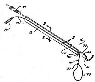
Laparoscopic~device 10 comprises~an elon~ated ~;
25:: b:ody:or handle~12, ~hich may be extruded from a `~
: medically compatible plastic or other s~it:able
:; :ma~erial. The~devioe can bei introduced through a
: krochar of an endoscope, such as trochar:14, shown in
Figure 3. A suitable device is shown in my U. S. ;~
Pat~nt No. 4,869,717, for "Gas Insu~flation Needle :~
With Instrument Port".
Handle 12 has a central passageway 16 through
which a suture 18 extends. The distal end of suture
18 is formed with ~ loop 20 by means of a slip ~not
~;
WO93/21834 2 1 3 ~ 6 6 2 P~T/VS93/03857
22. The slip knot has a diameter larger than that of ~ :
passageway 16. The proximal end of suture 18 has a ~--
pull in a form of a handle 24. ~.
A first channel 26 runs entirely along and
intersects the surface o~ handle 12 to form a
longitudinal groove which is generally parallel to
passageway 16. This channel slidably receives an ~
electrosurgical wire 28 which is covered by electrical -~.
insulation 30 an~ terminates at the distal end in an ~:
exposed wire hook`32. A cutting blade 34 can be
provided across the bight of the hook, as best ~een in `~
Figure 3. The proximal end of wire 28 is connected to
an electrical connector 36 for attachment to a source
of electrosurgical power (not shown). Wire 28 is
longitudinally lidable and rotatabl~ within channel .~.
26 so that the tip of hook 32 can be used to grasp the ~
tissue to be ligate~ and~draw it through loop 20, as :-.";.
will be described more fully below. The cutting blade
34 can be used to cu~ he suture just beyond sl~p knot
: 20 22 after t~e loop is drawn ~ig~t, as ~urther explained `~
below. ConYenien~ly, when not in use, wire 28 can be ~`~
drawn in the proxlmal direction so that the hook 32
and::~blade 34 are received in a recess 38 formed in the
distal end of handle 12 as a transverse slot across
2S~ the convex end~of~handle 12 and intersecting th~ end
: ~ : o~:channel 26, as best seen in Figures 3-7. `~
~ An option~l:seoond channel 40 can be pro~ided ...
: ~ ~ : which also runs:the entire length of hand~e 12, but ` `-
spaced from channel 26, such as on the opposite side
o~ passageway 16 ~om channel 26. This se~ond channel
40 in~ersects the surface of handle 12 to form a `.
longitudinal groove which is al~o generally parallel .~.
: ~ to passageway 16. Channel 40 can be used seleatively
~or receiYing o~her ligation devices. For ~xample, in
: 35 Fi~ure 8, a hypodermic syringe 42 is shown with a long
`' ;, ~
i`
..~,' ,^
WO93/218~ !2~1`.3 4 6 6 2 PCT/US93/03857
needle 44 attached thereto which extends through
channel 40. It can be used initially to inject
anesthesia to the tissue to be ligated, if this -
procedure is deemed necessary. Usually, the :
anesthesia used t~ deaden the area of the abdomen
where the trochars are inserted is suffic}ent and
additional anes~hesia is not required.
Alternatively, channel 40 can be used to receive
one or more optical fibers, such as optical fiber 46
Conveniently, the optical fiber can be connected to a
suitable source 48 o~ laser light, as shown in Figure -~;
9, for providing laser light to the operative site to :
fulgurate the ligated tissue. ~:
The method of preforming a tubal ligation is
diagrammatically illus~rated in Figures 10-17. In
Figure 10, tissue to be liga~ed, such as fallopian
tube 50 is shown~ The device 10 is positioned so that
suture loop 20 is brought into proximity to a tubal
section 52 of the tube 50 which is to be ligated. As
0 previously mentioned, this positioning is viewed
through an endoscope inserted through a sep~rate
tro~har. Once loop 20 is positioned, hook 32 is
~ ~ exte~ded to grasp the tubal section 52 and pull it
: through suture loop 20 as shown in Figure 11. The
: 25 physioian then pulls on handle 22 to place suture 18
under traction so:that loop 20 begins to pull tight
around tubal section 52/ as shown~in Figure 12. Thu~
slip knot 22~ ~ngages the distal en~ of handle 12 and
is held by ~lt as:the ~uture is drawn through knok 22 :`
to dr~w loop 20 tightly about tubal section 52. Hook ;-
32 then i5 retracted, as shown in Figure 3, as the -~
final tightening of the slip knot is complet~d.
Once suture loop 20 has ~een pulled ti~ht, the ~;
hook 32 can be extended again so that blade 34 can be
used to cut suture 18 just above sIip knot 22, as
".''',.':
: .
WO93/21834 `2 13 ~ 6 6 2 PCT/US93/03857
13 ~-
~ .
shown in Figure 14. The completed ligation is shown :~
in Figure 15. `
For many physicians, this constitutes the end of . -.
the procedure. However, other physicians may choose .
to use the hook 32 as an electrosurgical device for
cauterizing the ligated portion 52. This device may
be used as either a mono-polar or a bi-polar unit. As
illustrated in Figure 16, the wire hook is positioned ~:
adjacent ligated tissue 52 and electrosurgical current -~
is supplied to the hook to carry out the cauterizing
procedure.
If desired, an additional or alternative ~- <
procedure may be undertaken wherein the laser f iber 46 ..
i5 used for photo coagulation of ligated section 52 or
for photo ~aporization of the tissue, as illustrated
: in Fiyure 17.
Other common surgical procedures are simplîf ied
: by devices fur~her disclosed as modifications and
enhancaments of the present invention. For example, ~ .
~: 2Q fixation sutures are used extensively in surgery to ~ .
hold an organ, temporarily, to a body cavity wall.
: ~ In~the abdominal cavity, fixation sutures are used to ~;~
: temporarily hold organs such as the stomach, large
bowel, small bowel and gall ~ladder to the abdominal
~wall. In accordance with this disclosure, a
laparoscopic ~ixation suture device 70 is provided.
Thi~ de~ice has an elonga~ed handle 12~ shown with a `.
; : : tapered dis~al end, having a cenkra. pa~sageway 16, as ~--
il in the present invention, but only a single first
.~channel ~6 is.utiliz d. A suture l8~extends through
aid central passageway and has a suture needle 72 on
the distal end thereof and a pull 24 on the proximal
end thereo~. A nesting tu~e 74 is slida~ly received
in the first ch~nnel 26 and has a distal opening to -;~
frictionally receive the suture needle 72O The
~: '
W093/~l834 ; ~ 1 3 ~ ~ ~ 2 PCT/US93/03857
14 :
'
nesting tube 74 may be made o~ medically compatible
plastic, stainless steel, or other suitable material.
A second laparoscopic device provided with
forceps 76 can be used to remove the suture needle
from the nesting tube 74 and pass i' through the organ -
77 to be fixed t;: the body cavity wall 75. When the
needle 72 is replaced in the nesting tube 74, both
ends of the su~ure 18 can be withdrawn into the device
70 and the dPViCe pulled out from the body cavity wall
75. Thereby the two su~ure 18 ends may be pas~ed
through the body c~vity wall 75 and tied over a
bolster 78. Thus, the applica~ion o~ a fixation
suture is greatly simplified. The suture may be cut ~ ~
: at a later time, allowing the organ to return to its
normal position. The procedure may be.viewed by an
endoscope 7 9 .
The method of f ixing an organ to a body cavity
wall i~ diagrammatically illus~rated in Figures 18-23.
Figure lB shows the laparoscopic fixation 5U ure `~
device 70 with needle 72 nested in tube 74. The `
needl~ 72 is fric::tionally received in tube 74 as sh :>wn
in Figure 19. Figure 20 shows the in~roduction of
laparoscopic fixa;tion desrice 70 through a ~firs~ :
:trochar and a:laparoscopic device provided:with
25 :~forceps 76 through a second troc~ar. As seen in
Figures 20 and 21, the second laparoscopic device 7~
: is ~used to remo~Je~ ~he needle 72 and pass the suture 18 ::
thrc~u~h the organ 77. Figure 21 shows the procedure ~`
b~ing viewed through endoscope 79 inserted through a
; 30 .s~parate trochar. Figure 2~ shows the two ends of
su~ure 18 being drawn ~hrough the body cavity wall 75
after needle 72 has bae~ replaced in nestinq tube 74. ~;
Th~ suture 18 ends are ~hen tied over bols:~ex 78 to
hold the organ in place as shown in Figure 23,
''~ ~;',':
~'' .
W093~21834 2 1 3 1 6 g 2 PCT/US93/03857
Frequently, organs in the abdominal, peritoneum
or chest cavities are torn and require sewing of
double, triple or more stitches. Doctors frequently
have trouble learning and executing the tedious and :~
time consuming procedure of ligature knotting through .
a laparoscope. Therefore, in accordance with this ~ .
disclosure, a laparoscopic body part repair device 80 .
is provided to simplify the repair of structures
inside a body cavity. This device, as shown in Figure .
24, also utilities a handle 12, central passageway 16, .
and a first channel 26. The handle 12 is shown as
tapered on its distal end to provide an oblique ``~
surface to frictionally fit a nee~le 72 into first .~.
; channel ~6. It is understood that t~e distal end ~`
: 15 shape of handle 12 may be varied and still accomplish
this~purpose. A suture 18 ex~ends through the central .`
passageway 16 and has a 1ODP 20 with a slip knot 22 on ~.
the~distal end thereof and a pull 24 on the proximal
end thereof. A portion 82 of the suture 18 ext.ends :;
bey~nd the slip~knot 22 and has the suture needle 72 :~
a~tached thereto.~ The method of repairing structures
in a body cavity is shown in Figures 24-27.
Originally, the needle 72 is frictionally nestsd in ~;
firs~ channel 26.:;~The defert is repaired ~y utilizing
: 25~:~ a~:1aparoscopic~;~orceps device 76 introduced through a :;:
sec~nd trochar (shown in Figur s 24-27~: to rem~Ye the :`
suture n~edle 72~:from first channel~:26 and make the ~.
stitches. The~laparoscopic fo~ceps~devi~e 76 is then
utilized to pass the suture needle 72 through th~ loop ~;
~ 30~ ... 20. By simultaneously pulling pull 24 while holding ~:
: ~ tension on needle 72 with forceps device 76, the loop
easily closed, thus completing the suture. The
: ~ needle 72 may then be replaced in first channel ~6.
As shown in Fi~ure 27, the laparoscopic forceps davice
76 can be replaced with a laparoscopic cutti~g devlce .`~
...
WO93/218~ ~ 1 3 ~ 6 ~ 2 PCT/US93/03857
16
86 to cut suture 1$ and portion 82 just beyond knot
22, and allow the removal of needle 72 and device 80.
For the ligation of larger and medium sized blood
vessels, cystic ducts, trachea and bronchi, clip
appliers and staplers are not always reliable.
Therefore, ligating must he used to adequately close
off the vessel. However, often the surgeon has to
first free the vessel from a body part attached to it
with fibrous tissue by blunt dissection with a
dissecting instrument. Typically, this has to be done -~
to free a space for three ligatures. A device which ~:
can carry a ligature behind a vessel greatly
simplifies this procedure. Therefore, in accordance -~
with this disclosure, a laparoscopic suture carrying
de~ice so is provided t~ facilitate the ~igation of
certain vessels with a high assurance of complete
surgical closure. ~s~shown in Figuxes 28 and 29, ~his .-~
: ~ de~ice utilizes a handle 1~, shown with a tapered
: distal~end, central pa~sageway 16, first channel ~6,
and second channel 40. A suture 1~ extends through
; the central passageway 16 and has a loop ~0 with a
slip; knot 22 on the~distal end thereof and a pull 24
on the proxima1~end~thereof. A por ion 82 of the
suture 18 extends~beyond the slip knot 22 and has a
Z5~ ab 96 a~tached there~o.~ A carrier device ~2 is
;51idably recei~ed~in the second channel 40. The
aarrier device 92:has a generally U-shaped curve~ .
portion 93 at its dista1 end with a slit 94 along the
inner surface thereof, as shown. As seen in Fig~res ~;~
.29 and 30, the suture end 82 with ~he tab 96 can be
loaded in the carrier device 92 through the s1it 94O
The carri:er device 92 can be made of pr~-formed
plastic, having a memory, such that the curvature 93
.
can be s~raightened when retracted into se ond channel ;~.:
40. As shown in Figure 33, a laparo~copic forceps ~
:-
W093/2~834 PCT/US93/03857
213~662
17 ~:
device 76 is slidably received in the first channel
26.
The method of ligation for major vessels or ducts --~
is shown in Figures 31-35. The suture end 82 and tab .~
96 are brought behind the vessel 99 to a ligated by : :
tilting device 90 and then straightening it. This
sufficiently pierces fibrous tissue 108 connecting
ves~el 9~ to body part 110 to allow for the through
placement of curved portion 93 of carrier device 92.
The laparo~copic forceps device 76 in first channel 26 ~ :
i~ used then to draw the tab 96 ~hrough the loop 20.
The device 90 can again be tilted to withdrawn carrier
device 92 from ~ehind vessel 99 and swing it out of
the way. Then the ligation 92 and suture 20 loops are
closed by simultaneously pulling on the tab 96 with -~
the forceps 8~ and the pull 24 by hand. As shown in ~:
~ Figure 35, once the ligature is closed, the forceps ~:
;: ~ device 76 can be replaced with a laparoscopic scissors `~-~
: device ~6 to cut the excess suture beyond the knot 22.
: 20 : As seen in Fi~ures 36 and 37, the carrier device ~ ~,
~ . .
~92 may be replac d by a j-guide carrier 104 with a
slotted tip 106 to releasably hold the suture tab 96.
The j-guide carri:er:104 may either be of pre-formed :`~
plastic or may be~hollow with a guide wire running
25~ through it such that the memory o~ the pl~stic forms
h~: ~urvature when the guide wire is removed. ~-~
From the foregoing, the advantages of this -
invention are readily apparent. A laparoscopi~al -`.
; i surgical li~ation, repair, and electrosur~ical j .;
; 30 Go~gulation and:cutting device has been provided which
i8 ~imple in construction, yet versatile î~ use. It :~
can be used for ligating tissue and provides a ready
means for cutting the suture onc- a slip~Xnot has been ;
drawn tight around the tissue to be liga~ed. The hook ~.
has ~hree uses: ~1) to position the tissue to be
~ :,
~. .~.
W093J218~ PCT/VS93/03857
2 1 3 ~ ~ 6 ~
liy ted, (2) to use a blade connected thereto for :
cutting the suture material after ligation and (3) to
serve as an electrosurgical device to cauterize the
ligated tissue. In addition, the handle has a channel
for initially, slidably receiving a hypodermic needle
for anesthetizing the tissue to be ligated and
subsequently for slidably receiving a laser fiber for
coagulation or photo vaporiza~ion of the ligated ;~
tissue.
Additional advantages of various adaptations of
the presen~ invention are also readily apparent. One
adaptation of the device, utilizing a nesting tube ~.
provided in one of the channels to receiv~ a suture
needle attached to a loopless suture, provides for
body part to be more easily sutured to a body caYity
:wallO Another adaptation, utilizing a tapered handle
t~ rictionally receive suture needle attached to a :~:
looped and slip knotted suture, provides for a tear or
:: rupture in a body~part to be repaired by suturing
through one or more laparoscopies wi~hout the :'~
~; : necessity of tying ligature knots inside the body
~: . cavi:ty. A third adaptation, utilizing a curved
carrying device in one channel and a ligation assist :~
:: device in the other channel, provides for ligation of
: 1arge~vessels, often attached to a body part by
: fibrous tissue, to be accomplished in a much more
facile manner~
This in~ention has been described in detail with
~: I reference to particular embodiments thereof, but it
.will be understood that various other modifications
can be ~ffec~ed within the spirit and ~cope of this
:
:~ ~ invention. : ~
-,1