Note: Descriptions are shown in the official language in which they were submitted.
WO 9~/~20û~ Z 1 3 5 q 2 3 PC~/EP94/00914
"~SURING C~MBER FOR F~OW CYTOM~TER"
The present invention refe~s to a measuring chamber
for a flow cytometer to measure the fluorescent and
`~ , 5scattered light of individual cells or microscopic
particles, comprising: a body with a flat surface, an
~ inlet chamber and an outlet chamber within the body,
i which op~en on the ~lat sur~ace, the chambers being
p~sitioned ~ne in front of the other and belng connected
10by a channel in the body, the inlet chamber being
eouipped with an inlet tube and a~ injectior~ tube ar~
the outlet chamber being equipped with an ou~let tube,
a transparent plate placed in front of the flat surface,
the plate being movea~le from a Iirst position detached
15~rom the flat surface to a second pos~tion in which the
plate closes to seal the chambers and the channel,
~ptical means placed in front of the transparent plate
to direct light on the particle contained in the channel
and receive fluorescent light emi~ted from tne same
~0particle.
In a flow cytometer, some biological cells or o~her
types of microscopic particles are carried by a laminal
fi.ow of a li~uid, like water, through the focus of a~
intens~, luminous source~ The f~uorescent ana scattered
j 25light em1tted from each single cell when lt passes
i ~ through the focus are collected by suitable Opticâl
meanci and are directed onto proper light detectors.
I The scattered light, obtained in this way, supplies
, ~ ',
.,
'!i;'
~ :,
wo94n2000 PCT~4/Q0914
6 ~?,3 ; ~
~~orma~ion on the sl7e and s-ructure of the cell, ~rhile
the fluorescent light is a measure of the contents OI
the cell, which had been previously colored wlth a
fluoxescent coloring substance whlch is bound to it in
s
`i ~ a particular manner.
To obtain the highest le~el of excitation possible
at the focus and, consequently, the maximum sensltivity,
' the light ls concentrated in a very small focus, usually
with a length around lO0 micrometers. The cells must
follow the same rou~e ~hrough the focus in order that
they can be analyzed in a reproducable manner. To
obtaln this, the principle of "hydrodynamic focussing"
is used (P.J. Crossland-Taylor, Nature, Volume 171, Pgs.
37-38, 1953).
Hydrodynamic focussing is obtained in a cone
noz~le, filled with water. The water, which is pu~ped
in the nozzle, flows with laminal flow toward an opening
at the poin~ed tip of the noz-le so as to form a lamina
jet with a typical velocity of a few meters per second.
~0 The sample, that is, the suspension of the cells to be
analyzed, is injected in the noz71e ~hrough a thin tube
which has one of i~s end openings at or very close to
the a:~is of the nozzle. Since the flow inide the no_~le
is laminar, the sample is confined n the central pa~L
I ~5 of the flow, both ir. the nozzle and the je~.
¦ ~ Consequently, it follows a route of the jet that is
`~¦ always reproducible in an exact way. The cells are
measured when this jet passes through the e~citation
~. .
' ~ WO g4t~000 213 6 '1~ 3 PCT~4/00914
~cus. The principle of hydrodvnamic focussing is used
~n all of the types of flow cytometers. In some ~low
cytometers, the opening of the nozzle connects with the
atmospheric air, so that the emitted jet will contac~
~ 5 said air. An inconvenience of this type OI cytometer is
- that the user can come into contact with the noxious or
infected substances.
In other types of cytometers, the opening connects
with a narrow tube, preferably of rectangular section
(~nown as "closed measuring cha~ber"). ~ sign~icant
inconvenience with this last type is that the thln lube
is difficult to clean. However, this has the advantage
that the user is protected from e;~posuxe to disinfecting
and/or toxic agents present in the sample.
There are fundamental~y two types of flow
cytometers: those which use a laser as a source of light~
of excitation and those which use a high pressure arc
lamp which contains xenon or mercury.
In the cytometers which use laser, the scatrering
~0 of light can be measured ln a direction close to that o_
; the laser beam ("low angle light scattering"). Th~s can
also be measured at a righ' angle relative~to the laser
bQam ~"large angle light scattering") whereas the
fluorescence is collected at a risht angle to the laser
~S beam. S~id lnstruments can use either a~nozzle tha~
s
emits an àir jet or in a~closed measuring chamber.
The cytometers which use the arc lamps often have
a microscope~lens with oil immersion for the purpose of
, : ~:
`~: ~ ~ 3
W0~4/~00~ PC~4/00914 ~`~
36~3
concentrating as much eV.citlng light as possible on the
ftow containing the sample. The fluorescent li~ht is
usually collected with the same microscopic lens, which,
thus, is called epimodal because it is adapted to
collect also the fluorescent light emitted from the
~ sample, while the scat~ered light of the sample can be
i highlighted in a darX field ln the lower part of the
cytometer ~documents EP-~-0,229,815, US-A-4,315,501).
The e:.istence of an open measuring chamber IO use
with cytometers which use an arc lamp is alreadv known
(EP-A-0,0~6,770) In this measuring chamber, a --low of
the liquid is cGnfined in a flat layer of water on the
ope~ surface of a microscope cover glass. The flow is
observed from the opposire side through a microscope
lens with oil immiersion.
This type of measuring chamber has a serious
i~convenie~ice because it exposes the user to the risk of
contamination by toxic and/or infected subs~ances.
Moreover, the functioning principle implies a limiLa~iorl
~0 of the rate of sample flow, in some cases therebv
limitlng the measuring velocity.
On the other hand, this measuring chamber is the
Gnly one which allows for a precise measurement of the
light scattering in cytometers which use arc lamps.
``~ 45 Other measuring chambers for this type of
instruments do not permit an efficient measurement o~
~¦ the light s~cattering, which represents a serious
limi~ation of ~heir possibility of application
~`
~ 4 ~
. . '.
. 11
`- ~o 94/220no 2 1 3 6 4 2 3 PCT~4/00914
. .
(documents US-A~ 5,~.9 and US-~-4,954,715).
The objecl of the present invention is that of
` realizing a measuring cha~ber for a flow cytometer
:i ~ characterized i~ that the a~.is of the injection tube
~¦ 5 intersects the optical axis of the optical means at a
point outside the channel whère~y a liquid and some
particles, injected into the inlet chamber throughout
I the inlet tube and the injec~ion tube respectively, flow
with laminar flow through the channel.
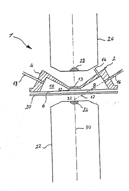
The advantages of the present in~ention wlll be
clarified by the following description and by the sole
diagrammatic figure of the annaxed drawing, which
illustrates the de~ice of the invention from an ai:ial
section ~iew.
Referlnq to the drawing, reference numeral (1)
. indicates a flow cytometer. A fundamental pa-t of the
.; cytometer is the measuring chamber, which is comprised
~; essentially of a body (4), preferably of circular form
with a lower flat surface (6). In the body, there is an
inlet chamber (8) and an outlet chamber (lC), which open
on the flat surface (6). The chambers (8, 10) are
` positioned~one in front of the other and each chamber
has a cross section which is reduced progressively when
: appro2ching the o~her cham~er. In particular, in ~he
~ ~ 25 ~orm of the invention illustrated in the drawlng, each
~ ~; ~
~:: ~ ~ ~ chamber has~the form:of a semicone. The chambers are
.
:;~ ~ ~ ~ connected through a~channel (12) which lS lnslde the
;~ ; body:(2)~
:.~ : ~
. ~ . .
': ~
WO 94/220nO ~ PCT/EE~4/009:14
?~3~
The channel (1~) preferably has a cross sect'onal
rectangular ~orm. On the bottom of the channel there is
a transparent element t13) which generally is made of
artificial ~uartz.
The inlet chamber (8) is equipped with an inle
. tube (14) which serves to inject `liquid under pressure
'~ int3 said chamber. Usually, this liquid consists Of
J distilled or filtered water. Moreovex, the chamber ~8)
is equipped with an injection tube ~16) which Qerves to
introduce cells or other microscopic matter to be .
analy7ed into the measuring chamber. The tube (16) is
moveable inside the body (~) along its axls (17) for
reasons explained below. -~
The outlet chamber ~10) is eq~ipped with an ou~let
1~ tube ~18) which serves to eject water and the analyzed
cells which entered through the tubes 14 and 16,
respectively.
A transparent plate (~0) is positioned in ront of
the flat surface ~6) of the body ( ). Also this plate
~O is preferably made or artificial quartz. The plate is
moveable from a first position ln which it ~s detached
rom the flat surface (as illustrated in the drawing) to -;
a second position in which it is laying against the
~ ; surface and closed to seal the inlet chamber (8), thej ~5 outlet chamber (10) and the channel (12). ~-
, ~ The c~ytometer ls further equipped with optical
means to direct the light against a particle contained
-:
in the channèI (12) nd to receive the fluorescent light
:
.. 6
..
.; :
S. : -
-- W0~4/22000 213 6 4 2 ~ PCT~4/00914
!
emit~ed by the same partlcle. Usually, these optical
means are comprised of a micrcsco~e objective ~ which
~ is placed in front of the transparent plate (20). The
¦ mlcroscope lens is equipped wi~h a lens (24) which
¦ 5 serves to concentrate the light emitted from a light
source, ln this case an arc lamp (not illustrated), on
~ the particle contained in the channel (12). It is
¦ observed that the position of the body (~) can be moved
clo~er to or further away from the lens (24) through
suitabel means so that the focus of the lens falls on
the interlor of the channel ~1 ).
The volume comprised between the transpa$ent plate
~20) and the extremity of the microscope ob]ective (~ G)
is filled with the corresponding immersion oil which
serves to adjust the refractive index of the lens t24)
to the refractive index of the material which makes up
the plate.
The cytometer (1) is further equipped with a second
mlcroscope objective (26) placed on the other slde w~th
~0 respect to the first microscope objective (~.) relatlve
to the body ~2). The ~.icroscope objectlve ~ 6) is also
equlpped with a objective (28) placed in front of the
transparent element (13). The lens ~(,4) and the lens
(28) are on the same optical axis (30). The function of
.. ~
5 ~ the second mlcroscope objective (26? will be clarlfied~
below.
The operation of the measuring chamber of the flow
~¦~ cytometer is the following: ~
, ,
~ 7
W094/2200~ 2~3~ 42 PCT~4/00914
Through the in~et tube (14), dis~llled or filtered
water is introduced and some cells to be analy~ed are
injected th~oughout the injection tube (16). The tube
~ (16) is positioned inside the chamber (8) in such a way
?~ 5 that its axis intersects the optical axis (3) at a point
, (32) outside the channel (12), ln ~his way a laminar
' flow can be realized, made of liquid and some particles
i ln the channel (12). Thanks to the laminar flow, it is
possible, through ~he microscope objectives (~2 and 26),
to analy7e the cells one by one on the inside of the
same channel. In fact, while a cell passes thrsugh the
channelj the light emitted on it is concentrated by the
first microscope objective (22). ~he cell difracts the
light rays in all dlrections and emits a fluorescent
light which ls collected again by the first microscopic
objective. Only one part of the diffracted rays, that
; is, those which form particular angles with respect ~o
the op~ical axis (30), are able to pass through the
transparent element (13) an~ are collected by the second
~0 microscope objective (26). The second microscope
objective, ln turn, transmits these rays t~ determlned
~: known devices, which carry out the analysis of th~
characteristics of ~he cell, such as size, granulalion
and s~ructure.
: ~ ~5 As the point of intersection of the axis (17) of
.. ~; :
the tube (16j with the optical axis ~30~ lies outside
¦ ~the channel (12), it 1s possible to move the flow of the
' cells to be analyzed in the channel in the direction o~
..~
:;;~ ~ 8
; ~ ~ :
~:.
~. W094/~000 PCT~4/0~914
21 36~23
!
the optical axis, moving the tube (16) along its axis.
Thus, the flow can be positioned in a way to pass
exactly through the focus of the mic~oscope objective
(2,) without moving the cell relative to this microscope
~bjective. In this way, the layer of the immersion oil
; provided between the microscope objective (~2) and the
transparent plate t20) can be maintained very thin to
reduce the background fluorescent light which is emitted
from the same oil and, thus, the disturbance caused by
this is reduced. Thus, the object of the inven~ion has
been attained, which is that of sendin~ the flow against
the transparent plate and not in the true and same
measuring point which is found on the inside of the
channel (1.~), because only in this way is it possible to
get a laminar flow and, thus, an accurate reading of the
movement of the particles along the Iocal plane.
. .
~"
: .
; ~ , .
.
,,~ ~ ~ ~ ; , 9
: ,. : : : ~,
~ : : .
