Note: Descriptions are shown in the official language in which they were submitted.
~ 1~7 1 ~ 4 ' ~
8IZI~JG AND CIJTTING GlJIDE FOR ~:
RE8ECTING T~IE DISTAL END OF THE FEM~JR :
This invention relates to implantation of
5 artificial knee ~oints, ~nd more particularly to a sizing ~ `
and cutting guide f c ; p r e p a r i n ~ e f e m u r t o ~ .:
receive the implant.
The geometry of a condylar knee implant is
optimized to transmit the normal forces experienced during
the walking process, from the femur, through the implant
and down through the joint to the tibia. The complex
geometry of the normal knee provides an articulating `
surface with a constantly varying radius of curvature to
allow for this transmission of force to be optimized for
all angles of bend. An implant aims to recreate this.
To assist in this, the inner surface of the
condylar implant should ideally follow a similar varying ~-~
radius of curvature to maintain the same thickness of
material throughout the implant. Because it is impractical
to achieve this degree of shaping of the distal end of the
femur, the design compromise adopted uses a number of flat -~
surfaces approximating the curved surface.
The usual design adopted is made up of five flat
surfaces, with the posterior three divided by the cruciate
cleft. Five surfaces must therefore be prepared on the `
distal condyles to exactly match the implant. This is `~`
especially important for a press-fit implant where good `~
30 bone apposition is critical. `~
It is also important to choose a correctly sized
knee implant from the range of sizes available to fit the `~
particular individual since individuals knee joints do
vary substantially. The choice of the correct implant i5
i~oortan. because if too small a;~ ?lant is used, then
the articulation of the knee wili be seriously affected
and may be grossly unstable with tensioning problems at
the extremes of motion. The correctly sized implant w
~ENDED CHEET
71a~ . -
` -2-
i
imitate the exact dimensions and radii of curvature of the
normal knee, by replacing only that bone which has been ~
- removed. Joint tensioning will be constant throughout the
full range of movements and the joint will be stable. With
a larger implant the joint will be "tight" with either
restricted flexion or laxity and instability during leg
extension.
The approximate size of a knee implant can be ~ -
determined prior to the operation by analysis of X-rays of
the knee. However, the surgeon will need to make his
final size assessment during the process of preparing the
femoral condyles.
The initial preparationjof the condylar bone
involves remo~al of the most distal bone surface
perpendicular to the mechanical axis of the knee
articulation and is accomplished by means of a device
which relates the cut surface to a varus angle to the axis
of the femur. This preparation affects the final -~
orientation of the implant, but does not determine the
size or position (mediolateral or anterior-posterior) of
the fina} implant. "
The positioning of the implant should be `
relative to available anatomic landmarks, and the ~`
correctly sized cutting guide should be placed with -
respect to these landmarks in order to align the implant
correctly. `~
Implant location is in the sagittal plane of the ``
leg and therefore the anatomic landmarks should, ideally, --
be picked up from this plane for the most satisfactory `~
30 ~ositioning data. ~
:
AMENDED SL;EET ` ` ``
. . . ~
~-~ 2l37la4 .. ,.,~
2a
EP-A-380 451 describes a guide for -..aking resections to the
distal end of the femur, comprising a ~lock (comprising a fixed
body and a movable body) having guide slots, posterior guide .
means for positioning the block in the posterior/anterior sense,
means for mounting the block on the femur, anterior guide means
projecting upwardly from the side ant~rior of the block, the
anterior guide means having a tip which is at a predetermined
location relative to the upper surface of the block, and means
for indicating the distance between the' posterior condyles and
a reference point at the frontal side of the femur. .
US-4 759 350 describes a guide for r,a~ing resections to the
distal end of a femur during the replacement of a knee joint,
lS comprising a miter block having a plurality of guide slots for
making the cuts to be made to fit a particular sized implant, and
means for mounting the miter block to the resected surface of the ~?
distal femur. :.
~E~ EEl
`21~7~
.... , .
The invention has therefore been made with .h~se points in
mind and according to the invention -n one asvect there is
provided a guide for making resections to the di~.al end of a
10 femur during the replacement of a knee joint, the cuide being
aaapted to be mounted on ~ distally r§ed end Gr^ the emur
that has been resected along a plane generally ?e~?endicular tO
the mechanical axis of the femur, the suide compr_sing:
a miter block having posterior and anterior sides, a
lS generally planar upper surface adapted to be placed face-to-.ace
with a distally resected femur, and a ?lurality of guide slots
for alignment and orientation of a blaae for making ~he cu~s to
be made to fit a particular sized implant;
posterior guide means, project ng upwarciy r-rom _`n.e
posterior side of the miter block, for positioning the ~irer
block with the said upper surface in face-to-face contact wi~.~
a distally resected femur and with the posterior g~ide means
contacting the posterior curved surface of -h- emur and
therefore position the miter block in the posterior/anterior
sense against the distally resected femur;
means for mounting the miter block on the resected surface
of the distally resected femur, with ~he upper su-face of the
'. - miter block mating face-to-face with the resected surface and the
i, . AAhENO~D SHEET
~1~71~4
posterior guide means contacting the posterior curved surface of
the femur; and
an anterior guice means, projecti~ng upwardly from the side
'. anterior of the miter block, for gauging the size of the femur
relative to the size of an artificial knee implant corresponding
to the guide, the anterior guide means having a tip which is at
a predetermined locat on relative the upper surface of the miter
block such that the ?osition of the tip will coincide with the
anterior end of the articulation zone of the femur when the
posterior guide means is properly positioned if the correct size
miter block and accordingly implant have been chosen, the said
predetermined location being such that when the miter block is
~; later used as a guide to the anterior resection, the cut will
- exit the anterior condyle at that end of the articulation area.
15Also according to the invention there is provided a system
comprising a plurality of knee implants of various sizes, and a
: plurality of guides of ~arious sizes, corresponding to the sizes
of the knee implants, for making resections to the distal end of
a femur during the replacement of a knee joint, each guide being
adapted to be mounted on a distally resected end of the femur
that has been resected along a plane generally perpendicular to
the mechanical axis of the femur, each of the guides comprising:
- a miter block having posterior and anterior sides, a
generally planar upper surface adapted to be placed face-to-face
with a distally resected femur, and having a plurality of guide
¦ slots for alignment and orientation of a blade for making the
cuts to be made to fit a particular sized implant;
posterior guide means, projecting upwardly from the
, ~L.ir.~t.. ' j ;r~~
.. . .. ~............ .. ...
- 2137~ a~
- 4a
posterior of the miter block, -or positioning the miter block
with the said upper surface n fa_e-to-face contact with a
distally resected femur and ~ith ~he posterior guide means :::
contacting the posterior curved surface of the femur and
therefore position the miter block i~ the posterior/anterior
sense against the distally resected fe~ur;
means for mounting the miter block on the resected surface
of the distally resected femur, with the upper surface of the ;
miter block mating face-to-face with _he resected surface and the
posterior guide means contacting the pogterior curved surface of
the femur; and
an anterior guide means, prcjecting upwardly from the
anterior of the miter block, f5r gauging the size of the femur :
: relative to the size of an arti~icial knee implant corresponding
:
to the guide, the anterior guide means having a tip which is at
a predetermined location relative the upper surface of the miter
block such that the position of the tip will coincide with the
anterior end of the articulation zone of the femur when the
posterior guide means is properly pcsitioned if the correct size
miter block and accordingly implant have been chosen, the said
predetermined location being such that when the miter block is
later used as a guide to the anterior resection, the cut will
exit the anterior condyle at that end of the articulation area.
AMENDED S~IEET
~ 21 3714~
4b
`-
,
Posteriorly, the articulating surfaces of the
knee are separated by the cruciate cleft into the lateral
and medial curved surfaces. The posterior part of the
articulating surface is therefore used as a reference
against which the posterior guide means abut. The
posterior guide means therefore indicates the most
posterior section of the subsequent implant relative the ';
posterior face of the miter block which has previously
been determined by X-ray overlay to be the preferred size.
Once this position has been determined, the `
locations for the subsequent cuts are fully referenced to
this point and should ensure a satisfactory matching of
the implant to the amount of bone which has been removed,
provided, as is explained below, the correct implant size
has been chosen. The most posterior cut will remove an
amount of articular bone equivalent to the thickness of
the posterior section of the implant. All other cuts are
angled and referenced to this.
Different anterior guide means may be provided
for each mi~er block. Preferably however a single anterior
guide means will be provided which can be removably
attached to each of the range of miter blocks
AMENDED SHEET
213714'1
~94/000s6 5 PCT/US93/06159
corresponding to the range of implant sizes, the position
of the tip of the anterior guide means being adjustable to `
alter the spacing of the tip from the upper surface of the
chosen miter block. In the latter case, the anterior guide
means desirably includes a main body having means for
ready attachment to the anterior face of a miter block, ~-~
supporting means for slidably supporting a stylus with the --
said tip located at one end thereof so that the spacing of -~
its tip can be adjusted relatiYe the upper surface of the `
miter block, and scale means providing a reading of the
correct adjustment of the tip relative the particular
guide block chosen. --
Anteriorly, the articulating surfaces of theknee, under the patella, form a single curved surface with
a prominent curved depression, the facia patellaris. This
articulating surface is asymmetric, the lateral portion
extending more proximal than the medial portion. The ~
margins of the articulating surfaces form a pronounced `
cusp at the point where the facia patellaris meets the `
cortical bone of-the knee.--This cusp is also manufactured
into the condylar knee implant and may therefore act as a -
secondary point of reference between the anatomic, and the
replacement device. The location of the cusp differs for
each knee size, being a proportionate perpendicular
distance from the cut surface of the bone.
As noted above it is preferred that the anterior
guide means fit directly on to the anterior face of all
the sizes of miter blocks. Its tip is adjustable in a
superior or inferior direction to align with the cusp on
the knee. A graduated scale on the movable stylus
represent the estimated size of the replacement knee. The
tip of the guide means is machined to lie a fixed distance
above the upper face of the miter-guide cutting block for
a set knee size. ~he most anterior cut indicated by the
miter guide cutting block is aligned to coincide with the
tip, which, for a set cut miter block size, will be a
fixed distance anterior, and superior, to the posterior
guide means.
~ . ~
213714~ :
W094/00056 6 PCT/US93/061~5 ;
When the anterior guide has been set the same as
the chosen miter block si~, then the tip should, if the
implant size has been correctly estimated, just touch the
margin of the cusp between the two articulatin~ spurs.
This will indicate that the anterior cut is in the correct
position to allow for optimum bone replacement by the
implant. If the tip does not touch the bone, or it
contacts the bone before locking into position on the
block, this is-indicative of an incorrectly estimated
lO implant size. In both cases, the anterior cut will result `
in less than optimal bone removal.
The mar~in of the cusp between the two
articulated spurs is a visible reference point since it -
marks the end of the articulation zone. In addition, this
is the point at which one wishes the cut made for the
anterior resection to exit from-the bone.
A tight fitting tip for the anterior guide
indicates overestimation of implant size, and if this
situation is not rectified, the anterior cut will remove
- 20 more bone than will be replaced by the anterior portion of
the implant. A loosely fitting tip indicates an
under-estimation of implant size and will lead to
insufficient bone removal and the likelihood that the
anterior back face of the implant will not be fully
supported by the bone.
In either case, remedial action is required. The
miter guide cutting block should be removed and the next
larger size tif the tip is too tight), or smaller size (if
the tip does not contact bone), be attached, again with
the posterior guide means in contact with the posterior
margin of the articulating surface. The anterior guide
should be adjusted according to the new block, and the
implant size reassessed.
If no satisfactory combination can be obtained,
i.e., an intermediate implant size between the available
sizes is required, then the surgeon must decide whether to
use a slightly smaller or larger implant than is optimum.
The arguments for either choice are a matter of surgical
21~714~ ~
~94/00056 PCT/US93/06159
-7- ~
practice. Obviously the larger the number of intermediate '
siz~s of implants available, the less this will be a '~'
~L~em.
3rt~. The posterior guide means is preferably in the
S form of one or more shoes which are attached to the ;'
posterior side of the miter block and are upstanding
therefrom to be brought into contact with the posterior
surfaces of the two posterior condyles. Preferably ",
however, the posterior shoes are removable so that once
lO the miter block has been fixed in place, then the shoes '
are removed so as not to interfere with the subsequent
cutting of the bone. ',,~
The anterior guide means are also pre~erably -,
remo~ably attached to the miter block so that once it has''
lS ', been decided that the correct miter block and therefore ~
final implant have been chosen, this anterior guide can be '
removed to enable the anterior resection cuts to be made',`,
without damage to the guide. `',
The miter cutting block is known and should be
affixed firmly to the femoral condyle after the final
distal resection. This can be achieved by, for example,
using a pair of angled nails whose axes are angled to one
another. This ensures a good firm attachment of the block
to the condyle during the cutting process. The nails are
thereafter removed once the miter block itself is removed
when all of the cuts have been completed. ~-
The miter block itself is well known and
includes a number of grooves at the appropriate angles
relative the surface which contacts the first distal
resection on the femoral condyle and guides the surgeon in
making cuts at the required angle. In a preferred
embodiment of the invention there are five femoral cuts
which remove an approximately constant amount of bone
arou,nd the articulation surface of the femoral condyle.
The-larger the number of cuts the more closely this
approximation can be achieved and five cuts is a
satisfactory number.
Other features will be pointed out hereinafter.
213714~
W094/000~6 8 PCT/US93/061~5 .
Brief Description of th~ Drawina
The invention will now be described, by way of
example, with reference to the accompanying drawings ~:
wherein corresponding reference characters indicate -`
corresponding parts throughout the several views of the
drawing, and wherein:
Figure 1 is a perspective view of a miter block
- used to guide the making of the cuts in the distal end of
a femur;
lo Figure 2 is a diagram showing the initial
fitting of a miter block to the distal end of a femur
using the posterior guide to position the block;
Figure 3 is a diagram similar to Figure 2
showing the miter block attached in place; ~-
- 15 Figure 4 is a diagram taken at right angles to
the diagram of Figure 3;
Figure S is a diagram similar to Figure 3 but
showing the attachment and use of the anterior guide;
Figure 6 is a perspective detail showing the
fitting of the anterior guide to the miter block;
Figure 7 is a perspective detail showing the
fitting of the posterior guide to the miter block;
Figure 8 is top plan view of another embodiment
of the miter block of the invention;
Figure 8A is a perspective view of the miter
block of figure 8, with handles mounted on the miter
block; -
Figure 9 is a cross-sectional view substantially
along line 9-9 in figure 8;
- 30 Figures lOA, lOB and lOC are perspective views
of another embodiment of the invention comprising an
indicator block, illustrating respectively a correctly
sized indicator block relative to the femur, a large
indicator block relative to the femur and a small
indicator block relative to the femur, which thereby
indicate how the corresponding implant and miter block
would fit; and
21371~
94/00056 9 PCT/US93/06159 ;
Figure 11 is a cross-sectional view illustrating
the indicator block of figures lOA, lOB and lOC mounted on ~
the distal end of the femur that has been resected along a -
plane generally perpendicular to the mechanical axis of
. 5 the femur. `~
":.'
Detaile~ De~cr~tion of Preferred Embodiments
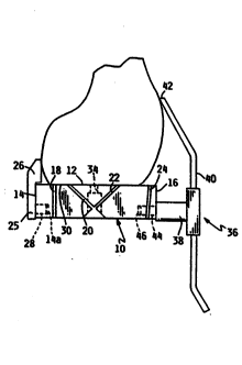
A miter block 10 as used to guide the making of
cuts through the femoral condyles during the preparation
of the femur to receive an implant is shown in Figure 1.
This is largely a known item and comprises a rectangular
shaped miter block 10 having an upper planar surface 12
which is designed to abut the resected end of the femur,
and posterior and anterior sides 14 and 16 at right angles
to the upper surface 12. As used herein, "upper" or
"lower" as in upper surface 12 means the surface of the
miter block 10 that faces generally upwardly or downwardly
relative to the body of the patient so that the "upper"
surface 12 faces generally toward the head of the patient
- 20 when the miter block 10 is mounted on the femur, and the
"lower" surface faces generally toward the feet of the
patient. In the body of the block 10 are provided a
number of guide slots 16 to 24, which are used to guide a
saw when resecting the end of the femur.
Removably fixed to the posterior side 14 of the
miter block 10, is a posterior guide means 25 comprising a
pair of shoes 26 which have guide surfaces 27 at right
angles to the surface 12. The posterior guide means 25
includes a rectangular shaped projection or tab 28, which
is engagable in a corresponding~shoe-mounting opening 14a
(Figure 5) in the posterior side 14 of the miter block 10.
The shoe-mounting opening 14a of the miter block 10 has a -
I generally rectangular cross section complementary to ~he
rectangular cross section of the tab 28 of the posterior
guide means 25 to closely receive the tab 28 in the show-
mounting opening 14a. At least one, but preferably a pair
of spring-loaded detent balls 29 are mounted in the miter
block 10 and biased into the shoe-mounting opening 14a for
2 1371 4~
W094/000~6 -lo- PCT/US93/0615~ .
releasably securing the posterior guide means 25 on the ;~:
miter block 10. The spring-loaded detent balls 29 engage .
small detent depressions in the tab 28.
Initially a cut 30 is made across the distal end
of the femur, that cut having been made in a plane at
right angles to the mechanical axis of the knee .-~-
articulation, and a guide which forms no part of the .-:
present invention is used to determine the position of the
cut and so the amount of bone removed. The resulting femur ~.
is shown if Figure 2.
Having previously examined X-rays of the knee
and by laying transparencies of a size corresponding to -
the various sizes of implants available over this, the
surgeon has chosen a knee implant size. Then he takes a
miter block 10 corresponding to the size of that implant
and, as best shown in Figure 2, places this against the ~.
cut end 30, whilst using the posterior shoes 26 as a .~:
guide. Thus the surfaces 28 contact the posterior
articulation regions of the condyle and together with the
. =. 20 .abutment of the.cut end 30 against the surface 12 locate .
the block 10.
Next an anterior guide means 36 is attached to
the anterior side 16 of the miter block 10. The anterior
guide means 36 includes a bracket 38 removably attached to
the anterior side 16 of the miter block 10, and an
elongate stylus 40 slidably supported in the bracket 38 so
that the spacing of the tip 42 of the stylus 40 can be
adjusted relative to the upper surface 12 of the miter
block 10. A scale 43 is provided between the stylus 40
and the bracket 38 to show the position of the stylus 40
¦ to provide an indication of the coxrect adjustment of the
¦ tip 42 relative to the bracket 38 and thus the miter block
1 10. The stylus 40 can be adjusted to and temporarily held
at a number of preselected positions, each corresponding
to a particular sized miter block 10, by means of a
spring-loaded ball (not shown) housed in the body 38 and
engaging in one of a number of detents (not shown) in the
rear surface of the stylus 40.
213714~ ~
394/000~6 -ll- PCT/US93/061~9
The bracket 38 also has a projection 44 which
can be fixed in a hole 46 in the block lO. As best shown
in Figure 6, the projection 44 has a reduced diameter
portion 44a and a small ball 48 is held in a bore 50 and ~
r 5 loaded by a spring 52 held by a grid screw 34 against the -
projection 44. The ball 48 therefore acts as snap-fit to
allow the guide 36 to be fixed to or removed from the
- block lO- and engages in the portion 44a to hold the guide
36 temporarily in place.
If the anterior guide 36 has been set the same
as the chosen miter block size, then the tip 42 of the
stylus 40 should, if the implant size has been correctly
estimated, just touch the margin of the cusp between the
two articulating spurs. This will indicate that the -
- 15 anterior cut is in the correct position to allow for
optimum bone replacement by the implant. If the tip 42 of ;;
the stylus 40 does not touch the bone, or it contacts the
bone before locking into position on the miter block lO,
this is indicative of an incorrectly estimated implant
2~=~ size.--In both cases, the anterior cut will result in less
than optimal bone removal. If the implant size has not
been correctly estimated the surgeon will need to replace
the miter block lO with the next largest or smallest miter
block lO corresponding to the next largest or smallest
25 implant and the above steps repeated until he is satisfied
he has the best fit.
When satisfied of this, the miter block lO is
fixed in place by a pair of nails 32 passing through holes
34 in the miter block lO. This orientation of the nails 32
- 30 is best shown in Figure 4 and is such that their axes are
crossed, so rigidly fixing the miter block lO in place.
Thereafter the surgeon removes the shoes 26 and
the anterior guide 36 so that these do not interfere with
cutting, an then makes the required cuts in the femur
35 guided by the slots 18 to 24.
Figures 8, 8A and 9 illustrate another
embodiment of the miter block, herein designated lO0,
similar in many respects to the miter block lO of figures
~1~7~ 4 1
W094/000~6 -12- PCT/US93~061~
1-7. ~iter bloc~ lOO includes a plurality, e.g., four,
guide slots 102, 104, 106 and 108 for guiding a saw blade
to make the desired cuts to the femur. Each of these
guide slots 102, 104, 106 and 108 are generally continuous
S and stop short of the opposite ends 110 and 112 of the
miter block lO0~
The miter block lOO also includes at least tand
preferably) two generally elongate locating pegs 114 that ~,
are adapted to be closely received in locating holes
drilled into the distal end of the femur. Also provided
~re two nail-guiding bores 116 provided in lugs 118
extending from the opposite ends 110 and 112 of the miter
block lO0~ The nail-guiding bores 116 are provided at an
anglet e.g., approximately 30-45 degrees, from the
longitudinal axis of the locating pegs 114 to guide the
nails 117 into the distal end of the femur. Most
preferably, the lower faces of the lugs 118 are provided
at an angle such that the lower faces are generally "
përpendicular to the longitudinal axis of the nails 117. ,
The locating,pegs 114 and nail-guiding bores 116
constitute one preferred embodiment of the means for
mounting the miter block 100 on the resected surface of
the distally resected femur, with the upper surface 120 of
the miter block lO0 mating face-to-face with the resected
2S surface.
The miter block 100 also has an opening 124
therethrough between the upper and lower surfaces 120 and
122 of the miter block 100 defining a window 124 for
visualization of the posterior cruciate ligament when
mounting the miter block 100 and when making the cuts to
the femur. The window 124 is preferably generally
rectangular in cross section and somewhat elongate in the
I direction extending between the posterior and anterior
sides 126 and 128 of the miter block 100. The guiding
slot 108 is intersected by the window 124, but is still
considered generally continuous because the two segments
of the slot 108 are not separated by a solid section of
the miter block lO0.
21~144 ~-`
b 94/00056 -13- PCI/US93/061S9
The miter block loO may includè two threaded
bores 130 in the opposite ends 110 and 112 of the miter
block 100 for receiving two handles 132. The threaded
bores 130 are preferably provided at an angle to the upper
surface 120 of the miter block 100.
The miter block 100 may also include a tab-
receiving slot or recess 134 having a generally T-shaped
cross section for receiving a generally T-shaped-cross-
section tab on a shoe. If used with the sizing guide 200
illustrated in figures lOA, lOB, lOC and 11, the miter
block 100 may be formed without such a tab-receiving
recess 134. The locating pegs 114 are sufficient to
locate the miter block 100 relative to the distal end of
the femur.
Figures lOA, lOB, lOC and 11 illustrate yet
another embodiment of the invention, in which a novel i~
sizing guide, herein indicated 200 is provided. Many
aspects of the sizing guide 200 are similar to the miter
blocks 10 and 100 described above with reference to ~--
- 20 figures ~-9. The sizing guide 200, however, does not --
include guide slots for guiding a saw blade. As
illustrated in figures lOA-lOC, the sizing guide 200
comprises an indicator block 202, anterior guide means 204
and posterior guide means 206.
The anterior guide means 204 is similar to the
anterior guide means 36 of figures 1-7, and as such
comprises a generally elongate stylus 208 and a bracket
210 for adjustably mounting the stylus 208 relative to the
upper planar surface 212 of the indicator block 202. Most
preferably, a single anterior guide means 204 is provided
in a system comprising a plurality, e.g., 5, indicator
blocks 202 of various sizes each corresponding to a size
miter block 100 and implant.
As described above with reference to anterior
guide means 36, the tip 214 of the stylus 208 is
adjustable relative to the upper surface 212 of the
indicator block 202, and a suitable means is provided to
releasably lock the stylus 208 in a number (e.g., 5)
~ 1 3 7 ~
W094/000~6 -14- PCT/VS93/06159- -
predetermined locations relative to the upper surface 212
of the indicator block 202. A suitable scale means
(similar to that shown at reference numeral 43 in figure
6) is provided to provide an indication of the correct
adjustment of the tip 214 relative to the bracket 210 and
thus relative to the upper surface 212 of the indicator
block 202. Most preferably, the stylus 208 is generally
elongate, with the end portion 216 being bent at an angle
relative to the main portion 218 of the stylus 208 in the
10- direction toward the femur 220. The end portion 216 is
tapered toward the tip 214 of the stylus 208.
The posterior guide means 206 is similar to the
posterior guide means illustrated at reference numeral 25
¦ ` in figure 7. As such, the posterior guide means 206
~5 comprises one or more guide shoes, preferably two guide
shoes similar to shoes 26 which are spaced apart but have
co-planar guide surfaces. The posterior guide means 206
is preferably removably attached to the posterior side of
the indicator block 202, and is upstanding from the
20 indicator block 202 to adapt the shoes for contact with `~
the two posterior condyles.
Most preferably, posterior guide means 206 is
provided with a tab 222 having a generally T-shaped cross
section as illustrated in figures lOA-lOC to adapt the tab
222 to be slid into a complementary shoe-mounting recess
224 having a generally T-shaped configuration. The shoe-
`mounting recess 224 is elongate in the direction extending
between the medial and lat~eral sides of the indicator
block 202. A depression to provided in the tab 222, and a
spring-loaded detent ball is provided in the shoe-mounting
recess 224, with the detent ball received in the
depression to lock the posterior guide means 206 in place.
! Preferably, three different size posterior guide
means 206 are provided as part of the system. The small
size posterior guide means 206 would correspond to the #3
and #5 positions indicated on the scale in figure 6. The
medium size or large size posterior guide means 206 would
correspond to the #7, #9 or #11 positions. These
213714~
~94/000~6 15 PCT/US93/06159
positions correspond to a known sizing convention for
femoral knee implants.
As illustrated in figure 11, the indicator block
202 is provided with two drill guide holes 226 for guiding ~,~
S a bone drill to drill locating holes in the distal end of
the femur 220. The locating holes in the femur 220 are
then used to receive the locating pegs 114 of the miter ;~
block 100.
- An opening 227 may be provided in the indicator
block 202 to receive a femoral intramedullary rod (not
shown) to ensure that the indicator block 202 is
positioned centrally with respect to the medullary canal.
The opening 227 is elongate in the direction between the
posterior and anterior sides 229 and 231 of the indicatorr~
block 202 so that the position of the indicator block 202
in the posterior/anterior direction is determined by the
posterior guide means 206 and not by an intramedullary
rod. `
Two threaded bores 228 may be provided in the
indicator block 202 to allow mounting handles similar to
handles 132 of figure 8A on the indicator block 202.
OPERATION `
- The sizing or gauging aspect of the invention
will be described with reference to the sizing guide 200
of figures 10A-lOC. Use of the miter block 10 as a gauge
is similar. In figures 10A-lOC, the stylus 208 has been
adjusted and locked in place in its appropriate position
relative to the upper surface 212 of the indicator block
202. The distal end of the femur 220 has been resected
¦ along a plane generally perpendicular to the mechanical
axis of the femur. To facilitate placement of the
apparatus 200, the knee should be ful~ly flexed and all
debris removed from the condylar region.
X-ray analysis will have provided an indication
of the likely size of the femoral condylar implant
required. A sizing guide 200 corresponding to this size
should be selected, and the handles mounted on the
21~7~4~
WOg4/000~6 -16- PCT/US93/0615~ .
selected indicator block 202. The corresponding posterior :
guide means is slid into position on the indicator block ...
202.
Figure lOA illustrates the situation when the
correct sized indicator block 202 and posterior guide
means 206 has been selected. The shoes of the posterior ~.:
guide means 206 abut the most posterior aspect of the :~:
femoral condyles, the upper surface 212 of the indicator
block 202 is in face-to-face engagement with the resected ``
end of the femur 220, and the tip 214 of the anterior ..
guide means 204 engages the femur 220 at the visually
discernable point 230 on the anterior condyle that is the
end of the articulation area of the joint. The phantom
line 232 indicates that the anterior cut would intersect :
the visually discernable point 230.
Figure lOB illustrates the situation where the
indicator block 202 is too large for the femur 220, which
~- means that the corresponding miter block and implant would ;.
: ~ also be too }arge. In figure lOB, the shoes of the
-posterior guide means 206 abut the most posterior aspect
- - of the femoral condyles, and the upper surface 212 of the :::
. indicator block 202 is in face-to-face engagement with the ~-
. resected end of the femur 220. The tip 214 of the
anterior guide means 204, however, is spaced more than 2mm
from the visually discernable point 230 on the anterior
condyle that is the end of the articulation area of the
joint, and does not engage the femur 220. Another,
smaller indicator block 202 and posterior guide means 206
should be selected and the stylus 208 adjusted until a
better fit is obtained. ~~.
.,
Figure lOC illustrates the situation where the
indicator block 202 is too small for the femur 220, which
means that.the corresponding miter block and implant would
also be too small. In figure lOC, the shoes of the
posterior guide means 206 abut the most posterior aspect
of the femoral condyles, and the upper surface 212 of the
indicator block 202 is in face-to-face engagement with the
resected end of the femur 220. The anterior guide means
-- . 21'371~4
. ` .. ~ .
-17-
204, however, cannot be mounted on the indicator block 202
without bending the stylus 208. The reference numeral 234
indicates the amount of interference between the stylus
208 and the femur 220. Another, larger indicator block
202 should be selected, the stylus 208 adjusted and a
corresponding posterior guide means 206 selected until a ;~,
better fit is obtained.
After the proper sizing guide 200 has been
selected, the indicator block 202 is held flush to the cut
end of the femur 220 using the attached handles, and two
locating holes are drilled into the femur 220 with the
guidance of the drill guide holes 226. The appropriate '~
miter block 100 is then placed on the resected end of the
femur 200, with its locating pegs 114 inserted into the
15 locating holes in the femur. The miter block 100 is then ~
used as described above to guide a saw blade in making the!.`
additional cuts to the distal end of the femur.
:`.
. ; .
- 30
,
.
-
A~Et~D~ ~'nEE~