Note: Descriptions are shown in the official language in which they were submitted.
,
METHOD AND APPARATUS FOR PREVENTING
POSTERIOR CAPSULAR OPACIFICATION
BACRGROUND OF THE INVENTION
The present invention relates to a method
for preventing the occurrence of posterior capsular
opacification (PCO) or secondary cataract formation
following the extracapsular extraction of a
cataractous lens. More particularly, the present
invention is directed to a method for preventing the
occurrence of PCO by destroying residual lens
epithelial cells on the interior surface of the lens
capsule of the eye through the application of energy
thereto. In addition, the present invention is
directed to a device configured to deliver energy to
residual lens epithelial cells on the lens capsule of
the eye in accordance with the method of the present
invention.
Cataract extraction is among the most
commonly performed operations in the United States and
the world. The cataractous lens is located within a
capsular sac or lens capsule which is positioned
within the posterior chamber of the eye. In order to
gain access to the cataractous lens, an incision is
made at the limbus of the eye for the purpose of
introducing a surgical instrument into the anterior
chamber of the eye. In the case of extracapsular
cataract extraction, a capsularhexis procedure is
performed in which a portion of the anterior membrane
of the lens capsule adjacent to the iris is removed
using a surgical cutting instrument in order to
2
provide direct access to the cataractous lens from the
anterior chamber. The lens is then removed through
various known methods, including phacoemulsification
which entails the application of ultrasonic energy to
the lens in order o break it into small pieces which
can be aspirated from the lens capsule. With the
exception of the portion of the anterior membrane of
the lens capsule that is removed in order to gain
access to the cataractous lens, the lens capsule
remains substantially intact throughout an
extracapsular cataract extraction. Following removal
of the cataractous lens, an artificial intraocular
lens typically is implanted within the~lens capsule in
order to mimic the refractive function of the original
lens.
Although cataractous lens removal and
intraocular lens implantation provide significant
benefits to most cataract patients, it is estimated
that up to fifty percent (50%) of all patients who
have intraocular lenses implanted within the lens
capsule will develop Posterior Capsular Opacification
( ~~ PCO!~ ) or secondary cataracts within f ive years of ter
surgery. PCO is caused by the deposit of cells and
fibers on the intraocular lens and on the posterior
capsular membrane, thereby obstructing light passing
through the intraocular lens and obscuring the
patient's vision. These cell deposits originate from
two sources: (1) the proliferation of residual lens
epithelial cells after surgery; and (2) the
accumulation of inflammatory cells and protein
deposits on the intraocular lens. Of these two
sources, the major cause of PCO by far is the
proliferation and migration of the residual lens
epithelial cells on the capsular membrane.
Ophthalmic surgeons, aware of the problems
associated with residual lens epithelial cells,
~w~~~~~.~
3
typically take considerable care in trying to remove
all residual lens epithelial cells prior to
implantation of the artificial intraocular lens.
However, despite these efforts, a significant number
of lens epithelial cells usually are left on the
interior surface of the lens capsule due to the fact
that these cells are difficult to identify and are
often difficult to reach due to their position on the
inside surface of the lens capsule.
The most common treatment for PCO entails
the application of laser energy to the posterior
membrane of the lens capsule for the purpose of
destroying the, lens epithelial cells propagating
thereon. However, the laser energy applied to the
posterior membrane of the lens capsule is ordinarily
directed through the implanted intraocular lens,
possibly resulting in damage to the optical and/or
structural characteristics of the intraocular lens.
The application of laser energy to the posterior
membrane of the lens capsule also typically results in
the destruction of a portion of the lens capsule as
well as the residual lens epithelial cells propagating
thereon. The destruction of a portion of the lens
capsule creates a risk of exposure to the vitreous;
possibly resulting in serious or irreparable damage to
the eye. In addition, the destruction of a portion of
the lens capsule creates a risk of shrinkage of the
lens capsule, possibly resulting in a compromising of
the optical characteristics of the intraocular lens.
In certain cases, the destroyed posterior capsular
tissue may regrow, e.g., as a result of a fibrin clot,
thereby creating a renewed possibility of PCO.
Accordingly, it is preferable to prevent the
occurrence of PCO rather than attempting to treat it.
Various procedures for the prevention of PCO
have been suggested in recent years. Many of these
~~.'~J"?'~2~:.1
4
procedures have included the application of chemicals
to the interior surface of the lens capsule in order
to destroy residual lens epithelial cells. However,
none of these procedures has proven to be particularly
successful in the prevention of PCO due to the fact
that it is extremely difficult to destroy residual
lens epithelial cells without simultaneously
destroying other cells within the eye, including the
possible destruction of the corneal endothelium.
Selective destruction of residual lens epithelial ,-
cells thus appears to be the key to the prevention of
PCO.
SU1~IARY OF THE INVENTION
The method of the present invention is
directed to the application of energy to the interior
surface of the lens capsule following extracapsular
cataract extraction for the purpose of preventing 'the
occurrence of PCO through the destruction of residual
lens epithelial cells. In one embodiment of the
method of the present invention, a surgical probe
having a capacity to emit energy therefrom in a
directionally controlled manner is inserted into the
eye following extracapsular cataract extraction such
that the distal end portion of the probe is anterior
to the anterior membrane of the lens capsule. Energy
is then directed to the probe such that energy is
emitted therefrom in a predetermined direction through
the anterior membrane of the lens capsule in order to
destroy residual lens epithelial cells disposed on the
interior surface of the lens capsule. The surgical
probe can be moved in order to ensure that energy is
delivered to substantially all portions of the lens
capsule, thereby destroying as many residual lens
epithelial cells as possible. The surgical probe is
CA 02137211 2004-05-12
71009-9
then deactivated and removed from the eye when the surgeon
is satisfied that the requisite residual lens epithelial
cells have been destroyed through the application of energy
from the surgical probe. This embodiment of the present
5 invention can be practiced either before or after
extracapsular cataract extraction.
The apparatus of the present invention is directed
to a surgical probe configured for insertion into the eye
such that a distal end portion of the probe can be
positioned between the iris and the lens capsule. The probe
includes an electrical conductor configured to deliver
energy outwardly therefrom. The probe further includes a
non-conductive covering defining a port therethrough whereby
energy from the electrical conductor can be emitted
outwardly from the probe in a directionally controlled
manner. The probe also includes an electrical connector for
connecting the electrical conductor to an electrical energy
source.
According to one aspect of the present invention,
there is provided an instrument for destroying residual lens
epithelial cells within a lens capsule of an eye, said
instrument comprising: an electrical energy source; a probe
comprising an electrode electrically coupled to said
electrical energy source, said probe having a distal end
portion configured for insertion into said eye between an
iris of said eye and said lens capsule; and an insulating
sleeve surrounding said distal end portion of said probe,
said insulating sleeve defining an aperture therethrough
whereby electrical energy delivered from said electrical
energy source to said electrode is emitted outwardly from
said probe through said aperture defined through said
insulating sleeve, and whereby electrical energy is emitted
CA 02137211 2004-05-12
71009-9
5a
outwardly from said probe in a directionally controlled
manner.
According to another aspect of the present
invention, there is provided an instrument for destroying
residual lens epithelial cells within a lens capsule of an
eye, said instrument comprising: a handpiece, said
handpiece having a proximal end portion and a distal end
portion; an energy source connected to said proximal end
portion of said handpiece whereby energy from said energy
source is passed through said handpiece from said proximal
end portion to said distal end portion; a probe having a
proximal end portion and a distal end portion, said proximal
end portion of said probe being mounted on said distal end
portion of said handpiece, said distal end portion of said
probe being configured for insertion into said lens capsule
of said eye, said distal end portion of said probe being
configured to emit energy therefrom, said proximal end
portion of said probe being mounted on said distal end
portion of said handpiece whereby energy passed through said
handpiece from said proximal end portion to said distal end
portion is delivered to said probe, and whereby said probe
emits energy to and thereby destroys residual lens
epithelial cells within said lens capsule.
According to still another aspect of the present
invention, there is provided an instrument for destroying
residual lens epithelial cells within a lens capsule of an
eye as described herein, wherein said energy source is an
electrical energy source, and wherein said probe further
comprises a first electrode mounted at said distal end
portion of said probe, said first electrode being mounted on
said probe whereby electrical energy delivered from said
handpiece to said probe is delivered to said first electrode
and whereby electrical energy delivered to said first
CA 02137211 2004-05-12
71009-9
5b
electrode can be emitted to residual lens epithelial cells
within said lens capsule for the purpose of destroying such
residual lens epithelial cells.
According to yet another aspect of the present
invention, there is provided an instrument for destroying
residual lens epithelial cells within a lens capsule of an
eye as described herein, wherein said probe further
comprises a second electrode mounted at said distal end
portion of said probe, said first and second electrodes
being mounted on said probe whereby electrical energy
delivered from said handpiece to said probe is delivered to
said first and second electrodes and whereby electrical
energy delivered to said first and second electrodes can be
emitted to residual lens epithelial cells within said lens
capsule for the purpose of destroying such residual lens
epithelial cells.
According to a further aspect of the present
invention, there is provided use of an instrument as
described herein for preventing capsular opacification by
destroying residual lens epithelial cells within a lens
capsule of an eye.
BRIEF DESCRIPTION OF THE DRAWINGS
For a more complete understanding of the present
invention, reference may be had to the following Detailed
Description read in connection with the accompanying
drawings in which:
FIGURE 1 is an elevational view of a surgical
device constructed in accordance with a first embodiment of
the device of the present invention;
CA 02137211 2004-05-12
71009-9
5c
FIGURE 2 is an end view of a surgical device
constructed in accordance with a first embodiment of the
device of the present invention;
FIGURE 3 is an elevational view of a second
embodiment of a device constructed in accordance with the
present invention;
~~.~'i:r:~.~.
6
FIGURE 4 is a bottom view of the probe of
the second embodiment of the device of the present
invention depicted in FIGURE 3; and
FIGURE 5 is a view of an eye undergoing
treatment in accordance with the method of the present
invention.
FIGURE 6 is a partial cross-sectional view
of a surgical device constructed in accordance with a
third embodiment of the present invention.
DETAILED DESCRIPTION
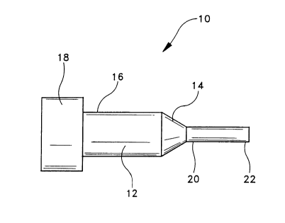
A surgical probe constructed~in accordance
with the present invention is generally indicated at
10 of FIG. 1. Surgical probe l0 is constructed to be
mounted on handpiece 12 at distal end portion 14 of
handpiece 12. Proximal end portion 16 of handpiece 12
is configured to be attached to an energy source 18.
Energy supplied by energy source 18 to handpiece 12 is
directed from proximal end portion 16 to distal end
portion 14.~ It will be appreciated that the manner in
which energy is conducted through handpiece 12 will
vary dependent upon the type of energy produced by
source 18. For example, when probe 10 is configured
to direct electrical energy to residual lens
epithelial cells within the lens capsule, electrical
energy from source 18 can be directed through
handpiece 12 through the use of electrical wiring or
through the use of other known electrical conductors.
However, it is important that the energy be delivered
to distal end portion 14 in a controlled manner in
order to prevent the unwanted delivery of energy to
the patient or to the surgeon using probe 10 of the
present invention.
Probe 10 is dimensioned such that it can be
inserted into the anterior chamber of the eye through
an incision formed at the limbus in conjunction with
the removal of a cataractous lens. Proximal end
portion 20 of probe 10 is cinounted on distal end
portion 14 of handpiece 12. Proximal end portion 20
of probe 10 and distal end portion 14 of handpiece 12
are constructed such that energy directed through
handpiece 12 is transmitted to probe l0. Probe 10 can
be integrally formed on handpiece 12.
Probe 10 further includes a distal end
portion 22. In one embodiment of the present
invention, distal end portion 22 is dimensioned and
configured to be inserted into the lens capsule of the
eye following extracapsular cataract extraction. The
lens capsule is within the posterior chamber of the
eye. Probe 10 can have a variety of configurations
without departing from the spirit and scope of the
present invention. For example, as depicted in FIG.
1, probe 10 can be straight and coaxially mounted on
handpiece 12. However, it will be appreciated that it
may be preferable to configure probe 10 such that it
includes one or more bends along its length in order
to enable a surgeon to reach otherwise difficult-to-
reach areas of the lens capsule. This is particularly
true when the target lens epithelial cells are located
on interior surface 100 of anterior membrane 102 of
lens capsule 104, as depicted in FIG. 5. The second
embodiment of probe 210 depicted in FIGS. 3 and 4 is
provided with a single bend in order to provide the
surgeon with an enhanced ability to reach these
difficult-to-reach areas of lens capsule 104. It will
be appreciated that probe 10 can have a variety of
other configurations having one or more bends for the
purpose of facilitating the application of energy to
lens capsule 104 without departing from the spirit and
scope of the present invention.
Probe 10 is constructed to deliver energy
....
8
along its length from proximal end portion 20 to
distal end portion 22, and then to deliver such energy
to interior surface 100 of lens capsule 104 for the
purpose of destroying residual lens epithelial cells
on interior surface 100. When probe 10 is configured
to deliver electrical energy to interior surface 100
of lens capsule 104, it can include a single
electrode, in which case electrical energy delivered
by the electrode to interior surface 100 of lens
capsule 104 travels outwardly from the electrode along
the length of the electrode until it reaches a ground
state. In this configuration of the present
invention, electrical energy emanating~from the single
electrode of probe 10 will tend to destroy cells
nearer to probe 10 where the electrical energy is at
its greatest level.
In the embodiment of the present invention
depicted in FIGS. 1 and 2, probe 10 includes first
electrode 24 and second electrode 26 which are
oriented such that electrical energy will tend to flow
from one electrode to the other. Although first
electrode 24 and second electrode 26 are depicted as
being coaxial in FIGS. 1 and 2, it will be appreciated
that the electrodes can be configured in various ways.
For example, in the second embodiment of probe 210
depicted in FIGS. 3 and 4, first electrode 224 and
second electrode 226 are not coaxially mounted. It is
also to be appreciated that more than two electrodes
can be used in conjunction with the device and method
of the present invention.
In the embodiments of the present invention
depicted in FIGS. 1 - 4, non-conducting zones 28, 228
separate first electrodes 24; 224 and second
electrodes 26, 226. Thus, electrical current directed
through one of the electrodes will enter the other
electrode only after being transmitted through a
~~.~'~;w.3.
9
medium other than non-conducting zones 28, 228. For
example, electrical current directed through one
electrode can be conducted by residual lens epithelial
cells within lens capsule 104 in order to effect the
destruction of such residual lens epithelial cells.
In the alternative, electrical current can be
transmitted through a conductor such as a balanced
salt solution that can be introduced into the eye
prior to application of power from energy source 18.
This aspect of the present invention will be discussed
in greater detail below in connection with the method
of the present invention.
In the embodiment of the invention depicted
in FIG. 6, distal end portion 322 of probe 3i0 is
configured such that the direction of emission of
energy therefrom can be limited by a non-conductive
cover 313 positioned about distal end portion 322 of
probe 310. Non-conductive cover 313 can be formed
from a variety of biocompatible, non-conductive
materials, including, but not limited to, silicone.
In this third embodiment, one or more portholes 311
are formed through non-conductive cover 313 proximal
distal end portion 322 such that electrode 324 is
exposed therethrough to an external environment of
probe 310. It is believed to be preferable to form
portholes 311 such that they are relatively close to
tip 315 of probe 310, thereby minimizing the amount of
probe 310 that must be inserted into the posterior
chamber. Dependent upon the desired direction of
emission of energy from probe 310, it may be desirable
to insulate tip 315 of probe 310 in order to prevent
energy from being emitted therefrom. It has been
found in certain cases to be preferable to configure
non-conductive cover 313 such that it covers tip 315
of probe 310, thereby preventing the emission of
energy outwardly therefrom, when the embodiment of the
to
present invention depicted in FIG. 6 is used to
destroy residual lens epithelial cells from a position
anterior to the lens capsule, as described in detail
below with respect to the method of the present
invention.
One of ordinary skill in the art will also
recognize that it may be desirable to include a non-
conductive cover in the first and second embodiments
of the present invention depicted in FIGS. 1 - 4 in
order to control the direction of emission of energy
from probe 10, 210.
As above-discussed, non-conductive cover 313
can be constructed of silicone. It is~preferable that
the non-conductive cover 313 be secured to probe 10,
210, 310 such that it will not slide during use. It
will be appreciated that movement of non-conductive
cover 313 may cause damage to eye tissues. In
addition, movement of non-conductive cover 313 may
result in the unintended application of energy to
ocular tissues other than the target tissues, thereby
resulting in further ocular damage.
A desirable method for securing a silicone
sleeve to a probe includes the step of providing a
silicone sleeve having an internal diameter less than
an external diameter of the distal end portion of the
probe to which it is to be attached. The silicone
sleeve is then immersed in ACS grade hexane until the
sleeve has expanded sufficiently such that the sleeve
can be placed over the distal end portion of the
probe. Upon placement of the silicone sleeve over the
distal end portion of the probe, the silicone sleeve
is permitted to dry under a fume hood. As the
silicone sleeve dries, it will tend to return to its
original size, thereby securing itself to the probe.
In the event that non-conductive covering 313 is to
cover tip 315 of probe 310, a drop of silicone can be
~~1.~'i~~.3.
11
placed at tip 315 and allowed to dry. Alternatively,
the sleeve can be a closed-ended sleeve, thereby
obviating the need to apply a drop of silicone to tip
315. One or more portholes 311 can then be formed as
desired through the non-conductive covering 313 using
known cutting tools.
It will be appreciated that energy emitted
from electrodes 24, 26 as depicted in FIGS. 1 - 4 will
emanate outwardly from the tip of probe 10 in
substantially all directions. In contrast, energy
emitted from electrode324 as depicted in FIG. 6 will
be directed outwardly in a limited fashion. One of
ordinary skill in the art will appreciate that energy
will emanate from electrode 324 and through portholes
311 in a substantially conical pattern, such conical
pattern having an axis lying substantially
perpendicular to a longitudinal axis of probe 310.
The direction of energy emitted from probe 310 can
thus be controlled by selectively forming portholes
311 in probe 310. The direction of energy emitted
from probe 310 can also be controlled by rotating
probe 310 about its axis such that energy is emitted
therefrom in the desired direction.
Energy source 18 can be any of a variety of
sources of electrical or thermal energy. It has been
found that electrical energy is preferable when used
in conjunction with the device and method of the
present invention due to the greater on/off
capabilities associated with a source of electrical
energy and due to the general availability of
electrical energy sources in operating rooms. For
example, most phacoemulsification systems have the
capability of providing the requisite electrical power
required by the device and method of the present
invention. Energy source 18 can also be provided by a
standard operating room system designed for bipolar
~~.~'~~~.3.
12
cautery systems. The voltage and current limitations
of such bipolar cautery systems have been shown to be
safe and effective when used in conjunction with the
device and method of the present invention. In
addition, the alternating current produced by power
supplies of this type tend to induce the_oscillation
of charged particles in balanced salt solutions,
thereby resulting in a heating of the solution. The
importance of this phenomenon will be discussed in
greater detail below with respect to the method of the
present invention. However, it is to be appreciated
that the device and method of the present invention
can also be used in conjunction with DC electrical
power sources.
Distal end portion 22 of probe 10 is rounded
in the embodiment depicted in FIGS. 1 and 2. The
rounded configuration of distal end portion 22
facilitates the delivery of energy from electrodes 24,
26 to residual lens epithelial cells while
simultaneously reducing the possibility of damaging
lens capsule 104. However, various configurations of
distal end portion 22 can be employed in conjunction
with the present invention. For example, distal end
portion 22 can be configured such that a plurality of
electrodes can be extended therefrom when distal end
portion 22 is disposed within lens capsule 104. The
plurality of electrodes can be positioned relative to
each other such that all or substantially all of
interior surface 100 of lens capsule 104 can be
subjected to energy at the same time using probe l0.
In another possible configuration, distal
end portion 22 can include an inflatable tip which can
be inflated when distal end portion 22 is in place
within lens capsule 104. This embodiment can be used
in connection with either electrical or thermal
energy. When used in conjunction with electrical
~~.~'~ a:~.l.
13
energy, the inflatable tip would preferably be
constructed of a material having a capacity to conduct
electricity such that electrical current could be
passed therethrough in order to effect the destruction
of residual lens epithelial cells on interior surface
100 of lens capsule 104. When the inflatable tip is
used in connection with the application of thermal
energy, it is preferably constructed of a heat
conducting material such that heat generated within
the inflatable tip is delivered to interior surface
100 and to the residual lens epithelial cells disposed
thereon. Heating can be effected through a variety of
known mechanisms, including the introduction of a
heated fluid into the inflatable tip or through the
application of energy from an energy source such as a
laser to the contents of the inflatable tip.
Probe 10 can include temperature probe 30
disposed at distal end portion 22. Temperature probe
30 has a capacity to measure the temperature at distal
end portion 22 and send a signal to deactivate energy
source 18 when the temperature reaches a predetermined
level, thereby preventing the possible application of
excessive energy levels to the eye. Alternative
mechanisms for preventing the application of excessive
energy to the eye can also be utilized. For example,
energy source 18 can be configured to provide energy
pulses of relatively short duration, thereby reducing
the likelihood that excessive energy will be delivered
to the eye.
Probe 10 can also have ain
irrigation/aspiration capability whereby irrigating
fluid can be introduced into the eye and tissue
fragments and fluids can be removed from the eye
during use of probe 10 in accordance with the method
of the present invention.
The above-described device of the present
.. f .
14
invention is constructed for use in conjunction with
the extracapsular extraction of a cataractous lens and
the subsequent implantation of an artificial
intraocular lens for the purpose of destroying
residual lens epithelial cells prior to the
implantation of the artificial intraocular lens.
Extracapsular cataract extraction generally is
performed by making an incision through the limbus of
the eye in order to provide access to the anterior
chamber of the eye. A surgical cutting tool is then
inserted through the incision and into the anterior
chamber. The surgical cutting tool is used to cut
portion 106 from the anterior membrane~102 of lens
capsule 104, thereby providing the surgeon direct
access to lens 108 within lens capsule 104. Lens 108
is then removed through a known procedure such as
phacoemulsification in which ultrasonic energy is
imparted to lens 108 in order to break lens 108 into
fragments which can then be aspirated from lens
capsule 104 through the use of a phacoemulsification
system having irrigation/aspiration capabilities.
In a first embodiment of the method of the
present invention, a surgeon will employ the device of
the present invention in order to remove any residual
lens epithelial cells from lens capsule 104 following
the removal of lens 108 from lens capsule 104. Probe
10 is inserted into the eye such that distal end
portion 22 is positioned at a predetermined location
within lens capsule 104. Probe 10 can be inserted
through a newly formed incision, but preferably is
inserted through the incision created in conjunction
with the removal of the cataractous lens, thereby
minimizing the trauma to the eye. Energy source 18 is
then activated in order to provide energy to distal
end portion 22 of probe 10. As discussed above,
energy source 18 can be either an electrical energy
~~1.~'i~~~.
source or a thermal energy source. However, in the
preferred embodiment of the method of the present
invention, electrical energy is used due to the above-
discussed beneficial aspects of using such energy.
5 It will be appreciated that the energy
directed to distal end portion 22 from energy source
18 will be transmitted into the tissues immediately
surrounding distal end portion 22. Residual lens
epithelial cells thus will tend to be destroyed by the
10 application of energy from probe 10 due to their
position on interior surface 100 of lens capsule 104.
It has been found that the application of excessive
energy to probe 10 will tend to damage~lens capsule
104 itself. In particular, it has been discovered
15 that distal end portion 22 of probe 10 will tend to
stick to lens capsule 104 in the event that too~much
energy is directed to a single site on interior
surface 100. The further delivery of energy from
probe 10 to such a site on interior surface 100 will
result in permanent, localized damage to lens capsule
104, including the possible perforation of lens
capsule 104. For this reason, it is imperative that
energy be supplied by probe 10 to lens capsule 104 in
a controlled manner.
One technique for limiting the amount of
energy delivered to a single site on interior surface
100 of lens capsule 104 is to move distal end portion
22 of probe 10 about lens capsule 104 rather than
localizing the delivery of energy, thereby minimizing
the possibility that too much energy will be delivered
to a single site. In the event that this technique is
used, it is preferable that probe 10 be moved about
lens capsule 104 in a regimented or patterned manner
in order to ensure that all areas of interior surface
100 are treated.
It has also been found that the use of
._
16
balanced salt solutions such as interstitial fluids,
osmotically balanced salt solutions, and viscoelastic
solutions, can minimize the possibility of probe 10
sticking to interior surface 100 of lens capsule 104
if such solutions are placed in lens capsule 104 prior
to the application of power to probe 10. Balanced
salt solutions are commonly used in ophthalmic
procedures such as extracapsular cataract extraction
for the purpose of preventing the collapse of the
anterior chamber due to the loss of fluid through the
incision. Such balanced salt solutions not only
provide a buffer between probe 10 and interior surface
100, but also provide a conducting medium through
which electrical energy from probe 10 can pass,
thereby facilitating the transfer of energy from probe
10 to the residual lens epithelial cells.
Particularly beneficial results have been
achieved through the use of viscoelastic solutions
containing 2-hydroxypropylmethyl cellulose, such as
the solutions sold by Storz Instrument Company, a
wholly-owned subsidiary of the assignee of this
invention, under the trademarks "OCCUCOAT" and
"OCCUCOAT PF". It has been discovered that probe 10
is less likely to stick to interior surface 100 of
lens capsule 104 at a given power setting when
"OCCUCOAT" viscoelastic solutions are used as compared
to other balanced salt solutions or water. This
benefit may be the result of the fact that the
application of electrical energy from probe 10 to
interior surface 100 in the presence of "OCCUCOAT"
viscoelastic solution causes the viscoelastic solution
to form a precipitate or gel which acts as a barrier
between probe 10 and interior surface 100. The
resulting.precipitate or gel dissipates a few seconds
after terminating the application of electrical energy
and therefore does not pose any complications in the
17
surgical procedure. In addition, the size and
duration of this precipitate or gel has been found to
be reproducible and proportional to the intensity of
the power and the duration of application of power
from probe 10. This predictable change in the
physical characteristics and appearance of the
"OCCUCOAT" viscoelastic material thus enables a
surgeon to identify the areas that have been treated
with energy from probe 10 for the purpose of
destroying residual lens epithelial cells.
In addition to the above-described benefits,
it has also been discovered that the use of a
viscoelastic solution containing 2-hydroxypropylmethyl
cellulose, such as "OCCUCOAT" viscoelastic material,
in conjunction with the method of the present
invention results in significantly greater temperature
increases when compared to other balanced salt
solutions and water. Alternating current produced by
energy source 18 causes the oscillation of the charged
particles in a viscoelastic solution containing 2-
hydroxypropylmethyl cellulose, thereby resulting in
the heating of the viscoelastic solution. The maximum
temperature achieved using "OCCUCOAT" viscoelastic
solution used in conjunction with the method of the
present invention is 100 C. Such heat serves to
destroy residual lens epithelial cells within the lens
capsule. It is believed that the oscillation of
charged particles within the viscoelastic solution
caused by the application of AC current thereto, as
well as the local osmotic differences resulting from
such oscillations, further facilitates the destruction
of the lens epithelial cells within the lens capsule.
It has been found that energy emitted from
probe l0.in conjunction with the method and device of
the present invention may reach the iris, thereby
causing tissue damage to the iris. For this reason,
is
it may be desirable to provide an iris shield that can
be placed between the lens capsule and the iris prior
to directing energy through probe 10. In one
embodiment of the method of the present invention, an
iris shield formed from a hydrogel material is placed
between the iris and the lens capsule in order to
prevent energy from probe 10 from adversely affecting
the iris. One of ordinary skill in the art will
recognize that other materials can be used to form an
iris shield in accordance with the teachings of the
present invention, so long as such material is
biocompatible, is capable of shielding the iris from
energy emitted from probe 10, and is not structurally
compromised by energy emitted from probe 10.
It will be appreciated that the amount of
energy required to perform the method of the present
invention will vary dependent upon a number of
factors, including the size and configuration of the
electrodes) of probe 10 and the presence or absence
of a conducting medium within lens capsule 104.
Devices with larger electrode surface areas will have
higher power requirements. In addition, the desirable
power level will vary dependent upon each surgeon's
chosen technique. For example, if probe 10 is used in
a relatively quick, sweeping motion within lens
'capsule 104, a higher power may be used due to the
fact that there will be less power delivered to any
single site on interior surface 100 of lens capsule
104. Similarly, greater power levels can be used when
a viscoelastic solution containing 2-
hydroxypropylmethyl cellulose is present due to the
above-referenced characteristics of such viscoelastic
solutions. However, if the surgeon prefers to treat
individual sites on a methodical or sequential basis,
it may be desirable to utilize lower power levels in
order to minimize the possibility of damage to lens
~~...3'~~~~~.3.
19
capsule 104.
In some cases it may be preferable to
utilize two or more different configurations of probe
in conjunction with the method of the present
5 invention in order to ensure that all areas of
interior surface 100 are subjected to the energy
emanating from probe 10. For example, probe 210 may
be inserted for use following use of probe l0 in order
to ensure that areas of interior surface 100 that may
10 not have been heated using probe l0 are subjected to
energy from probe 210. It may also be necessary in
certain cases to form a second incision through the
limbus in order to provide a different angle of attack
for probe 10, thereby ensuring that all areas of
interior surface 100 are subjected to the energy
emanating from probe 10.
Following the application of energy to lens
capsule 104 and the resulting destruction of residual
lens epithelial cells, the surgeon deactivates and
removes probe 10 from the eye. Upon the removal of
particulate matter and any balanced salt solutions
from lens capsule 104 using known
irrigation/aspiration techniques, the surgeon can
proceed with the implantation of an artificial
intraocular lens 104 using a variety of known methods.
In a second embodiment of the method of the
present invention, a probe 310 capable of emitting
energy from its distal end portion in a predetermined
direction is provided. Although probes of various
configurations can be used, it has been found to be
advantageous to employ a probe such as that depicted
in FIG. 6 and disclosed in detail herein in order to
control the emission of energy from the probe. It is
to be appreciated that the second embodiment of the
method of the present invention can be used either
before or after the extracapsular extraction of the
~~.3'~s~.~.
cataractous lens. In the second embodiment of the
method of the present invention, distal end portion
322 is inserted into the eye such that it is
positioned within the posterior chamber of the eye
5 between the iris 101 and the anterior membrane 102 of
lens capsule 104 and such that porthole 311 is
directed posteriorly towards the lens capsule. Energy
is then directed to probe 310 such that energy is
emitted therefrom at a level sufficient to destroy
10 residual lens epithelial cells on lens capsule 104.
It will be appreciated that energy emanating from
probe 310 will pass through lens capsule 104 and
destroy residual lens epithelial cells.disposed on
interior surface 100 thereof. Dependent upon the
15 configuration of probe 310, it may be necessary to
move probe 310 about in order to ensure that energy is
directed to all portions of lens capsule 104, thereby
ensuring that as many residual lens epithelial cells
as possible are destroyed: The delivery of energy to
20 probe 310 is then ceased and the probe is withdrawn
from the eye. It is to be appreciated that a second
probe having a different configuration can be inserted
as above-discussed in order to reach portions of lens
capsule 104 not reachable using probe 310. A second
probe also can be inserted through a second incision
formed at the limbus as above-discussed in order to
reach portions of lens capsule 104 not reachable using
probe 310. Furthermore, balanced salt solutions such
as interstitial fluids, osmotically balanced salt
solutions, and viscoelastic solutions, as above-
discussed, can be used in connection with the second
embodiment of the method of the present invention.
Although the device and method of the
present invention have been disclosed herein with
respect to certain preferred embodiments, it will be
apparent to one of ordinary skill in the art that
CA 02137211 2004-05-12
71009-9
21
various modifications can be made to the invention
without departing from the spirit and scope of the
invention disclosed and claimed herein.