Note: Descriptions are shown in the official language in which they were submitted.
2 ~ 3 ~
VACCINE COMPRISING AUTOLOGOUS EPIDERMAL GROWTH FACTOR
F I E L D O F T ~ E I N V E N T I O N
This invention is relates to the field of immunology, in particular to vaccine compositions able to
produce an autoimmune reaction against autologous (self) Epidermal Growth Factor (EGF).
An important object of this invention is to obtain a vaccine composition for the active
immunotherapy of EGF dependent malignant tumors, which can inhibit the proliferation of those
tumors, and which therefore are useful for the treatment of malignant neoplasms, and of other
EGF related diseases. Thus, the invention is also related to the field of cancer therapy.
I~ESCRIPTION OF T~E PRIOR Al~T
Epidermal Growth Factor, a polypeptide that stimulates epithelial cell proliferation, has been
considered to be one of the growth factors involved in malignant transformations. Its action is
mainly performed via its membrane receptors.
Epidermal Growth Factor (EGF) is a 53 amino acid polypeptide, its molecular weight is about
6045 D. It was isolated and purified for the first time from murine submaxillary gland (Cohen S.
J.Biol Chem (1962) 237,1.55~ ), later a similar molecule was obtained from human urine (Cohen
S. Human Epidermal Growth factor: Isolation and Chemical and Biological Properties PNA USA
72~1975 1 3]7).
EGF is capable of stimulating the proliferation of epithelial and mesenchymal cells, both in vitl O
and in vivo (Cohen S.l Carpenter G., PNAS USA 72, ]3]7, 1975) and EGF gives a specific
stimulation in some breast cancer cell lines (Osborne C.K. et als Can Res. 40,2.361 (19~0). A
role of the EGF in the differentiation process in the mammary gland, mainly for the development
ofthe lobule alveolar system has been demonstrated (Tonelli C.J. Nature (1980) 285, 250-25~)
This bio-regulating action is exerted via a membrane receptor (EGF-R), a 1,186 amino acid
glycoprotein of about 170 kD, the gene of which has been cloned and sequenced. The
intracellular domain of the receptor is associated with an activity of Tyrosine specific protein
kinase wich shows an structural homology to the oncogene product v-erb-B showing the relation
to the malignant transformation process ~Heldin C.H Cell 37, 9-20 (1984))
EGF and its receptor constitutes a molecular complex of high specificity and the interaction
between them develops important mechanisms of cell growth regulation
High levels of EGF-R have been detected in malignant tumors of epithelial origin, such as breast,
~..
!..A
2 213'7 6~9
bladder, ovarian, vulva, colonic, pulmonary, brain and esophagus cancers. Tlle role played by
EGF and its receptor in regulating tumor growth is unknown, but there are suggestions that the
EGF-R expression in tumor cells provides a mechanism for autocrine growth stimulation which
leads to uncontrolled proliferation (Schlessinger J., Schreiber A.B., Levi A, Liberman T.,
Yarden Y. Crit. Rev. Biochem. 1983, 14 (2) 93-111).
The presence of EGF-R in tumor cells has proven to be an indication of a poor prognosis in
human breast cancer. Approximately 40 % of the breast tumors shows specific binding sites of
high affinity for the EGF, there is also an inverse correlation with the presence of estrogen
receptor indicating EGF-R as an dediferentiation marker or an indicator of the potential capacity
of proliferation of the malignant cells. (Perez R., Pascual M.R., Macias A., Lage A., Breast
Cancer Research and Treatment 4, ~ 89- 193,1984).
It also has been reported that the EGF-R expression is higher in regional ganglional metastasis
that in the primary breast carcinomas (Sainsbury J.R., et al.(l985): Lancet 1(8.425), 364-366),
and that the expression of the receptor is dif~erent in the different histologic subtypes of brc~lst
carcinomas cells, which also makes their presence a signal of bad prognosis( Macias A., et al
(1986). AnticancerRes. 6:849-852).
Previous studies performed in the Ehrlich Ascitic Tumor (EAT) model in Balb-C mouse,
demonstrated the in vivo inhibitory effect of the EGF .(Lombardero J., et als Neoplasma 33, 4
(1987), suggesting the possibility of considering this molecule as a biological response modifier.
It has been previously reported the presence of an EGF precursor molecule in the cell membr~ne
of EGF dependent tumors. The authors of the present invention have reported this as an
important fact to consider this molecule as a target for the action of autoantibodies. The
results obtained in different studies, have prompted the consideration of the EGF\EGF-R
system a possible target for therapeutic actions.
Passive immunotherapy using monoclonal antibodies against the EGF-R, have been object of
multiple investigations, which have demonstrated that the specific recognition by the antibody of
the receptor inhibits the EGF binding, with an inhibitory effect on the mitogenic stimulation of
malignant cells (SATO J.D.,et al. Methods in Enzymology, vol 146 pp 63-81.(1987) however
these antibodies which are of murine origin will usually produce a human anti mouse antibody
response (HAMA).
Until the present invention no active immunotherapy against EGF dependent tumors capable of
inhibiting proliferation has been proposed, since the art consistently reports that "self~' molecules
will not induce any immune reaction because the host has been educated to be tolerant to self
The present invention provides a vaccine composition containing autologous EGF coupled to a
carrier protein, which complex will inhibit the EGF dependent tumors growth, through an
autoimmune effect, without the collateral effects of the introduction of a heterologous protein in
the human body.
3 ~4137~3g
This vaccine composition can be used in the treatmenl of EGF dependent tumors or any
malignant disease associated to EGF.
It will be understood that in this specification EGF is to be read as including any fragment and/or
derivative of EGF which have similar immunological properties and/or ef~ects as the original
molecule. Derivatives include, but are not limited to, conventional aminoacid substitutions, site-
directed replacement of aminoacids for enhanced stability and/or activity, chemical modifications
and the like.
D E T A I L E D D II S C R I ~ T I O N O F T H E I N V E N T I O N
I. Obtaining an immunogenic preparation:
Two preparations were obtained. The first is murine EGF ( mu-EGF) coupled to a protein
carrier and the second is human recombinant EGF ( hu-re-EGF) (National Medicament Register
Office from Cuba, HEBERMIN, No. 1 266),also coupled to a carrier protein.
The preparation containing hu-rec-EGF coupled to a carrier protein was obtained to be used
only in non human primates and in humans. A proper adjuvant is applied.
The mu-EGF conjugated to a protein carrier is used in the studies performed in mice as a model
to determine the immunogenicity and the antitumoral effect of a preparation containing an
autologous EGF molecule.
Immunogenicity studies were performed in primates with the conjugate hu-EGF-carrier protein
since the hu-EGF is very similar to the primate EGF though it is recognized as a self mo1ecule.
These results allowed to demonstrate the immunogenic response an autologous molecule can
elicit .
To obtain the preparations, a solution of murine or hu-rec-EGF in PBS/MgC12 10mM, is mi~;ed
with a solution of the carrier protein in the same solvent, in a ratio of between 1 and 5 moles of
EGF per mol of protein.
Afterwards glutaraldehyde 0,5 %, is added to obtain a final concentration between 0.1% and
0.05%.
The mixture is incubated between 1 and 3 hours at room temperature and subsequently dialyzed
in PBS /MgC12 10mM with, at least, 3 changes of dialysis solution.
Conjugate Characterization:
The test of the conjugation efficiency and of the maintenance of the antigenicity, is performed
through an ELISA assay.
3 ~
ELISA plates of activated PVC (NUNC) were coated with 50 ~1 of a antiserum against the
carrier protein utilized, in a concentration between I and 10 llg/mL. In the case of cholera toxin
chaim B (CTB) as carrier, the plates were coated with the ganglioside GMI.
Subsequently 3 washes with PBS/Tween were carried out and then, the plates were blocked
with a solution of BSA between 0 5 and 1% in PBS /Tween, incubated them during a period of
30 minutes to I hour at 370C. Dilutions between 0.1 and 0.001 mg/ml of the conjugates to be
assayed were added to the plates, 50 ~ /well, and incubated for I to 2 hours at 37~C
In the next step a mouse anti hu-rec-EGF antiserum was added in a dilution between 1:500 and
1:1000, 50 ~,ll/well, and incubated for between 30 minutes and I hour at 37~C.
As the last step, the plates were incubated with a antimouse- alkaline phosphatase antiserum, in a
dilution between 1:500 and 1:1000, 50 IlVwell, for 30 minutes to 1 hour at 37~C.
Reaction color was developed with p-nitrophenylphosphate, at a concentration of I mg/ml in
diethanolamine, 50 IlVwell, incubated for 30 minutes at 37~C. Optical density was measured at
405 nm in an ELISA plates reader.
The results demonstrated the activity of the molecule and the efficiency of the conjugation,
because, the conjugate maintains its recognition site for the molecule coating the plates, which
specifically recognizes the carrier protein and, at the same time, can be recognized by a anti EGF
antiserum.
II.-Ch~ractel ization of cl~ects produced by the prep;lr~tion COllt;linillg mu-EG~.
Preclinical studies:
II;I) Endogenous EGF immunogenicity: inductio-l of autoimmunity in mice.
In order to demonstrate the capacity of the immunogenic preparation containing mu-E(i~
obtained through the technique described in item I of inducing autoimmunity against the endogen
EGF, a test was performed in Balb/c mice.
Groups of animals were inoculated with different doses in the range of 50 to 100 ~lg of mu-EGF
conjugated to the carrier protein per animal per week during 4 to 6 weeks.
The first week the immunogenic preparation was prepared in a ratio 1:1 with complete Freund
adjuvant, all the following doses were prepared with incomplete Freund adjuvant.The same procedure was performed in a control group, but only adjuvant was administered to
the animals. One week after the last immunization, blood was extracted to the animals, the serum
separated from the remainder of the blood and the titer of antibodies against mu-EGF was
determined by an ELISA technique.
II b) Immunogenicity of hu-rec-EGF:
In order to demonstrate the immunogenicity of hu-rec-EGF in mice and to show that the
antibodies against hu-rec-EGF recognize the mu-EGF, the experiment was performed in Balb/c
* Trade-mark
~ 7 ;~
mice.
Groups of animals were inoculated with different doses in the range of 50 to 100 ~g of hu-lec
EGF-protein per animal per week during 4 to 6 weeks.
The first week the immunogenic preparation was prepared in a ratio 1:1 with complete Freund
adjuvant, all the following doses were prepared with incomplete Freund adjuvant.
The same procedure was performed in a control group, but only adjuvant was administered to
the animals.
One week after the last immunization, blood was extracted from the animals, the serum was
separated from the rest of the blood and the titer of antibodies against mu-EGF was determined
by an ELISA technique.
IIc) Antitumor Activity:
The main objective of this experimental procedure is to determine whether the immune response
obtained against the autologous EGF is able to elicit any antitumor effect in EGF dependent
tumors.
The animals with higher antibody titer, determined according to the technique described
previously, were inoculated with Ehrlich Ascitic Tumor (EAT) in cellular concentrations between
0.2 to 2 million cells per animal. The control group was treated in the same manner.
The animals were observed for grafting as well as for survival.
III. Characterizatio-l of the imnlunc response:
IIIa) Isotype of the antibody response
In order to determine whether the autoimmune response obtained upon the immunization of
mice with the autologous EGF is a response producing antibodies of the isotype IgM or IgG, an
ELISA assay was performed testing the sera of immunized animals with EGF according the
techniques described in items II a) and b). In the case of IgM characterization an antiserum
against this molecule was incubated with the samples. IgG characterization is performed with an
antiserum against IgG.
IIIb) Characterization of the memory of the immune response
The development of a product to be used as a vaccine in an active therapy requires the
determination of its capability of inducing an immunological memory, and the determination of
the duration of said memory if it is induced.
This information allows the possibility of a correct design of the immunization schemes that can
be implemented with the product.
Groups of mice were immunized with one dose between 50 and 100 llg of hu-rec-EGF per
A
animal in complete Freund adjuvant in a proportion 1:1.
The kinetic of antibody production against mu-EGF was studied in different groups of anil11als
This study was performed after the first immunization and after the re-immunizations whel1 tl-e
titer is declinin~. The antibody levels were determined according an ELIS~ techniclue.
IV. Immunogenicity studies irl noll hum;~lll prinlAtcs:
The criteria of immunogenicity of the immunogenic preparation are based on results obtained in
non human primates because these are the species that are closest to human.
A group of primates were immunized with the immunogenic preparation containing hu-rec-EGF
in a dose of 50û llg (conjugated with tetanic toxoid) and together with the adjuvant. After the
last immunization a sample of blood was extracted and antibody titers against hu-EGF were
determined.
EXPERIMENTS
Example 1.
Study of the pres~nc~ of ~n EGF precursor molecule in th~ c~ll membr~ne Or ~gr
dependent tUlllol-S.
This study was performed through a Western Blotting technique. Samples of S ductal carchlc)ma
of the breast in different stages, one head and neck tumor, four samples of fibrocystic dysplasia
and five normal samples obtained as controls were studied.
Cell membranes were obtained from the samples through the procedure described elsewhere (
GrimauxM.,RevNeurol. 1988,144: 101-103).
Electrophoresis was performed at 250 v, 10 Ma, 3. OW, 15 ~C and 150 Vh. Molecular weight
standards were used in the range of 14 300 D (Lyso~yme) to 340 000 D (alpl1a 2 macroglobuli~
Proteins separat*ed during electrophoresis were transferred to a nitrocellulose membrane of 0.45
~,lm in a Phast system equipment in a buffer transfer solution. After the transference the
membrane was blocked overnight with 10 % skimmed milk with constant stirring.
AP~er three washes in buffer solution a mouse monoclonal antibody recognizing human EGF was
added and incubated four one hour.
A~er three washes a biotynilated anti mouse antibody was added and incubated during one hour.
Peroxidase streptavidine conjugate was added and reaction developed with diaminobencidine and
hydrogen peroxide after one hour incubation.
The results obtained demonstrated that the samples studied corresponding to normal tissues did
* Trade-mark
~ ~ ~ 7 ~ ~ ~
not show any band in the zone of high molecular weight according to the standards. However tlle
samples corresponding to breast pathology (dysplasia and carcinomas) showed a diffuse banding
in the high molecular weight zone. This is an experimental evidence of the presence of a high
MW EGF precursor in the tumor membranes.
Example 2.
Obtaining the mu-EGF/CTB conjug~te.
One ml of mu-EGF in PBS/MgC12 10mM at a concentration of I mg/ml, was mixed with 2 ml of
a solution of CTB in the same solvent at a ratio of 1 mol of mu EGF per mol of CTB.
Glutaraldehyde (3 ml, 0.5%) was added to obtain a final concentration of 0.05%.
Incubation was performed during 1 hour at room temperature and subseguently dialyzed in PBS
/MgC12 1 OmM with, at least, 3 changes of the dialysis solution.
Example 3.
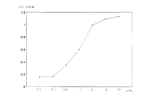
mu-EGF/CTB conjug;lte characterizatioll.
ELISA assay for conjugate test: PVC activated ELISA plates (NUNC) were coated with 50 ~1
of the GM1 ganglioside (recognizing the CTB molecule) in a concentration of 4 llg/ml in
methanol, which was left to dry off in the flow during I hour.
Subsequently 3 washes with PBS /Tw*een were carried out and then the plates were blocked with
a solution of BSA 1% in PBS /Tween, and incubated during 30 minutes at 37~C.
Conjugate dilutions between 0.1 and 0.001 mg/ml were added to the plates at 50 ,ul/well and
incubated during I hour at 3 7~C.
Next a mouse anti-mu-EGF antiserum in a 1:1000 dilution, 50 ,ul/well was added and incubated
for I hour at 37~C.
Then, the plates were incubated with anti mouse antiserum alkaline phosphatase conjugate
(dilution 1:1000), 50 lli/well for 1 hour at 37~C. The color was developed with p nitropheny-
lphosphate at a concentration of I mg/ml in diethanolamine, 50~11/well, incubated for 30 minutes
at 37~C; optical density was measured at 405nm.
The results demonstrated a direct relationship between the concentration of the conjugate and
the absorbance values. This demonstrates the activity of the conjugated and the efficiency of the
conjugation, since, the molecule maintains the recognition for the GM1 ganglioside (identifies
CTB) and, at the same time, is recognized by a anti mu EGF antiserum (Figure 1)
Example 4.
~mmunogenicity of autologous EGF: induction of autoimmunity in mice.
In order to demonstrate that the immunogenic preparation containing autologous EGF is
* Trade-mark
.~
8 ~ 3 ~
capable of inducing au~oh~ unity, the experimentation was performed in Balb/c mice
Groups of animals were inoculated with a dose of 50 llg of conjugated mu-EGF per aninlal
subcutaneously weekly during 4 to 6 weeks.
The first week the immunogenic preparation was prepared in a proportion 1:1 with complete
Freund adjuvant, all the following doses were prepared with incomplete Freund adjuvant.
The same procedure was performed in a control group, but only adjuvant was administered to
the animals. One week after the last immunization, blood was extracted from the animals, the
serum obtained and the titer of antibodies against mu-EGF was determined by an ELISA
technique.
Costar plates were coated with mu-EGF at a concentration of 10 ~lg/ml in carbonate bicarbonate
buffer (pH 9.6), and incubated overnight. After the plates were washed the samples were added
in different dilutions. Incubation took place during one hour. Alkaline phosphatase anti-mouse
antibody conjugate was added and incubated during one hour after which color was developed
and optical density measured at 405 nm in an ELISA reader.
All the animals immunized with the mu-EGF-CTB preparation developed antibody titer against
the mu-EGF up to 1:1000 dilution. The control group did not show any antibody titer (Figure 2)
Example 5.
Immunogenicity Or hu-rec EGF: inductioll of autoimmunity in mice.
In order to demonstrate that the immunogenic preparation containing hum-rec EGF was capable
of producing antibody titer against mu-EGF, the experimentation was performed in Balb\c mice.
Groups of animals were inoculated with a dose of 50 ~,lg of hum-rec-EGF per animal
subcutaneously, weekly during 4 to 6 weeks.
The first week the immunogenic preparation was prepared in a proportion 1:1 with complete
Freund adjuvant, all the following doses were prepared with incomplete Freund adjuvant.
The same procedure was performed in a control group, but only adjuvant was administered to
the animals.
One week after the last immunization, blood was extracted from the animals, the serum obtained
and the titer of antibodies against mu-EGF was determined by an ELISA technique.
Costar plates were coated with mu-EGF at a concentration of 10 ~g/mL in carbonate
bicarbonate buffer (pH 9.6), and incubated overnight. After the plates were washed the samples
were added in different dilutions. Incubation took place during one hour. The alkaline
phosphatase antimouse antibody conjugate was added and incubated during one hour af~er which
color was developed and optical density measured at 405 nm in an ELISA reader.
.A
~ 3 ~
All the animals immunized with the hum-rec EGF preparation developed antibody titer against
the mu-EGF up tO 1:20000 dilution. The control group did not show any antibody titer ( Figul-e
3).
Exalllplc 6.
Immunogenicity of hu-rec EGF ill a prepalatioll ~vith alulllillllm llydroxide.
In order to demonstrate that the immunogenic preparation containing hum-rec EGF and
Aluminum Hydroxide as adjuvant was capable of producing antibody titer against mu-EGF, the
experimentation was performed in Balb\c mice.
Groups of animals were inoculated with a dose of 50 llg of hum-rec-EGF (with Aluminum
Hydroxide as adjuvant) per animal subcutaneously, weekly during 4 to 6 weeks.
The same procedure was performed in a control group, but only adjuvant was administered to
the animals. One week after the last immunization, blood was extracted from the animals, the
serum obtained and the titer of antibodies against mu- EGF was determined by an ELISA
technique.
Costar plates were coated with mu-EGF at a concentration of 10 ~g/ml in carbonate bicarbonate
buffer (pH 9.6), and incubated overnight. After the plates were washed the samples were added
in different dilutions. Incubation took place during one hour. The all~aline phosphatase
antimouse antibody conjugate was added and incubated during one hour after which color was
developed and optical density measured at 405 nm in an ELISA reader.
All the animals immunized with the hum-rec EGF/ Al OH preparation developed antibody titer
against the mu-EGF up to 1:4000 dilution. The control group did not show any antibody
titer.(Figure 4)
Example 7.
Anti-tumour activity.
The main ojective of this experimental procedure was to determine whether the immune response
obtained against the autologous EGF was able to elicit any antitumor effect in EGF dependent
tumors .
The animals with higher antibody titer, determined according to the technigue described
previously in the example 5, were inoculated with Ehrlich Ascitic Tumor (EAT) in cellular
concentrations of 2 million cells EAT per animal. The control group (nonimmunized mice) was
treated in the same manner.
The animals were observed for grafting as well as for survival. Survival curves of treated and
control animals are shown in Figure 5.
, . .. -
~ ~ 3 ~
The Increase in Life Span Index was 22.5% showing and increase in surYival for the treated
animals in relation to tlle control statistically significant according to the Mantel l~aenszel and
Wilcoxon tests.
Example 8.
Association between antibody titer against mu EGI~ and 125-I EGF biodistribution.
This experiment was performed to demonstrate that there is a different biodistribution of 125-I
EGF in animals with antibody titer against mu- EGF in relation to animals that did not have
antibody titer against mu-EGF.
An experiment with 4 groups of mice was performed for this purpose:
Group 1: 30 mice with antibody titer against mu-EGF.
Group 2: 30 mice without antibody titer against mu-EGF.
Group 3: 30 mice with antibody titer against mu-EGF grafted with EAT.
Group 4: 30 mice without antibody titer against mu-EGF grafted with EAT.
Samples from group I and 2 were taken from blood, lung, kidneys, liver, and skin at the
following times: 2, 5, 8, 11, 15, 20, 30, 60, 120 and 150 minutes and 3 animals were sacrificed at
every corresponding time, counting the radioactivity in the organs extracted.
The results obtained have shown a difference in the accumulation of I-125 EGF in time mainly hl
kidney and liver (Figure 6 a,b), indicated that the presence of antibodies against EGF do alter the
biodistribution of this molecule.
Samples from group 3 and 4 were taken from blood, lung, kidneys, liver, skin and from the
ascitic fluid at the following times: 2, 5, 8, I l, 15, 20, 30, 60, 120 and 150 minutes and 3 animals
were sacrificed at every corresponding time, and the radioactivity counted in the organs
extracted.
It could be appreciated less accumulation of the labeled EGF in the ascitic fluid of animals with
antibody titer than in the animals without antibody titer (Figure 7), indicating a more rapid
depuration of the EGF present in the ascitic fluid in these animals, and/or a limitation in EGF
access to the ascites.
Example 9.
Immune response characterization: isotype obtained against autologous EGF.
In order to know wether the autoimmune response obtained upon the immunization of mice with
the autologous EGF was a response producing antibodies of the isotype IgM or IgG, an ELIS,~
assay was performed,in which the plates were coated with mu EGF to concentration of 10 llg/ml
, 50 lli/well, and incubated for I hour at 37~C.
Subsequently, dilutions between 1:10 and 1:1000 of the serums of animals immunized with Inu
a
~ ~ 3 ~
EGF- CTB according to Example 5 were applied, 50 Ill/well, and were incubated for 1 hour at
37~C.
Parallel design of plates were performed to measured IgG or IgM response witll the
corresponding antiserum (anti-IgG or anti-IgM respectively).
Color in the plates was developed with p nitrophenylphosphate, at concentration of 1 mg/mL in
diethanolamine, incubated during 30 minutes at 37~C and values of optical density at 405nm
were read.
IgG response was obtained in all treated animals (Figure 8).
Example 10.
Characterization of thc memory of the immune response against autologous EGF.
Two groups of 10 mice were studied with a single immunization of 50 ,ug hu-re-EGF in
complete Freund adjuvant.
Group I.- The kinetics of antibody production against mu EGF was studied in this group of
animals. Every 4 days blood samples were extracted. The antibody levels were determined by an
ELISA technique.
Group II.- Was conformed with the animals with declining antibody titer from Group I and
immunized again. Every 2 days blood samples were extracted. The antibody levels were
determined by an ELISA technique.
Results have shown a memory response when the animals were immunized again Wit}l the
preparation (Figure 9).
Example 11.
Obtention of the immunogenic preparatiol1: hu-rec EGF toxoid tetanic.
A solution of hu-EGF in PBS/MgC12 1 OmM at a concentration of 1,4 mg/ml, was mixed with 2
ml of a solution of TT in the same solvent at a concentration of 4 mg/mL. Glutaraldehyde (3 ml
,0.5%),was added to obtain a final concentration of 0.05%.
Incubation was performed during 1 hour at room temperature and subsequently dialyzed in PBS
/MgC12 10mM with, at least, 3 changes ofthe dialysis solution.
Example 12.
Conjugated characterization: hu-rec EGF toxoid tetanic.
ELISA assay for conjugate test:
,~
12
Costar plates (High Binding) were coated with 50 Ill of an antiserum anti TT obtained in sheep,
in a concentration of l O llg/ml, and incubated overnight.
Subsequently 3 washes with PBS /Tween were carried out and then the plates were blocked Witl
a solution of BSA 1% in PBS /Tween, and incubated during 30 minutes at 37~C.
Conjugate dilutions between O. l and 0.001 mg/mL were added to the plates at 50 Ill/well, and
incubated during l hour at 37~C.
Next a mouse anti-hu-EGF antiserum in a l:lO00 dilution, 50 IlUwell was added, and incubated
for l hour at 3 7~C.
Then, the plates were incubated with anti mouse antiserum alkaline phosphatase conjugate
(dilution l:lO00), 50 ,ul/well for 1 hour at 37~C. The color was developed with p nitropheny-
lphosphate at a concentration of ] mg/mL in diethanolamine, 50 !ll/well, incubated for 30
minutes at 37~C, and optical density was measured at 405nm.
The results demonstrated a direct relationship between the concentration of the conjugated and
the absorbance values.
This demonstrates the activity of the conjugate and the efficiency of the conjugation, since, the
molecule maintains the recognition for anti serum anti TT, and at the same time, is recognized by
a anti mu EGF antiserum (Figure l O).
Example 13.
Study of the immunogcnicity of hu-EGF coupled to a carrier protein (TT) in noll hllman
primates.
The study was performed with 4 Rhesus monkeys, being submitted to a clinical veterinary
examination including:
Physical examination
Thorax X rays
Blood tests.
These animals were immunized with a hu-EGF coupled to TT, according to Example l O.
Immunization was performed subcutaneously in weeks l, 2, 3, 4, 6 and 12. Complete Freund's
adjuvant was used in the first immunization and incomplete Freund's adjuvant in all others.
Blood was extracted from the animals and antibody titers were determined through an ELISA
Costar plates were coated with hu-EGF at a concentration of lO llg/mL in carbonate bicarbonate
buffer (pH 9.6), and incubated overnight. Af~er the plates were washed the samples were added
in different dilutions. Incubation took place during one hour. The alkaline phosphatase antihuman
* Trade-mark
~ 3~fi;~
13
antibody conjugate was added and incubated during one hour after which color was developed
and optical density measured at 405 nm in an ELISA reader.
All the animals immunized with the hu-EGF-TT preparation developed antibody titer against tl1e
hu-EGF up to 1:20000 dilution (Figure I I ).
B~IEF DESCRIPTION OF THE DRAWINGS
Figure 1: ELISA assay for determination of conjugation efficiency between CTB and hu rec
EGF.
X axis: serial dilutions ofthe conjugate (from Img/ml to 0.001mg/ml)
Y axis: optieal density at 405 nm, measured in an ELISA plates reader.
Figure 2: ELISA assay for the determination of antibody titer against the mu EGF in 5 mice
immunized with the conjugated mu EGF - CTB.
X axis: Antiserum dilutions (1: 10, 1: 100, 1: 1000)
Y axis: optieal density at 405 nm, measured in an ELISA plates reader .
The curves represent the titer of 5 tested animals, compared with the same animals before
immunization.
Figure 3: ELISA assay for the determination of antibody titer against mu-EGF, in S mice
immunized with hu-rec-EGF.
x axis: sera dilutions 1 :100, 1:1000, 1:10000, 1:20000.
y axis: optical density at 405 nm
Figure 4: ELISA assay for the determination of antibody titer against mu-EGF, in 5 mice
immunized with hu-rec-EGF in Aluminum Hydroxide as adjuvant
xaxis:seradilutions 1:100, 1:500, 1:1000, 1:2000, 1:4000, 1:8000.
y axis: optical density at 405 nm
Figure 5: Survival of animals immunized with mu EGF-CTB and subsequently inoculated with
Ehrlich Ascites Tumour, in comparison with control animals (nonimmunized) and inoculated
with same tumor.
Figure 6 a. Aeeumulation of 125-I EGF in liver of miee immunized with hu-ree-EGF, in relation
to nonimmunized eontrols.
Figure 6 b. Aeeumulation of 1~5-I EGF in kidneys of miee immunized with hu-rec-EGF, in
relation to nonimmunized controls.
Figure 7: Accumulation of mu-EGF 1125, in ascitic fluid of animals grafted with EAT,
previously immunized with hu-rec-EGF.
,~,
,'~
14
Figure B: ELISA assay for determination of IgG or IgM of the immune response in animals
immunized with n-u EGF-CTB.
Figure 9: Antibody response kinetic (IgG) against mu EGF, in animals immunized with
hu-rec-EGF (~emmory).
x axis: time (days).
y axis: Inverse logaritllm of the antibody titer.(mean value).
Figure 10: ELISA assay for the determination of the efficiency of conjugation with Tetanic
toxoid and hu-rec-EGI~.
x axis: dilutions of conjugate.
~ axis: optical densiIy at 405 nm
Figllre I 1: antibody titer against hu-rec-EGF in non human primates immunized with
hu-rec-EGF, coupled to tetanic toxoid.