Note: Descriptions are shown in the official language in which they were submitted.
2139244
1
TITLE OF T8E INVENTION:
Haemostatic Pressure Pad
NAME ~( S )~ OF INVENTOR i( 8 )r
Anthony Hk Lam
FIELD OF T8E INVENTION
The present invention relates to a haemostatic pressure
pad intended to be used with a haemostatic compression device.
BACKGROUND OF T8E INVENTION
Haemostatic pressure pads intended for use in a
haemostatic compression device come in a variety of shapes and
configurations. An example of a haemostatic pressure pad that
is in common use is United States Patent 4, 572,182 which issued
to Royse in 1986. The Royse patent discloses a haemostatic
pressure pad which is a circular disk. The circular disk has
a symmetrically positioned mounting boss and a "V" shaped
notch. The mounting boss is used to mount the haemostatic
pressure pad on an arterial clamp. The "V" shaped notch
facilitates the placement of the haemostatic pressure pad over
a catheter prior to removal of the catheter from a patient's
artery.
None of the haemostatic pressure pads commercially
available at the present time are capable of applying
simultaneous pressure to both the femoral artery and the
femoral vein.
. 21 39244
SUI~tARY OF THE INVENTION
What is required is haemostatic pad that is capable of applying
simultaneous pressure to both the femoral artery and the femoral
vein.
'rJ According to one aspect of the present invention there is
provided a haemostatic pressure pad comprising a generally U-shaped
body having a first finger portion and a second finger portion. The
first finger portion has a first end and a second end. The second
finger portion has a first end and a second end. The first finger
portion and the second finger portion are conjoined at the respective
second ends. The first finger portion is substantially broader or
wider than the second finger portion. The body has a lower contact
face and an upper attachment face. Means are positioned on the
attachment face of the first finger portion for attaching the body to
15 a haemostatic compression device. A channel is formed between the
first finger and the second finger. The channel extends inwardly
from the respective first ends of the first finger and the second
finger, terminating at the conjoined respective second ends. The
channel extends through the body from the contact face to the
20 attachment face. The pad is comparable in configuration to a hand
having the fingers together and the thumb spaced therefrom - there is
a broad portion (the fingers) and a narrow portion (the thumb). The
channel preferably extends inwardly to at least about the middle of
the body.
25 The haemostatic pressure pad, as described above, is capable of
staunching blood flow from both a femoral artery and a femoral vein
when used in accordance with the method that will hereinafter be
further described. It is preferred that the channel have
substantially parallel sidewalls. It is also preferred that an arrow
be positioned on the attachment face of the first finger. The arrow
extends away from the second finger substantially perpendicularly to
the channel. This assists health care professionals in positioning
the body in accordance with the teachings of the method.
According to another aspect of the present invention there is
35 provided a method of positioning a haemostatic pressure pad.
x
r'
2139244
Firstly, provide a haemostatic compression device. Secondly, provide
a haemostatic pressure pad as described above. Thirdly, attach the
haemostatic pressure pad to the haemostatic compression device.
Fourthly, position the contact face of the haemostatic pressure pad
'rJ on a leg of a patient with the wound positioned between the sidewalls
of the channel. The haemostatic compression device is used to apply
sufficient force to staunch blood flow. The channel extends
substantially perpendicularly to both a femoral artery and a femoral
vein in the leg of the patient with the first finger staunching blood
flow from the femoral artery and the second finger staunching blood
flow from the femoral vein.
In order to ensure the intended positioning it is preferred
that the haemostatic pad have directional indicator, such as an
arrow, on the attachment face perpendicular to the channel. The
15 health care professional is then able to ensure correct positioning
merely by pointing the arrow toward the patient's umbilicus.
According to another aspect of the present invention there is
provided an haemostatic compression device/haemostatic pad
combination. The haemostatic compression device has a piston with a
key shaped terminus. The haemostatic pressure pad, as described
above, has an added feature of a boss having a key hole shaped
aperture positioned on the attachment face of the first finger
portion. The key shaped terminus of the piston from the haemostatic
compression device is insertable into the key hole shaped aperture to
25 attach the body to an haemostatic compression device.
Other types of haemostatic pads are attached to a piston
of an haemostatic compression device in such a manner that
they are free to rotate. However, in accordance with the
teachings of the method of a particular positioning of the
haemostatic pad is preferred. It is, therefore, preferable that
the haemostatic pad be non-rotatably mounted. The combination,
as described above, achieves that objective through the use of the
_~ 21392'44
4
key to key hole engagement.
BRIEF DESCRIPTION OF THE DRAInTIN(38
These and other features of the invention will become more
apparent from the following description in which reference is
made to the appended drawings, wherein:
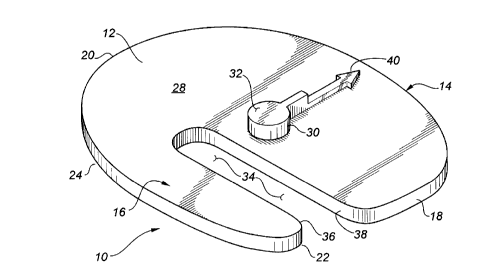
FIGURE 1 is a perspective view of a haemostatic pad
constructed in accordance with the teachings of the present
invention.
FIGURE 2 is top plan view of the haemostatic pad
illustrated in FIGURE 1.
FIGURE 3 is side elevation view of the haemostatic pad
illustrated in FIGURE 1.
FIGURE 4 is top plan view of the haemostatic pad
illustrated in FIGURE l, positioned in accordance with the
teachings of the preferred method.
FIGURE 5 is a side elevation view of the haemostatic pad
illustrated in FIGURE 1 in combination with an haemostatic
compression device.
DETAILED DESCRIPTION OF THE PREFERRED EMBODIMENT
The preferred embodiment, a haemostatic pressure pad
generally identified by reference numeral 10, will now be
described with reference to FIGURES 1 through 5.
The primary procedure for which a haemostatic compression
device is used is catheterization via a femoral artery or vein.
One example of an instance in which catheterization would be
used in when a patient is experiencing cardiogenic shock. A
catheter is introduced through puncture wounds in the leg into
a femoral artery, a femoral vein, or both. Often a hollow
sheath, termed an "introduces" is placed into the puncture
wound in the leg as a preliminary step. The catheter is
21 39244
J
extended through the introducer, along the artery (or vein) and into
the patient's heart. Haemostatic pressure pad 10 is intended for use
in such procedures.
Referring to FIGURES 1 and 2, haemostatic pressure pad 10
'rJ includes a generally U-shaped body 12 having a first finger portion
14 and a second finger portion 16. First finger portion 14 has a
first end 18 and a second end 20. Second finger portion 16 has a
first end 22 and a second end 24. First finger portion 14 and second
finger portion 16 are conjoined at respective second ends 20 and 24.
First finger portion 14 is broader than second finger portion
16. The reason for this difference in size will become apparent from
the description of use and operation. The blood flow through a
femoral artery is from the heart to the leg. The blood flow through
a femoral vein is form the leg to the heart. The pressure in the
15 femoral artery is much greater than the pressure in the femoral vein,
and, therefore, a larger first finger portion is required.
Referring to FIGURES 3 and 5, the body 12 has a lower contact
face 26 and an upper attachment face 28. Referring to FIGURES 1 and
2, a boss 30 having a key hole shaped aperture 32 is positioned on
the attachment face 28 of the first finger portion 14.
A channel 34 having substantially parallel sidewalls 36 and 38
is formed between first finger portion 14 and second finger 16,
terminating at conjoined respective second ends 20 and 24. Channel
34 extends through the body 12 from contact face 26 to attachment
25 face 28.
An arrow 40 is positioned on the attachment face 28 of the
first finger portion 14 of the body 12. Arrow 40 extends away from
second finger portion 16 substantially perpendicularly to channel 34.
This assists health care professionals in positioning body 12 as will
hereinafter be described in relation to the preferred method.
2139244
Referring to FIGURE 5, it is preferred that haemostatic
pressure pad 10 be used in combination with an haemostatic
compression device having a piston 42 with a key shaped terminus 44.
Key shaped terminus 44 of piston 42 from the haemostatic compression
~J device is insertable into key hole shaped aperture 32 of boss 30 to
attach the body 12 to the haemostatic compression device.
Referring to FIGURE 4, the preferred method of positioning the
haemostatic pressure pad 10 will now be described. It will be
understood that preparatory steps would include: providing an
haemostatic compression device; providing a form of haemostatic
pressure pad 10; and attaching haemostatic pressure pad 10 to the
haemostatic compression device.
Referring to FIGURE 3, contact face 26 of haemostatic pressure
pad 10 is placed in contact with a leg 46 of a patient 48. Referring
15 to FIGURE 4, the puncture wounds are positioned between sidewalls 36
and 38 of channel 34. Arrow 40 is positioned so that it points
toward an umbilicus 52 of patient 48. Referring to FIGURE 3, piston
42of the haemostatic compression device is used to apply sufficient
force to haemostatic pressure pad 10 to staunch blood flow.
Referring to FIGURES 2 and 4, channel 34 extends substantially
perpendicularly to both a femoral artery 54 and a femoral vein 56 in
leg 46 of patient 48. When positioned as described first finger
portion 14 stauncher blood flow from femoral artery 54 and second
finger portion 16 stauncher blood flow from femoral vein 56.
25 Referring to FIGURE 3, in order to staunch the blood flow from
femoral artery 54 and femoral vein 56, haemostatic pressure pad 10
must exert a force upon skin 58, and a subcutaneous layer of fat 60.
Femoral artery 54 and femoral vein 56 are positioned between
subcutaneous layer of fat 60 and muscle 62.
2139244
It will be apparent to one skilled in the art that
modifications may be made to the illustrated embodiment without
departing from the spirit and scope of the invention as hereinafter
defined in the Claims. The use of arrow 40 is preferred, but is not
rJ absolutely essential. Similarly, the use of boss 30 with key hole
shaped aperture 32 is preferred, but is not absolutely essential.