Note: Descriptions are shown in the official language in which they were submitted.
B&P File No. 3158-030/LlrBc
-- ~mosg~
1
Title: Device for Separating Magnetically Labelled Cells
FIELD OF THE INVENTION
The present invention relates to a device for
separating magnetically labelled cells in a sample using
an applied magnetic field, and methods of using the device
to prepare purified cell preparations, preferably
hematopoietic stem cell preparations depleted of selected
cells such as T lymphocytes, tumor cells and/or red blood
cells. The invention also relates to purified
hematopoietic stem cell preparations.
BACKGROUND OF THE INVENTION
Blood cells have a relatively short life span
and need to be replenished throughout life. In adults,
blood cell formation or hematopoiesis takes place in the
bone marrow, but blood-forming stem cells can also be
found in peripheral blood. Iiematopoietic cells represent
a hierarchy of proliferating and differentiating cells.
The most abundant are the differentiating cells. These
cells have limited or no proliferative capacity and
represent the immediate precursors of the specialized end
cells that are found in blood. The immediate precursors of
the differentiating cells are the progenitor cells. Most
of these cells are restricted to differentiate along a
single lineage but they may have quite extensive
proliferative capacity. Progenitor cells appear
morphologically as blast cells and they typically do not
have specific features of the hematopoietic lineage to
which they are committed. Progenitor cells are derived
from stem cells. Stem cells have been historically defined
by their ability to self-renew as well as to generate
daughter cells of any of the hematopoietic lineages. The
presence of stem and progenitor cells may be detected by
their ability to produce colony-forming cells in culture.
They may also be detected by screening for the CD34
antigen which is a positive marker for early hematopoietic
cells including colony forming cells and stem cells.
._
~14068~
- 2 -
There is a continued interest in developing stem
cell purification techniques. Pure populations of stem
cells will facilitate studies of hematopoiesis.
Transplantation of hematopoietic cells from peripheral
blood and/or bone marrow is also increasingly used in
combination with high-dose chemo- and/or radiotherapy for
the treatment of a variety of disorders including
malignant, non-malignant and genetic disorders. Very few
cells in such transplants are capable of long-term
hematopoietic reconstitution and thus there is a strong
stimulus to develop techniques for purification of
hematopoietic stem cells. Furthermore, serious
complications and indeed the success of a transplant
procedure is to a large degree dependent on the
effectiveness of the procedures that are used for the
removal of cells in the transplant that pose a risk to the
transplant recipient. Such cells include T lymphocytes
that are responsible for graft versus host disease (GVIiD)
in allogeneic grafts and tumour cells in autologous
transplants that may cause recurrence of the malignant
growth.
A variety of techniques have been described for
the removal of either T cells or tumour cells from
transplants (See for example Bone Marrow Processing and
Purging: A Practical Guide, (ed. A.P. Gee), CRC Press,
Boca Raton (1991)). Most of the techniques involve
purification of the hematopoietic cells ("positive
selection") or the depletion or "purging" of tumour cells
("negative selection") in the cell preparation used for
transplantation.
The two most important variables in either
positive or negative selection techniques are (1) the
efficiency of removal of undesirable cells (either T cells
or tumor cells) and (2) the recovery of hematopoietic
cells (most readily assessed by measurement of CD34
positive cells before and after the separation). These
variables are typically expressed as (1) the logarithm
~~~o~~~
- 3 -
(log) of the depletion and (2) the percentage recovery of
the CD34 positive cells. For example, a technique for
depleting T cells in a cell suspension that results in a
two log depletion of T cells, and a 30$ recovery of CD34
positive cells, would provide a cell suspension containing
1$ of the T cells and 30~ of CD34 positive cells that were
present in the cell suspension before the T cell depletion
procedure.
High gradient magnetic separation (HGMS) has
been used for the removal of magnetically labelled cells
from suspensions of bone marrow cells for research use
(Bieva et al., Exp. Hematol. (1989) 17: 914; Miltenyi et
al., Cytometry (1990) 11: 231; and Rogler et al., Bone
Marrow Transplant. (1990) 6: 163 and Thomas et al., J.
Hematother. (1993) 2: 297; and clinical use (Yau et al.,
Exp. Hematol. (1990) 18: 219; Poynton et al., The Lancet
(1983) March: 524; and Reading et al., Leukemia Res.
(1987) 11: 1067).
HGMS separation involves placing a filter of
fine magnetisable wires in a strong magnetic field. High
gradient magnetic fields are produced around the wires,
allowing the capture of even very weakly magnetic
particles upon the magnetisable wires.
There have been several attempts to apply HGMS
to the separation and isolation of magnetically labelled
CD34 positive cells (i.e. positive selection techniques),
although the recoveries and purities achieved have been
undesirably low (For example, see Rato, R., and Radbruch,
A., Cytometry 14:384, 1993). Typically, attempts have
employed an HGMS filter which consists of a random or
semi-random array of stainless steel wire wound loosely
into a column located in a strong magnetic field
(Miltenyi, S. et al., Cytometry 11:231, 1990; Molday, R.S.
and Molday, L., FEBS. Lett. 170:232, 1984; Kato,R and
Radbruch, A., supra; Kemshead, J.T. in Hematotherapy 1:35,
1992; and Remshead, J.T. in Bone Marrow Processing and
Purging, 293, Gee, A.P. Ed., C.R.C. Press, Inc., Boca
_21~06~~
- 4 -
Baton, Florida, 1991). The positive selection procedures
suffer from many disadvantages including the presence of
materials such as antibodies and/or magnetic beads on the
CD34 positive cells, and damage to the cells resulting
from the removal of these materials.
It has been assumed that pure hematopoietic stem
cells can be numerically expanded in the laboratory.
Accordingly, investigators typically have not focused on
the recovery or yield of hematopoietic stem cells that can
be obtained with the available cell purification or cell
"purging" methods. However, recent studies with highly
purified candidate stem cells from human and murine bone
marrow have shown that it may not be possible to achieve
such numerical expansion of stem cells derived from adult
hematopoietic tissue in vitro (Lansdorp et al., J. Exp.
Med. (1993) 178: 787, and Rebel et al., Blood (1994)
83:128). As a result, techniques that optimize the use of
the available hematopoietic cells for transplantation are
of considerable interest. Unfortunately, all currently
available methods for the removal of either T cells or
tumour cells from transplants suffer from deficiencies.
These include the following: 1) the methods that allow
for effective (i.e. >2 log) depletion of T or tumour cells
typically recover generally far less than 50$ of the
normal blood-forming or hematopoietic cells initially
present in the cell suspension available for
transplantation (i.e. as a result of centrifugation,
density separation, wash procedures and other pre-
processing required prior to the actual separation process
or during the immunological selection procedure itself);
and/or 2) the methods that recover >50% of the
hematopoietic cells fail to reproducibly achieve effective
(>2 log) depletion of undesirable cells.
SUMMARY OF THE INVENTION
The present inventors have developed a device
for removing magnetically labelled cells from a sample
containing magnetically labelled cells and non-
2.~4068~
- 5 -
magnetically labelled cells, without significant loss of
non-magnetically labelled cells present in the sample.
The separation of weakly magnetic cells by HGMS from non-
magnetic cells, without significant loss of the non-
magnetic cells was found by the present inventors to be
dependent on the following variables: (1) magnetic forces
that attract magnetically labelled cells to the magnetized
wires; (2) fluid shear forces acting on all cells in the
solution; (3) non-specific entrapment of non-magnetic
cells; and (4) contact between magnetic cells and the
magnetic matrix. Hitherto no one else has described a
single device that addresses all of these important
variables. However, the four variables are controlled in
an embodiment of the device of the present invention in
that the device has a) an ordered magnetic HGMS matrix
with a design that incorporates spacing of the magnetic
wires to increase HGMS efficiency; (b) a flow distributor
that ensures an even fluid flow around all magnetic wires;
and (c) a filter design that ensures close approximation
of magnetic cells to the magnetized wires of the magnetic
matrix and yet does not entrap many non-magnetic cells.
The present inventors have shown that the use of
magnetic wires in an ordered array in the device of the
present invention offers many advantages over a random
packing of wires. The use of alternating layers of
magnetic and non-magnetic mesh also has been found to
ensure optimal spacing of the magnetizable wires, minimal
entrapment of non-magnetic cells as well as a rigid
architecture of the device that enhances reproducibility
of construction and behaviour.
The present inventors have also found that the
random distribution of flow around all magnetized wires
could be maximized using a flow distributor. The present
inventors prepared a simple and effective flow distributor
by inserting particles, preferably rigid spherical
particles ranging in size between 50Eun to 1000 ~m in
diameter, in the inlet and outlet of the device. While the
21~0~68
- 6 -
use of spherical flow distributor beads or particles is
preferred as the flow distribution means, any suitable
arrangements which produces a uniform flow through the
filter chamber may be used. For example, effective and
even expansion and contraction of fluid flow containing
suspended cells may also be achieved using carefully
designed inserts containing vertical and horizontal flow
dividers and openings. The angle of flow
expansion/contraction could also be decreased by
elongation of the filter inlet and outlet.
Further, the present inventors have found that
connecting the peripheries of the magnetic meshes to the
housing prevented flow around the meshes and increased the
retention of magnetically labelled cells allowing for
increased purity of non-magnetically labelled materials.
The peripheries of the magnetic meshes may be connected to
the housing by encasing the stack of meshes in heat-shrink
tubing, a moulded plastic housing or, by precise
mechanical matching of magnetic mesh and filter housing at
a tolerance of 0.2 mm or less.
The device and methods described herein are
preferably used to deplete T lymphocytes and tumor cells
from samples to prepare hematopoietic cell preparations
for use as transplants as well as other therapeutic
methods. Removal of the T lymphocytes from an allogeneic
transplant is an effective way to prevent Graft Versus
Host Disease (GvHD) which is the major problem in patients
receiving allogeneic bone marrow transplants (Champlin,
R.J., Hematother. 2:27-42, 1993). Cell dose in the
transplant has also been shown in model studies in rats to
be a critical factor in prevention of graft rejection
(Uharek et al., Blood 79:1612-1621, 1992). Accordingly,
the goal of T cell depletion techniques is to effectively
deplete T cells without significant losses of the
hematopoietic cells that express CD34. Methods that
effectively (>2 log) deplete T cells and recover a high
proportion (>50%) of the CD34' cells are highly desirable.
~l~oss3
_,_
In contrast to hitherto known techniques, the device of
the present invention allows such a cell population to be
obtained without using multi-step procedures which are
laborious and time-consuming. The cell preparations that
can be obtained with the methods of the present invention,
represent a significant advance in the art of bone marrow
transplantation. The methods of the present invention are
fast (less than two hours), require minimal processing of
the sample and yet deplete CD3+ T cells at a good
efficiency (>2 log depletion) while recovering >50% of
CD34' cells .
Accordingly, broadly stated the present
invention relates to a device for removing magnetically
labelled cells from a sample using an applied magnetic
field, comprising:
(a) a housing;
(b) an inlet element at the top portion of the
housing having an input end and an output end;
(c) a filter chamber adjacent to the output end
of the inlet element for filtering the magnetically
labelled cells from the fluid while allowing unlabelled
cells to pass through when a magnetic field is applied
thereto, and containing a multiplicity of magnetic matrix
elements extending transversely across the filter chamber;
(d) an outlet element for collecting the fluid
which passes through the filter chamber having an input
end coupled to the filter chamber and an output end;
and which device has one or more of the
following features:
(i) the inlet and/or outlet elements having
flow distribution means for distributing the flow
generally uniformly across the filter chamber;
(ii) the peripheries of the magnetic matrix
elements are connected to the housing by a junction which
is substantially impenetrable to the fluid; and
(iii) the magnetic matrix elements are ordered
and spaced apart so as to maximize the magnetic capture of
~~4os~~
- 8 -
magnetically labelled cells onto the magnetic matrix when
a magnetic field is applied.
The present invention also broadly contemplates
a method of using the device of the invention to deplete
selected cells from a sample. Accordingly, in a preferred
embodiment the invention provides a method of using the
device to deplete selected cells, preferably T
lymphocytes, tumor cells, or red blood cells from a
sample, preferably blood or bone marrow comprising:
a) magnetically labelling the selected cells to
obtain magnetically labelled cells;
(b) passing the sample containing the
magnetically labelled cells through a device as described
above in the presence of a magnetic field;
c) collecting a preparation which is
substantially depleted of the magnetically labelled cells.
The present invention still further contemplates
a hematopoietic cell preparation comprising hematopoietic
cells and which is characterized as follows:
ZO a) it is obtained by high gradient magnetic cell
separation from a sample which contains hematopoietic
cells and T lymphocytes or tumor cells;
b) it contains greater than 50$ of the
hematopoietic cells present in the sample; and
c) T lymphocytes or tumour cells in the sample
are depleted by greater than 2 logarithms.
These and other aspects of the present
invention will become evident upon reference to the
following detailed description and attached drawings. In
addition, reference is made herein to various
publications, which are hereby incorporated by reference
in their entirety.
BRIEF DESCRIPTION OF THE DRAWINGS
The invention will now be described with
reference to the accompanying drawings, in which:
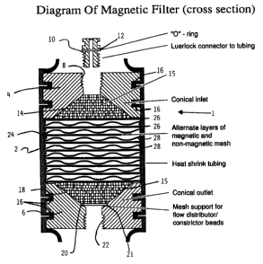
Figure 1 is a cross-sectional view of a device
including a filter according to the present invention;
g _
Figure 2 is a schematic chart, comparing
different filter materials and their spacing;
Figure 3A is a profile of magnetic cell
separation of CD8+ cells using alternating magnetic and
non-magnetic screens;
Figure 3B is a profile of magnetic cell
separation of CD3+ cells using alternating magnetic and
non-magnetic screens which are sealed to the housing using
heat shrink material;
Figure 4 shows the flow distribution achieved by
different sizes of glass beads; and
Figure 5 are graphs showing the indirect
immunomagnetic removal of lineage positive hematopoietic
cells from murine bone marrow using anti-biotin x anti
dextran complexes.
DETAILED DESCRIPTION OF THE INVENTION
I. Description of a Preferred Device of the Invention
The device of the invention will now be
described with reference to the Figures. In Figure 1, the
whole device is denoted by the reference 1, and
incorporates a housing 2. The material for the housing
can be selected depending on the application. Suitable
materials include tubular forms of non-magnetic metals or
synthetic polymers, such as regular or heat-shrinkable
plastics. For clinical applications, medical grade tubing,
which is relatively puncture-proof should be used. In a
preferred embodiment, the housing 2 is formed from tubular
material for example, heat shrinkable material, as
detailed below.
Within the housing 2, there is an inlet element
4 and an outlet element 6. The inlet element 4 has a
threaded inlet port 8. A connector 10 includes an 0 ring
12 , and is adapted to engage the threaded inlet port 8 .
In known manner, the connector 10 provides a Luerlock
connector for connection to tubing.
The inlet element 4 defines a flow distribution
chamber 14, which is generally frusto-conical. The
_ ~~.~46~~
-lo-
chamber 14 extends from an input end of relatively small
diameter to an output end having a larger diameter cone
angle between 135° and 90°. Within the chamber 14, there
are a plurality of spherical particles 15 which are
preferably rigid or semi-rigid, for example, glass beads,
and polyacrylamide beads, that cause flow through the
input end to be evenly distributed across the lower,
output end of the chamber 14. The spherical particles are
generally 50N.m to 1000 4un in diameter, preferably
spherical particles of 700-1000um in diameter are used.
In this embodiment, at the outside of the inlet
element 4, there are two annular grooves 16, to ensure
positive engagement with the housing 2, again as detailed
below.
It is anticipated that, for large scale
production, the housing 2 would be moulded in plastic, in
which case a variety of connection arrangements could be
employed between the housing 2 and the inlet and outlet
elements 4,6.
The outlet element 6 generally corresponds to
the inlet element 4. It defines a frusto-conical chamber
18, and also has annular grooves 16. The frusto-conical
chamber 18 has at its top a first input end of relatively
large diameter, and at its bottom a second output end of
relatively small diameter. Again, within the chamber 18
there are spherical particles 15 such as glass beads,
intended to ensure that the flow into the chamber 18 is
uniformly distributed across the inlet end. To support
the spherical particles 15, there is an output mesh
support 20, engaged in an annular step 21 at the outlet
element 6. Since flow is downwards through the device 1,
no such mesh support is required in the inlet chamber 14.
The outlet element 6 has a threaded port 22, for
connection to a Luerlock connector and tubing, as for the
port 8 at the inlet.
Between the inlet and outlet elements 4, 6,
there is defined a filter chamber 24. Within this filter
214068
- 11 -
chamber 24, layers of magnetic mesh 26 are alternately
vertically spaced with layers of non-magnetic mesh 28. As
shown, there are nine layers of magnetic mesh and eight
layers of non-magnetic mesh.
The layers of magnetic mesh 26 are generally
planar. Each layer comprises, in known manner, magnetic
wires extending in generally perpendicular directions and
interwoven. The topmost magnetic layer 26 defines the
bottom of the inlet chamber 14 and serves to retain the
spherical particles 15 in position. Similarly, the
lowermost magnetic mesh 26 defines the top of the outlet
chamber 18. Further, although the spherical particles 15
will naturally be retained in the outer chamber 18 by
gravity and downward flow of any fluid, this layer of mesh
26 also serves to retain these spherical particles 15 in
position.
The non-magnetic mesh layers 28 are made of
interwoven wires with a threefold larger diameter than the
wires in the magnetic mesh layers 26 to provide desired
spacing between magnetic mesh layers 26. As such, the
layers 28 should be dimensioned so as to provide a spacing
between the magnetic mesh layers 26 of approximately 6 to
9 times the diameter of the magnetic wires of the magnetic
meshes 26. The number of magnetic layers depends on the
separation requirements and can vary from 5 to 100. Where
the device is used to deplete T lymphocytes and/or tumor
cells from a sample, 40 to 60 layers are generally
required to obtain a greater than 3 log depletion, and
about 10 layers are generally required to obtain a 1 to 2
log depletion. Figure 2 is a schematic diagram indicating
the effect of spacing of the magnetic matrix elements or
screens on the separation efficiency. As shown at the
top, with 23 layers of magnetic screens, with no spaces,
the percentage depletion for CD8~ cells is 90%. The most
efficient separation, 96%, is obtained with ten layers
(note 9 in Figure 1) of magnetic screens and spacers. The
lowest separation efficiency was obtained (59%) when three
2~1~~~8~
- 12 -
layers of magnetic screens are used with spacers located
at the top of the filter chamber 24, i.e. adjacent the
inlet. With the three magnetic screens spaced evenly in
the top, middle and bottom of the filter chamber 24, an
efficiency of 80% was achieved, indicating that spacing of
the magnetic screens inside the filter chamber 24 is
desirable. Finally, with knit mesh magnetic wires of 50
microns in diameter, which were not supported by non-
magnetic screens but by non-magnetic wires that extend
through the filter housing, an efficiency of 72% was
achieved.
To assemble the device 1, the housing 2 is
located vertically as shown in Figure 1, but with the heat
shrink material of the housing in an initially, unshrunk
state, so that it has a diameter generally larger than the
various elements within the housing 2, to permit these
elements to be freely inserted and assembled. The housing
2 is initially purely cylindrical. The outlet element 6
is placed within the housing 2, the mesh 20 placed in
position, and the spherical particles 15 evenly
distributed on top of the mesh 20. The magnetic and non-
magnetic meshes are then placed on top of the output
element 6 alternately as shown. The input element 4 is
then placed in position on the top most magnetic mesh 26,
and the appropriate quantity of spherical particles 15
inserted through the threaded inlet port 8.
Then, it is ensured that the various elements
are aligned and the input and output elements 4, 6 are
pressed together to maintain the various elements in the
desired alignment. The housing 2 is then subjected to
sufficient temperature to cause it to shrink in known
manner. As shown, this will cause the material of the
housing 2 to engage the annular grooves 16 of the input
and output elements 4, 6. Also, as shown, the peripheries
of the meshes 26, 28 become embedded in the housing 2
without penetrating it, while supporting the housing 2, so
as to provide a junction which is substantially
_~1~~~8
- 13 -
impenetrable to fluid.
It will be appreciated that the spherical
particles 15 may be inserted in chamber 14 before or after
the housing is subjected to sufficient temperature to
cause it to shrink, or just prior to use of device 1. The
ends of the tubular housing 2 will extend radially
inwardly, as shown in Figure 1.
The device 1 is then fully assembled and ready
for connection with the connectors 10 in known manner. The
fully assembled device may be sterilized before use by
conventional techniques such as autoclaving.
In use, a sample containing magnetically
labelled cells is directed into device 1 at input element
4. The sample enters the flow distribution chamber 14 and
passes through the plurality of spherical particles 15.
The sample enters the filter chamber 24 and travels
through the layers of magnetic mesh and non-magnetic mesh
in the presence of a uniform strong magnetic field
produced by a magnet, for example, a solenoid
electromagnet. In the process, magnetised cells are
retained on the magnetised magnetic mesh 26. The sample,
including non-magnetic materials, passes through filter
chamber 24 and into the outlet element 6.' The sample
enters the first input end of outlet element 6 and its
flow is uniformly distributed by the plurality of
spherical particles 15. Use of the filter device provides
efficient separation of magnetically labelled cells.
II. Removal of Cells from a Sample
The device of the invention may be used to
deplete selected cells from a sample, such as cells which
express cell surface antigens recognized by antibodies,
preferably monoclonal antibodies. In one embodiment of the
invention the method is used to deplete selected cells
from cell suspensions obtained from blood and bone marrow.
In particular, the method may be used to deplete tumor
cells from bone marrow or blood samples harvested for
autologous transplantation, or deplete T lymphocytes from
CA 02140683 2004-O1-21
- 14 -
bone marrow or blood samples harvested for allogeneic
transplantation. The device of the invention may also be
used to remove virus particles from a sample.
The selected cells to be depleted in the sample
may be magnetically labelled by conjugating the cells to
magnetic particles. Suitable magnetic particles include
particles in ferrofluids and other colloidal magnetic
solutions. "Ferrofluid" refers to a colloidal solution
containing particles consisting of a magnetic core, such
io as magnetite (Fe304) coated or embedded in material that
prevents the crystals from interacting. Examples of such
materials include proteins, such as ferritin,
polysaccharides, such as dextrans, or synthetic polymers
such as sulfonated polystyrene cross-linked with
i5 divinylbenzene. The core portion is generally too small to
hold a permanent magnetic field. The ferrofluids become
magnetized when placed in a magnetic field. Examples of
ferrofluids and methods for preparing them are described
by Kemshead J.T. in J. Hematotherapy, 1:35, 1992, at pages
20 36 to 39, and Ziolo et al. Science (1994) 257:219.
Colloidal particles of dextran-iron complex (See Molday,
R.S. and McEnzie, L.L. FEBS Lett. 170:232, 1984; Miltenyi
et al., Cytometry 11:231, 1990; and Molday, R.S. and
MacKenzie, D., J.Immunol. Methods 52:353, 1982; Thomas et
25 al., J. Hematother. 2:297 (1993); and U.S. Patent No.
4,452,733) are preferably used in the method of the
invention.
Substances which are capable of binding to the
selected cells to be depleted such as lectins,
3o carbohydrates, proteins, and antibodies specific to an
antigen on the surface of the cells, preferably
antibodies, may be chemically bound to the surface of the
magnetic particles for example, using cyanogen bromide.
When the magnetic particles are reacted with a sample
35 containing the selected cells, conjugates will form
between the selected cells and the magnetic particles with
'-- _
- 15 -
bound substances. The reaction conditions are suitable to
allow the selected cells to bind to the magnetic particles
to form conjugates.
Alternatively, the selected cells to be depleted
may be magnetically labelled by indirectly conjugating the
selected cells to the magnetic particles by means of
antibody reagents. Examples of antibody reagents are
bispecific antibodies, tetrameric antibody complexes, and
biotinylated antibodies.
Bispecific antibodies may contain a variable
region of an antibody, for example, murine antibody,
specific for at least one antigen on the surface of the
magnetic particles, and a variable region of a second
antibody which is capable of binding to at least one
antigen on the surface of the selected cells. The
bispecific antibodies may be prepared by forming hybrid
hybridomas. The hybrid hybridomas may be prepared using
the procedures known in the art such as those disclosed in
Staerz & Bevan, (1986, PNAS (USA) 83: 1453) and Staerz &
Bevan, (1986, Immunology Today, 7:241). Bispecific
antibodies may also be constructed by chemical means using
procedures such as those described by Staerz et al.,
(1985, Nature, 314:628) and Perez et al., (1985 Nature
316:354), or by expression of recombinant immunoglobulin
gene constructs.
A tetrameric immunological complex may be
prepared by mixing a first monoclonal antibody which is
capable of binding to at least one antigen on the surface
of the magnetic particles and a second monoclonal antibody
which is capable of binding to at least one antigen on the
surface of the selected cells. The first and second
antibody are from a first animal species. The first and
second antibody are reacted with an about equimolar amount
of monoclonal antibodies of a second animal species which
are directed against the Fc-fragments of the antibodies of
the first animal species. The first and second antibody
may also be reacted with an about equimolar amount of the
CA 02140683 2004-O1-21
- 16 -
F(ab')2 fragments of monoclonal antibodies of a second
animal species which are directed against the Fc-fragments
of the antibodies of the first animal species. (See U.S.
Patent No. 4,868,109 to Lansdorp for a description of
methods for preparing tetrameric antibody complexes).
The use of biotinylated antibodies in
combination with magnetic iron-dextran particles that are
covalently labelled with (strept) avidin for the indirect
magnetic labelling of selected cells recognized by
io monoclonal antibodies is described by Miltenyi, S. et al.,
Cytometry 11:231, 1990. Many alternative indirect ways to
specifically cross-link colloidal magnetic particles to
selected cells would also be apparent to those skilled in
the art.
i5 When the method of the invention is used to
remove tumor cells from autologous blood or bone marrow
grafts, antibodies specific for different cell surface
antigens on the tumor cells may be conjugated to the
magnetic particles or used in the antibody reagents. For
2o example, for removal of lymphoma cells antibodies to cell
surface antigens that are expressed on lymphoma cells such
as CD10, CD19 and CD20 may be used. Monoclonal antibodies
specific for epithelial antigens such as high molecular
weight mucins may be used for the depletion of carcinoma
25 cells. Where the method is used to deplete T lymphocytes
from a sample, antibodies to cell surface antigens such as
CD2, CD3, CD5, CD4, CD6, CD8 and CD28 may be used.
The sample containing the selected cells is
reacted with the antibody reagents so that the selected
3o cells present in the sample bind to the antibody reagents
to form conjugates of the selected cells and the antibody
reagents. The reaction conditions are selected to provide
the desired level of binding of the selected cells and the
antibody reagents. For depletion of cells targeted by
35 antibody reagents, a sample containing the selected cells
to be depleted and cells to be isolated is preferably
21~~~8~
- 17 -
incubated with antibody reagents for a period of 5 to 60
minutes at either 4° or ambient room temperature. The
concentration of the antibody reagents is selected
depending on the estimated concentration of the selected
cells in the sample and the specificity of the antibodies
of the antibody reagents. Generally, the concentration is
between about 0.1 to 50 ~g/ml of sample. The magnetic
particles are then added and the mixture is incubated for
a period of about 5 minutes to 30 minutes at the selected
temperature. The sample is then ready to be separated
over the device of the present invention.
The sample containing the magnetically labelled
conjugates is passed through a device of the invention in
the presence of a magnetic field using the procedures
outlined in detail above. In an embodiment of the
invention, the fluid flow, gravity and the external
magnetic field are generally in the same direction and the
magnetic matrix elements are perpendicular to this. The
magnet is preferably a solenoid electromagnet with a 3"
diameter bore and having a magnetic field of 0.5-2 Tesla.
The magnetically labelled conjugates are
retained in the high gradient magnetic column and the
materials which are not magnetically labelled flow through
the column after washing with a buffer. The preparation
containing non-magnetically labelled cells may be further
analyzed using procedures such as flow cytometry.
III. Uses of the Device and Methods of the Invention
The device and methods of the invention may be
used in the processing of biological samples including
bone marrow, cord blood and whole blood.
The device and methods of the invention are
preferably used to deplete or purge tumour cells or T
lymphocytes from samples to prepare hematopoietic cell
preparations for use in transplantation as well as other
therapeutic methods that are readily apparent to those of
skill in the art. For example, in the case of an
autologous transplant, bone marrow can be harvested from
- 18 -
a patient suffering from lymphoma or other malignancies,
the sample may be substantially depleted of any tumor
cells using the device and methods described herein, and
the resulting hematopoietic cell preparation may be used
in therapeutic methods. Bone marrow or blood can also be
harvested from a donor in the case of an allogenic
transplant and depleted of T lymphocytes by the methods
described herein.
Using the method of the invention it is possible
to recover a highly purified preparation of hematopoietic
cells. In particular, a hematopoietic cell population
containing greater than 50% of the hematopoietic cells
present in the original sample, and which is depleted of
T lymphocytes or tumour cells in the original sample by
greater than 2 logarithms may be obtained. The
hematopoietic cells in the preparation are not coated with
antibodies or modified making them highly suitable for
transplantation and other therapeutic uses that are
readily apparent to those of skill in the art.
The method and device of the invention may also
be used to remove red blood cells from samples such as
blood and bone marrow. Half of the volume of normal blood
consists of mature red blood cells. Typically these cells
exceed nucleated cells by >100 fold. For many clinical and
research applications, removal of red blood cells is
required or desirable. The present invention provides
more efficient procedures for removing red blood cells
with higher recovery of cells than conventional methods
such as Ficoll-Hypaque density centrifugation (See Example
7 herein).
In a particular application of the invention,
samples may be processed using the methods and device
described herein for diagnostic flow cytometry of
leukocyte subpopulations. For example, the methods may be
used to prepare blood samples of patients infected with
the Human Immuno Deficiency (HIV) virus for monitoring
lymphocyte populations in such patients. Enumeration of
CA 02140683 2004-O1-21
- 19 -
the absolute numbers of leukocyte subpopulation by
conventional immunofluorescence measurements and flow
cytometry has been complicated by the abundant presence of
red blood cells in peripheral blood and consequently, such
s enumerations are most often derived from separate
measurements of nucleated cells numbers and immuno-phenotype
(Hoffmann, R.A. et al.Proc. Natl. Acad. Sci. U.S.A. 77:
4914, 1980.). A variety of procedures have been proposed and
are used to remove red blood cells from blood for
io immunophenotypic measurements but these procedures are
labour intensive and difficult to automate and in some cases
the procedure itself may interfere with immunofluorescence
measurements (Caldwell, C.W, and Taylor, H.M. Am. J. Clin.
Path. 86: 600, 1986). In contrast, the present invention
i5 provides an efficient and direct method for removing red
blood cells from blood samples that can readily be automated
as no centrifugation or wash steps are involved.
The following non-limiting examples are
illustrative of the present invention:
2o EXAMPLE 1
The Effects of Changes in the Construction of the Device On
Cell Separation
The ability of a filter device with various
constructions to separate T-cells from peripheral blood
2s mononuclear cell suspensions was tested. The following
materials and methods were used in the investigation:
Materials and Methods
Magnetic Colloidal Dextran Iron
The procedure for making dextran iron particles
3o described by Molday and MacKenzie (Molday and MacKenzie, J.
Immunol. Meth. (1982) 52: 353-368) was modified as follows.
1.518 FeC12.6H20 and 0.648 FeC124H20 were dissolved in 20 ml
distilled water. This solution was heated to 60°C and lOg
of Dextran T-40T"" (Pharmacia, Uppsala, Sweden) was added.
35 The mixture was stirred continuously and the dextran iron
dissolved (temperature can be
21~06~3
- 20 -
increased to 80°C if necessary) . Aqueous ammonium ( 25$ )
was added to the solution dropwise to titrate the pH to
10. The first two mls were added very slowly and
approximately 15 ml total was required. After the pH was
set at 10 the mixture was heated to 75-80°C for 15 min.
with continuous stirring. The resulting colloidal
suspension was cooled to room temperature and centrifuged
at 1,OOOg for 5 min. to remove aggregates. The
supernatant was filtered first through a course Watman
filter and then a 0.2~r membrane filter. The non-magnetic
iron and free dextran were removed from the magnetic
colloidal particles using High Gradient Magnetic
Separation (see filter construction and separation
conditions below). The dextran-iron suspension was passed
through a 1.5 inch diameter 40 layer HGMS filter as
described herein in a 0.5 Tesla magnetic field (solenoid
magnet) at 2.5cm/min. The filter was washed with 200 ml
of water and 300 ml phosphate buffered saline pH 7.4
(PBS). The magnetic field was then reduced to zero and
the magnetic particles collected by washing the filter
with 100 ml of PBS. The suspension of magnetic particles
was filtered (0.2~ filter), placed in a sterile tube and
the optical density (1 cm) at 450 nanometer (OD45o)
recorded. Within 48 hours of use, the magnetic colloidal,
particles were separated again using the same HGMS
procedure without the 200 ml water wash.
Antibodies
The mouse IgG~ anti-human CD3 monoclonal antibody
(UCHT1) was provided by Dr. P. Beverley, ICRF, London and
is described by Beverley and Callard (Beverley and
Callard, Eur. J. Immunol., (1981) 11: 329-334). The mouse
IgG~ anti-human CD8 monoclonal antibody OKTS (Basch, In:
Leucocyte Typing, eds. Bernard, Boumsell, Dausset,
Milstein, Schlossman (1984) pp. 661-664) was purified from
culture supernatants of the OKTS hybridoma line obtained
from American Tissue Culture Collection (ATCC). The mouse
IgG~ anti-dextran monoclonal antibody (DX1) was purified
~mass3
- 21 -
from culture supernatants produced by a switch variant of
the hybridoma 34166 as previously described (Thomas et
al., J. Immunol. Meth. (1992), 154: 245-252). F(ab')2
fragments of the rat monoclonal IgG~ antibody TFL-P9
specific for the Fc portion of the mouse IgG~ molecule were
obtained by pepsin digestion of purified immunoglobulin as
described previously (Thomas et al., J. Immunol. Meth.
(1989), 120: 221-231). Tetramolecular antibody complexes
(US patent 4,868,109) were prepared by mixing the ORTS or
UCHT1 antibody with the anti-dextran antibody (DX1) and
then adding the F(ab')Z rat anti-mouse IgG~ antibody in a
molar ratio of 1:4:5 respectively for OKT5, and 1:2:3
respectively for UCHT1. A significant proportion of the
resulting tetramolecular antibody complexes have dual
specificity for T cells and dextran.
Magnetic Labelling of Cells
Leukapheresis collections of normal human
peripheral blood were washed with PBS and either
resuspended to the original volume in Hank's buffered
saline plus 2% (v/v) fetal calf serum (experiments using
UCHT1 ) or the mononuclear cells were isolated using Ficoll
Hypaque density separation as described by Wognum et al.
(Wognum et al., Cytometry (1987), 8: 366-371). The
mononuclear cells were suspended (2X10⁷cells/ml) in Hank's
buffered saline plus 2% fetal calf serum (HF). The
leukapheresis suspensions were approximately 2X10⁷cells/ml
with a packed red blood cell volume of 20% (v/v).
CD3' cells in the peripheral blood leukapheresis
suspensions were magnetically labelled by incubating the
cell suspension with anti-dextran X anti-CD3 tetrameric
antibody complexes (3~g UCHT1/ml) on ice for 20 min.
Magnetic colloidal dextran iron (final OD4so 0~2) was then
added to the suspension which was mixed and incubated for
another 30 min. This "start" suspension was then
separated directly using HGMS (see below).
Peripheral blood mononuclear cells were
incubated with anti-CD8 X anti-dextran tetrameric antibody
~I4~~83
- 22 -
complexes (0.5~g OKTS/ml) on ice for 30 min. After one
wash the cells were again resuspended at 2X10⁷cells/ml
( HF ) and mixed with magnetic colloidal dextran iron ( final
OD45o=0.1) and incubated on ice for 30 min. CD8~ were then
separated from this labelled cell suspension using HGMS
(see below).
Heparinized whole blood was diluted 1:1 with
0.9% NaCl (USP saline) and labelled with anti-dextran X
anti-CD3 tetrameric antibody complexes and colloidal
dextran iron as described above.
A buffy coat suspension was prepared of whole
bone marrow. The cells were diluted with a sufficient
volume of USP saline to obtain a packed red blood cell
volume of 20%. Cells were labelled with anti-dextran X
anti-CD3 tetrameric antibody complexes and colloidal
dextran iron as described above.
HGMS Filter Construction
Cylindrical filters were assembled using
alternating layers of magnetic stainless steel wire mesh
(430ss, 80 mesh, 0.0055" wire, 0.0070" openings, Separator
Engineering, Pointe Claire, Que., Canada) and non-
magnetic stainless steel wire mesh (304ss, 16 mesh,
0.018" wire, 0.045" openings, Western Canadian Screen,
New Westminster, B.C., Canada). In initial separations
1.125" discs were cut from the mesh and stacked
(alternating magnetic and non-magnetic) in a non-magnetic
(316ss) stainless steel tube (1.125" ID) with conical (90
degree apex) end pieces. It was later found that a small
percentage of the cells would flow around the edges of the
screen discs inside this stainless steel housing. This
problem has not been recognized by numerous other workers
in this field, and it is believed that reports of
relatively modest depletion using HGMS techniques may
partly be explained by this effect. To solve this problem
several types of heat shrink tubing were explored, and the
problem was solved by encasing the stack of screens and
conical end pieces in two layers of medical grade heat-
214~~8~
- 23 -
shrink tubing (FEP roll cover, Zeus Industrial Products
Inc., Orangeburg, SC). A detailed diagram of the HGMS
filters is shown in Figure 1. Grooves were cut around the
circumference of the end pieces to facilitate forming a
seal with the shrink tubing. To shrink each layer of
shrink tubing, the tubing was heated for fifteen minutes
in a 250°C oven. The end pieces and stack of mesh discs
were held together during this time with a standard C-
clamp. Shrink tubing filters were made in various sizes
(0.5". 1.125". 1.25". 1.5" and 2.0" diameters) and lengths
(5-60 layers of non-magnetic and magnetic screen or 10-120
screens in total). The end pieces were connected to
standard medical tubing and blood bags via a 10-32
standard thread to Luerlock connector (Popper and Sons
Inc., New Hyde Park, NY).
The advantages of some sort of flow distributor
became evident in a 2" diameter model filter made with
clear plexiglass end pieces, 120 degree cones and a single
magnetic screen. With this system it was observed that at
flow rates that were calculated to give an effective flow
rate of 1 cm/minute at the wires of the magnetic screen,
red cells or an indicator dye raced through the middle of
the filter at a very high flow rate. Only a very small
area of the available screen was used and at a much higher
than calculated flow rate under these circumstances
(Figure 4A). After numerous failures to solve this
problem by various means, a relatively simple solution was
found in that insertion of glass beads between 200 and
1000 um in diameter on top and below the mesh, resulted in
an even and predictable flow pattern at the site of the
magnetic mesh at the cross-section of the filter (Figure
4E and F). In subsequent experiments glass beads (16-20
mesh, Potters Industries Inc., Brownwood, TX) were loaded
into the top cone (approx. 3/4 of the volume of the cone)
to act as a simple flow distributor.
Solenoid Magnet
The configuration for a High Gradient Magnetic
CA 02140683 2004-O1-21
- 24 -
Separation in the present invention has the fluid flow,
gravity and the external magnetic field in the same direction
and the wires perpendicular to this. To achieve this with
long (40 layers plus end pieces - 4 inches) 2" diameter
filters a solenoid electromagnet with a 3" diameter bore was
designed by A.J. Otter (Eng.) TRIUMFTM (Vancouver, B.C.
Canada). The magnetic field in the 3" bore is 0.5 Tesla and
extends for 6" vertically. This magnet is water cooled and
requires a 4.5KW power supply.
to Cell Separations
Medium and cell suspensions were passed through
the HGMS filters using a peristaltic pump. The dry filters
were primed bottom to top at a slow flow rate of approximately
lcm/min. Care was taken to remove all bubbles that might
i5 affect the pattern of fluid flow in the filter. After
reversal of the flow direction, five column volumes of medium
were passed through the filter before passage of the labelled
cell suspension. As the cells were passed through the filter
(at flow rates ranging from 1.0-2. 0 cm/min. ) , the magnetic
2o field was maintained at 0.5 Tesla and the non-magnetic cells
were collected in the Flow Through fraction. The filter was
washed with 3 column volumes of medium (HF) with the magnetic
field on. After this the magnetic field was reduced to zero
and the magnetic cells were washed off the filter. The
25 magnetic cells were typically recovered in 3 column volumes.
Flow Cytometry
Start, flow through and purified cell suspensions
were stained with FITC-conjugated F(ab')2 fragments of sheep
anti-mouse IgG (SAM-FITC, Cappell Cat.-No. 1311-1744) to
3o detect the presence of antibody labelled cells in these
suspensions. Cells (106) were suspended in 100,1 of SAM-FITC
diluted 1:100 in staining buffer and incubated for 30 min. on
ice, then washed and resuspended. Stained and unstained
samples were analyzed by flow cytometry using a FACScanT"'
35 (Becton Dickinson, San Jose, CA) flow cytometer. Cells
labelled with antibodies were
~l~asss
- 25 -
quantitated in the Start, Flow Through and Purified
fractions. The % depletion = (# of positive cells in the
flow through - # of positive cells in the start) x 100.
% recovery of negative (non-target cells) - ( # of
negative cells in the flow through - # of negative cells
in the start) X 100.
Magnetic Matrix in HGMS Filter
Five separations were run selecting CD8+ cells
from peripheral blood mononuclear cells. In four
separations 23 screens were stacked in the stainless steel
tube described above. The order and number of screens
was as follows: A) 23 magnetic screens; B) alternating
magnetic and non-magnetic screens with a non-magnetic
spacer at each end; C) one non-magnetic screen then
alternating three layers of magnetic and non-magnetic
screens at the top then 15 non-magnetic screens; D) 3
magnetic screens spaced apart with non-magnetic screens to
the top, middle and bottom of the chamber (Figure 2). All
four filters were 1.125" in diameter and 0.5" long
excluding end pieces. The fifth separation was with a
knit mesh filter (430ss, 50~m diameter wire, in a plastic
cylinder, 2lmm diameter and 27mm in length) as described
by Thomas et a1. (Thomas et al., Hematotherapy (1993) 2:
297). A total of 4x10⁷ mononuclear cells were passed
through the filters at a flow rate of lcm/min. It was
previously determined that 10 layers of magnetic mesh
could bind 10 times this number of cells, indicating that
the available surface for magnetic capture was by no means
saturated in these experiments. The performance of each
filter was evaluated by its ability to capture
magnetically labelled CD8' cells (% depletion of CD8'
cells). The results of this experiment showed that the
highest efficiency of capture was achieved with 10
alternating layers of magnetic and non-magnetic screens
3.5 (filter B). This array out performed twice the number of
magnetic screens without spacers (non-magnetic screens)
suggesting that the fields of magnetic attraction around
-- ~~.~068~
- 26 -
the wires interfere with each other if the layers are not
spaced. Three layers of screens at the top of the filter
proved less efficient than three magnetic screens spaced
one at the top, middle and bottom of the filter. This
could be explained by an uneven flow distribution at the
top of the filter (see below). For all subsequent
experiments, alternate layers of 430ss mesh and non-
magnetic ss mesh were used.
Efficiency of Magnetic Capture in HGMS Filters
i0 Twenty alternating layers of magnetic and non-
magnetic screens (1.124" diameter, 40 screens in total)
were stacked in the stainless steel housing tube and used
to separate CD8+ cells (ORT5 labelling see above) from
4.8X10⁹ peripheral blood mononuclear cells (18.7% CD8~).
The start volume was 50 ml. Fractions (6 ml) of the flow
through were collected. The total number of cells and the
% ORTS' cells were determined for each fraction. The first
two fractions collected had a CD8~ cell content very
similar to unseparated cells, suggesting that these first
cells to come through had missed the magnetic matrix
entirely (Figure 3A). Later observations of red blood
cells passing through a filter housed in a clear tube
confirmed that some cells were passing around the edges of
the screens avoiding close contact with the magnetic
matrix. Based on these observations, the design of the
filter housing was modified in order to by-pass this
phenomenon. It was conceived that a close contact between
the filter matrix and the filter walls could be ensured by
using heat shrink tubing as a material for the filter
wall. Suitable material was identified (FEP roll cover,
Zeus Inc., Orangeburg, SC) and used to assemble HGMS
filters. An example of an experiment showing that with
such heat-shrink tubing the by-pass phenomena described
above is avoided is illustrated in Figure 3B. In this
experiment, a time course study was done with a 20 layer
1.125" diameter sealed shrink tubing HGMS filter.
Peripheral blood leukapheresis cells (7.2X10⁹, 120 ml,
.,_ _
- 27 -
46.6% CD3~, packed red blood cell volume - 20%) were
labelled with anti-dextran X anti-CD3 tetrameric antibody
complexes and colloidal dextran iron and passed through
the filter at lcm/min. 10 ml fractions of the flow
through were collected and the total number of cells and
the %CD3' determined. The first few fractions were just as
efficiently depleted of CD3+ cells as subsequent fractions
indicating that sealing the edges of the screens to the
housing had effectively prevented cells from by-passing
the magnetic matrix. In this experiment, the filter was
swamped with magnetic cells resulting in a decrease in the
efficiency of magnetic capture after binding 5-7X10$ cells.
Table 1 summarizes the log depletion of T cells
and the % recovery of negative cells during four HGMS cell
separations. All filters were composed of alternating
layers of magnetic and non-magnetic 1.125" screens and
were run a lcm/min. Two separations used an unsealed
filter in a stainless steel housing (Expt. 1) and the
other two used sealed shrink tubing filters (Expt. 2+3).
Duplicate separations with identical cell suspensions were
performed in the first two experiments. The separations
with unsealed filters selected labelled CD8+ cells from
peripheral blood mononuclear cells (7.2 X 10T per
separation). Magnetically labelled CD3' cells (UCHT1)
were used to test the sealed filters. In the first
instance (Expt. 2) leukapheresis collections (115 X
l0~cells, packed red cell volume=18%) were directly
labelled and separated and in the second (Expt. 3)
mononuclear cells (37 X lO7cells) were used. In view of
these results, suitable heat-shrink tubing was used for
the construction of HGMS filters in all subsequent cell
separation experiments.
Effect of Flow Distribution on Separation Efficiency
Transparent perspex end pieces with a 90° cone
were used to study the distribution of red blood cells or
an indicator dye upon entry into a 2" HGMS filter.
Initial studies were conducted with a mock HGMS filter
~~.~OfiB~
- 28 -
containing no matrix elements at a flow rate of 8m1/min.
At a selected time point 0.5 ml of either an indicator dye
(Trypan Blue 1$) or a suspension of red blood cells were
injected and the absorbance at 280 nm of the column
effluent was monitored. Results with the indicator dye or
red blood cells were identical and are summarized in
Figure 4. Without beads the fluid did not distribute over
the entire 2 inch surface of the mock HGMS filter but
rapidly passed through the center, appearing in the exit
long before the time required to flush the entire filter
volume (Figure 4A). In this situation the effective flow
rate in the filter is many times higher than planned and
only a fraction of the magnetic matrix is effectively
used. Both these factors will contribute to poor
depletion of labelled cells. The experiment was repeated
with beads ranging from 8/10 mesh (B), 12/14 mesh (C),
12/16 mesh (D), 16/20 mesh (E) and 20/30 mesh (F).
Optimum flow distribution was indicated by cells exiting
in a smooth curve over the same time required to flush the
void volume of the filter. 16/20 mesh beads (700-1000~m
diameter) were the largest beads still achieving
satisfactory flow distribution and were chosen for
subsequent experiments in order to keep the available
surface area for non-specific entrapment of cells to a
minimum.
A direct comparison of the efficiency of
magnetic cell separation using end pieces with and without
glass beads to distribute flow in the top of the filter
was made in two large scale CD3' separations of peripheral
blood leukapheresis cells (Table 2). The first experiment
(12.0 X 10⁸ cells) used a 40 layer sealed filter and the
second (17.2 X 10⁸ cells) a 60 layer sealed filter. Both
experiments compared a separation with a standard heat
shrink tubing filter (2" diameter) to the same filter with
the top cone (part of the end piece) filled with 16/20
glass beads (to approx. 3/4 volume of the entry cone).
The flow rate in all separations was l.5cm/min. In both
CA 02140683 2004-O1-21
- 29 -
experiments the glass beads substantially improved the %
depletion of positive cells with little or no loss of
negative cells (Table 2).
wrrnnr~ ~
Separation of Whole Blood
A 17 layer 1.25" diameter sealed filter with
16/20 glass beads in the top cone as described in example 1
was used to separate CD3+ cells from 10 ml of whole blood (5.4
X 10~ nucleated cells, packed red cell volume of 50~). Two
to separations were run one with undiluted blood and the second
with the 10 mls of blood diluted to 50 ml. One of the
filters was coated with silicon by passage of 10 ml Methyl
Hydrogen Polysiloxane (Dow CorningTM 1107 Fluid) at 1% (v/v)
in acetone through a pre-assembled filter, followed by air
(>100 ml) and further drying/heat curing for 1-2 hrs. at 80°C.
The HGMS system efficiently separated undiluted and diluted
(5X) whole blood (See Table 3). This finding is important
because it indicates that the cell fractionation techniques
that typically precedes immunomagnetic separation (i.e.
zo Ficoll HypaqueT"" or other forms of density separation) and
which invariably result in loss of time and cells may be
avoided using the HGMS filters and methods described herein.
The very good recovery of cells with this method could be
very useful for the purification or pre-enrichment of rare
2s cells from peripheral blood or bone marrow.
wTwrnr ~ ~
Large Scale Separations
The High Gradient Magnetic Cell Separation
technique as described in example 1 was scaled up to deal
3o with the numbers of cells required in an allogeneic bone
marrow transplant. An average graft (2 X 101° nucleated cells
total) typically contains approximately 10% or 2 x 10⁹
T-cells. To accommodate this number of cells a 2" diameter
40 layer (80 screens total) sealed filter was assembled.
35 This filter was connected by two three-way valves to several
blood bags containing the cells, wash
~.... _
- 30 -
medium, or empty bags for the collection of flow through
and purified cell fractions. The cell labelling was
performed in a blood bag by injection of the anti-dextran
X anti-CD3 tetrameric antibody complexes to the bone
marrow cell suspension (150 ml buffy coat cells) followed
by a 30 min. incubation on ice and addition of colloidal
dextran iron and a further 30 min. incubation. The
results of 9 separations of peripheral blood leukapheresis
cells and one bone marrow separation are presented in
Table 4. All separations were run at lcm/minute. These
results show that the HGMS device and methods described
here as well as the cell suspensions that can be generated
with this device and methods are readily adapted to
clinical scale separations. The high degree of target
cell depletion required in clinical T-cell depletions with
excellent (i.e. >50%) recovery of hematopoietic progenitor
cells described here is novel and should prevent or
attenuate GVHD and result in rapid engraftment.
EXAMPLE 4
Depletion of CD3+ T cells from Allogeneic Bone Marrow Using
the Device of the Invention
The usefulness of the device for the depletion
of T cells from allogeneic bone marrow transplants is
illustrated in the laboratory results of eight clinical T
cell depletion procedures (Table 5). For these
separations, a buffy coat fraction (" 150 ml) of the bone
marrow suspensions (" 1.5 1) was prepared on a COBS 2991
cell centrifuge. This suspension Was diluted with saline
to set the packed red blood cell volume at approximately
15% ("300 ml total volume). UCHT1 (mouse IgG~-anti-CD3) x
DX-1 (mouse IgG~-anti-dextran) x P9 (F(ab')Z (rat anti-
mouse IgG~ ) tetrameric complexes ( at a 1: 2 : 3 molar ratio of
respectively UCHTl, DX1 and P9 F(ab')Z antibodies) were
added to give a final concentration of 3~g/ml UCHT1, after
a 30 minute incubation period on ice, magnetic iron
dextran particles were added to a final OD4so of the
particles of 0.~2 AUFS and incubation on ice was continued
21408
- 31 -
for another 30 minutes . The cells were then passed through
a 2" magnetic filter (as described in Example 1)
positioned in a 0.5 T vertical magnetic field containing,
40-60 layers of magnetic screens at the indicated flow
rate using a peristaltic pump. Details of the magnetic
filter device used for these studies are provided in
Example 1. The cells that were recovered in the flow
through (depleted of CD3' cells) as well as samples of the
cells prior to magnetic separation were assayed for their
content of CD3 and CD34~ cells using flow cytometry as
described in Example 1. The results of the eight clinical
T cell depletion procedures are shown in Table 5. As shown
in the Table, the method of the present invention depleted
CD3+ cells at a good efficiency (>2 log depletion) while
recovering >50% of the CD34+ cells.
EXAMPLE 5
Removal of CD45RA+ Lymyhoma Cells from Bone Marrow
This example illustrates an improved method for
removing lymphoma cells from bone marrow or blood. Table
6 shows that immunomagnetic removal of cells that express
CD45RA using the methods and device described herein
results in highly efficient (>4 log) removal of spiked
Daudi lymphoma cells from either peripheral blood or bone
marrow cells without significant loss (< 40% of start
material) of the cells that do not express CD45RA. The
recovery of colony-forming cells that do not express
CD45RA (BFU-e) was also >60% in these experiments.
In the experiments, CD45RA+ lymphoma cells
( Daudi, cell line cells ) were labelled with FITC and mixed
with peripheral blood or bone marrow cells at the
indicated percentage. The cells were then incubated with
8d2 (mouse IgG~-anti-CD45RA) x DX1 (mouse IgG~-anti-
dextran) x P9 F'(ab)2 (rat anti-mouse IgG~) tetrameric
antibody complexes at 0.5 ~g/ml of 8d2. The labelled cell
suspensions were then passed through a 0.5 inch, 40 layer
filter at lcm/minute. A total of three experiments were
performed that illustrated the unique advantages of the
- 32 -
method of the invention. The results of these experiments
are shown in Table 6. For removal of lymphoma cells from
clinical autologous peripheral blood or bone marrow
grafts, a combination of antibodies specific for
different cell surface antigens on lymphoma cells such as
CD10, CD19 and CD20 can be used to minimize the chance
that variant lymphoma cells would escape magnetic capture.
EXAMPLE 6
Pursing of breast cancer cells from peripheral blood and
bone marrow.
Autologous transplantation of peripheral blood
stem cells (PBSC) or bone marrow is increasingly used in
the treatment of poor prognosis breast cancer patients.
A concern in such transplant procedures is the possible
contamination of tumour cells in the autologous graft. To
examine the efficiency of the device/filter and methods
disclosed herein for the immunomagnetic removal of breast
cancer cells, tumour cells from breast carcinoma cell line
BT20 (ATCC) were labelled with fluorescein in order to
allow rapid and sensitive detection among unlabelled
normal peripheral blood and bone marrow cells. The cell
mixture was then labelled with tetrameric antibody
complexes containing mouse IgG~ anti-breast carcinoma
antibody H23, mouse IgG~, anti-dextran DX1 and F(ab~)2
fragments of rat anti-mouse IgG~ P9 at a 1:2:3 molar ratio,
at 3 ~g/ml of H23. After 15 minutes at 4°C, magnetic iron-
dextran was added to the cell suspension to give an OD45o
of these particles of 0.2 AUFS and incubation was
continued for another 30 minutes prior to passage over 0.5
inch filters containing 40 layers of magnetic screens at
a flow rate of 0.5 cm/min. The cells in the flow through
as well as the cells prior to magnetic labelling were
counted and analyzed by flow cytometry to generate the
data shown in Table 7.
The results of these experiments clearly
indicate that even with a single antibody highly efficient
21068
- 33 -
(> 3 log) removal of breast cancer cells can be obtained
with the methods disclosed herein and that application of
this method allows recovery of the majority of H23', CD34'
cells. Clinical applications as described for CD3+ T cells
would probably use multiple antibodies with specificity
for different antigens expressed on breast cancer cells.
EXAMPLE 7
Purification of CD34~ cells b~ i~unomagnetic removal of
CD34' cells .
This example illustrates the results of an
experiment in which a single step immunomagnetic
purification of CD34+ CD38' cells was performed with three
different bone marrow cell suspensions (Table 8). In this
experiment, a mixture of tetrameric antibody complexes
with dual specificity for dextran and respectively
glycophorin (lOF7MN, l~g/ml), CD45RA (8d2, l~rg/ml), CD67
(B13 29, 5 ~g/ml), CD3 (UCHT1, 3 ~g/ml) and an undefined
platelet antigen (3H2, 5~g/ml) was added to the bone
marrow cells (1-3 x 10~ cells/ml). After a 15 minute
incubation at 4°C, iron-dextran complexes were added to
the cells (to an OD4so of the magnetic particles of 0.2
AUFS) and incubation was continued for another 30 minutes.
The cells were then passed over a 0.5 inch diameter
magnetic filter containing 40 layers of magnetic mesh at
a flow rate of 1 cm/min. The cells that did not bind to
the filter (flow through) as well as cells prior to
magnetic labelling were counted and analyzed by flow
cytometry. The results of this experiment are shown in
Table 8. Note that the purity of CD34+ cells in this
experiment was comparable to that obtained by the
immunoadsorption technique mentioned above and yet the
majority of CD34'CD38~°" cells were recovered without
modifications or antibody at their surface.
EXAMPLE 8
Depletion of murine cells that express lineage markers.
Murine hematopoietic stem cells can be enriched
from bone marrow by removal of cells that express markers
2~.~~~8;
_.. _
- 34 -
which correspond to a particular differentiation lineage
(Spangrude and Scollay, Exp. Hematol. 18: 920-926, 1990).
Indirect immunomagnetic removal of lineage positive cells
from murine bone marrow using anti-biotin x anti-dextran
complexes is shown in Figure 5. Mouse bone marrow cells
were labelled with a cocktail of five biotinylated
monoclonal antibodies with specificity for different
lineage-specific antigens as described by Spangrude and
Scollay (supra). After a wash step the cells were
incubated with anti-biotin x anti-dextran tetrameric
antibody complexes followed by magnetic dextran-iron
( 0 . D . '50 0 . 2 ) . The cel l s were pas sed through a 4 0 1 ayer
0.5" diameter filter (described in example 1) at a flow
rate of 1 cm/min. For analysis cells were stained with
anti-dextran antibodies conjugated to R-Phycoerythrin.
Figure 5 illustrates the efficient removal of lineage
positive cells. This separation of murine bone marrow
resulted in a 10-fold enrichment of lineage negative cells
including candidate hematopoietic stem cells with a
Sca''Lin'Rh123d"« phenotype and approximately 75~ recovery of
lineage negative cells. No inhibitory effects on in vitro
colony-formation or in vivo repopulation by the separation
procedure were observed. This experiment illustrates that
rapid depletion of lineage positive cells can be achieved
using the method and device disclosed herein.
EXAMPLE 9
Immunomagnetic removal of red blood cells.
Approximately half of the volume of normal peripheral
blood consists of mature red blood cells. Typically these
cells exceed nucleated cells by >100 fold. For many
clinical as well as research applications, removal of red
blood cells is required or desirable and such red blood
cell depletion can be achieved using a variety of
techniques. One of the most common ways to remove red
blood cells from peripheral blood or bone marrow is by
means of Ficoll Hypaque density .centrifugation. This
example illustrates the immunomagnetic removal of
~~.4U68~
- 35 -
erythrocytes using a single antibody with specificity for
glycophorin (lOF7MN). As shown in Table 9, the method of
the invention resulted in efficient depletion (>3 log) of
red blood cells and recovers most (>50%) of the nucleated
cells (Table 9). In contrast, the more laborious Ficoll
procedure results in a much poorer recovery of nucleated
cells. Because the immunomagnetic procedure disclosed here
does not involve any wash steps, it is very suitable for
automation.
While what is shown and described herein
constitutes various preferred embodiments of the device
and method subject invention, it will be understood that
various changes can be made to such embodiments without
departing from the subject invention, the scope of which
is defined in the appended claims.
2I40683
"._ _ _
36 -
Table 1. Depletion of T-cell from peripheral blood using High Gradient
Magnetic
Separation. Duplicate separations with identical cell suspensions were
performed in Expt. 1 and 2. In Expts. 2 and 3, the edges of the wire
matrix were embedded in the filter housing (heat shrink tubing).
Log Depletion °~6 Recovery of
Expt-# # of Layers of T-Cells Negative Cells
1 . 10 0.64/0.54 78/90
20 1.05/ 1.05 60/65
2 15 2.55/2.44 88/72
3 15 3.05 73
~~~o~s
- 37 -
Table 2. Comparison of large scale HGMS T-cell depletions with or without
glass
beads to distribute flow in the top of the inter. CD3+ cells were depleted
from peripheral blood leukapheresis cells.
Log Depletion °r6 Recovery
CD3+ Cells Negative Cells
Exit. 1
40 layers of screen
without glass beads 1.9 58
with glass beads 2.4 53
Exit-2
60 layers of screen
without glass beads 2.5 54
with glass beads 3.5 64
~~.4U6~3
- 38 -
..
b c
E
a V ~ N
~ ~ et'
~ +
U
'p
v, '0
U
O
rr
C ~ U ~. r~
t~ M ca 1~
Q"'
A
: o oe
U
A. v
~'
a
~U
3 o >
'
~..
~ Z
.C
'~
a
0
_~
~
E~
00
' o
v
:r
U
",; v .,
'' a $ ~ ao
~ N N
a ~ ~
bC
C
~
3
o
w
o
.o
~. o
a
0 0 ~
~,
M
O
M -r N
V
s
m
C
A o~
G
U
"' U et tD
o a. ui ai
s~ +
o Ca '
c G
~~U
~
C7
ox
M -~ N
H
~1~U68~~
TABLE 4. Large scale separations of CD3+ cells using High Gradient Magnetic
Separation.
Peripheral Blood (Leukapheresis Collection)
Log Depletion of % Recovery of
Expt. CD3+ Cells 108 CD3+ Cells Negative Cells
#
1 8.6 2.90 71
2 8.6 3.50 71
3 9.9 3.22 58
4 20.6 2.00 70
5.7 2.46 82
6 . 20.1 2.10 93
7 8.8 3.20 88
8 17.8 2.57 89
9 16.5 2.64 100
Percolled Bone Marrow
Log Depletion of °r6 Recovery of
Expt. # CD3+ Cells 108 CD3+ Cells Negative Cells
1 6.2 2.75 68'
' % recovery of CD34+ cells was 84°~ and the recovery of hemopoietic
colony forming cells was 126°~.
- 40 -
Table 5. Removal to CD3* T-cells from clinical bone marrow
grafts from matched unrelated donors.
Patient # Total # of Cells Log Depletion of I Recovery of
TCDM- Processed 108 CD3* Cells CD34*Cells
8* 58 1.55 95
9 99 2.50 93
10** 98 >3.50 45
12 141 >3.90 85
14 187 3.47 73
16 505 2.19 77
17 150 >3.13 66
18 293 >3.76 73
Mean 191 >3.00 76
*Magnetic separation flow rate - 1.0 cm/min. All other separations
were run at 0.5 cm/min.
**Marrow suspension was density separated using Percoll prior to
magnetic separation.
~~~oos~
- 41 - -
Table 6 Purging lymphoma cells from peripheral blood and bone marrow using
high gradient magnetic separation.
Log Depletion orb Recovery 96 Recovery
Total # of of Tumor Cells of CD45RA' of CD34+ o~ Recovery
Sample Cells 107 (Daudi') Cells Cells of BFU-E
Peripheral Blood 79.5 > 4.2 65 ND ND
Leukapheresis
0.5% Daudi
Normal Bone 7.0 > 5.8 75 33 63
Marrow 15.796 Daudi
Lymphoma Bone 4.4 > 6.0 74 24 60
Marrow 7.8~r6 Daudi
ND = Not Done. CD34+ cells are normally extremely rare in peripheral blood
making
the determination of the recovery of CD34+ cells using FRCS difficult.
'Daudi cells were first labelled with FITC and added to the start cell
suspension.
These Daudi cells were then detected in the separated fractions using FACS as
described.
~I~4683
- 42 -
Table 7 Purging breast carcinoma cells (BT20) from peripheral blood and bone
marrow using High Gradient Magnetic Separation.
Total # of Log Depletion 9~6 Recovery of ~r6 Recovery of
Sample Cells 10~ BT20 Cells H23-Lymphocytes CD34+ Cells
Peripheral blood 2.5 3.21 85 ND
Leukapheresis
Normal Bone 2.2 3.80 80 68
Marrow
~1~Q683
- 43 -
Table 8 Depletion
of cells
expressing
lineage
markers
from human
bone marrow
to enrich
for primitive
hematopoietic
progenitors
(CD34+,
CD381o),
Total 1t Flow Through
( 107)
of NucleatedStart FracUon 9~ CD34 tn Fraction9% RecoveryEnrichment
of
Bone MarrowCells in 96 CD34+ Flow Through 96 CD34+ of CD34+ CD34+ CD381
the ~
Sample Start FractionCD381 Fraction CD381 CD381 CellsCells
Organ Donor1.94 0.6 67.2 16.6 72 28x
Marrow
87
Organ Donor2.48 0 . 3 53.1 11.2 60 3 ~x
Marrow
f16
Aspirated 0.56 0.4 75.0 21.1 59 53x
Bone Marrow
~.~468
- 44 -
Table 9. A comparison of erythrocyte removal from whole blood using
High Gradient Magnetic Separation or Ficoll Density Separation.
Log Depletion of I Recovery I Recovery CD3'
Technique Erythrocytes Nucleated Cells Lymphocytes
HGMS* 3.1 65.7 79.2
Ficoll 3.9 21.3 44.8
*iThole blood was magnetically labelled with anti-glycophorin x anti-
dextran tetrameric antibody complexes and magnetic dextran iron as
described in the methods section.
Each technique processed 10 ml of whole blood with approximately 5 x
10~~ erythrocytes.