Note: Descriptions are shown in the official language in which they were submitted.
~ WO 94/03202 ~140879 PCF/US93/07471
~
INTERLEUKIN-4 STIMULATED T LYMPHOCYTE CELL DEATH
BACKGROUND OF THE INVENTION
Field of the Invention
The present invention relates to the treatment and
prevention of diseases that are primarily due to T cell immune
responses. In particular, it relates to the suppression or
elimination of certain autoimmune diseases, graft rejection,
and allergic disorders by treatment with interleukin-4 (IL-4)
and the specific antigen involved, thus allowing the killing
of only the subpopulation of T cells that recognizes this
specific antigen. In this manner, IL-4 pretreatment
sensitizes T cells to undergo programmed cell death following
T cell receptor engagement.
Description of Related Art
Apoptosis is a form of programmed cell death that
occurs in many biological systems (1-5). An apoptotic cell
undergoes a specific program of events dependent upon active
metabolism that contributes to its own self-destruction.
Distinct morphological changes occur during this process such
as membrane blobbing and cytoplasmic and nuclear condensation.
These changes are accompanied by fragmentation of genomic DNA
into pieces constituting one to several nucleosomes. In the
final stages, the cell disintegrates into apoptotic bodies
that are specifically recognized and phagocytozed by
neighboring cells.
= T lymphocytes are sensitive to apoptotic cell death
induced by a variety of stimuli at multiple points in their
lifespan. Experimental evidence strongly suggests that
programmed cell death normally plays a large role in shaping
and maintaining the T cell repertoire. Repertoire here is
WO 94/03202 2 1, 4 0 3*7 o PCT/US93/07471
2
defined by the number of distinct antigen receptor
specificities contained in the entire pool of T lymphocytes in
the organism. Each T lymphocyte bears surface receptors for
antigen that are all of identical structure on that cell and 5 therefore are
said to represent a single antigen specificity.
Since each T cell has a unique specificity, the total collection of antigen
specificities in an organism is the sum
of different individual T cells, thus the T cell repertoire.
By eliminating or expanding the number of individual T cells,
the responsiveness of an organism.to a particular antigen can
be either curtailed or enhanced,.respectively. These changes
have been documented to occur and are known as changes in the
T cell repertoire. Alterations in the T cell repertoire occur
naturally during T cell development such that only a small
fraction of thymocytes (or immature T cells) survive the
intrathymic development and selection events that allow
emigration of developing T cells to the peripheral circulation
(6,7). The majority of thymocytes appear to undergo apoptotic
cell death in the thymus because they bear particular
receptors. This "editing" of the T cell repertoire is thought
to be the result of two processes: lack of positive
selection, and negative selection or clonal deletion. The
latter is fundamental to the establishment of self-tolerance
as cells expressing potentially autoreactive receptors are
actively eliminated. Fetal thymic organ culture (8), in vivo
(9), and in vitro (10,11) experiments have shown that the
double positive (CD4+,CD8+) thymocytes appear to be more
sensitive to apoptotic death induced by T cell receptor
occupancy than more mature single positive cells. These
double positive cells are also sensitive to programmed cell
death induced by glucocorticoids (12).
Transformed T cells undergo activation-induced death from stimuli that are
normally mitogenic for T cells (13-19).
These include antigen, anti-TCR or CD3 mAb binding, the
combination of phorbol ester and Ca2+ ionophore, and mAb
modulation of alternative activation molecules Thy-i and Ly-6.
These cells are also susceptible to glucocorticoid-induced
apoptosis. The processes of activation- and
~
WO 94/03202 2140878 PCT/US93/07471
3
glucocorticoid-induced programmed cell death are mutually
antagonistic in transformed T cells (20-22).
Mature untransformed T cells have been shown to
undergo apoptosis in response to various stimuli, such as IL-2
deprivation in the case of cells requiring IL-2 for viability
(23), and modulation of the Fas antigen by the APO-1 mAb
(24,25). Additionally, it has recently been demonstrated that
IL-2 programs mature T lymphocytes to undergo apoptosis in
response to antigen receptor stimulation both in vitro and jn
vivo (26). T cells must be under the influence of IL-2 prior
to T cell receptor stimulation for apoptosis to occur, and the
amount of cell death rises with increased amounts of IL-2.
This process is selective, such that only stimulated T cells
triggered by their specific antigen receptor and not by
bystander cells undergo cell death. This apparent feedback
pathway, termed propriocidal regulation, may represent a
mechanism by which T cell responses are regulated (26).
The discovery that interleukin-4 (IL-4) predisposes
T lymphocytes to programmed cell death, or apoptosis, allows
for a novel method of therapeutic intervention in disease
processes in humans and animals primarily caused by the action
of IL-4-responsive T cells (27). In essence, this involves
specifically inducing the death of a subpopulation of T
lymphocytes that are capable of causing disease, while leaving
the majority of T lymphocytes substantially unaffected. This
method of intervention contrasts with, and is potentially far
superior to, currently used therapeutic methods that cause a
general suppression or death of T lymphocytes. Examples of
widely-used general immunosuppressive agents are
corticosteroids, such as prednisone, which are used to treat
autoimmune diseases and allergic conditions, and cyclosporin
A, which is used for treating graft rejection (28). These
treatments suffer from the drawback of severely compromising
immune defenses, by debilitating a large portion, if not the
entire T cell repertoire. This leaves the patient vulnerable
to infectious diseases. The two key elements of the present
process are that: i) only the subset of T cells that reacts
with antigens that cause the disease are affected by the
WO 94/03202 PCT/US93/07471 ~
4
treatment; and ii) the T cells affected by the treatment are
killed, i.e., they are permanently removed from the
repertoire.
Several general principles underlie the present 5 process. T cells recognize
antigen in the form of short
peptides that form noncovalent complexes w#h major . -~
histocompatibility complex (MHC) proteinson the surface of
antigen-presenting cells found throughout the body (29).
. ,,
Antigens may also take the form of polysaccharides, organic
molecules, or nucleic acids. Each T cell bears a unique
antigen receptor called the T cell receptor (TCR) that is
capable of recognizing a specific antigen-MHC complex.
Through rearrangement of the gene segments containing the
protein-coding segments of the TCR, a vast array, perhaps a
virtually unlimited number of combinations, of different TCRs
are generated (30). By a mechanism termed "allelic
exclusion", each T cell bears a single unique TCR. The T cell
repertoire is therefore a large number of T cells, each with a
distinct TCR that recognizes a specific antigen-MHC complex.
It is this vast array of T cells that allows immunological
responses to the diversity of antigenic structures on invading
micro-organisms, tumor cells, and allografts, thus preserving
the integrity of the organism.
Most antigens are able to elicit a response in only
a very tiny fraction of the T cell repertoire (31). For
example, the initial response to protein antigens may involve
as few as 1 in 1000 to 1 in 10,000 T lymphocytes (32). For
this reason, diseases caused by T cell reactivity are mediated
by only a small subset of the large repertoire of T cells
(33). In particular, in those cases where, it has been
directly measured, such as in multiple sclerosis, the fraction
of the T cell repertoire which mediates disease is quite small
(33). The important feature of the T cell subset that
participates in disease is"that it involves T cells which
specifically recognize an antigen that provokes the disease.
In allergic conditions, the antigen causes the release of
inflammatory response molecules. For example, "helper" T
cells secrete lymphokines such as IL-4 that cause B cells to
WO 94/03202 2140878 PCT/US93/07471
produce the inflammatory antibody IgE. In autoimmune
diseases, the antigen may be derived from a specific organ in
the body and, when recognized by a subset of T cells,
stimulates the T cells to attack that organ. A similar effect
5 occurs during graft rejection. Antigenic proteins in the
transplanted organ evoke a response in a subset of T cells
that attacks the.engrafted tissue. For unknown reasons, the
fraction of T cells recognizing foreign or 'allo1 tissue is
significantly higher than the number that will typically
recognize a protein antigen. Nonetheless, the number of
responding T cells is still a distinct minority (1-10%) of the
overall T cell repertoire (34).
In a typical T cell response to a specific antigen-
MHC complex, stimulation of the TCR (35) results in a cascade
of gene activation events. These have been extensively
characterized at the molecular level, and two such activation
events are especially germane to the present invention i)
production of growth lymphokines such as IL-4 and ii)
expression of the cell surface proteins that constitute
high-affinity receptors for IL4. Resting T cells express
small numbers of high affinity IL-4 receptors; this number
increases following activation (36). IL-4 is a 15,000 dalton
protein that causes T cells bearing the appropriate high
affinity receptor to divide (37,38). The production of IL-4
followed by its interaction with its receptor causes an
autocrine mechanism that drives the T cells into the cell
cycle. This leads to an initial expansion of T cells that are
specifically reactive with the antigen. At present, evidence
indicates that in both the human and murine immune systems, a
subclass of T lymphocytes called CD4+ TH2 cells may
proliferate after antigen activation by producing and
responding to IL-4 (39). This subset plays an important and
perhaps unique role in stimulating B cells to produce
immunoglobulin (Ig) (40). "This is because IL-4 and other
lymphokines produced by Tx2 cells, such as IL-5 and L-6, act
as differentiation factors for B cells that are crucial for Ig
production. Therefore, in autoimmune diseases in which Ig
plays a pathogenetic role, the elimination of CD4+ TH2
WO 94/03202 214UO(('~ npryp. PCT/US93/07471 ~
6
lymphocytes represents a highly effective way to halt disease.
Our results and those of others (27,40-43) show that other
classes of T cells including CD4+ TH1 type lymphocytes that
mediate delayed-type hypersensitivity as well as CD8+ cells
that mediate cytotoxicity will also proliferate in response to
IL-4 and are predisposed to TCR induced apoptosis. Therefore, IL-4 has a
potentially broad role in T.cell growth during
immune responses. Thus, IL-4 could be'.broadly active in
different classes of T cells to predispose them to apoptosis.
The present inventive discovery indicates that IL-4 also has
the surprising effect of predisposing the expanded pool of
either human or mouse T cells to apoptosis or programmed cell
death if they are again stimulated or rechallenged through the
TCR (27). In the present work described supra, the degree of
apoptosis achieved in T cells is correlated positively with
both the level of IL-4 the cells experience during their
initial expansion, the strength of the TCR stimulation upon
rechallenge, and the timing of the rechallenge. In
lymphokine-predisposed apoptosis, the effects wear off 2-3
days after lymphokine is no longer present, hence rechallenge
must occur within that period (44). The process of activation
and apoptosis eventually depletes the antigen-reactive subset
of the T cell repertoire.
Apoptosis is a type of programmed cell death in
which the T cell nucleus shrinks, the genetic material (DNA)
progressively degrades, and the cell collapses (1-5).
Evidence suggests that cells cannot recover from apoptosis,
and that it results in irreversible killing (1-5). T cells
that do not undergo apoptosis but which have become activated
will carry out their "effector" functions by causing
cytolysis, or by secreting lymphokines that cause B cell
responses or other immune effects (45). These "effector"
functions are the cause of tissue damage in autoimmune and
allergic diseases or graft'rejection. A powerful approach to
avoiding disease would therefore be to permanently eliminate
by apoptosis only those T cells reactive with the disease-
inciting antigens, while leaving the majority of the T cell
repertoire intact. Apoptosis and T cell deletion caused by
WO 94/03202 21-4r~8( PGT/US93/07471
U-y~
7
antigenic stimulation have been demonstrated in model systems,
but since a mechanism for this phenomenon was not previously
known, it was not possible to use this in a therapeutically
effective way (46-50).
By using IL-4 as an agent that predisposes T cells
to death by TCR stimulation in appropriate cycle with
immunization with the antigen(s) leading to autoimmune disease
or graft rejection, the death of disease-causing T cells can
be invoked. Specific methods are described for i) treatment
of autoimmune or allergic diseases by identified protein
antigen and IL-4, and ii) treatment of graft rejection by
blood cell antigens and IL-4. Such methods, by logical
extension, can be further developed for other diseases of man
or animals that result from the effects of T cells activated
by specific antigens. Because the vast majority of immune
responses depend on T cell activation, it is predicted that
this form of therapy could be applied to a wide variety of
autoimmune and allergic conditions especially where antibody
production is involved (51,52).
In several human autoimmune diseases, data have
indicated that antigen-activated T cells play a key role in
the production of disease. These include but are not limited
to: 1) multiple sclerosis (53-58); 2) uveitis (59,60); 3)
arthritis (61-63); 4) Type I (insulin-dependent) diabetes
(64,65); 5) Hashimoto's and Grave's thyroiditis (66-68); and
6) autoimmune myocartiditis (69). The ethical limits on human
experimentation have made it very difficult to prove that T
reactivity is the sole inciting agent of these diseases.
Nonetheless, a large body of experimental work on animal
models -- murine experimental allergic encephalitis as a model
for multiple sclerosis (70,71), BB diabetic rats for human
diabetes (72,73), murine collagen-induced arthritis for
rheumatoid arthritis (74,75), and S antigen disease in rats
and guinea pigs for human autoimmune uveitis (76, 77), among
others -- suggests that T cells are the critical agent of
these diseases. From recent work, the identity of
disease-causing proteins or peptide antigens is emerging: i)
multiple sclerosis: the peptide epitopes of myelin basic
WO 94/03202 PCT/US93/07471 0
c~J&OSj9 8
protein~+ (MBP) residues 84-102 and 143-168 (54,57,78,79); ii)
autoimmune uveitis: the human S antigen, which has been
recently molecularly cloned (59,80); iii) type II collagen in
rheumatoid arthritis (81); and iv) thyroglobulin in
thyroiditis (82). Similarly, a wide variety of proteins have
been identified which stimulate the proauction of the allergic ;.:
immunoglobulin IgE, which is the unde,rlying immunological
reaction for common allergies. IgE is produced by B
lymphocytes in a process that requires lymphokines produced by
antigen-activated T cells known as "T cell help". The class
of CD4+ "helper" T cells that stimulate B cells (TH2 cells)
typically produce and respond to IL-4 (39,40).
The basic concept of the present therapeutic
approach is very simple. Disease-causing T cells are first
challenged by immunization to cause the activated T cells to
express high affinity IL-4 receptors and, for TH2 cells, to
begin producing and secreting IL-4. When the cells are
expressing high levels of IL-4 receptor, additional human IL-4
is infused to very efficiently drive all the activated cells
into the cell cycle. The cells under the influence of IL-4
are then caused to undergo apoptosis by re-immunization with
antigenic peptide or protein. Further, if the antigen is
capable of stimulating sufficient IL-4 production, it may not
be necessary to administer exogenous IL-4. In either case,
the timing of rechallenge is important -- it must occur within
a short interval such as 2-3 days after the first stimulus
when cells bear high levels of the IL-4 receptor and are
responding to exogenous or endogenous IL-4.
The conceptual advance provided by the inventive
discovery that underlies the present methods is that T cell
immunity works as a balance between the production and
destruction of antigen-specific T lymphocytes. Previously,
investigators have focused on the use of lymphokine growth
factors such as IL-4 to increase the proliferation and
responsiveness of T lymphocytes (38,43,83-87). It is now
proposed that the opposing T cell mechanisms be used
therapeutically. The discovery that IL-4 predisposes T cells
to death is contrary to the previously understood properties
WO 94/03202 - 2'' 408( Q PCT/US93/07471
9
of IL-4, and provides a radically new approach to the
treatment of diseases caused by T cell reactivity. By
providing physicians and medical researchers with the basis of
the present inventive discovery, the processes of immune
autoregulation leading to T cell destruction can be exploited
in combatting disease.
It has been previously known for some time that
prior activation and lymphokine production were capable of
diminishing immune responsiveness both in vivo and in vitro
(46-48). The mechanisms underlying these effects were not
understood. Absent the knowledge that IL-4 predisposes T
lymphocytes to antigen-dependent apoptosis, it was not
possible to manipulate this phenomenon for medical or
therapeutic purposes. It is now possible to rigorously study
the kinetics and dose requirements of IL-4 in the
predisposition phase, and antigen in the apoptosis phase, to
routinely optimize the treatment cycle for a given disease
following the guidance provided herein.
That this process depends on the discovery of a
novel property of IL-4 is particularly auspicious. IL-4 has
been thoroughly studied since its discovery in 1982 (85,86).
It is well-understood genetically, its cDNA and gene have been
molecularly cloned, and antibodies against the protein for
immunodetection have been prepared (87,88). IL-4 is already
available pharmaceutically in a form for use in humans and
studies in human cancer victims have given insights into how
IL-4 affects human physiology at different doses (89-92). All
of these features significantly enhance the feasibility of its
novel use to cause auto-destruction of disease-causing T
lymphocytes for the treatment of a wide variety of diseases in
humans and other mammals.
SUMMARY OF THE INVENTION
The present invention arose from the discovery that
IL-4 programs mature T cells for antigen-driven death. The T
cell death caused by IL-4 followed by antigen stimulation has
hallmarks, such as DNA fragmentation, of "programmed cell
death" or apoptosis. Thus, IL-4 acts as a death cytokine that
WO 94/03202 10 PC'T/US93/07471
triggers the demise only of T cells that are specifically
stimulated through their antigen receptor. This invention
therefore allows the capability of altering the T cell
repertoire much the same way that negative selection in the
thymus naturally eliminates T cells having certain antigen
specificities. This novel use of a previously undiscovered ,.. property of IL-
4 will allow the specific elimination of
certain classes of antigen receptor-bearing T cells, forming
the basis for new clinical applications of IL-4.
IL-4 is a lymphokine produced by T lymphocytes that
was originally discovered to cause the growth of B lymphocytes
(85). Later it as found that this molecule had pleiotropic
activities on B cells such as increasing surface expression of
MHC class II molecules, elevating immunoglobulin secretion and
class switching and inducing the presence of Fce receptors
(93). Most importantly, IL-4 had powerful effects on both
CD4+ and CD8+ T cells (37-43). IL-4 strongly enhanced the
activity of cytolytic T cells which are involved in graft
rejection (42). Also, IL-4 is a potent T cell growth factor
(37,38,43). Among helper T lymphocytes, IL-4 promotes the
growth of TH2 cells that produce IL-4 in response to antigen
stimulation and help B cells mount an antibody response (40).
A critical determinant of the choice between T
lymphocyte proliferation or programmed cell death is the prior
exposure of these cells to IL-4. Antigen receptor stimulation
in T cells not exposed to IL-4 causes normal activation,
'leading to lymphokine production and growth. In contrast, T
cells previously exposed to IL-4 undergo apoptosis after
antigen receptor stimulation. Therefore, antigen-activated T
cells that are under the immediate influence of IL-4 will
respond to rechallenge by antigen by undergoing apoptosis.
The timing is significant because later antigenic stimulation
can cause growth rather than apoptosis if the cells are no
longer under the influence-of IL-4 if, for example, IL-4 is
removed and the T cells are allowed to return to their resting
state.
At least three uses for this novel property of IL-4
can be envisioned.
WO 94/03202 2140878
PCT/US93/07471
11
First, there is an emerging set of findings that
show that infusion of peptides derived from antigens involved
in autoimmune diseases leads to the lessening of severity of
such diseases (cf. 94). A variety of studies of the
autoimmune disease experimental allergic encephalitis (EAE)
shows that it is caused by the activation of T cells by
immunization with myelin basic protein (MBP). Interestingly,
infusion of peptides derived from the MBP sequence that
stimulate the T cells that generate the disease are effective
at blocking the disease (71). The discovery disclosed herein
provides an explanation for these seemingly paradoxical
observations, which is that the T cells are activated and are
potentially stimulated by IL-4 during peptide infusion, and
then undergo apoptosis when they are restimulated by the MBP
antigen. Human diseases that have been associated with T cell
activation by peptide antigens include multiple sclerosis and
autoimmune uveitis (78,80). It is envisioned that these
diseases, and, for example, systemic lupus erytaematosus,
systemic vasculitis, polymyositis-dermatomyositis, systemic
sclerosis (scieroderma), Sjogren's Syndrome, ankylosing
spondylitis and related spondyloarthropathies, rheumatic
fever, hypersensitivity pneumonitis, allergic bronchopulmonary
aspergillosis, inorganic dust pneumoconioses, sarcoidosis,
autoimmune hemolytic anemia, immunological platelet disorders,
cryopathies such as cryofibrinogenemia, autoimmune polyendo-
crinopathies, and myasthenia gravis can be approached by
therapy which can now be potentially modulated in a rationale
way using IL-4 and the relevant peptide to cause apoptosis of
the T cells responsible for the disease. Not all T cells have
a propensity to produce and respond to IL-4. However, the TH2
class of T lymphocytes which produce and respond to IL-4 are
crucial as "helper" cells for immunological responses that
involve the production of antibody. Many of the autoimmune
diseases mentioned above have an antibody component that leads
directly to pathology (as in myasthenia gravis) or indirectly
to pathology by immune complexes (as in systemic lupus) (51).
Therefore, the elimination of TH2 "helper" cells may provide a
significant amelioration or cure of these diseases. By
WO 94/03202 12 PCT/US93/07471 ~
targeting a population of T cells that respond to IL-4 for
apoptosis, this invention significantly extends a previous
discovery that IL-2 predisposes T cells to apoptosis (26).
The appropriate time of IL-4 infusion or a repetitive
immunization schedule could substantiallyaugment the
protective effect of the infused peptid~,S:- Secondly, there is a signifi.c~nt
body of literature
that suggests that pre-immunization G~"an animal or man prior
to engraftment with a foreign tissue prolongs the survival
time of the graft (cf. 95). One example of this phenomenon is
the "donor-transfusion effect," in which transfusing a patient
about to receive an organ transplant with blood from the organ
donor decreases rejection of the transplant. Studies have
shown that CD8+ cells will grow in response to IL-4 (42,43)
thereby potentially rendering CD8+ cells susceptible to IL-4-
mediated apoptosis. This is the primary class of T cells
involved in graft rejection. Based on the discovery of this
novel property of IL-4, CD8+ T cells may be induced to undergo
IL-4-mediated apoptosis; administering IL-4 during and
immediately after the pre-immunization/transfusion phase, or
repetitive immunization with MHC antigen at appropriately
short intervals, could augment T cell death, leading to
greater tolerance of grafts.
Thirdly, a wide variety of atopic or allergic
disorders, commonly known as asthma or allergies, results from
the effects of activating T cells, which causes both the
release of harmful lymphokines and the production of IgE by B
cells (96,97,98). Over the past few decades, clinicians have
made primitive attempts to treat these diseases by a
"desensitization" process consisting of repetitive exposure to
the same antigen that elicited the allergy (97). Despite the
fact that very little is known about the mechanisms set in
play by this procedure, in some cases such treatments were
highly successful (97). An important scientific by-product of
this work in clinical allergy is that considerable effort has
gone into identifying proteins and other molecules that cause
allergic responses (98). This has led to the identification
of protein sequences for antigens such as Amb a V and Amb t V,
WO 94/03202 2140878 PCT/US93/07471
13
which are ragweed allergens that cause hay fever, the protein
sequence and characterization of antigenic peptides from
allergen M that causes allergy to codfish (99), and the
molecular cloning of the cDNA for antigen 5 of white-face
hornet venom, associated with allergy to hornet stings (100).
Drugs that can cause allergy are typically small organic
molecules that may become immunogenic by forming covalent
complexes with host proteins. In addition, a large variety of
allergens have been prepared as protein extracts to be
administered clinically to humans under the supervision of the
Food and Drug Administration, and evaluated by a Panel on
Review of Allergenic Extracts (97). With the molecular
identification of these and other allergy-evoking antigens, it
will be possible to immunize in cycle with IL-4 (See page 12
and Figure 3) to induce apoptosis of T cells involved in
allergic disorders such as allergic rhinitis, bronchial
asthma, anaphylactic syndrome, urticaria, angioedema, atopic
dermatitis, allergic contact dermatitis, erythema nodosum,
erythema multiforme, Stevens-Johnson Syndrome, cutaneous
necrotizing venulitis, and bullous skin diseases.
Because a key component of the allergic response is
the production IgE antibody that depends on "helper" T cells
that respond to IL-4 (96), it is likely that IL-4 mediated
apoptosis of T cells could have a significant effect on
allergic disease processes.
The key feature of each of these treatment protocols
is that only the antigen-specific T cells, which comprise only
a small component of the patient's T cell repertoire, would be
eliminated. The treatment would leave the patient's immune
system largely intact. This is in contrast to present
treatments that rely upon general immunosuppression that
= seriously incapacitates the host's immune function (see 101).
Moreover, because this treatment causes death of the T
lymphocytes, it is superior to other recently discovered
mechanisms which do not kill T cells but rather cause
functional inactivation or anergy that is typically reversible
(102-104). The experimental results described infra therefore
WO 94/03202 Q'~ g PCT/US93/07471
14
have broad clinical significance in applications to human
immunological diseases.
Throughout the history of immunological approaches
to human and animal diseases, beginning with the first
vaccination against smallpox carried out by Edward Jenner in
1798, the emphasis has been on stimulating a positive and protective antigen-
specific immune response. In modern
immunology, this is known to be due to activating lymphocytes.
Hence, causing the activation and proliferation of
antigen-specific immune cells, especially T lymphocytes, forms
the basis of most of the clinical applications of immunology.
In particular, the recent advent of molecularly cloned
cytokines, especially those with the ability to cause the
proliferation of immune cells, has furthered the clinical
application of immunology. Such molecularly cloned cytokines
can be readily prepared pharmacologically, and are powerful
agents for stimulating the growth and division of lymphocytes.
The conceptual and practical advance offered by the discovery
disclosed herein is that cytokines such as IL-4, when given in
sufficient quantity, also cause negative regulatory effects
such as T cell apoptosis. These regulatory effects represent
built-in mechanisms to limit or suppress the immune response.
Thus, the recognition that these mechanisms exist, and the
identification of a biologic, IL-4, that potently evokes
antigen-specific T cell death, offers the opportunity to
exploit the negative regulation of the immune response for the
treatment of disease.
Accordingly, it is an object of the present
invention to provide a method for treating or preventing
disease in a human or animal caused by antigen-activated T
cells. This method induces the death by apoptosis of a
subpopulation of T lymphocytes that is capable of causing said
disease to an extent greater than that of other T lymphocytes.
Said disease can include an autoimmune disease, graft
rejection, or an allergic or atopic disorder, and said
apoptosis can be achieved either by exploiting endogenous
IL-4, or by administering this substance exogenously. When
IL-4 is administered exogenously, apoptosis can be achieved by
CA 02140878 2003-05-01
a cycle comprised of challenging specific T cells via
immunization with a substance selected from the. group
consisting of an antigen, a peptide, a protein, a
polysaccharide, an organic molecule, and a nucleic acid,
5 followed by administering a high dose of IL-4 when said T
cells are expressing high levels of IL-4 receptor, so as to
cause said T cells to undergo apoptosis upon reimmunization
with said substance. When endogenous IL-4 is employed to
achieve apoptosis, said cycle comprises challenging said T
.0 cells via immunization by repeated administration of said
substance at intervals appropriate to cause apoptosis without
the subsequent administration of a high dose of IL-4, relying
instead on endogenous levels of IL-4.
Further scope of the applicability of the present
.5 invention will become apparent from the detailed description
and drawings provided below. However, it should be understood
that the detailed description and specific examples, while
indicating preferred embodiments of the invention, are given
by way of illustration only, since various changes and
:0 modifications within the spirit and scope of the invention
will become apparent to those skilled in the art from this
detailed description.
CA 02140878 2005-04-05
15a
Various embodiments of this invention provide use of interleukin-4 in
conjunction
with an antigen associated with a T cell immune ri;sponse for inhibiting the T
cell immune
response in a human or animal, wherein the antigen is for contacting the T
cell after
contacting of the T cell by the interleukin-4. The 'I' cells associated with
the immune
response may be eliminated. A pre-selected sub-population of the T cells which
recognize
the antigen may be eliminated. The antigen may b e for repetitive
administration.
Various embodiments of this invention provide use of interleukin-4 in
conjunction
with an antigen associated with a T cell immune response for preparation of a
pair of
medicaments for inhibiting the T cell immune response in a human or animal,
wherein a
first medicament of the pair comprises interleukin-4 and a second medicament
of the pair
comprises the antigen and wherein the second medicament is for contacting the
T cell after
contacting of the T cell by the first medicament.
Various embodiments of this invention provide an in vitro method for
eliminating a
pre-selected sub-population of T cells in a sample comprising antigen
presenting cells, the
method comprising contacting the sample with an antigen recognized by the T
cells after
contacting the sample with interleukin-4.
BRIEF DESCRIPTION OF 7'HE DRAWINGS
The above and other objects, features, and advantages of the present invention
will
be better understood from the following detailed descriptions taken in
conjunction with the
accompanying drawings, all of which are given by way of illustration only, and
are not
limitative of the present invention, in which:
Figure 1. Photomicrographs of IL-4 treated A.E7 cells in the presence or
absence of
T cell receptor stimulation. IL-4 responsive A.E7 c.-lls were cultured in
medium alone or
with platebound anti-CD3E mAb (145-2C11) for 48 hours. For photomicroscopy,
the
medium was replaced with 0.4% trypan blue in phosphate buffered saline (PBS,
0.8 M
potassium phosphate, 154 mM sodium chloride, and 2.9 mM sodium phosphate, pH
7.4).
The trypan blue stain was removed
CA 02140878 2003-05-01
16
wells were gently washed three times with PBS only.
Photomicrographs were made on a Zeiss Axiovert 405 M
microscope using Hoffman modulation contrast optics.
Figure 2. A.E7 cells treated with IL-2 (14 U/ml) or
IL-4 (1000 U/ml) undergo apoptosis when cultured with
anti-CD3E mAb (145-2C11). Analysis of DNA integrity by
agarose gel electrophoresis of IL-2 stimulated (lanes 1 and
3), and IL-4 stimulated (lanes 2 and 4) A.E7 cells that were
cultured in medium alone (lanes 1 and 2), or with platebound
anti-CD3c mAb (lanes 3 and 4).
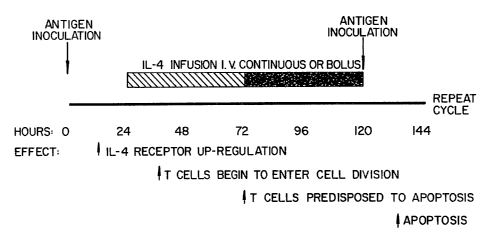
Figure 3. Indicated is a chronological sequence of
treatments (above the line) and expected outcomes (below the
line). Times for antigen inoculation and IL-4 infusion are
shown. Antigen may consist of protein or peptide molecules as
discussed for the treatment of autoimmune diseases or red
blood cells for preventing graft rejection. The hatched box
indicates the earliest time frame for IL-4 treatment; the
shaded box indicates optimal time for IL-4 treatment.
DETAILED DESCRIPTION OF THE INVENTION
The following detailed description of the invention
is provided to aid those skilled in the art in practicing the
same. Even so, the following detailed description should not
be construed to unduly limit the present invention, as
modifications and variations in the embodiments herein
discussed may be made by those of ordinary skill in the art
without departing from the spirit or scope of the present
inventive discovery.
MATERIALS AND GENERAL METHODS
Materials. Female B10.A and BALB/c mice were
purchased from Charles River. Pigeon cytochrome c, and
propidium iodide, as purchased from Sigma Chemical Co. (St.
Louis, MO). Purified murine rIL-4 was kindly provided by Dr.
W. Paul (National Institute of Allergy and Infectious
WO 94/03202 2140878 PCT/US93/07471
17
Diseases, NIH). The anti-murine IL-2 monoclonal antibody
(mAb) S4B6.1 was generously provided by Dr. J. Ziiftiga-Pfliicker
(National Institute of Allergy and Infectious Diseases, NIH).
Anti-murine CD3E mAb 145-2C11 (105) used in experiments was
immobilized by coating either 12- or 96- well culture plates
(500 l or 100 l, respectively) (Costar, Cambridge MA), at a
concentration of either 1 g/ml or 10 g/ml in
phosphate-buffered saline (PBS) for 120 minutes or overnight
at 37 C. The plates were washed three times with medium
(Click's or Eagle's Hank's amino acid, EHAA, and 10% FCS, 2 mM
glutamine, 50 M /3-mercaptoethanol, penicillin and
streptomycin; Biofluids, Inc., Rockville, MD) before use.
mAbs specific for MHC class II molecules (Ak, 10.2.16; and Ek,
Y17), were a gift from Dr. R. Schwartz (National Institute of
Allergy and Infectious Diseases, NIH).
Cell culture. The murine nontransformed T cell
clone A.E7 was carried as described previously (106). For
experimentation, either resting cells (>2 weeks following
antigen stimulation) or antigen stimulated cells were used.
Antigen stimulation consisted of culturing 1x10⁶ resting T
cells with 1x10⁷ B10.A irradiated (3000R) splenocytes and 5 M
pigeon cytochrome p in 2 mis total volume. After 48 hours,
the antigen presenting cells (APCs) were removed by MHC class
II mAb mediated complement lysis (low-tox-M rabbit complement,
Cedarlane Laboratories, Westbury, NY), and the cells were
recovered by Lymphocyte M density centrifugation (Cedar Lane
Laboratories) as previously described (41,104). The cells
were then recultured for 48 hours in medium with 1% MLA
(gibbon ape leukemia cell supernatant containing 140 U/mi of
IL-2 activity) or rIL-4 (10-1000 U/ml). T cells were then
harvested and washed and assays were carried out in 96-well
flat-bottomed plates in triplicate, with 200 l total volume
for 48 hours. 5x10⁴ cells/well (with the exception of Table
I, Experiment 1 which was 1x10⁵ ceils/weil) were added with
the designated lymphokine, either in the absence or presence
of T cell receptor stimuli. The live cell number was then
determined as described below.
WO 94/03202 PGT/US93/07471
18
T cell proliferation assays. A parallel culture of
5x10⁴ cells, without T cell receptor (TCR) stimulation, was
pulsed by the addition of 1 Ci of [3H] thymidine (3H-TdR)
(6.7 Ci/mmol, New England Nuclear) for 16-24 hours. Cells
were subsequently harvested onto glass filter paper, and the
samples counted by liquid scintillation on an L=?CB betaplate
counter. Data are expressed as the mean cpm of triplicates.
Cell viability. Viable cell number was determined
by manual counting of trypan blue excluding cells using a
hemocytometer by flow cytometry (FACS) with propidium iodide
stained cells. For flow cytometry quantitation, cells were
harvested by pipetting, washed once in phosphate buffered
saline (PBS), and each sample was suspended in a constant
volume of PBS with propidium iodide (2 g/ml). The
fluorescence intensity of samples collected for a constant
amount of time (100 sec.) was determined using a FACSCAN II
analyzer with Lysis II software (Becton Dickenson, Mountain
View, CA).
For this procedure, each sample is kept in a
constant volume and the cells are collected for a constant
amount of time, independent of the number of events. Only the
live cell number, as gaged by forward scatter and propidium
iodide exclusion, is quantitated. A comparison between
duplicate cultures analyzed for live cell number by trypan
blue exclusion or flow cytometric analysis reveals that the
relationship between these two quantitation methods is linear,
evidenced by an R value >0.97.
Analysis of DNA fracgmentation in agarose ctels.
1x10⁶ cells were incubated in 12-well plates coated
with anti-CD3e mAb for 48 hours, at which time the cells were
harvested by gentle scraping and prepared by a modification of
a previous procedure (8). Briefly, cells were washed once in
PBS and incubated in 20 l'*of lysis buffer (50 mM Tris, pH
8.0, 10 mM EDTA, 500 g/ml proteinase K, and 0.5% sodium
sarkosyl) for 1 hour at 50 C. RNase A (50 g/ml) (Boehringer
Mannheim) was added and the cells were incubated for an
additional hour at 50 C. Dye buffer (10 mM EDTA, 1% (w/v) low
WO 94/03202 2~ ~ ~ ~ 78 PCT/US93/07471
19
melting point agarose, 0.25% (w/v) bromphenol blue, and 40%
(w/v) sucrose) was added, the samples were heated to 70 C for
five minutes, quenched on ice, and electrophoresed in a 2%
Nusieve agarose, 1% ultra-pure agarose gel with ethidium
bromide.
Example I
IL-4 bredisposes T cells to antigen-induced
apoptosis.
We have previously shown that IL-2 participates in
an apparent feedback pathway, termed propriocidal regulation,
by predisposing T lymphocytes to antigen-induced apoptosis
(26). We therefore determined if IL-4, another.T cell growth
factor, would induce this pathway. We first studied the
nontransformed CD4+ TH1 clone A.E7 that responds to pigeon
cytochrome c in the context of an Ek MHC class II molecule.
This clcine has been shown to upregulate its IL-4 receptor in
response to antigenic stimulation and proliferate in response
to IL-4 (41). As shown in Table 1, antigen stimulated A.E7
cells proliferate to IL-4 in a dose dependent manner, as
indicated by tritiated thymidine (3H-TdR) incorporation
(experiment 1). Moreover, there was a dramatic cell loss when
the proliferating cells were subsequently placed onto
anti-CD3e-coated plates for 48 hours, as compared to the
uncoated plate control. The reduction in cell number was
minimal with no growth lymphokine added and increased roughly
in proportion to the degree of proliferation achieved with
'increasing amounts of lymphokine. Cells treated with 1000
U/ml IL-4 showed an 84% decrease in cell number following TCR
stimulation. The overall cell loss found with IL-4 was as
great as that obtained with IL-2 stimulation (85% versus 87%,
respectively).
We observed a similar phenomenon with the
lymphokine-dependent T cel7.'lines, CR.4R and CT.4S. We could
not detect any T cell receptor surface expression in either
cell line and anti-CD3e stimulation had no effect on these
cells (S.B. and M.L., unpublished results). Nonetheless, when
TCR occupancy was mimicked by a combination of phorbol
PC'T/US93/07471 ~
WO 94~/0~3~~~=~
myristic acetate (PMA) and ionomycin, extensive cell loss was
observed after 48 hours (Table 1, experiment 2). Greater than
90% cell loss was observed for CT.4R cells exposed to either
IL-2 or IL4, and 87% cell loss was seen for CT.4S cells
5 incubated with IL-4.
Several features of the c'ell loss in these
experiments suggested that cell .4eath was occurring. First,
microscopic examination in all cases revealed cells appearing
to undergo apoptosis. As shown in Figure 1, trypan blue
10 staining can detect non-viable cells (dark colored) in the
cells stimulated with anti-CD3e antibody more than in
untreated samples. Second, the number of A.E7 cells following
1000 U/ml IL-4 and anti-CD3c stimulation was less than the
number of cells put into the wells implying cell loss in
15 addition to any potential block in proliferation (Table 1,
experiment 1). Third, ladders of nucleosomal length DNA were
obtained following IL-4 and anti-CD3c treatment of A.E7 cells,
indicating the occurrence of apoptosis. As shown in Figure 2,
DNA fragmentation was observed in cells cultured with
20 platebound anti-CD3e mAb (lanes 3 and 4), and was not observed
'in cells cultured with medium alone (lanes 1 and 2). We also
observed cell death when IL-4 treated A.E7 cells were
co-cultured with irradiated splenocytes and antigen (Table 1,
experiment 3). Because IL-4 can stimulate the release of IL-2
under certain conditions (107), a mAb capable of binding IL-2
(S4B6.1) was included in the IL-4 stimulation. This did not
inhibit subsequent T cell stimulation-induced apoptosis (Table
1, experiment 4), suggesting IL-4 treatment alone predisposes
T cells to apoptosis.
Because these experiments were carried out in T cell
clones that had been carried in vitro for a long period of
time, we investigated whether IL-4 could predispose lymph node
cells to apoptosis. Conditions for stimulating lymph node T
(LNT) cells to produce lymphokines and proliferate in response
to either IL-2 or IL-4 have recently been determined (108).
Treatment with TCR stimulation and IL-2 produces cultures
exhibiting a predominantly TH1 phenotype producing and
responding to IL-2, whereas the inclusion of IL-4 leads to a
WO 94/03202 _2140878 PCT/US93/07471
21
TH2 phenotype of cells producing and responding to IL-4 (108).
Freshly isolated lymph node cells were treated for 72 hours
with either soluble anti-CD3e mAb or concavalin A, and IL-2 or
IL-4. The LNT cells proliferated significantly in response to
lymphokine in all samples (Table 2, CPM). There was large
decrease in the number of live cells recovered following a
48-hour incubation on anti-CD3e-coated plates compared to the
plastic control at all conditions tested (Table 2). These
results show that IL-4 has the ability to predispose LNT cells
to apoptosis. Furthermore, as was previously observed with
IL-2 (26), IL-4 by itself can evoke the propriocidal pathway
that leads to apoptosis following antigen receptor
stimulation.
WO 94/03202 PCT/US93/07471
~jk4s7s 22
Table 1. The effect of IL-4 and T cell receptor
stimulation on T cell viability.
f:xa lls I'rctrcatmrnlt ('M Ccll Nunibcr(x1015/ml) Cell Loss
~ ontr l Anti- 3
I A.E7 Nonc 2,060 7.3 + 0.7 5.4 0.7 27
A.E7 14 U nil-t IL-2 173.845 37.3 4.1 4.7 + 0.4 87
A.E7 10 U mi-t IL-4 2,725 6.3 0.3 4.7 0.4 25
A.1:7 100 U nil-t 1L-4 17,833 10.1 4-0.8 5.5 0.8 45
A.j7 1000Utnl=l IL-4 89,159 18.6 0.4 3.0 0.4 84
C'ontr l PNIA/12
2 CT.4R 28 U nil-t IL-2 313,574 55.3 5.4 2.9 0.9 95
CT.4R 1000 U nil-t IL-4 317,227 34.3 4.6 3.6 0.8 90
CT.4S 1000 U ml-t IL-4 155,982 27.7 f 1.8 3.6 0.7 87
FACS Cell Number3
-Az 8g-.
3 A.E7 100011 mi-I IL-4 257,568 22,703 5,348 76
Control nti-CD3E
4 A.E7 14U mi-I IL-2 266,137 11.7 1.6 2.6 0.4 78
A.E7 1000U mi-I IL-4 257,913 9.4 1.7 1.9 # 0.3 80
A.B7 1000U mi-I IL-4 257,568 9.3 1.3 2.0 0.5 79
+ S4B6.1
tIndicates the treatment of A.137 cells following 48 hour antigen stimulation.
CI'.4R and CT.4S
cell lines did not undergo antigen stimulation but were pretteated as
described. The concentration
of lympholone indicated was kept constant for each sample during the 48 hour
ptr.treatment and
the 48 hour duration of the experiment.
WO 94/03202 2140878 PCT/US93/07471
23
Table 1. (continued)
2 these cells do not express TCR on the cell surface, so were
cultured with PMA (10 ng/ml) and ionophonre (2 M).
3 Indicates the live A.E7 cell number as determined by forward
scatter profile, propidium iodide dye exclusion and surface
staining with anti-mouse Vail mAb (Pharmingen) (see materials
and Methods). Experimental conditions consisted of a 10-fold
excess of B10. A irradiated spleen cells, l M pigeon
cytochrome c(+ Ag) and 30 g of anti-mouse IL-2 mAb S4B6.1.
Table 2. The effect of lymphokines and antigen
receptor stimulation on lymph node cell
viability.
Pretre:unxnt CPM Cell Number % Cell Loss
TCR Stini. Wmphokinc Contml Anti-CD3e
Anti-CD3C IL-2 85.912 16,li76 3.775 7,917 1,31() 53
Con A IL-2 54,835 28?26 60() 8,489 2.804 70
Anti-CD3c IL-4 93,725 42,920 3,4()9 4,628 475 89
Con A IL-4 81502 36,930 2.705 3,510 887 91
BALB/c lymph node cells were cultured at a
concentration of 1x10⁶ cells/ml for 72 hours with soluble
anti-CD3E (3 g/ml) or Concavalin A (3 g/ml) in the presences
of IL-2 (14 U/ml) or IL-4 (1000 U/mi). The cells (5x10⁴
cells/well) were washed extensively and incubated with medium
or anti-CD3-coated plates for an additional 48 hours in the
presence of lymphokine. The cells were then harvested and the
live cell number was determined by FACS analysis.
WO 94/03202 PCT/US93/07471 0
24
We previously proposed the term propriocidal
regulation for the antigen receptor-stimulated apoptosis of
mature T lymphocytes that were induced into the cell cycle by
IL-2 (26). This mechanism would result in the elimination of
any T lymphocyte that had a sufficient affinity for the
inciting antigen. Our previous results~suggest that in order
for the T lymphocyte to undergo propriocidal cell death, it
must be responding to IL-2 treatment, but not necessarily
producing the lymphokine. We now extend these results to show
that A.E7, a TH1 clone that can respond to, but not produce
IL-4, will undergo apoptosis by TCR stimulation if actively
cycling in response to IL-4 treatment. We have also shown
that normal lymph node cells driven into cell cycle by antigen
receptor stimulation and either IL-2 or IL-4 treatment,
undergo cell death upon subsequent TCR stimulation. Cell
cycling per se is not required, because we have found that
certain'blocking agents do not prevent TCR-mediated apoptosis
(27). Nonetheless, agents that prevent progression beyond
late Gi, and not those that block proliferation in S phase,
were found to be capable of inhibiting apoptosis (27) . Thus,
we favor the hypothesis that cell cycle progression beyond
late G1, stimulated by growth lymphokines such as IL-2 or IL-
4, is permissive for TCR-mediated death in T lymphocytes. It
is likely that cell cycle progression beyond the late G1 stage
due to the transformed phenotype of T cell hybridomas and
lymphomas accounts for their sensitivity to TCR-mediated
apoptosis without lymphokine treatment (13-18). Our results
suggest that an intrinsic property of the T lymphocyte
response to a growth lymphokine such as IL-4 is the
susceptibility to apoptosis upon further TCR stimulation.
Moreover, this response portrays a mechanism by which an
immune response to specific antigens may be naturally
suppressed.
Example II
Method for IL-4/Peptide-Medicated Apoptosis of T Lymphocytes
As shown in Figure 3, immunization with a specific
peptide or protein is carried out on day one. In the case of
WO 94/03202 PCT/US93/07471
multiple sclerosis, for example, there is evidence that either
of two immunodominant peptides from myelin basic protein (MBP)
are encephalitogenic in man; MBP 84-102 (the preferred
peptide), or MBP 143-168 (78,79). Either or both of these
5 peptides, coupled to tetanus toxoid, can be given in alum
adjuvant intramuscularly (IM), at a dose between about 10 to
about 1000 g. Early immunization experience using proteins
or peptides has suggested that intramuscular administration is
optimal (109-113). Newer data suggest that oral
10 administration may also be effective (94). As with any
medicinal substance, or biologic, tests on any peptides and
proteins used for the immunization would need to be routinely
carried out over a range of doses to determine: 1) the
pharmacokinetic behavior of these substances; 2) their
15 immunogenicity; and 3) safety and identification of any
untoward effects. This would constitute a Phase I clinical
trial (114). Thus, the particular proteins or peptides
employed in this protocol (for example, in multiple sclerosis,
MBP 84-102, or MBP 143-168; in uveitis, the S Antigen; or in
20 rheumatoid arthritis, type II collagen) would require
individual routine optimization. Similar intervention could
be used with preparations of allergy-inducing proteins. These
could be derived from a variety of allergen protein extracts
that are now used clinically, or could be generated by
25 recombinant DNA technology for those such as hornet venom
antigen 5, for which cloned DNA is available (100). Ample
evidence from the development of vaccines suggests that either
synthetic peptides or recombinant DNA-derived proteins are
effective in eliciting an immune response in humans (109-112).
These studies also provide guidance as to the range of doses
effective for immunization.
Proteins:
1) Hepatitis B surface antigen, produced as a
recombinant protein in yeast. Adults 2.5 to 20 g; children
1.25 to 5 g intramuscularly (IM). 90-96% of vaccines showed
an immune response, with the best response at 10-20 g (109).
Further studies showed the efficacy of a 10 g dose, with
better results when given IM rather than subcutaneously (110).
WO 94/03202 PCT/US93/07471 0
26
20 g doses in alum adjuvant given IM were found to be
effective at preventing infection in clinical trials (111).
2) HIV gp 120, either natural or recombinant
molecules. Doses in chimpanzees between 50-1000 g elicit T
cell responses (115).
Peptides:
i
1) Chorionic gonadotropih. Several studies have
indicated successful immune respon"s against a human
chorionic gonadotropin-fi subunit peptide (residues 109-145)
coupled to cholera or tetanus toxoid and given in doses from
50-1000 g in alum adjuvant (112).
2) Malaria sporozoite antigen. Studies of a
Plasmodium falciparum peptide (NANP)3 coupled to tetanus
toxoid showed an immune response to doses of 20-160 g of
peptide conjugate given IM, with the best response at 160 g
(113).
Immunization is then followed by a waiting period
during which the antigen activates the subset of T cells
bearing reactive TCRS, causing them to express IL-4 receptors
and possibly IL-4. This process will only upregulate IL-4
receptors on cells that have been antigenically-stimulated
(36). Based on studies of both human and mouse T cells in
vitro, between about 12 to about 24 hours after antigen
exposure are required to express significant increases in the
numbers of IL-4 receptors, and as long as about 72 hours are
required to express optimal numbers of lymphokine receptors on
the majority of T cells (36). Thus, the waiting period can be
as short as about 12 hours or as long as about 72 hours,
becoming increasingly optimal toward the upper end of this
range.
This is then followed by an infusion of high doses
of IL-4. Though only very limited data exists on the clinical
use of IL-4 (89-92), a great deal of information has been
obtained from clinical studies using IL-2. The administration
of high-doses of the related T cell growth lymphokine IL-2 to
humans has been well-studied in cancer patients, and various
doses have been evaluated (116-120). Data indicate that IL-2
should be given intravenously (I.V.) either as frequent bolus
+
WO 94/03202 2140878 PCT/US93/07471
27
doses or as a continuous infusion (116-118). Doses that have
been previously established range between about 300 to about
3000 units/kg/hour continuous infusion, or from 104 to 106
units/kg I.V. bolus (117). Units are defined by standards
available from the Biological Response Modifiers Program at
the National Institutes of Health, and are defined as the
quantity of IL-2 or IL-4 that gave 50% maximal thymidine
incorporation in the bioassay under standard conditions. Side
effects of these doses included chills, fever, malaise,
headache, nausea and vomiting, weight gain due to fluid
retention, diarrhea, rash, and pruritis, which can all be
treated with acetaminophen or indomethacin; no serious
morbidity or mortality was observed. Studies of IL-4
administration to humans used human recombinant IL-4 of
specific activity 1.5 X 10⁷ units/ug, given in doses of 10-20
ug/kg body weight, three times/day. (89-91, 120,121) The
side effects with IL-4 were similar to those observed with
IL-2 and included weight gain due to water retention and
nausea. After IL-4 treatment, the patient can be immediately
reimmunized with an equivalent dose of antigen. For example,
for multiple sclerosis, treatment can be carried out with
about 10 to about 1000 g of peptide MBP 84-102 coupled to
tetanus toxoid and given in alum adjuvant IM. It is likely
that the preferred dose would be near the upper end of this
range since greater TCR stimulation produces a greater level
of apoptosis (26,27). IL-4 treatment would have stimulated
the T cells bearing IL-4 receptors -- predominantly the
disease-causing T cells -- and these cells would then be
re-stimulated through their TCR. These cells will then
undergo apoptosis. After an immunization period of about 12
to about 72 hours, the cycle would begin again with reinfusion
of IL-4. As will be described below, increased efficacy would
likely result from multiple cycles of therapy. The treatment
endpoints would be: i) elintination of in vitro reactivity to
the antigen, which can be easily measured where possible by
various mixed lymphocyte or proliferation assays using
peripheral blood lymphocytes; ii) amelioration of clinical
symptoms; or iii) toxicity. The treatment endpoints for
PCT/US93/07471 0
WO 94/03202
28
allergic diseases would be: i) improvement of clinical
symptoms; ii) normalization of an allergic skin test; iii)
reduction in serum IgE levels; and iv) where possible to
measure, reduced T cell responses to the allergenic protein.
Several features of the present therapy require
further explanation. First, it is expected that T cells
besides those antigenically stimulated may express high
affinity IL-4 receptors. However, this should not diminish
the specificity of the therapy because only those cells whose
TCRs are stimulated by rechallenge with antigen will undergo
apoptosis, as described sunra. The effectiveness of the
therapy could be variable depending on the nature of the
antigen and the exact protocol employed. Extensive in vitro
studies indicate that between 50-80% of the antigen-specific
IL-4 stimulated T cells will undergo apoptosis when
rechallenged by TCR stimulation (supra, 27). Second, the
reduction in number of antigen-specific T cells determines the
overall effectiveness of the therapy. Therefore, repeated
cycles can substantially increase efficacy even if the level
of killing in each cycle is only 50-70% (Table 3). As shown
in the mouse studies, supra, the level of antigen-reactive T
cells will decrease below the number of such cells prior to
the first immunization with repetitive immunization.
Furthermore, the expected toxicity of this protocol at
moderate doses or lymphokine should be minor, and previous
studies of the therapeutic use of growth lymphokines such as
IL-2 or IL-4 in humans indicates that all side effects
dissipate promptly following discontinuation of lymphokine
treatment (89-91,116,117). The most serious side effect,
fluid retention, should be minimized by the intermittent
nature of IL-4 treatment. The 2-3 day rest period between
doses would allow for diuresis of the fluid built up during
IL-4 administration. Finally, the repeated administration of
antigen will cause production of some endogenous IL-4, which
will predispose some cells to apoptosis. While it is
extremely unlikely that endogenous levels can reach the very
high levels of IL-4 that can be administered
pharmacologically, it is possible that empirically-determined
WO 94/03202 '2140878 PCr/US93/07471
29
decreases in the IL-4 dose could be achieved because of
endogenous IL-4 effects. The level of killing is dependent on
the total level of IL-4 to which the T cell is exposed, and
this will reflect a combination of endogenous and exogenous
sources (supra, 26,27).
With certain antigens, the predisposition of cells
to apoptosis may be sufficiently induced by the endogenous
production of IL-4. In these cases, appropriate immunization
with antigen, in the absence of exogenously administered IL-4,
could produce T cell apoptosis and a protective effect. Based
on the studies of the timing of susceptibility to apoptosis
disclosed supra, immunizations repeated at specific intervals
would be crucial for effective therapy. To effect IL-4-
mediated apoptosis, immunizations would have to be repeated at
about 24 to about 120 hour intervals, preferably at about 24
to about 72 hour intervals, and would have to be repeated
multiple times using antigen doses at about the high end of
the ranges discussed above. T cell reactivity or
cell-mediated immunity for the specific antigen could then be
monitored by in vitro assays to determine that T cells had
undergone apoptosis. Absent the knowledge provided by the
discovery disclosed herein, previous attempts to decrease
immune responsiveness by repetitive immunization have not been
optimal. For example, donor transfusion protocols to
ameliorate graft rejection involved 3 transfusions given at 2
week intervals (122, 123). Allergy shots, i.e.,
desensitization therapy, are typically given initially at 4-7
day intervals, after which intervals are progressively
increased in length to 2 to 4 weeks (97). Based on the
present novel understanding of T cell apoptosis, the most
effective immunization protocol would involve repetitive
administrations of antigen at about 24 to 72 hour intervals.
WO 94/03202 PCr/US93/07471 0
(rik"sls 30
Table 3
Theoretical number of reactive cells after
fractional killina using IL-4 and T=cell
receptor stimulation
Reactive Cells
Cvcle Fractional RillinQ Remaining
Start None 100,000
1 70% 30,000
2 70% 9,000
3 70% 2,700
4 70% 810 =
5 70% 243
6 70% 73
Theoretical values are based on starting with 100,000 cells
and a constant killing efficiency of 70%. A reduction of over
100-fold is seen in 4 cycles and over 1000-fold in 6 cycles.
At a fractional killing of 50%, a reduction of nearly 100-fold
would be seen in 6 cycles. A first order kinetics is
represented here because the process of apoptosis involves a
single lethal hit delivery as has been shown for apoptosis
induced by antimetabolites (1-5).
Example III
Method for transplantation antiuen/IL-4-mediated
anoptosis.
In medical procedures in which tissue is transferred
between individuals who are genetically non-identical at their
relevant histocompatibility antigen loci, herein referred to
as allografting, and the transplanted tissue referred to as an
allograft, the major problem encountered is rejection of the
donor allograft by the host. The term 1 host" refers to the
individual who is the recipient of the allograft, and the term
"donor" refers to the individual from whom the allograft is
derived. Studies of the process of graft rejection have shown
that it is due to the antigen-specific activation of T
WO 94/03202 2140878 PCT/US93/07471
31
lymphocytes, especially those bearing CD8 surface molecules
(124). More importantly, agents that block the ability of T
cells to mount an immune response in humans effectively
prevent or lessen graft rejection (125). Since CD8+ T cells
have been shown to be susceptible to apoptosis by IL-4, supra,
this phenomenon can be used as a specific means to eliminate
the reactive T cells, thereby avoiding graft rejection.
Essentially the same protocol with respect to timing
and IL-4 dose can be used for this therapy as was described
su ra for the therapy of autoimmune diseases. The major
difference between this therapy and that described above is
the source of antigen. Major histocompatibility complex (MHC)
antigens are cell surface proteins that are tremendously
polymorphic among individuals. Each individual's cells bear a
genetically determined set, or haplotype, of such antigens
which serve as an immunological "fingerprint" on each cell
(126). This allows one's immune system, in particular those
responses generated by T cells, to recognize one's own cells,
and to attack only cells that do not bear the self
"fingerprint" (127). There are two classes of MHC -- class I
antigens, found on all cells in the body; and class II
antigens, found predominantly on monocytes, macrophages, B
lymphocytes, dendritic cells, and activated T cells (126). It
is the class I MHC antigens that are recognized by CD8+ T
cells that are the predominant influence in allograft
rejection (124,1127). Because of this complexity of MHC
antigens, the simplest source is cells from the allograft
donor. It has been empirically observed that transfusion of a
graft recipient with donor blood suppresses graft rejection,
although the mechanism of this effect is unknown, and the
clinical effectiveness in many cases is modest (123). These
protocols provide evidence that three transfusions of 200 ml
of whole blood or packed cell equivalent from the donor is
= easily tolerated by the recipient with minimal side effects
(122). There is evidence that the donor-transfusion in some
cases elicited sensitizing antibody responses in the allograft
host, and these patients were not given allografts (122).
These studies possibly represent an empirical observation that
WO 94/03202 32 PCT/US93/074710
preexposure to donor antigen suppresses the T cell response,
although this is controversial (124). The present method
includes administration of blood as a source of MHC antigens
in doses of about 50 to about 200 ml to patients in cycle with
IL-4, as indicated in Fig. 4. In the case of kidney
transplants, the amount of blood coqld be determined by the fluid tolerance of
end-stage renaldisease patients. The
blood can be given as either whole blood, packed cells, or
washed packed cell transfusions (123). The success of
treatment can be assessed by: i) a decreased requirement for
general immunosuppressive medications; ii) graft survival; arid
iii) adequate function of the allograft. For example, the
function of a transplanted kidney can be established by
determining serum levels of creatinine and blood urea nitrogen
(125). This can be followed by IL-4 infusion and rechallenge
with blood cells as antigen as shown in Figure 3.
The invention being thus described, it will be
obvious that the same may be varied in many ways. Such
variations are not to be regarded as a departure from the
spirit and scope of the invention, and all such modifications
as would be obvious to one skilled in the art are intended to
be included within the scope of the following claims.
WO 94/03202 2140878 PCT/US93/07471
33
LIST OF REFERENCES CITED
1. Kerr, J.F.R, and B.V. Harmon. 1991. Definition and
incidence of apoptosis: an historical perspective. In
Apoptosis:the molecular basis of cell death, L.D. Tomei
and F.O. Cope, ed. Cold Spring Harbor Laboratory Press,
' Plainview, New York, p. 5.
2. Lockshin, R.A., and Z. Zakeri. 1991. Programmed cell
death and apoptosis. In Apoptosis: the molecular basis
of cell death, L.D. Tomei and F.O. Cope, ed., Cold Spring
Harbor Press, Plainview, New York, p. 47.
3. Cohen, J.J., R.C. Duke, V.A. Fadok, and K.S. Sellins.
1992. Apoptosis and programmed cell death in immunity.
Ann. Rev. Immunol. 10:267.
4. Duvall, E. and A. H. Wyllie. 1986. Death and the cell.
Immunoloay Today 7:115.
5. Cotter, T. G., S. V. Lennon, J. G. Glynn, and S. J.
Martin. 1990. Cell death via apoptosis and its
relationship to growth. Development and differentiation
of both tumor and normal cells. Anticancer Research
10:1153.
6. von Boehmer, H. 1988. The developmental biology of T
lymphocytes. Ann. Rev. Immunol. 6:309.
7. Marrack, P., and J. Kappler, 1987. The T cell receptor.
Science 238:1073.
8. Smith, C. A., G. T. Williams, R. Kingston, E. J.
Jenkinson, and J. J. T. Owen. 1989. Nature 337:181.
9. Shi, Y., R. P. Bissonnette, N. Parfrey, M. Szalay, R. T.
Kubo, and D. R. Green. 1991. In vivo administration of
monoclonal antibodies to the CD3 T cell receptor complex
induces cell death (apoptosis) in immature thymocytes.
J. Immunol. 146:3340.
10. McConkey, D. J., P. Hartzell, J. F. Amador-Perez, S.
Orrenius, and M. Jondal. 1989. Calcium-dependent killing
of immature thymocytes by stimulation via the CD3/T cell
receptor complex. J. Immunol. 143:1801.
11. Nieto, M. A., A. Gonzalez, A. Lopez-Rivas, F.
Diaz-Espada, and F. Gambon. 1990. IL-2 protects against
anti-CD3-induced cell>death in human medullary
thymocytes. J. Immunol. 145:1364
12. Wyllie, A. H. 1980. Glucocorticoid-induced thymocyte
apoptosis is associated with endogenous endonuclease
activation. Nature 284:555.
WO 94/03202 PCT/US93/07471
2JO(jIS 34
13. A"shwell, J. D., R. E. Cunningham, P. D. Noguchi, and D.
Hernandez. 1987. cell growth cycle block of T cell
hybridomas upon activation with antigen. J. Exp. Med.
165'173.
14. Mercep, M., J. A. Bluestone, P. D. Noguchi, and J. D.
Ashwell. 1988. Inhibition of transformed T cell growth
in vitro by monoclonal antibodies directed against
distinct activating molecules. J. Immunol. 140:324.
15. Ucker, D. S., J. D. Ashwell,'' and G. Nickas. 1989,
Activation-driven T cell death. 1. Requirements for de
novo transcription and translation and association with
genome fragmentation. J. Immunol. 143:3461.
16. Nickas, G. J. Meyers, L. D. Hebshi, J. D. Ashwell, D. P.
Gold, B. Sydora, and D.S. licker. 1992. Susceptibility
to cell death is a dominant phenotype: triggering of
activation-driven T-cell death independent of the T-cell
antigen receptor complex. Mol. Cell. Biol. 12:379.
17. Shi, Y., M. G. Szalay, L. Paskar, M. Boyer, B. Singh, and
D. R. Green. 1990. Activation-induced cell death in T
cell hybridoma is due to apoptosis. J. Immunol.
144:3326.
18. Odaka, C., H. Kizaki, and T. Tadakuma. 1990. T cell
receptor-mediated DNA fragmentation and cell death in T
cell hybridomas. J. Immunol. 144:2096.
19. Takahashi, S., H. T. Maecker and R. Levy. 1989. DNA
fragmentation and cell death mediated by T cell antigen
receptor/CD3 complex on a leukemia T cell line. Eur. J.
Immunol. 19:1911.
20. Zacharchuk, C. M., M. Mercep, P. K. Chakraborti, S. S.
Simons, Jr., and J. D. Ashwell. 1990. Programmed T
lymphocyte death. Cell activation and steroid-induced
pathways are mutually antagonistic. J. Immunol.
145:4037.
21. Iseki, R., M. Mukai, and M. Iwata. 1991. Regulation of T
lymphocyte apoptosis. Signals for the antagonism between
activation- and glucocorticoid-induced death. J.
Immunol.. 147:4286.
22. Iwata, M., S. Hanaoka, and K. Sato. 1991. Rescue of
thymocytes and T cell hybridomas from glucocorticoid-
induced apoptosis by simulation via the T cell
receptor/CD3 complex:,:a possible in vitro model for
positive selection of the T cell repertoirE.. Eur. J.
Immunol. 21:643.
23. Duke, R. C. and J. J. Cohen. 1986. IL-2 addiction:
withdrawal of growth factor activates a suicide program
in dependent T cells. Lymphokine Research 5:289.
WO 94/03202 2140878 PCT/US93/07471
24. Watanabe-Fukunaga, R., C.I. Brannan, N. G. Copeland, N.
A. Jenkins, and'S. Nagata. 1992. Lymphoproliferation
disorder in mice explained by defects in Fas antigen that
mediates apoptosis. Nature 356:314.
5
25. Trauth, B. C., C. Klas, A. M. J. Peters, S. Matzku, P.
Moller, W. Falk, K-M. Debatin, and P. H. Krammer. 1989.
Monoclonal antibody-mediated tumor regression by
induction of apoptosis. Science 245:301.
26. Lenardo, M. J. 1991. Interleukin-2 programs mouse a/3 T
lymphocytes for apoptosis. Nature 353:858.
27. Boehme, S. and Lenardo, M.J. (1992) Antigen
receptor=induced apoptosis of nontransformed, mature T
lymphocytes (propriocidal regulation) but not
glucocorticoid-induced apoptosis, requires a distinct
stage of the cell cycle (manuscript submitted).
28. Katz, P. and A.S. Fauci, Immunosuppressives and
immunoadjuvants, Immunological Diseases, M. Somter et
al., eds. (Boston: Little, Brown and Company), pp.
675-698 (1989).
29. Weiss, A., T lymphocyte activation, Fundamental
Immunologv, Second Ed., W,.E. Paul, ed. (New York: Raven
Press), pp. 359-384 (1989).
30. Hedrick, S.M., T lymphocyte receptors, Fundamental
Immunology, Second Ed., W.E. Paul, ed. (New York: Raven
Press), pp. 291-358 (1989).
31. Fink, P.J., M.J. Blair, L.A. Matis and S.M. Hedrick,
Molecular analysis of the influences of positive
selection, tolerance induction, and antigen presentation
on the T cell repertoire. J. Exp. Med., 172:139 (1990).
32. Tse, H.Y., R.H. Schwartz and W.E. Paul, Cell-cell
interactions in the T cell proliferative response, J.
Immunol., 125:491-500 (1980).
33. Oksenberg, J.R., S. Stuart, A.B. Begovich, R.B. Bell,
H.A. Erlich, L. Steinman and C.C.A. Bernard, Limited
heterogeneity of rearranged T-cell receptor
Va-transcripts in brains of multiple sclerosis patients,
Nature 345:344 (1990).
34. Lindahl, K.F. and D.B. Wilson, Histocompatibility
antigen-activated cytotoxic T lymphocytes, J. Exp. Med.
145:508-522 (1977).
35. Crabtree, J., Contingent genetic regulatory events in T
lymphocyte activation, Science 243:355-361 (1989).
36. Foxwell, B.M., Woerly, G., and Ryffel, B. (1989).
Identification of interleukin-4 receptor=associated
WO 94/03202 PC'T/US93/07471
% 1 36
proteins and expression of high and low affinity binding
on human lynphoid cells, Eur. J. Immunol. 19:1637-41.
37. Spits, H. et al. 1987. J. Immunol. 139:1142.
38. Hu-Li, J. et al. 1987. J. Exp. Med. 165:157.
39. Mosmann, T.R. and Coffman, R.L.,..1987. Two types of
mouse helper T cell clone, Immunology Today 89:223-227.
40. Bottomly, K. 1988. Immunoloq)r'Today 9:268.
41. Mueller, D. L., L. Chiodetti, P. A. Bacon, and R. H.
Schwartz. 1991. Clonal anergy blocks the response to
IL-4, as well as the production of IL-2, in
dual-producing T helper cell clones. J. Immunol.
146:4118.
42. Widmer, M.B. and Grabstein, K.H. 1987. Nature: 326:795.
43. Grabstein, K.H. et al. 1987. J. Immunol. 139:1148.
44. Boehme, S. and Lenardo, M.J., 1992. Manuscript in
preparation.
45. Paul, W.E., The immune system: an introduction.
Fundamental Immunology, Second Ed., W.E. Paul, ed. (New
York: Raven Press) pp. 3-38 (1989).
46. Russell, J.H., White, C.L., Loh, D.Y. & Meleedy-Rey, P.
Proc. Natl. Acad. Sci. 88, 2151-2155 (1991); and Kawabe,
Y. & Ochi, A. Nature 349, 245-248 (1991).
47. Liu, Y. & Janeway, C.A. Jr. J. Exp. Med. 172, 1735-1739
(1990).
48. Webb, S., Morris, C. & Sprent, J. Cell 63, 1249-1256
(1990).
49. Jones, L.A., Chin, L.T., Longo, D.L. & Kruisbeek, A.M.
Science 250, 1726-1729 (1990).
50. Rocha, B. & von Boehmer, H. Science 251, 1225-1228
(1991).
51. Marsh, D.G. and Norman, P.S., Antigens that cause atopic
diseases, Immunological Diseases, Fourth Edit., M. Samter
et al, Eds. Vol. II, pp. 981-1002.
52. Middleton, E. et al, Eds., Allergy: Principles and
Practice, Third edition, (St. Louis: C.V. Mosby) (1988).
53. Johnson, D, D.A. Hafler, R.J. Fallis, M.B. Lees, R.O.
Brady, R.H. Quarles and H.L. Weiner, Cell-mediated
immunity to myelin-associated glycoprotein, proteolipid
protein, and myelin basic protein in multiple sclerosis,
J. Neuroimmunology 13:99-108 (1986).
WO 94/03202 214087 PCT/US93/07471
37
54. Martin, R., M.D. Howell, D. Jaraquemada, M. Flerlage, J.
Richert, S. Brostoff, E.O. Long, D.E. McFarlin and H.F.
McFarland, A myelin basic protein peptide is recognized
by cytotoxic T cells in the context of four HLA-DR types
associated with multiple sclerosis, J. Exp. Med.,
173:19-24 (1991).
55. Pette, M., K. Fujita, D. Wilkinson, D.M. Altmann, J.
Trowsdale, G. Giegerich, A. Hinkkanen, J.T. Epplen, L.
Kappos and H. Wekerle, Myelin autoreactivity in multiple
sclerosis: recognition of myelin basic protein in the
context of HLA-DR2 products by T lymphocytes of
multiple-sclerosis patients and healthy donors. Proc.
Natl. Acad. Sci. USA 87:7968-7972 (1990).
56. Jaraquemada, D., R. Martin, S. Rosen-Bronson, M.
Flerlage, H.F. McFarland and E.O. Long, HLA-DR2a is the
dominant restriction molecule for the cytotoxic T cell
response to myelin basic protein in DR2Dw2 individuals,
J. Immunol. 145:2880-2885 (1990).
57. Martin, R., D. Jaraquemada, M. Flerlage, J. Richert, J.
Whitaker, E.O. Long, D.E. McFarlin and H.F. McFarland,
Fine specificity and HLA restriction of myelin basic
protein-specific cytotoxic T cell lines from multiple
sclerosis patients and healthy individuals, J. Immunol.
145:540-548 (1990).
58. Brinkman, C.J.J., W.M. Nillesen, O.R. Hommes, K.J.B.
Lamers, B.E. dePauw, and P. Delmotte, Cell-mediated
immunity in multiple sclerosis as determined by
sensitivity of different lymphocyte populations to
various brain tissue antigens, Ann. Neuroloav 11:450-455
(1981).
59. Hirose, S., L.A. Donoso, T. Shinohara, A.G. Palestine,
R.B. Nussenblatt and I. Gery, Lymphocyte responses to
peptide M and retinal S antigen in uveitis patients, JPn.
J. Ophthalmol. 34:298-305 (1990).
.60. de Smet, M.D., J.H. Yamamoto, M. Mochizuki, I. Gery, V.K.
Singh, T. Shinohara, B. Wiggert, C.J. Chader and R.B.
Nussenblatt, Cellular immune responses of patients with
uveitis to retinal antigens and their fragments, Am. J.
Ophthalmol. 110:135-142 (1990).
61. Hawrylko, E., A. Spertus, C.A. Mele, N. Oster and M.
Frieri, Increased interleukin-2 production in response to
human Type I collagen stimulation in systemic sclerosis
patients, Arthritis Rheum., 34:580-587 (1991).
62. Abdel-Nour, A.N., C.J. Elson and P.A. Dieppe,
Proliferative responses of T-cell lines grown from joint
fluids of patients with rheumatoid arthritis and other
arthritides, Immunol. Lett. 12:329-33 (1986).
WO 94/03202 2jO PCT/US93/07471 ~
- 38
63. Paliard, X., S.G. West, J.A. Lafferty, J.R. Clements,
J.W. Kappler, P. Marrack and B.L. Kotzin, Evidence for
the effects of a superantigen in rheumatoid arthritis,
Science 253:325-329 (1991).
64. Bottazzo, G.F., B.M. Dean, i.M. McNally, E.H. MacKay,
P.G.F. Swift and D.R- Gamble, In situ characterization of
autoimmune phenomena and expression of HLA molecules in
the pancreas in diabetic insulitis, New EncLl. J. Med.
313:353-360 (1985).
65. Lundkin, K.E., G. Gaudernack, E. Qvigstad, L.M. Sollid
and E. Thorsby, T lymphocyte clones recognizing an
HLA-DQw3.2-associated epitope involving residue 57 on the
DQ beta chain, Human Immunol., 22:235:46 (1988).
66. Davies, T., A. Martin, E.S. Concepcion, P. Graves, L.
Cohen and A. Ben-nun, Evidence of limited variability of
antigen receptors on intrathyroidal T cells in autoimmune
thyroid disease, New Enct. J. Med. 325:238-244 (1991).
67. Volpe, R., Immunoregulation in autoimmune thyroid
disease, New Eng. J. Med. 316:44-46 (1987).
68. Londei, M., G.F. Bottazzo and M. Feldman, Human T-cell
clones from autoimmune thyroid glands: specific
recognition of autologous thyroid cells, Science
228:85-89 (1985).
69. Dale, J.B. and E.H. Beachey, Sequence of myosin-
crossreactive epitopes of streptococcal M. protein, J.
Exp. Med., 164:1785-1790 (1986).
70. Sobel, R.A., V.K. Tuohy and M.B. Lees, Parental MHC
molecule haplotpe expression in (SJL/J x SWR)F, mice with
acute experimental allergic encephalomyelitis induced
with two different synthetic peptides of myeline
proteolipid protein, J. Immunol. 146:543-549 (1991).
71. Wraith, D.C., D.E. Smilek, D.J. Mitchell, L. Steinman and
H.O. McDevitt, Antigen recognition in autoimmune
encephalomyelitis and the potential for peptide-mediated
immunotherapy, Cell 59:247-255 (1989).
72. Woda, B.A., E.S. Handler, D.L. Greiner, C. Reynolds, J.P.
Mordes and A.A. Rossini, T-lymphocyte requirement for
diabetes in RT6-depleted diabetes-resistant BB rats,
Diabetes 40:423-8 (1991).
73. Metroz-Dayer, M.D., A.> Mouland, C. Budeau, D. Duhmel and
P. Poussier, Adoptive transfer of diabetes in BB rats
induced by CD4 T lymphocytes, Diabetes 39:928-32 (1990).
74. Seki, N., Y. Sudo, A. Yamane, S. Sugihara, Y. Takai, K.
Ishihara, S. Ono, T. Hamaoka, H. Senoh and H. Fujiwara,
Type II collagen-induced murine arthritis. IL Genetic
control of arthritis induction is expressed on L3T4+ T
to
WO 94/03202 2140878 PCT/US93/07471
39
cells required for humoral as well as cell-mediated
immune responses to type II collagen, Rea. Immunol.,
2:203-212 (1989).
75. Chiocchia, G., M.C. Boissier, M.C. Ronziere, D. Herbage
and C. Fournier, T cell regulation of collagen-induced
arthritis in mice. I. Isolation of type II
collagen-reactive T cell hybridomas with specific
cytotoxic function, J. Immunol., 145:519-25 (1990).
76. Caspi, R.R., F.G. Rfoberge, C.G. Mcallister, M. El Saied,
T. Kuwabara, I. Gery, E. Hanna and R.B. Nussenblatt, T
cell lines mediating experimental autoimmune
uveoretinitis (EAU) in the rat, J. Immunol.,
136:9928-933 (1986).
77. Merryman, C.F., L. Donoso, X.M. Zhang, E. Heber-Katz and
D.S. Gregerson, Characterization of a new potent
immunopathogenic epitope in S-antigen that elicits T
cells expressing Vfl8 and Va2-like genes, J. Immunol.
146:75-80 (1991).
78. Ota, K., M. Matsui, E.L. Milford, G.A. Mackin, H.L.
Weiner and D.A. Halfer, T-cell recognition of an
immune-dominant myelin basic protein epitope in multiple
sclerosis, Nature 346:183-187 (1990).
79. Pette, M., K. Fujita, D. Wilkinson, D.M. Altmann, J.
Trowsdale, G. Giegerich, A. Hinkkanen, J.T. Epplen, L.
Kappas and H. Wekerle, Myelin autoreactivity in multiple
sclerosis: recognition of myelin basic protein in the
context of HLA-DR2 products by T lymphocytes of
multiple-sclerosis patients and healthy donors, Proc.
Natl. Acad. Sci. USA 87:7968-72 (1990).
80. Shinohara, T. V.K. Singh, M. Tsuda, K. Yamaki, T. Abe and
S. Suzuki, S-Antigen: from gene to autoimmune uveitis,
Exptl. Eye Res. 50:751-757 (1990).
B1. Klimink, P.S., R.B. Claque, D.M. Grennan, P.A. Dyer, I.
Smeaton and R. Harris, Autoimmunity to native type II
collagen - a distinct subset of rheumatoid arthritis, J.
eum., 12:865-70 (1985).
82. Canonica, G.W., M.E. Cosulich, R. Croci, S. Ferrini, M.
Bognasco, W. Dirienzo, D. Ferrini, A. Bargellesi and G.
Giordano, TITLE, Clinical Immunol. Immunopathol.
32:132-41 (1984).
83. Kupper, T. et al. Autocrine growth of T cells independent
of Interleukin-2: identification of interleukin-4 (IL4,
BSF-1) as an autocrine growth factor for a cloned
antigen-specific helper T cell, J. Immunol. 138:
4280-4287 (1987).
WO 94/03202 PCT/LJS93/07471 ~
10AW) 40
84. Varma, C. et a., Interleukin 7 and interleukin 4
stimulate human thymocyte growth through distinct
mechanisms, Cytokine 2:55-59 (1990).
85. Howard, M. et al., Identification of a T cell-derived B
cell growth factor distinct from interleukin 2, J. Exp.
Med., 155, 914-923 (1982).
86. Street, N.E. and Mosmann, T.R., Biotherapv 2, 347-362
(1990).
87. Solari, R. et al., Purification and characterization of
recombinant human interleukin 4, Biochemical J. 262,
897-908 (1989).
88. Chretien, I., et al., Development of polyclonal and
monoclonal antibodies for immunoassay and neutralization
of interleukin-4, J. Immunol. Methods 117, 67-81 (1989).
89. White, M.V. et al., Blood, 79, 1491-1495 (1992).
90. Rubin, J.T. and Loize, M.T., Surqerv 111, 274-280 (1992).
91. Loize, M.T., T cell growth factors and the treatment of
patients with cancer, Clin. Immunol. Immunopath., 62,
547-54 (1992). ,
92. Maher, D.W., Davis, I., Boyd, A.W. and Morstyn, G., Human
interleukin-4: an immunomodulator with potential
therapeutic applications, Proct. Growth Factor Res.,
3,43-56 (1991).
93. Gillis, S. "T cell derived lymphokines" in Fundamental
Immunology, 2nd Edition, Ed. W. Paul, New York: Raven
Press, 1989, pp- 629-630.
94. Marx, J., Testing of autoimmune therapy begins, Science
252:27-28 (1991).
95. El-Malik et al., Transplantation, 38:213-216 (1984).
96. Toda, T., Regulation of reagin formation. Proa. Allergy,
19:122 (1975).
97. Grammer, L.C., Principles of immunologic management of
allergic diseases due to extrinsic antigens, Allergic
Diseases Diagnosis and Management, Third Edit., R.
Patterson, Ed., PP-358-373.
98. Marsh, D.G. and Norman, P.S., Antigens that caused atopic
diseases, Immunological Diseases, 4th Ed., M. Somter et
al., Eds. Vol. II, pp. 981-1002.
99. Elsayed, S., Titlestead, K., Apold, J. et al. A
synthetic hexapeptide derived from allergen M imposing
allergenic and antigenic reactivity. Scand. J. Immunol.,
12:171 (1980).
WO 94/03202 2140878 PCr/US93/07471
41
100. Fang, K.S.Y., Vitale, M., Fehiner, P. and King, T.P.,
CDNA cloning and primary structure of a white-face hornet
venom allergen, antigen 5, Proc. Natl. Acad. Sci. USA,
85:895-899 (1988).
101. M. Wayne Flye, Principles of Organ Transplantation,
(Philadelphia: W.B. Saunders) (1989).
102. Schwartz, R.H., A cell culture model for T lymphocyte
clonal anergy, Science, 248:1349-1356 (1990).
103. Morahan, G., Allison, J. and Miller, J.F.A.P., Tolerance
of Class I histocompatibility antigens expressed
extrathymically, Nature, 339:622-624 (1989).
104. Beverly, B., Kong, S.M., Lenardo, M.J. and Schwartz,
R.G., Reversal of in vitro T cell cloncal energy by IL-2
stimulation, Int'l. Immunol., 4, 661-671 (1992).
105. Leo, Ob., M. Foo, D. H. Sachs, L. E. Samelson, and J. A.
Bluestone. 1987. Identification of a monoclonal antibody
specific for a murine T3 polypeptide. Proc. Natl. Acad.
Sci. USA 84:1374.
106. Hecht, T. T., D. L. Longo, and L. A. Matis. 1983. The
relationship between immune interferon production and
proliferation in antigen-specific, MHC-restricted T cell
lines and clones. J. Immunol. 131:1049.
107. Tanaka, T., S.Z. Ben-Sasson, and W.E. Paul. 1991. IL-4
increases IL-2 production by T cells in response to
accessory cell-independent stimuli. J. Immunol.
146:3831.
108. Seder, R., Paul, W., Davis, M., and de St. Groth, B.F.,
J. Exp. Med., in press.
109. Zajoc, B.A., D.J. West, W.J. McAleer and E.M. Scolnick,
Overview of clinical studies with Hepatitis B vaccine
made by recombinant DNA, J. Infect. 13:(Suppl A)39-45
(1986).
110. Yamamoto, S., T. Kuroki, K. Kurai and S. Iino, Comparison
of results for phase I studies with recombinant and
plasma-derived hepatitis B vaccines, and controlled study
comparing intramuscular and subcutaneous injections of
recombinant hepatitis B vaccine, J. Infect. 13:(Suppl A)
53-60 (1986).
111. Francis, D.P. et a1.,:.The prevention of Hepatitis B with
vaccine, Ann. Int. Med. 97:362-366 (1982).
112. Steven, V.C. and W.R. Jones, Vaccines to prevent
pregnancy, New Generation Vaccines, G.C. Woodrow and M.M.
Levine, eds. (New York: Dekker) pp. 879-900 (1990).
WO 94/032"~ PCr/US93/07471
42
113. Herrington et al., Safety and immunogenicity in man of a
synthetic peptide malaria vaccine against Plasmodium
Falciparium sporozoites, Nature, 328:257-259 (1987).
114. Owen, J.A. Jr., Managing and conducting Phase I and Phase =
II clinical trials, Drug Development, Second Ed., C.E.
Hamner, ed. (Boca Raton: CRC Press) pp. 159-174 (1989).
115. Putney et al., Features of HIV envelope and development
of a subunit vaccine, AIDS Vaccine Research and Clinical
Trials, S. Putney and B. Bolognesi, eds. (New York:
Dekker) pp. 3-62 (1990).
116. Loize, M.T., L.W. Frana, S.O. Sharrow, R.J. Robb and S.A.
Rosenberg, In vivo administration of purified human
interleukin 2. I. Half-life and immunologic effects of
the Jurkat cell line-derived interleukin 2. J. Immunol.
134:157-166 (1985).
117. Loize, J.T., Y.L. Malory, S.E. Ettinghausen, A.A. Rayner,
S.O. Sharrow, C.A.Y. Seipp, M.C. Custer and S.A.
Rosenberg, In vivo administration of purified human
interleukin 2. II. Half-life, immunologic effects, and
expansion of peripheral lymphoid cells in vivo with
re6ombinant IL 2. J. Immunol. 135:2865-2875 (1985).
118. Donohue, J.H. and S.A. Rosenberg, The fate of
interleukin-2 after in vivo administration, J. Immunol.
130:2203-2208 (1983).
119. Belldegrun, A., M.M. Muul and S-A Rosenberg, Interleukin
2 expanded tumor-infiltrating lymphocytes in human renal
cell cancer: isolation, characterization, and antitumor
activity, Cancer Research 48:206-214 (1988).
120. Rosenberg, S.A., M.T. Lotze, L.M. Muul, S. Leitman, A.E.
Chang, S.E. Ettinghausen, Y.L. Malory, i.M. Skibber, E.
Shiloni, J.T. Vetto, C.A. Seipp, C. Simpson and C.M.
Reichert, Observations on the systemic administration of
autologous lymphokine-activated killer cells and
recombinant interleukin-2 to patients with metastatic
cancer, New Eng. J. Med. 313:1485-1492 (1985).
121. Wong, H. et al, J. Immunol., 148:2118 (1991).
122. Salvatierra, O. et al., Deliberate donor-specific blood
transfusions prior to living-related renal
transplantation, Ann. Sura. 192:543-551 (1980).
123. Opelz, G., M.R. Mickey and P.I. Terasaki, Blood
transfusions and kidney transplants: remaining
controversies, Transpi. Proc. 13:136-141 (1981).
124. Ruiz et al., Evidence that pre-transplant donor blood
transfusion prevents rat renal allograft dysfunction but
not the in situ autoimmune or morphologic manifestations
of rejection, Transplantation 45:1-7 (1988); and
= WO 94/03202 _ 214O 878 PCT/US93/07471
43
Auchincloss, H. and D.H. Sachs, Transplantation and graft
rejection, Fundamental Immunology, Second Ed., W.E. Paul,
ed. (New York: Raven Press) pp. 889-922 (1989).
125. Cosimi, A.B., R.C. Burton, R.B. Colvin, G. Goldstein,
J.T. Herrin and P.S. Russell, Treatment of acute renal
allograft rejection with OKT3 monoclonal antibody,
Transplantation, 32:535-539 (1981); and Simmons, R.L. et
al, Transplantation, Principies of Surgery, Fifth Edit.,
S.I. Schwartz, G.T. Shires, and F.C. Spencer, Eds., pp.
387-458.
126. Robinson, M.A. and T.J. Kindt, Major histocompatibility
antigens and genes, Fundamental Immunology, Second Ed.,
W.E. Paul, ed. (New York: Raven Press) pp. 489-540
(1989).
127. Carbone, F.J. and M.J. Bevan, Major histocompatibility
complex control of T cell recognition, Fundamental
Immunology, Second Ed., W.E. Paul, ed. (New York: Raven
Press) pp. 541-5701 (1989).