Note: Descriptions are shown in the official language in which they were submitted.
30328-20
CA 02143999 2004-08-17
-1-
Device and method for combined bioaffinit~r assay and electrophoretic
seuaration
The invention concerns a device for combined bioaffinity assay and
electrophoretic
separation. The invention also concerns a method for accomplishing a combined
bioaffinity assay and electrophoretic separation.
In the past bioaffinity assays and more specifically immunochemical methods
have been mainly used
for qualitative and quantitative analysis of drugs and hormones present in
biological matrices in low
concentrations. Clean-up steps are often not required, because most endogenous
compounds do not
directly or indirectly interfere with the specific antigen-antibody binding.
An important side effect of
the selective, bioaffinity extraction of analytes from a biological matrix is
that at the same time
substantial concentration of analytes is obtained. An antibody only recognizes
a small part of the
antigen-molecule, the so-called epitope. Any molecule containing such an
epitope accessible for the
antibody, will bind as if it were the analyte of interest. The impact is
largely dependent on the
relative affinity to the antibody and the relative concentration of the
antibody molecule in
comparison with the affinity and concentration of the analyte. The cross-
reactivity in
antigen-antibody binding of structural analogues can not always be controlled
in a manner that only
a single analyte will interact with the antibody. A positive effect of cross-
reactivity of molecular
recognition elements is that they are nowadays often employed for the
preconcentration of analytes
either off line or on-line with chromatographic and/or spectroscopic
analytical procedures.
There have been attempts for a combination of immuno affinity and capillary
electrophoresis (CE)
hoping to achieve a different type of selectivity in comparison to immuno
affinity chromatography.
The resolving power of CE is large in comparison with liquid chromatography
while the utilisation
of other physico-chemical properties for the separation can in many cases
contribute to an increase
of the selectivity of the analytical system.
In a known attempt to use bioaffinity assay (BA), or, more specifically,
immuno assay
preconcentration in combination with CE antibodies were immobilized on the
surface of
30328-20
CA 02143999 2004-08-17
-2-
aminopropyltriethoxysilyl derivatized glass beads. After modification of the
surface of the
glass-beads with 1,4-phenylene diisothiocyanate monoclonal antibodies were
coupled thereto. The
glass-beads were filled into a capillary between two glass frits. Filling the
coated glass beads into the
capillary is usually performed by hand. This procedure is rather difficult to
achieve and, moreover, is
very labor-intensive. One major drawback of filling capillaries with glass
beads is, that the chance of
blocking the capillary is dramatically increased. Also, the binding of the
analyte molecules to the
antibodies is not uniform, due to the unpredictable and inhomogeneous flow
conditions in the glass
beads filled capillary. Thus, dependent on the location of the glass beads
within the capillary the
association and dissociation kinetics is different. Differences in mass-
transport of the analvte
molecules from the solution to the antibodies, however, can give rise to peak
broadening, which
results in a reduction of the resolution of the device.
While the state of the art is explained by way of example of an antigen-
antibody interaction with
subsequent capillary electrophoresis, it is to be understood that these above
identified disadvantages
apply to all comparable attempts of a general analyte molecule - molecular
recognition element
interaction. Such general interactions are, for example, antibody-antigen
complexation,
receptor-drug interactions, specific protein-protein interactions, DNA-protein
interactions,
DNA-hybridization assays and still further comparable interactions. It is
therefore an object of the
present invention to provide a device and a method for combined bioaffinity
assay and capillary
electrophoresis, which combines the advantages of each single concept and
overcomes the '
disadvantages of the known attempts. The chance of blocking the capillaries
shall be avoided. The
flow conditions for the analyte within the capillaries shall be predictable
and generally
homogeneous. Location-dependent effects of the association and dissociation
kinetics of the analyte
molecule - molecular recognition element interaction shall be avoided such,
that a high separation
efficiency can be achieved.
All these and still further objects are resolved by a device and a method for
combined bioaffinity
assay and electrophoretic separation which comprise the features listed in the
characterizing parts of
the respective independent patent claims. More specifically, according to an
aspect of the
present invention a device for combined bioaffinity assay and electrophoretic
separation
is provided, which comprises a capillary system having two stages, a first
stage in which
bioaffinity assay interaction of analyte molecules and molecular recognition
elements is
performed and a second stage, in which electrophoretic separation of the
analyte
molecules and subsequent detection of the separated species is accomplished.
Within the
CA 02143999 2004-08-17
30328-20
-3-
first capillary stage the molecular recognition elements are
attached and immobilized to the inside capillary wall, for
example, by adsorption or by covalent binding to the
capillary material. By having the molecular recognition
elements attached and immobilized directly to the inside
wall of the capillary, obstacles within the flowing path of
the analyte are avoided. Therefore, the danger of blocking
the capillary tube is practically removed. The flow
conditions are predictable and depend mainly only on the
flow velocity of the analyte within the capillary tube.
Location-dependent effects of the association and
dissociation kinetics of the analyte molecule-molecular
recogition element interaction are avoided. The molecular
recognition elements which are attached and immobilized to
the capillary inside wall are, for example, antibodies,
antigen, receptors, drugs,
DNA-strands, carbohydrates, and the like more recognition
elements, or combinations of two or more of these elements.
The attachment and immobilization of the molecular
recognition elements to the inside capillary wall can easily
be performed automatically such, that manual labor is
reduced. In addition this automatization results in a high
precision of the device, which thus can be identically mass-
produced.
According to one aspect of the present invention,
there is provided a device for combined bioaffinity assay
and electrophoretic separation, which comprises a capillary
system having two capillaries, a first capillary in which
bioaffinity interaction between analyte-molecules and
molecular recognition elements is performed and a second
capillary, in which electrophoretic separation of the
analyte molecules and subsequent detection of the separated
species is accomplished, wherein the molecular recognition
CA 02143999 2004-08-17
30328-20
-3a-
elements are attached and immobilized to the inside wall of
the first capillary.
In a preferred embodiment of the invention the
capillary system is established preferably planarly on a
small slab of glass, polymer, or semiconducting material by
michromachining or by standard techniques known from
microelectronics industry. This specific embodiment of the
invention has the advantage, that, if desired, even electric
couplings for electrodes for establishing an electric field
and for detecting signals from a detector for
electrophoretically separated species can be integrated on
the slab of glass, polymer, or semiconducting material. The
molecular recognition elements are attached to the inside
walls of the first stage of the capillary system. In order
to provide a sufficiently large surface for attaching the
molecular recognition elements, the total length of first
stage of the capillary system can be enlarged in a
controlled manner, for example, by providing a controlled
roughness of the side walls, or by providing a meander-
shaped channel. Thus, unlike to the situation in which the
molecular recognition elements, i.e. the antibodies, are
attached to the surface of glassbeads that are randomly
distributed within a capillary, the flow conditions are
controllable and predictable. The overall dimensions of
this chip-embodiment of the invention are very small; such
chip-solutions at the most have the size of a conventional
semiconductor wafer, and usually they are considerably
smaller such, that a number of chips can be established
simultaneously on one wafer. This particularly contributes
to an easy and cheap manufacture of the device according to
the invention.
The method for combined bioaffinity assay and
electrophoretic separation according to an aspect of the
CA 02143999 2004-08-17
30328-20
-3b-
present invention comprises flowing an analyte through a
capillary system having two stages. In the first capillary
stage the analyte molecules are captured by respective
molecular recognition elements present in that stage. More
particularly the analyte molecules are captured by molecular
recognition elements which are attached and immobilized to
the inside wall of that capillary stage, for example, by
adsorption or by covalent binding to the capillary material.
After a predetermined time the analyte-molecules are
dissociated from the molecular recognition elements.
Subsequently the analyte-molecules are separated in a second
stage of the capillary system by electrophoresis and finally
the separated species are detected at the terminal part of
the capillary system.
According to another aspect of the present
invention, there is provided a method for combined
bioaffinity assay and electrophoretic separation, wherein an
analyte comprising one or more species of analyte molecules
is transported through a capillary system having two
capillaries, a first capillary in which the analyte
molecules are captured by respective molecular recognition
elements present in the first capillary and are dissociated
from the molecular recognition elements after a
predetermined time, and a second capillary of the capillary
system, in which the analyte-molecules are separated by
capillary electrophoresis and finally the separated species
of analyte-molecules are detected a the terminal part of the
capillary system, wherein the analyte molecules are captured
by molecular recognition elements which are attached and
immobilized to the inside wall of the first capillary.
30328-20
CA 02143999 2004-08-17
-4-
Further preferred embodiments of the device and the method according to the
invention are subject
of the respective dependent claims. The invention will be explained in more
detail in the following
description of preferred embodiments with reference to the accompanying
drawings. In the drawings
Fig. 1 is a schematic representation of a device for combined bioaffinity
assay and
electrophoretic separation, and
Figs. 2-6 illustrate schematically the method for combined bioaffinity assay
and
electrophoretic separation.
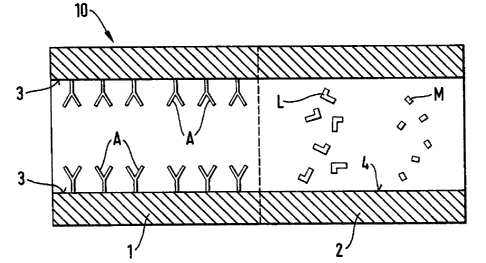
An exemplary embodiment of the device according to the invention is depicted
schematically in Fig.
1 and is generally designated with the reference number 10. It comprises a
capillary system of a
length from about 0.1 cm to about 200 cm, preferably about 1 cm to about 50 cm
and an internal
cross-sectional area from about 5 Etm2 to about 100000 EtmZ, The shape of the
internal cross-section
of the capillary system can be about circular, rectangular, trapezoidal, or
similar. The capillary
system also comprises entrance and exit openings for the introduction and the
removal of an analyze
and a carrier medium (if required). For reasons of simplification of the
schematic drawing the
respective entrance and exit openings are not depicted in Fig. I.
The capillary system comprises a first stage 1, in which bioaffinity
interaction and preconcentration
of analyte molecules are performed and a second stage 2, in which
electrophoretic separation of the
preconcentrated analyte molecules and subsequent detection of the separated
species is
accomplished. The electrodes for building up an electric field along the
longitudinal extension of the
second stage of the capillary system are not depicted in order to simplify the
drawing. However, one
of ordinary skill in the art will be aware of various embodiments for such
electrodes, such as, for
example, thin metal rings on the inside wall 4 of the capillary tube which are
connected with a thin
wire that extends through the capillary wall and ends in an electric coupling
provided on the outside
of the capillary tube. In a preferred embodiment the capillary system is
realized on a small slab of
glass or semiconducting material. In that case the electrodes and the
couplings could be integrated on
the "chip" applying well known manufacture techniques from microchip industry.
-s-
In accordance with the invention in the first capillary stage 1 molecular
recognition elements A are
attached and immobilized to the inside of the capillary wall 3. The molecular
recognition elements
can be attached to the capillary wall, for example, by adsorption or by
covalent binding. However
other physical and/or chemical techniques are equally applicable. Adsorptive
binding is generally
easier to achieve and theoretically opens possibilities for the regeneration
of the bioaffinity part of
the first stage 1 of the capillary system.
In case that the capillary system is made up of one capillary tube only, the
first capillary stage 1
should occupy only a limited length of the capillary. Thus, only a limited
length of the capillary
should be occupied by molecular recognition elements A, so that a sufficient
length of the capillary
is available for the actual electrophoredc separation of the analyte molecules
after release from the
molecular recognition elements A. By a variation of the length of the first
stage the amount of
molecular recognition elements, which are attached and immobilized to the
capillary wall, can be
easily controlled and thus the sensitivity of the device can be adapted to the
requirements. Preferably
the length of the first stage 1 amounts to about 1% to about 9s%, preferably
less than 2s% of the
total length of the capillary system. In Fig. 1 and in the subsequent Figs. 2-
6 the first and the second
stage are separated by a dotted.line.
In a preferred embodiment the capillary system comprises two capillary tubes
1,2. A first capillary
tube which is coated along its entire length on its inside wall 3 with
molecular recognition elements,
is coupled with a capillary 2 that enables optimal separation conditions.
Thus, the length of each
stage is easily controlled and can readily be adapted to the requirements. In
case that the capillary
system comprises two capillary tubes 1,2 the dotted lines in Figs. l-6 stand
for the ends of the two
capillary tubes along which they are attached with each other, and are
preferably glued together.
In Figs. 2-6 the method for combined bioaffinity assay and electrophoretic
separation is illustrated
for a single class of molecular recognition elements A and one species of
analyte molecules, which
consist of unlabeled analyte molecules M and labeled analyte molecules L. The
labeling of part of
the analyte molecules can be achieved by various methods. For example,
luminescent, ultraviolet
radiating, radioactive, or electrochemically active substances can be used.
The device depicted for
the illustration of the.method according to the invention corresponds to the
one shown in Fig. 1. It
can comprise one capillary tube only, or it can comprise two or more capillary
tubes, such as, for
example, a first tube 1 which is coated along its inside wall 3 with the
molecular recognition
elements A and a second one 2 for carrying out the separation on the principle
of capillary
electrophoresis. In the Figures the unlabeled analyte molecules M are
symbolyzed by the small
rectangles while the L-shaped symbols stand for the labeled analyte molecules
L. The molecular
-6-
recognition elements A axe bound to a limited length of the internal capillary
wall 3 at the injection
side of the capillary.
In Fig. 2 a mixture of the unlabeled and labeled analyte molecules M and L of
defined concentrations
is shown injected into the first stage 1 of the capillary system so, that
these can interact with the
molecular recognition elements A on the inside walls of the capillary. With
increasing
concentrations of the unlabeled analyte molecules M, the amount of the labeled
analyte molecules L
that is captured by the molecular recognition elements A, will decrease. After
an adequate incubation
time an equilibrium will be found between the bound and free fractions of the
labeled analyte
molecules L and of the unlabeled analyte molecules M. As long as standardized
assay conditions are
maintained, even non-equilibrium conditions can be used.
With a rinse procedure, which is indicated in Fig. 3, the unbound fractions of
the labeled analyte
molecules L and of the unlabeled analyte molecules M are removed from the
capillary tube.
In the next step, which is depicted in Fig. 4, the bound fractions of the
labeled and unlabeled analyte
molecules L and M are released by the molecular recognition elements A. By
injection of a
chaotropic agent, e.g. a salt-solution, an organic solvent or another buffer
solution, the
dissociation-rate can be increased and reassociation can be deminished.
The next step is the separation of the labeled analyte molecules L and the
unlabeled analyte
molecules M in the electrical field inside the second capillary stage 2. The
separation efficiency in
capillary electrophoresis CE is, among others, dependent on the size of the
injected sample plug.
Therefore ist is desirable to concentrate the labeled and unlabeled analyte
molecules L and M which
are spread over the length of the first capillary stage after the dissociation
step. This concentration
can be achieved by various methods, for example by Isotachophoresis or by
isoelectrical focussing.
Preferably a field amplified sample concentration is accomplished using the
electrical field within
the second stage 2. For that purpose the conductivity of the ample with the
chaotropic agent and the
analyte molecules L and M is chosen smaller than the conductivity of the
separation buffer. Then
under the condition that the analyte molecules L and M are charged, these are
concentrated due to
the higher electrical field, as is indicated in Fig. 5.
This stacking effect is essential for the efficiency of the separation in the
second stage 2 of the
capillary system, which is indicated in Fig. 6, and for a precise quantitation
of the labeled analyte
molecules L. In case the first stage comprises only one type of molecular
recognition element A
attached to its inside wall, and with one type of labeled analyte molecules L,
which can be
selectively detected, the separation efficiency does not play a major role.
However, for mufti-analyte
assays employing different molecular recognition elements and labels, it is
apparent that the
separation efficiency is very important.
For illustrative purposes only an exemplary embodiment of a device according
to the invention
which is coated along its inside capillary wall of the first stage with
antibodies is described
hereinafter:
Chemicals
A ready to use 20 mM sodium tetraborate buffer pH = 8:0 (BB8) can be obtained
from Fluka (Buchs,
Switzerland), a ready to use 69 mM sodium-potassium phosphate buffer pH = 7.0
(PB7) can be
obtained by Ciba-Geigy (Basel, Switzerland), methanol and toluol of chemical
grade and milli-Q
water should be used. Atrazine, 2-ethylamino-4-chloro-6-isopropylamino-1,3,5-
triazine, monoclonal
antibodies against atrazine and fluoresceine labeled atrazine (FA) are
obtained from internal sources
of the applicant. Bovine Serum Albumine (BSA) can be obtained from Fluka
(Buchs, Switzerland)
and may be used without further purification.
Instrumentation and capillary electrophoresis conditions
A PACE 2100 electropherograph equipped with a fluorescence detector or a UV
detector is used
(Beckman Instruments, Fullerton CA, USA). A 15 mW Argon laser (Spectra-
Physics, Mt. View CA,
USA) operating at 488 nm and a custom-build optical system, delivering 5 mW at
the end of the
optical fibcr positioned on the detection window of the capillary can be used
for excitation of the
fluoresceine labeled analyte molecules. Electrophoretic separations can be
made by applying, for
example, 20 to 30 kV over the capillary, which is kept at 30°C,
injections usually are made by
pressure.
Coadng_procedures
Coated capillaries am custom made after cleaning fused silica capillaries with
1 M KOH for 2 h,
rinsing with water for 10 min and rinsing with 0.1 M HCl for 10 min and drying
for 3 h at 200°C
during which the capillary is flushed with nitrogen. Coating with
(mercaptomethyl)
dimethylethoxysilane (MDS, Fluka, Buchs Switzerland) can be done by filling
the capillary with this
reagent and placing it for 18 h in an oven at 200°C under vacuum in
order to obtain a monolayer on
the capillary wall.
i~ ~ Cl
Coating with 3-aminopropyltrimethoxysilane (Aldrich, Steinheim, Germany) can
be done by filling
the capillary with a 2~'o solution in toluol and heating the capillary at
100°C for 3 h. After this the
capillary is rinsed with methanol for 10 min. This aminopropyl coated
capillary can be used for
covalent binding of antibodies and BSA after treatment with 0.5%
glutaraldehyde (Merck,
Darmstadt, Germany) in PB7 for 4 hours at room temperature and subsequently
the capillary is
rinsed with PB7.
Coating of capillaries with antibodies is achieved by filling the capillaries
with a mixture of the
antibody solution (10 wg protein/ml) and PB7 in case of capillary coupling or
by pressure injection
for 30 or 60 sec by means of an electropherograph. After injection the
capillaries are laid
horizontally in order to avoid siphoning of the antibodies through the
capillaries during the 3 h
incubation at room temperature. Then the capillaries are filled with a
solution of BSA in PB7 (1-2
mg/ml ) and incubated for another 3 hours at room temperature in an attempt to
reduce non-specific
binding.
For the covalent binding of antibodies, a fused silica capillary is first
modified with
3-amino-propyltrimethoxysilane function. Glutaraldehyde is used to bind the
antibodies to the
capillary surface.
Length of antibody coating
The length of the capillary coating preferably equals to the length of the
injected plug of antibody
solution and can be calculated when the column dimensions, the viscosity of
the medium and the
applied pressure are known. They also can be estimated by a determination of
the relation between
the length of the capillary and the break through time of a continuous
injection of an aqueous FA
solution. Under the assumption that the viscosity of the antibody solution
does not differ from that of
other aqueous solutions, the injected plug length can be calculated on basis
of the injection time.
Coupling_of the coated and uncoated capillaries
A 7 cm long coated capillary is coupled to an uncoated fused silica capillary
of 30 or 40 cm length,
the i.d. of both capillaries is either 50 or 75 wm. By means of a messing
holder the capillary can be
held in order to polish the capillary ends with a Beckman capillary cutter or
with polishing sheets. In
that way a zero dead-volume coupling can be obtained. By means of a microscope
the surface of the
capillary end can be checked. The easiests way to position the capillaries is
by using a metal wire
30328-20
CA 02143999 2004-08-17
-9-
with a diameter of 40 or 70 ~.m for capillaries with an i.d. of 50 or 75 p.m,
respectively, and push this
wire through the 7 cm coated capillary and another 1-2 cm in the fused silica
capillary. It is essential
that the metal wire is cut by a sharp knife in order to avoid distortion of
the end. Bending of the wire
should be avoided as well. A glass-fibre plate 10 x 15x 2 mm, normally used as
the basis for an
electronic print, with a 360 p.m deep V-shaped (90 degrees) groove, can be
used for the positioning
TM
of the capillaries. By means of Katiobond (Delo, Grafelfing, Germany), a UV-
polymerising,
non-flowing glue, the two capillary ends are glued together after positioning
of the connected
capillaries in the groove and placing a 250 N.m thick deck-glass on top. Then
the coupling device is
illuminated with an Opticure light gun (Norland, New Brunswick, NJ, USA) for 2-
4 min. In order to
allow complete polymerisation, it is advisable to wait for 1 hour before using
the capillary. After
illumination the wire can be withdrawn from the capillaries.
The stability and binding properties of antibodies to the inside wall of the
first stage of the capillary
system are virtually unaffected by the type of immobilisation and the
electrical field across the
capillary system. Immobilisation of different antibodies, either mixed in one
zone ar in separate
zones of the first stage of the capillary system provides for the development
of combined
multianalyte bioaffinity assay and capillary electrophoresis analysis. The
advantages of both
bioaffinity assay analysis and capillary electrophoresis are used. The claimed
invention overcomes
the disadvantages of the prior art approaches and provides a device and a
method which is readily
applicable and results in a high analytical sensitivity.