Note: Descriptions are shown in the official language in which they were submitted.
21~5380
-
6 BACKGROUND OF THE INVENTION
7 Field of the Invention:
8 The present invention pertains to instruments and methods for
9 use in endoscopic procedures and, more particularly, to an
endoscopic portal providing a variable size passage to an operative
11 site within an anatomical cavity to prevent undesired fluid flow
12 through the portal while allowing surgical instruments of various
13 sizes to be selectively introduced through the portal and to
14 methods therefor.
lS Description of the Prior Art:
16 Surgical procedures involving the placement of an endoscopic
17 portal, such as a sleeve or cannula, through a wall of an
18 anatomical cavity to provide a passage for insertion of instruments
19 into the cavity frequently require that the passage be sealed to
prevent undesired flow of fluids through the endoscopic portal.
21 For example, in many endoscopic medical procedures access to the
22 interior of an anatomical cavity is gained by utilizing a
23 penetrating instrument, such as a trocar, obturator or needle,
24 having a sharp penetrating point for penetrating a wall of the
'' 2l4s3sn
1 cavity to establish communication with the interior thereof. Upon
2 penetration of the cavity wall by the penetrating instrument, a
3 sleeve or cannula is left in place for utilization as a portal to
4 introduce instruments into the anatomical cavity. The penetrating
instrument is usually received within the sleeve, which passes
6 through the wall of the anatomical cavity with the penetrating
7 instrument and remains in situ a~ter withdrawal of the penetrating
8 instrument therefrom to provide a lumen establishing communication
9 with an operative site in the cavity. The sleeve typically has a
proximal end disposed externally of the anatomical cavity and
11 secured in a housing provided with a valve that allows the
12 penetrating instrument to be inserted into and removed from the
13 sleeve. Once the penetrating instrument has been removed from the
14 sleeve, various instruments can be introduced in the anatomical
cavity via the lumen of the sleeve dependent upon the operative
16 procedure to be performed.
17 It is extremely important in endoscopic procedures to prevent
18 undesired fluid flow to and from the surgical site; and,
19 accordingly, the portal must be sealed prior to and subsequent to
the introduction of instruments and while such instruments are in
21 place. In addition, fluids, such as gaseous phase carbon dioxide
22 or nitrous oxide, may be introduced in the anatomical cavity for
23 insufflation as part of the endoscopic procedure, and the escape of
24 such fluids must be prevented during penetration of the cavity as
well as during the operative procedure.
21~538~
1 The valves of endoscopic portals typically have a valve
2 passage with a size corresponding to an outer diameter or size of
3 the penetrating instrument to form a seal with the penetrating
4 instrument, the size of the penetrating instrument varying in
accordance with the endoscopic procedure being performed and the
6 type of anatomical cavity being penetrated. Furthermore, the
7 valves of endoscopic portals typically are designed to close when
8 the penetrating instrument is removed to prevent fluid flow through
9 the valves. Many prior art endoscopic portals utilize a flapper or
gate valve that is normally biased to a closed position and movable
11 to an open position to allow the penetrating instrument to be
12 inserted through the valve passage, which has a single,
13 predetermined size corresponding to the size of the penetrating
14 instrument. However, additional instruments to be introduced into
the anatomical cavity through the valve passage may be of diverse
16 types and sizes, and it will be appreciated that fluid can escape
17 past smaller size instruments.
18 Accordingly, many prior art endoscopic portals suffer from the
19 disadvantages of allowing the passage or leakage of fluids when
instruments smaller in size than the size of the single valve
21 passage are introduced therethrough or of limiting the instruments
22 to be introduced through the portal to a single size. Many
23 attempts have been made to variably seal endoscopic portals to
24 allow the introduction of various sized instruments therethrough;
however, there still exists a great need for an endoscopic portal
26 having a universal valve to prevent the escape of fluid from an
21~338~
" ~_
1 anatomical cavity by sealing variably sized instruments passing
2 through the portal without requiring placement in the portal of
3 seals of various sizes.
4 SUMMARY OF THE INVENTION
Accordingly, it is a primary object of the present invention
6 to overcome the above described disadvantages of prior art
7 endoscopic portals.
8 Another object of the present invention is to provide a valve
9 assembly for an endoscopic portal having a compressible valve
passage for sealingly contacting instruments of various sizes
11 inserted therethrough, the valve assembly being made in a fashion
12 to cause inversion of the valve passage in response to the
13 instruments being inserted along the valve passage to facilitate
14 insertion of the instruments in the endoscopic portal.
A further object of the present invention is to provide a
16 valve assembly for an endoscopic portal, the valve assembly being
17 pressurized to sealingly engage instruments of various sizes
18 inserted therethrough and having a wall forming a valve passage
19 movable along with instruments inserted through the valve a~sembly.
An additional object of the present invention is to provide a
21 valve assembly for an endoscopic portal formed as a bladder
22 containing a compressible material and defining a valve passage,
23 the bladder being movable by contact with instruments of various
24 sizes in response to movement of the instruments along the valve
passage to facilitate movement of the instruments along the valve
21~538~
1 passage without tearing of the bladder and while maintaining
2 sealing contact between the bladder and the instruments.
3 Yet a further object of the present invention is to provide a
4 method of inserting instruments through an endoscopic portal in
endoscopic procedures including the steps of inserting an
6 instrument in a valve passage of a bladder of a valve assembly of
7 the endoscopic portal and moving the walls of the bladder forming
8 the valve passage in response to movement of the instrument to seal
9 the instrument in the endoscopic portal.
Some of the advantages of the present invention over the prior
11 art are that various endoscopic procedures can be performed with a
12 single portal thusly reducing instrument cost and the time required
13 to complete endoscopic procedures, a single endoscopic portal can
14 be used with various sizes and types of instruments without
requiring manipulation of the endoscopic portal or the addition or
16 interchanging of different sized seals, insertion of instruments of
17 various sizes in the valve assembly is facilitated, tearing of the
18 valve assembly bladder is avoided while maintaining a compressive
19 or sealing force or contact between the bladder and instruments
inserted in the valve passage, the valve assembly can be of
21 simplified construction, and the endoscopic portal can be
22 inexpensively manufactured to be economically disposable for single
23 patient use.
24 The present invention is generally characterized in an
endoscopic portal for establishing communication with an anatomLcal
26 cavity through a wall of the cavity including an elongate tubular
-- 21~5380
1 sleeve having an open distal end for positioning in the cavity and
2 an open proximal end for positioning externally of the cavity with
3 the sleeve extending through the cavity wall and a valve assembly
4 disposed adjacent the sleeve proximal end for preventing undesired
passage of fluid through the sleeve and for forming a seal with
6 instruments of various sizes inserted in the sleeve via the valve
7 assembly. The valve assembly includes a bladder defining a valve
8 passage therethrough for permitting instruments of various sizes to
9 be inserted in and removed from the lumen of the sleeve via the
valve passage. The bladder contains a material which, in
11 combination with the bladder walls, causes the valve passage to be
12 normally closed or sealed and allows the valve passage to be opened
13 by instruments inserted in the valve passage. The bladder is
14 fashioned to roll or invert in response to the instruments being
moved along the valve passage to facilitate movement of the
16 instruments along the valve passage without tearing, snagging or
17 catching while maintaining sealing contact with the instruments.
18 A method of inserting instruments through an endoscopic portal
19 according to the present invention includes the steps of inserting
an instrument through a valve passage in a bladder of a valve
21 assembly of the endoscopic portal and moving the walls of the
22 bladder forming the valve passage in response to movement of the
23 instrument through the valve assembly.
24 Other objects and advantages of the present invention will
become apparent from the following description of the preferred
26 embodiment taken in conjunction with the accompanying drawings
'' 214538~
1 wherein identical reference numbers indicate identical parts or
2 parts providing identical functions.
3 BRIEF DESCRIPTION OF THE DRAWINGS
4 Fig. 1 is a side view, partly in section, of an endoscopic
portal according to the present invention.
6 Fig. 2 is an enlarged, broken sectional view of the endoscopic
7 portal of Fig. 1 showing the valve assembly therefor.
8 Fig. 3 is a broken perspective view of the valve assembly of9 Fig. 2 with an instrument extending therethrough.
Fig. 4 is an enlarged, broken sectional view of a modification
11 of an endoscopic portal according to the present invention showing
12 the valve assembly therefor.
13 Fig. 5 is a broken perspective view of the valve assembly of14 Fig. 4 with an instrument extending therethrough.
Fig. 6 is a side sectional view of the valve assembly of Fig.
16 4.
17 DESCRIPTION OF THE PREFERRED EMBODIMENT
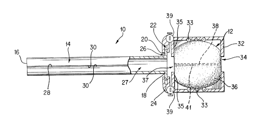
18 An endoscopic portal 10 including a valve assembly 12
19 according to the present invention is illustrated in Fig. 1.
Endoscopic portal 10 includes an elongate tubular or cylindrical
21 portal sleeve or cannula 14 for being positioned through a wall of
22 an anatomical cavity during puncture or penetration of the cavity23 wall by a penetrating instrument to provide access to an operative
24 site within the cavity. Sleeve 14 has an open distal end 16 for
being disposed within the anatomical cavity and an open proximal
26 end 18 for being disposed externally of the cavity with the sleeve
21~538~
_
1 14 extending through the cavity wall. Distal end 16 preferably
2 has a blunt configuration to prevent damage to tissue and organ
3 structure within the anatomical cavity and can be non-tapered as
4 shown in Fig. 1 or tapered, such as conically tapered. Sleeve 14
terminates proximally at a flange 20 at proximal end 18, the flange
620 being received in a recess 22 in a forward wall 24 of a housing
725 mounting the proximal end 18 of sleeve 14.
8Flange 20 and recess 22 can have various configurations
9 including annular configurations as shown in Fig. 1 with the flange
20 having an outer diameter or size smaller than the outer diameter
11 or size of recess 22 to permit diametric or lateral outward
12 expansion of sleeve 14 as will be explained further below. In
13addition to the flange 20 and recess 22 shown, sleeve 14 can be
14 permanently or removably coupled to housing 25 in many various ways
while allowing or not allowing diametric or outward expansion of
16sleeve 14. Forward wall 24 has an annular aperture 26 therein
17 allowing passage therethrough by sleeve 14, and the aperture 26 has
18 a diameter or size larger than the outer diameter or size of sleeve
19 14 to permit diametric expansion thereof.
20It will be appreciated that flange 20, recess 22 and aperture
21 26 can have various configurations with the peripheries of flange
2220 and sleeve 14 being disposed within the peripheries of recess 22
23 and aperture 26, respectively, to permit expansion of sleeve 14. It
24 will be further appreciated that the sizes of flange 20, recess 22
and aperture 26 can be varied in accordance with the amount of
26 expansion desired for sleeve 14 and that, by increasing the gaps or
214538~
.=~_
1 spaces between the periphery of flange 20 and the periphery of
2 recess 22 and between the periphery of sleeve 14 and the periphery
3 of aperture 26, greater expansion of sleeve 14 can be obtained.
4 Where it is desired to limit or control the amount of expansion for
sleeve 14, flange 20, recess 22 and aperture 26 can be sized such
6 that the peripheries of flange 20 and/or sleeve 14 contacting the
7 material of forward wall 24 serves as a positive stop or abutment
8 limiting diametric expansion of sleeve 14. It will be further
9 appreciated that where diametric or lateral expansion of sleeve 14
is not desired, no gaps or spaces are needed between flange 20 and
11 sleeve 14, respectively, and the housing forward wall 24.
12 Sleeve 14 can be made of a suitable rigid, semi-rigid,
13 flexible or bendable medical grade material such as metal or
14 plastic or a flexible, expandable or stretchable material such as
rubber permitting sleeve 14 to be normally disposed in a closed,
16 non-flexed, non-expanded or non-stretched state illustrated in Fig.
17 1 and to be diametrically or laterally expanded or moved in a
18 direction transverse to a longitudinal axis of the sleeve 14 to an
19 open, flexed, expanded or stretched state to increase the diameter
or cross-sectional size of the lumen 27 of the sleeve 14 to
21 accommodate instruments or objects therein larger than the diameter
22 or cross-sectional si~e of the lumen 27 in the closed state.
23 Depending on the material utilized for sleeve 14, a Longitudinal
24 slit 28 can be provided through the thickness of the wall of sleeve
14 to extend the entire length thereof to facilitate flexing,
26 expansion or stretching of the sleeve 14. Slit 28 defines opposing
21~5380
_ _
1 edges 30 that touch or are adjacent or substantially adjacent one
2 another in the closed state and are disposed further apart from one
3 another in the open state. It will be appreciated that sleeve 14
4 can be made of a rigid material without slit 28 where diametric
expansion of sleeve 14 is not desired.
6 Housing 25 can be made of any suitable material, such as
7 plastic, and can have various configurations including a
8 cylindrical configuration as illustrated in Fig. 1 with an enlarged
9 forward end to facilitate grasping by a surgeon. Preferably, at
least the inner surfaces of housing 25 are made of a smooth,
11 slippery material to promote movement of valve assembly 12 as
12 explained further below. Alternatively or in addition to the
13 housing inner surfaces being slippery, the walls of housing 25 can
14 have rollers 33 along the inner surfaces thereof to facilitate
movement of the valve assembly 12. Housing 25 has a rear wall 32
16 with an opening 34 therein longitudinally aligned with the lumen 27
17 of sleeve 14 to allow various instruments to be inserted through
18 portal 10 via the housing 25. Opening 34 has a diameter or
19 peripheral size larger than the diameter of the lumen 27 to
accommodate instruments or objects larger in size than the lumen
21 diameter in the closed state. Housing 25 has internal walls or
22 shoulders 35 spaced proximally from forward wall 24 for confining
23 valve assembly 12 between the shoulders 35 and the housing rear
24 wall 32. Shoulders 35 extend inwardly from the upper and lower
walls of housing 25, and the shoulders 35 can extend parallel with
26 forward wall 24 as shown in Fig. 1 or non-parallel. The distance
214538~
..., _
1 that shoulders 35 extend inwardly from the housing upper and lower
2 walls is sufficient to confine valve assembly 12 and to prevent
3 longitudinal movement thereof when instruments are inserted
4 therethrough as described further below. Preferably, shoulders 35
extend inwardly from the housing upper and lower walls to terminate
6 at an opening 37 that is larger in size than lumen 27 to allow
7 instruments or objects larger than the lumen 27 to be passed
8 through the housing 25. If desired, shoulders 35 can be angled
9 proximally or provided with flanges angled proximally therefrom to
enhance inversion or rolling movement of valve assembly 12 when
11 instruments of various sizes are inserted therein as explained
12 further below. Depending on the configuration of housing 25,
13 bladder 36 can be confined in housing 25 in many various ways such
14 as between the front wall and a rear wall or shoulder of the
housing. Valves 39 such as stopcocks communicating with the
16 interior of housing 25 can be provided for supplying fluids to the
17 anatomical cavity or for aspirating fluids from the anatom1cal
la cavity via lumen 27. The valves 39 can be mounted on housing 25 in
19 many various ways, such as between forward wall 24 and shoulders 35
to facilitate operation by the hand of the surgeon grasping hou~ing
21 25 as shown in Fig. 1.
22 Valve assembly 12 includes a bladder 36 disposed in hou81ng
23 25, the bladder 36 defining a longitudinal valve passage 38
24 therethrough aligned with lumen 27 and openings 34 and 37. Bladder
36 can be made of any suitable expandable material to form an
26 envelope for holding a material in the interior thereof and to
12
~ ~ 4 ~ ~ 8 Q
~ .,,
promote movement of bladder 36 when instruments of various
sizes are moved along valve passage 38. The bladder 36 can be
made of an expandable, medical grade membrane, such as Tecoflex
EG-85A manufactured by Thermedics, Inc., Teflon*, Goretex* or
rubber, allowing instruments or objects to pass easily
therethrough. However, the bladder does not have to be made
of a tear resistant material and can be made in its entirety
of les~ expensive materials for simplicity and cost reduction.
Bladder 36 contains a material, such as a compressible
fluid or solid material including air, water, saline, gel or
foam, for example, and has a size and configuration normally
closing, collapsing, sealing or compressing the walls of the
bladder forming valve passage 38 while allowing the valve
passage to open in response to pressure or force applied
externally to the bladder 36 by instruments inserted in the
valve passage 38. Accordingly, due to the material forming the
bladder and/or the compressible material within the bladder,
the shape, size and configuration of the bladder normally
closes or seals valve passage 38 while allowing temporary
deformation or contraction of the bladder 36 to open passage
38 in response to external pressure applied to the bladder 36.
Bladder 36 is fashioned to move or invert in response to
contact of the walls forming the valve passage with instruments
moved through the valve passage 38. Bladder 36 can be fashioned
in many ways to move with instruments inserted through the
valve passage and to roll, rotate or invert in response to
pa~sage of instruments along the valve passage 38; for example,
elastic
* Trade-mark
13
-
D ~
' i
21~5380
-
1 materials and/or a bias can be used in bladder 36 to promote such
2 movement. Bladder 36 is confined against longitudinal movement or
3 displacement by the shoulders 35 and the housing rear wall 32; and,
4 with the bladder 36 disposed in housing 25, the open proximal end
18 of sleeve 14 remains unsealed allowing fluid, such as
6 insufflation gas, to be supplied to the anatomical cavity through
7 the lumen 27. The bladder 36 can be sized and shaped to have
8 various predetermined si~es and configurations, including
9 spherical, partial spherical, heart-shaped, toroidal or donut-
shaped, disk-shaped, funnel-shaped, conical or nipple-shaped
11 configurations, the bladder 36 having a toroidal configuration in
12 ~ Fig. 3.
13 According to a method of operation for the endoscopic portal
14 10 and valve assembly 12 according to the present invention,
bladder 36 is filled by the compressible material such that the
16 bladder walls forming valve passage 38 are normally closed or
17 collapsed in a direction transverse to a longitudinal axis of the
18 valve passage 38 to form a seal along the length of the valve
19 passage 38 preventing fluid flow through the valve assembly 12 as
shown in Fig. 1, it being noted that valve passage 38 in Fig. 1 is
21 illustrated as being slightly open for the sake of clarity in
22 identifying the valve passage. Bladder 36 is confined in a
23 longitudinal direction between shoulders 35 and housing rear wall
24 32. opening 34 in housing rear wall 32 is covered by bladder 36 to
form a seal at the housing rear opening 34. Proximal end 18 of
26 sleeve 14 remains uncovered by the bladder 36 to allow fluid to be
14
supplied to or withdrawn from an anatomical cavity via valves
39. An instrument I, such as a penetrating instrument
including a trocar, obturator or needle having a sharp tip for
penetrating a wall of an anatomical cavity, is inserted through
the valve passage 38 to be received within sleeve 14 as
described in applicant'~ U.S. Patent No. 5,395,342, which
issued March 7, 1995. As shown in Fig. 2 and in Fig. 3,
wherein hou~ing 25 is not shown, insertion of the instrument
I in valve passage 38 applies external pressure to bladder 36
temporarily deforming the bladder 36 to open valve passage 38
to receive the instrument I. The instrument I is moved
forwardly or distally along the valve pas~age 38 and into
sleeve 14 while being sealingly contacted by bladder 36 with
a compressive sealing force along valve passage 38, the
longitudinal direction of insertion or movement of instrument
I being indicated by arrows in Figs. 2 and 3. Movement of
instrument I along the valve passage 38 in the direction of
insertion causes movement of bladder 36 with the instrument
cau~ing continuous rolling, rotational or inverting movement
of bladder 36 as indicated by the arrows in Fig~. 2 and 3 due
to the sealing grip of bladder 36 with the instrument I.
Movement of the in~trument I in the direction of insertion
causes the passage-defining portion or walls 41 of the bladder
36 to be moved di~tally or forwardly cauQing the valve pas~age
38 to invert at a forward or di8tal end thereof and causing the
bladder 36 to roll or rotate to accommodate mov -nt of the
instrument through the valve assembly. Movement of
Bl'~
... ..
21~5380
1 walls 41 in the direction of insertion causes the walls 43 of
2 bladder 36 opposite the .walls 41 to move in a direction opposite
3 the direction of movement of walls 41 as shown by the arrows in
4 Fig. 2. Movement of bladder 36 with instrument I facilitates
insertion and passage of instrument I through valve assembly 12
6 without tearing, snagging or catching of the bladder 36 while
7 allowing the bladder to maintain a compressive seal with the
8 instrument I. With the instrument I extending through valve
9 passage 38, bladder 36 conforms to the size and configuration of
the instrument I along the valve passage 38 to be in sealing
11 relation or contact with the instrument I and form a seal along the
12 length of the passage 38 preventing the flow of fluid through valve
13 assembly 12.
14 Where the instrument I is a penetrating instrument as shown,
the instrument I can be utilized to penetrate a wall of an
16 anatomical cavity with the sharp tip protruding beyond the sleeve
17 distal end 16 such that the sleeve 14 passes through the cavity
18 wall during penetration to position distal end 16 within the
19 anatomical cavity while proximal end 18 remains externally of the
cavity~ During penetration and while the penetrating instrument I
21 is in place, fluid flow to and from the cavity through valve
22 assembly 12 is prevented due to the seal formed by bladder 36 with
23 the penetrating instrument I. Where valves 39 are provided, fluid
24 can be supplied to the anatomical cavity, and such fluid cannot
escape through valve assembly 12. Once distal end 16 of sleeve 14
26 is within the anatomical cavity, the penetrating instrument I can
16
214~38~
1 be withdrawn from the endoscopic portal 10 leaving the endoscopic
2 portal 10 in place. It. will be appreciated that withdrawal of
3 instrument I through the valve assembly 12 in a direction opposite
4 the direction of insertion will cause bladder 36 to rotate and a
rearward end of valve passage 38 to invert in a direction opposite
6 the direction of withdrawal of instrument I. Upon withdrawal of
7 the penetrating instrument I, bladder 36 returns to its initial
8 configuration or state to cause valve passage 38 to automatically
9 close and thusly seal endoscopic portal 10. Instruments of various
sizes can be inserted in the anatomical cavity through the lumen 27
11 of the endoscopic portal 10 with bladder 36 deforming or
12 contracting in response to external pressure applied by the
13 instruments to open valve passage 38 to a size and shape to receive
14 the instruments with bladder 36 forming a seal therewith.
It will be appreciated that various sizes of instruments can
16 be introduced at an anatomical cavity via the endoscopic portal 10
17 in that the single valve passage 38 will open to a size just large
18 enough to receive the instruments with bladder 36 forming a seal
19 therewith. Instruments larger in size than the diameter of lumen
27 can be introduced into the anatomical cavity and tissue and
21 other objects can be removed from the anatomical cavity in that
22 sleeve 14 can be expanded diametrically or laterally outwardly by
23 the instruments or objects from the closed state wherein edges 30
24 touch or are separated from one another by a minimal gap to the
open state wherein the edges 30 are separated from one another or
_ 2145380
1 the gap is increased to expand lumen 27 to a size large enough to
2 receive the instruments or objects.
3 The bladder is shown confined within the housing; however, the
4 bladder can be extended distally to elongate the valve passage and
create a longer seal and more support for instruments passing
6 therethrough. To this end, the bladder can have a nipple-like
7 portion extending partially or entirely through the portal sleeve.
8 By designing the bladder such that the walls move along with the
9 instrument, tearing or other damage to the valve assembly is
minimized while permitting the use of elastic or other conformable
11 flexible materials to provide a valve capable of sealing engagement
12 with instruments of varying diameters since there is minimal
13 resistance to passage of the instruments.
14 A modification of an endoscopic portal according to the
present invention is illustrated at 110 in Figs. 4 - 6. Endoscopic
16 portal 110 is similar to endoscopic portal 10 except that the valve
17 assembly 112 for endoscopic portal 110 extends into portal sleeve
18 114. Valve assembly 112 includes bladder 136 disposed in housing
19 125, the bladder 136 defining a longitudinal valve passage 138
therethrough. Bladder 136 includes an elongate, distal extension
21 145 extending through opening 137 and aperture 126 to extend into
22 portal sleeve 114. Extension 145 can be nipple-like in
23 configuration and can extend partially or entirely through the
24 portal sleeve 114 to form a seal with instruments introduced
through the portal sleeve. Accordingly, valve passage 138 and
26 walls 141 defining valve passage 138 extend into the portal sleeve
18
214538Q
........ .
1 114 to form a seal with instruments or objects along all or part of
2 the length of the portal.sleeve 114.
3 Operation of endoscopic portal 110 is similar to that
4 described for endoscopic portal 10 in that walls 141 defining valve
passage 138 are normally closed to form a seal along the length of
6 the valve assembly 112 as shown in Fig. 6 and are opened by an
7 instrument I inserted through the valve passage 138 as shown in
8 Fig. 4. The instrument I is moved forwardly along the valve
9 passage 138 and into sleeve 114 while being sealingly contacted by
bladder 136 along valve passage 138. Movement of instrument I in
11 the direction of insertion shown by the arrow in Fig. 4 causes
12 movement of bladder 136 including extension 145 with the
13 instrument. Movement of bladder 136 with instrument I causes the
14 walls 141 to be moved in the direction of insertion and the walls
143 opposite the walls 141 to be moved in a direction opposite the
16 direction of movement of walls 141 as shown by the arrows in Figs.
17 5 and 6. With the valve assembly 112, a seal is obtained with
18 instrument I along the length of the portal sleeve 114 for enhanced
19 sealing and support of instrument I.
With the valve assemblies of the present invention, a single
21 valve passage for receiving instruments and objects can be opened
22 to various sizes corresponding to the sizes of instruments and
23 objects passing therethrough in sealing relation with the valve
24 assemblies. By providing the valve passage to be normally closed
and to be compressed around instruments passing therethrough, fluid
26 flow through the valve assemblies is prevented prior to insertion
19
' 21~5~8~
1 of, during insertion of and upon removal of the instruments.
2 Continuous rolling, rotation or inverting movement of the valve
3 assembly bladder in response to movement of instruments along the
4 valve passage permits enhanced gripping of the instruments by the
valve assembly while avoiding tearing, catching or snagging for
6 smooth insertion. Movement of the valve assembly bladder with the
7 inserted instruments allows the use of strong compressive or
8 sealing forces without damage to the bladder by the instruments
9 during insertion and withdrawal. The passage of the valve
assemblies can be caused to conform to the size and shape of
11 instruments passing therethrough such that more than one instrument
12 can be passed simultaneously through the valve assemblies as well
13 as irregularly shaped instruments and objects. By providing an
14 expandable endoscopic portal, the present invention permits
instruments and objects larger than the diameter of the lumen of
16 the portal sleeve to be inserted in and removed from an anatomical
17 cavity.
18 Inasmuch as the present invention is subject to many
19 variations, modifications and changes in detail, it is intended
that all subject matter discussed above or shown ln the
21 accompanying drawings be interpreted as illustrative only and not
22 be taken in a limiting sense.