Note: Descriptions are shown in the official language in which they were submitted.
WO 94/08655 ~ 1 4 6 0 1 ~ PCI/US93/09798
.. . ..
,, .
ULTR~SONIC TRANSDERMAL DRUG DELIVERY SYSTEM
Technical Area
This invention relates to drug delivery systems and, more particularly, to
tr~n.sd~rm~l drug delivery systems.
Backglound ofthe Invention
A vast majority of pharm~ceuticAl agents (e.g., drugs) in clinical use today aregiven either orally or by injection. While injection provides a fast and direct route to
the blood stream, injection often causes pain and anxiety and, occasionally,
co~ AI;on. Further, injection does not provide for a conslanl or s..~t~ined
10 delivery of drugs. Finally, when a drug is injected by a syringe, the entire dose is
placed in the body and cannot be withdrawn should an adverse reaction occur.
Oral ~.I...;I);~l.~ion ~ub;e t~ the pharm~ceutiç~l agent to hepatic metabolism.
~pAtic metabolism :~Ub~l~..l;Ally degrades the effectiveness of ph~rm~ce~lticAl agents,
up to 90 percent in some cases. More specifically, the first organ that receives an
15 ;"le~l;e absorbed drug taken orally is the liver. The liver detoxifies molecules that
are foreign to the body. Most drug molecules are considered by the liver to be
foreign. As a result, a .~i~nifi~nt quantity of a particular metlicine may never reach
the rest of the body due to the liver's deto~i~illg the drug's molecules. The degree of
detoxification varies from person to person and may account for adverse reactions in
20 some people and not others by infl~1~ncing the amount of a drug that is left for
absorption by the rem~in-l~r of the body. More importantly, the decrease in
effectiveness due to hepatic metabolism by the liver leads to increases in the amount
of the agent being ~dmini~tered, which leads to undesirable side effects and gastric
intolerance. That is, the amount taken by mouth is usually more than the body needs,
Wo 94/086~ PCr/US93/09798
6 ~ 2-
often res..lting in adverse side effects. Further, because dosage requilcl"e,lLs often
vary from individual to individual, it is difficult to tailor individual dosages using the
pred~finecl amounts dt;le,l"ined by m~mlf~ctllrers of orally a.l..,;..;~ .cd drugs.
Finally, as with a syringe injection, when a drug is taken by mouth and absorbed, the
entire dose is in the body. If an adverse reaction takes place it is difficult to remove
the drug to stop the reaction. Nevertheless, oral ~lminictration preselllly is the most
plerellcd way of giving pharm~ce ~tic~l agents due to the ease of ~rlminictration and
avoidance of the need for invasive vascular access, as required by injections.
The adult skin structure can be broken into three layers. The stratum
corneum, which is actually part of the epidermal layer, is the first layer of skin defense
against the exterior en~hol""~ L. The stratum COIllculll iS capable of absorbingsuperficial trauma while still . . .~inl s ;- ;- g adequate protection against loss of water and
ingress of micro or~ni.cm.c and other substances. The stratum corneum layer is 15-20
cells thick. In many areas of the human body, the stratum corneum layer is very thin,
often below several microns. The intercellular space of the stratum corneum is
appl,Jx;.~.A~ely 30percent by volume. The intercellular space is filled by lipidcomposition, which is ideally suited to form a transport barrier. The inner layer of the
stratum corneum is in contact with granular cells (very moist) and the outer layer is in
contact with a dry en~ on",e,ll. Thus a substantial water content gradient exists
across the stratum corneum.
The second layer is the epidermal layer, which consists of epidermal cells
bound together by tight junctions into a viscoelastic matrix. Between the junctions lie
heavily convoluted lipid-filled extracelllli~r spaces Co~ g a host of cellular
Iymphocytic factors, el~y~es and other anti-microbial agents. The epidermal layer is
the body's prime protective barrier. Its basal cells provide metabolic and additional
water barrier functions. The epidermal barrier provides a formidable defense structure
even in the absence ofthe stratum collleulll, especially to water-soluble agents that do
not possess a lipid extMcell~ r phase. Enzyme activity may be controlled or rendered
inactive by employing chemical, enzymic or heat lle~
The innermost layer is the dermal layer. The dermal layer consists of basal
germ cells positioned upon a basal ",~",b,~l1e with known permeability of
applox;...~lely 40 kilodaltons and below. Unless specific excitation factors arepresent, large molecular weight materials cannot cross the basal r"en~l~ne.
Below the basal membrane are the majority of the capillary loops that
35 collll lise the terminal states of the microcirculation tree (i.e., the blood vessels) of the
human O~g~ lllc. The capillary loops are the target of current passive transdermal
WO 94/0865~ Pcr/US93/09798
~3~ ~1~6~10
drug delivery systems (desc,il)ed below). Because a very large number of capillary
loops are present, large surface areas are available for the systemic ~xcllAI~ge of fluids.
Pentl~Lillg all three skin layers are l~ulllcrws hair follicles in various growth
states--telogen, anagen and c~t~g~n The hair follicle growth stage co,~elales with the
5 depth of the follicle, late-anagen follicles being the deepest and closest to the most
heavily developed capillary blood supply. The centerline of the hair follicle ispositioned less than five microns from the encircling capillary blood supply. The
stratum CGIIlculll follows the invagination ofthe follicles at the skin level, tel..,;..~l;..g
app,u~il"a~ely half-way down the follicles. The sensory nerve nclwolk that surrounds
the follicles responds to any physical P"~cit~tiQn on the hair shaft. Thus, a highly
sensitive responsive system is present in the hair follicle regions of the skin. Follicle
density on skin surfaces varies depending upon location from 100/cm2 to 900/cm2.Other than by syringe, there are two methods by which drugs can be delivered
through the skin--passive and active diffusion. Passive diffusion involves placing a
concellLlaLion of drug in a reservoir on the surface of the skin and allowing the drug
to passively diffuse through the skin into the body. Since there are natural barriers in
the skin which keep almost all molecules from entering the body through the skin,
only a few molecules from the reservoir of the drug pass through the skin and are
absoll,ed first by the blood stream and then by the body.
Due to natural skin barriers, few pharm~ceutic~lc have been succes~fillly
diffi.~ed through the skin and into the subdermal microcirculation regions of the
human body, i.e., the underlying blood vessels. The most succçc~fi.l drugs to berliffi~sed through the skin are clonidine, nitroglyce~hl, scopolamine, and estradiol.
Because these drugs are effective at very low plasma concentrations, they can beapplied using small passive skin patches. A 10 ng/ml plasma concentration has been
allJiLlalily adopted by the industry as a mean figure above which passive transdermal
drug delivery is not effective. This concentration level çl;...;..~es the possibility of
passive transdermal delivery of such highly succç~fi.l agents as aspirin, which
~e.~uhes a concentration of 150,000 ng/ml to be effective. Currently, ~cet~minophen,
cimeti~ine~ and indometh~r.in cannot be delivered by passive transdermal drug delivery
systems.
In addition to concentration level, molecular size is an issue with the passive
0 diffusion of drug absorption. The skin's natural barriers limit or prevent absorption of
metlis~mPnt~ that are composed of large molecules. Thelt;ro,e, with passive diffusion,
if a me~ ine is to be effective in the body, it must work well at very low dosages and
be of a molecular size that the skin will allow to enter the body. While çl-Pmic~l
~ 2 1 ~
en'nancers ha~e been investi_ated as solutions to allow for ~reater dosage absorption
throu~h the skin by passive diffusion, none have ~-orked w-ell enough to pass the
Federal Dru~ Administration (FDA) requirements and/or be successful cornrnercially.
A potentially more viable way for drugs to transcend the skin's barriers is to
5 use an active ener~ source that "pushes" or "pulls" drug molecules through the s~cin
and, at the same time controls, the rate of delivery. An energy driven system will
allow a greater quantity of the medicine to be delivered in a shorter or variable time
frame. Potentially an energy driven system will perrnit larger molecular weight drugs
to transcend the barriers of the skin in a short time period.
Two types of active transdermal drug delivery have been proposed. The first,
which is called iontophoresis, is a system that uses a direct current of electricity to
charge drugs Electrically charged drugs are driven into the skin. To date, there is
only one medicine, Lidocaine, used in such a device. Lidocaine is a drug used for
local anesthesia. Extensive investigation is presently being conducted by the
pharmaceutical industry on the use of iontophoresis for drug delivery. While this
method of delivery is slow, it probably will increase the number of medic~ments used
for transdermal drug delivery. Furthermore, delivery is better controlled, when
compared to passive diffusion.
The other method of active drug delivery uses ultrasound as the energy
source. For a variety of reasons, the results of drug delivery by this method have
traditionally been inconsistent. Results of previous experiments have been difficult to
repeat. More specifically, it has been known for several decades that ultrasoundradiation pressure applied to drug molecules in contact with skin can increase
transdermal penetration rate. The mech~nism of action has been unclear with someresearchers citing boundary stirring effect, some citing microchannel production via
cavitation and others citing direct radiation pressure onto the dru~, pumping it into
the skin.
Some researchers have conducted studies of the interaction of ultrasound and
specific drug formulations. Some researchers have applied an ultrasonic field to drug
molecules themselves, rather than to the skin and associated structures. Other
researchers have shown that ultrasound is effective. in shearin_ polymeric
compositions of drugs contained in transdermal patches. The intent of these
researchers was to modulate the release rate of a drug from a polymeric matrix.
Finally, some researchers have applied ultrasound to the skin itsel The following
U.S. patents describe some of the results of the foregoing research: 4,657,543;
4,767,402; 4,780,212; 4,787,888; 4,821,740; 4,948,587; 4,953,565; and 5,007,438.
~ 214601~
Also see Patent Cooperation Treaty (PCT) application ~o. 91/1277~ and German
Patent ~i-o. 27 56 460. ~rost, if not all, of the foregoing patents show a lack or no
control of application direction, little or no control of frequenc- and power le~els, no
control of duty cycle and ignorance of a host of other controlling factors.
Various criteria for drug delivery enhancer design have been established. They
are: (i) the enhancer should elicit no pharmacological response; (ii) the enhancer
should be specific in its action; (iii) the enhancer should act immediately with a
predictable duration and its action should be reversible; (iv) the enhancer should be
chemically and physically stable, and be compatible with all of the components of the
drug formulation; (v) the enhancer should be odorless, colorless, and tasteless; and
(vi) the enhancer should be nontoxic, nonallergenic, and a nonirritant. These criteria
can be conveniently applied with slight modification to all transdermal drug delivery
enh~ncPment approaches, both chemical and nonchernical. No single drug delivery
enh~ncement approach available today meets all of the foregoing criteria. Or anic
enhancers produce a characteristic foul taste in the mouth shortly after skin
application. Several alcohol or solvent-based enhancers cause severe skin irritation
and can lead to an e~7~m~tous reaction. Device-based enhancers such as
iontophoretic titrators come closer to satisfying all of the criteria, but fall short in
broad spectrum general applicability, specificity of action, reversibility of action and
nonirritabilit,v.
As will be better understood from the following discussion, the present
invention is directed to providing an active transdermal drug delivery system that
enhances the diffusion of large molecular wei~ht substances (e.g., large molecular
weight drugs) between an external device-based reservoir and the subdermal
microcirculation tree of an organism, such as the hurnan body. This result is achieved
by using ultrasonic energy to excite the skin system of the organism in a way that
allows multifrequency, multidirectional subsurface waves to diffuse large molecular
weight substances through the skin in an efficient and controllable manner.
Summary of the Invention
In accordance with this invention, an ultrasonic transdermal drug delivery
system is provided. ~lore specifically, a transdermal drug delivery system formed in
accordance with this invention in~ es ultrasonic transducers that create ultrasonic
waves. The ultrasonic waves release a stored pharrn~ce~ltical a ent (e.g., a drug) and
forcibly move the agent through the skin of an organism, such as the human body,into the blood vessels underlying the tr~n~ucers. The transdermal drug delivery
system incl~ldes a housing having a reservoir for storing the drug to be released. The
A~tEN~D S~IET
WO 94/0865~ PCr/US93/09798
-6- 21~6Dll~
reservoir is separated from the skin by an ultrasonically controllable polymeric."G-"I"~ne. ~It~ l;vc;ly, the ultrasonically controllable polyrneric me",b,~1e can
store the drug to be released. An adhesive ~tt~f.hçs the delivery system to the skin.
The cavity is defined by an assembly of ultrasonic tran.qd~lcPrs. The ultrasonictr~nqdllcPr assembly incllldes a stimuli tr~nqducPr for creating an ultrasonic stimuli
wave in the skin of an o,~;alfi~,n and at least one pumping (drug delivery) tr~nqd~lcer
for moving the drug through the polymeric ~c~b~ne and the skin into the blood
vessels of the or~ani~",. Control electronics, pi~re,~bly stored in the housing, control
the operation ofthe stimuli tr~n.qd-lcer and the at least one pumping tr~nqducP~r.
In accordance with other aspects of this invention, the tr~nqd~lc~r assembly hasthe shape of a trl.nc~ted cone.
In acco~-lal-ce with further aspects of this invention, the stimuli tr~n.qdllcçr is
located in the top of the cone and the at least one pumping tr~nqd~lcer is located in the
wall of the cone.
In acco~dal-ce with additional aspects of this invention, the top of the cone isdefined by the stimuli tr~nqd~lcçr~ which has a flat, circular shape, and the walls of the
cone are defined by a plurality of tr~n.qdllcçr se~ ; each of which forms a pumping
tr~nqd~lcer. Preferably, the resol1a,l~ frequency of the flat, circular tranqdllcer is less
than the reson~nt frequency of the tr~nqdllcP,r segmP.ntq
In acco,d~-ce with yet other aspects of this invention, located between the
tr~nqdllcPrs and the reservoir is a drug-impermeable l~min~te that also functions as a
focusing lens for the tr~n~d~lcers.
In accoidance with still other aspects of this invention, the control electronics
apply ultrasonic stimuli pulses to the skin by energizing the stimuli tr~nqducçr at a first
frequency, preferably Iying in the 5 KHz-1MHz range for a predetelll~ined period of
time (10-20 seconds). Between the stimuli pulse periods, the control electronicsapply variable frequency ultrasonic pumping pulses to the skin by ene,~ i,-g thel,ulllph~g tr~n.qdllcPr se~,nel"s. Preferably, the frequency of the variable frequency
ultrasonic ~,u"",i"g pulses lie in the 50MHz-300MHz range.
In accordance with yet further aspects of this invention, a skin temperature
sensor is positioned in the housing to sense the te"~pe,~ re of the skin leceiving the
drug. The temperature h~"a~ion is used by the control electronics to prevent theultrasonic waves from overheating the skin.
In acco~-lance with still further aspects of this invention, the drug delivery
system also inchldP,s one or more additional stimuli tr~n.qdllcers, such as infrared (~)
or laser emitters, in the housing. The additional stimuli tr~nqdllcP.rs emit stimuli pulses
WO 94/0865~ 2 1 4 6 0 1 o Pcr/US93/09798
at selccled intervals during the variable frequency ultrasonic pumping portion of the
operational cycle. The additional stimuli pulses enh~nce the operation of the drug
delivery system by heating the skin and/or creating additional ultrasonic waves in the
shn.
In acco~dance with yet still further aspects of this invention, the variable
frequency, ultrasonic ~JU~ J;llg pulses are applied to opposed tr~ncd~lc~r seg...~
In accordance with yet still other aspects of this invention, the variable
frequency, ultrasonic ~ulllp;l~ pulses are applied to alternate pairs of tr~ncd~lc~r
seg...~ in a rotational manner.
In acco-dance with yet still additional aspects of this invention, the drug
delivery system incl~ldes a sensor for sensing drug delivery effectiveness and using the
res~llt~nt i,~""alion to control the rate of drug delivery.
In acco~dance with still yet other aspects of this invention, the sensor, which
may function as a stand-alone device, in~ ldes a cavity, an ultrasonic tr~ncd~lcçr, a
focusing lens, and a substance sensing tr~ncdllcer located in the cavity, plus energizing
electronics for the ultrasonic tr~ncdllcer and a test data processor for evalu~ting the
output of the substance sensing tr~ncdllcer. The cavity is juxtaposed against the skin
and the ultrasonic tr~ncdllcer and focusing lens are sized evenly and ene-gi~ed to
cause body fluid to be drawn into the cavity.
In ~.. ~ly, the invention provides a new and improved transdermal drug
delivery system. The transdermal drug delivery system is an active system that uses
ultrasonic waves to çnh~nce drug delivery. Relatively low frequency ultrasonic pulses
excite or stimlll~te the nervous system of an Ol~;~S"I, such as the human body,
similarly to the way the nervous system is excited by skin trauma, such as heat or a
25 blow to the shn. As is well understood by those shlled in the medical arts, skin
trauma stimlll~tion causes both the dermal-epidermal junction (i.e., the basal)
",e",b,~ne and the capillary endothelial joints to open so that fluids can be moved to
the area of the trauma. The invention takes advantage of these opellings to pumpdrugs from a reservoir through the shn into the capillary loops which form the ends
30 of the microcirculation tree of the organism. Pumping is accomplished by applying
variable frequency ultrasonic ~uu~ Jing pulses to the skin by ene-~i,ing the pumping
tr~n~ducer se~ e b~lweell stim~ tion pulses. Because the tr~n~d~lcer seg~ s
that receive the variable frequency ultrasonic ~wll~;llg pulses form the walls of a
trllnc~ted cone, the ultrasonic waves produced by the tr~n.~dllcers impinge on the
35 underlying skin at an oblique angle. The ultrasonic waves create a pumping action
that first creates large openings on the surface of the skin for the initial receipt of
wo 94/086s2i ~6 ~ ~ PCI/US93/09798
-8-
drugs in a first layer of skin cells. The received drugs are pumped through the skin as
the wave alternately moves the skin cells away from and then toward one another in
an inward direction. The drug is also forced through the aperture surrounding the
hair follicles and through the sweat glands of the 01,~3alU~ln.
In ecs~nc~, ultrasound is used by this invention to open ~.h~nn~ in the skin
surface and then literally pushes a merliç~m~nt which has been dissolved in a fluid
through the r.h~nn~ls between the cells in the second layer of skin, the epidermis. The
ultrasound also opens the cells in the deepest layer of the skin, the dermis. The
dermis is a layer of cells one cell thick which controls the immllnology of the skin and
produces cells which migrate to the top surface of the skin to renew the stratumcorneum. Ordinarily, this layer is closed to permeation except for certain
stiml~l~tions~ such as trauma, local infection or ~hçmic~l irritation, for example
through an insect bite. The ultrasound opens the basal layer of the dermis. In
addition to the skin, the ultrasound pumps medicants through the çh~nn~l~
sullo~ ding hair follicles and sweat gland pores. Large quantities of many drugs(in~l~lrling those with large molecules) can be ~lmini~tlo.red through the skin using the
present invention.
Clusters or loops of blood vessel capillaries located directly beneath the skin
basal layer and surround the hair follicles and sweat gland por,es receive the
20 a~h;~ eled medicant. Once the merlic~nt enters the capillaries, it is absorbed into
the systemic or blood circulation of the body and delivered to where it is needed.
In some embodiments of the invention, additional stimlllation is provided by
IR or laser emiKers during the pumping portion of the cycle. Excess heating of the
skin is prevented by terrnin~ting stim~ tion of skin when heating eYceed~ a
predetermined level. Further, feedb~c~ control of drug delivery is provided in some
embodiments of the invention. A novel sensor determines the m~gnitl~de of a
substance conlained in the plasma and inlel~LiLial fluid of the body that relates to the
effort of the drug being delivered. The sensor inc.l~ldes an ultrasonic tran~dllcPr and a
focusing lens constructed and oriented in a way that withdraws fluid into a cavity that
houses a substance sensing tran~d~lcer. The sensor can be used as a stand-alone
device separate and apart from a drug delivery system.
Brief Description of the Drawings
The foregoing aspects and many of the ~ttend~nt advantages of this invention
will become more readily appleciaLed as the same becoll-es better understood by
rere.c;~ce to the following detailed description, when taken in conjunction with the
acco.llpallyillg drawings, wherein:
W O 94/0865~ PC~r/US93/09798
~ 9 21~6~10
FIGU R E 1 is a three-dimensional view of a section of the skin of the human
body;
FIGU RE 2 is a seqllçnce of diagrams illustrating the growth cycle of a hair
follicle;
FIG U RE 3 is a series of diagrams illustrating the typical angle of insertion of
hair follicles in di~renl regions of the human body;
FIGllR E 4 is a flow diagram illustrating the various paths a drug can take
through the skin into the cells of the human body;
FIGllRE 5 is a block diagram of an ultrasonic transdermal drug delivery
10 system formed in accordance with the invention;
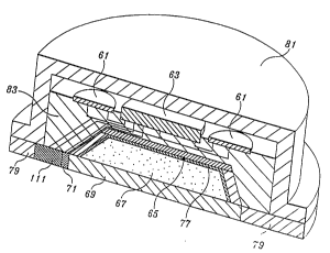
FIGIJRE 6is a cross-sectional, pictorial view ofthe structural ~l~ngelllenl of
transdermal drug delivery system illustrated in FIG U RE 3;
FIG U RE 7 is a pictorial view of the ultrasonic trancd~lcer assembly portion ofthe transdermal drug delivery system illustrated in FIG U RE 6;
FIG U RE 8 is a plan view of the tr~n.cduc~r 5eg~"~1 portion of the ultrasonic
tr~n.cd~lc~r assembly illustrated in FIG URE 5;
FIG U RE,9 is a block diagram of the electronic control portion of the
ultrasonic transdermal drug delivery system illustrated in FIGU RES 5 and 6;
FIG U RE 10 is a timing diagram illustrating the drug delivery cycle of the
ultrasonic transdermal drug delivery system illustrated in FIGU RES 5 and 6;
FIGU RE 11 is a cross-sectional view of the ultrasonic tr~nc~ cer assembly
illustrated in FIG U RE 7 and the drug reservoir enclosed by the assembly;
FIG U RE 12is a plan view showing the Illovelllenl of waves across the surface
of the skin produced by the ultrasonic tr~ncd-lcer assembly illustrated in FIG U RE 11;
FIG U RE 13 is a pictorial cell diagram illustrating the effect of applying an
ultrasonic tr~nedllcpr signal perpendicularly to the skin;
FIG U RE 14 is a pictorial cell diagram illustrating the effect of applying an
ultrasonic tr~ncduc~r signal to the skin at an oblique angle;
FIG U RE 15 is a cross-sectional, pictorial view of the structural arr~ng~mPnt
of an alternative embodiment of an ultrasonic transdermal drug delivery system
formed in accordallce with the invention;
FIG U RE 16 is a block diagram of the electronic control portion of the
ultrasonic transdermal drug delivery system illustrated in FIG U RE 15;
FIG U R E 17 is a timing diagram illustrating the drug delivery cycle of the
ultrasonic transdermal drug delivery system illustrated in FIG U R E 15;
W O 94/086~5 PC~r/US93/09798
214fi~l. -10- --
FIGU RE 18 is a block diagram of another alternative embodiment of an
ultrasonic transdermal drug delivery system formed in accordance with the invention;
FIG U RE 19 is a cross-sectional, pictorial view of the structural a,
ofthe embodiment ofthe invention illustrated in FIGllRE 18;
SFIG U RE 20 is a block diagram of the electronic control portion of the
ultrasonic transdermal drug delivery system illustrated in FIGU RES 18 and 19;
FIG U RE 21 is a timing diagram illusLl~ling the drug delivery cycle of the
embodiment ofthe invention illustrated in FIG U RES 18 and 19;
FIG U RE 22 is a partially pictorial, partially block diagram of a substance
10concentration sensor formed in accoldance with this invention;
FIG U RE 23 is a partially pictorial, partially sçhem~tic diagram of a substancesensing tr~n~d~lc~r suitable for use in the substance conce"L,~Lion sensor illustrated in
FIGIJRUE 23.
FIG U RE 24 is a pictorial diagram of an alternative substance sensing
15tran~dllcer suitable for use in the substance concentration sensor illustrated in
FIGIJRUE 22.
FIG U RE 25 is a cross-sectional, pictorial view of the structural arr~ng~m~nt
of a further alternative embodiment of an ultrasonic tr~n~d--c~r drug delivery system
formed in accordance with the invention; .-
20FIG U RE 26 is a block diagram of the electronic control portion of the
ultrasonic transdermal drug delivery system illustrated in FIG~ R E 25;
FIG U RE 27 is a timing diagram illustrating the drug delivery cycle of the
embodiment of the invention illustrated in FIGVRE 25;
FIGU R E 28 is a pictorial, exploded view illustrating a replaceable drug
25canister portion of the embo~im~nts of the invention shown separated from the
housing;
FIG U RE 29 is a master flow diagram illusLl~ling the operation of th
microprocessor of the embodiment of the invention illustrated in FIG U R E 26;
FIG U R E 30 is a flow diagram of the initi~li7~tit)n and det~rmine drug
30concentration portions of the master flow diagram illustrated in FIGURE 29.
FIG U RE 31 is a flow diagram of the determine initial skin temperature and
apply stimuli pulse portions of the master flow diagram illustrated in FIGU RE 30; and
FIG U RE 32 is a flow diagram of the delivery drug portion of the master flow
diagram illustrated in FIG U RE 29.
WO 94/0865~ PCr/US93/09798
-11- 2146~1~
Detailed Description of the Plerelled Embo~ e
As will be better understood from the following description, the present
invention provides an act*e tr~nederm~l drug delivery system that uses ultrasonic
energy to both excite the skin in a manner that "fools" the nervous system of anS organism and pump a pharm~ce ~tical agent (e.g., a drug) from a reservoir through the
skin to the capillary loops just below the skin surface. Prior to describing theplesel,lly p~ lled embo-lim~nte of the invention, in order for the invention to be
more easily understood, a brief description of the skin is set forth.
As shown in FIGURE 1, the skin incl~ldes two primary layers--the
epidermis31 and the dermis33. Located beneath the dermis33 is sub.;ulalleous
tissue 35 followed by deep fascia 37 and, then, muscle 39. F.xtçntling through the
epidermis31 and the dermis33 are hair follicles41. As shown in FIGURE2,
depending on their age, hair follicles te".linate in a dermis layer 33 or in the epidermis
or subcutaneous tissue near the dermis layer. As shown in FIGURE 3, the angle of15 hair follicles varies b~lween 30 and 60 depending upon the location of a follicle.
Sweat pores43 (FIGURE 1) extend through the dermis and epidermis
layers 31 and 33, te, ...;~ g at sweat glands 45 located in the subc~lt~neolle
tissue35. F.~t~n~ling through the muscle39, the fascia37 and the subc~lt~neous
tissue 35 are b,~nches 47a, 47b, 47c . . . and 49a, 49b, and 49c . . . of the venous and
20 arterial ~y~lc~ s of the o-gan;sThe veins and artery b.~nches terminate at capillary
loops 51, which comprise the terminal stages of the microcirculation tree of theorganism. Capillary loops are located at the dermal-epidermal interface, the bulbous
regions of the hair follicles, ~dj~c~nt the sweat glands 45, and in a variety of other
areas.
The present invention is directed to providing an active transdermal drug
delivery system that uses ultrasonic energy to ~nh~nce both the movement of drugs
and the type of drugs deliverable from a drug rese,~/oi, positioned on the surface of
the skin to the capillary loops 51 as well as directly to body cells. As shown in
FIGURE 4, various paths exist for drugs to move from the surface of the skin of an
30 or~,allis- such as the human body to the cells of the ol~ ani~lll. One path is through
the hair follicles and sweat glands to the capillary loops. Another path to the capillary
loops is through the extr~cçll~ r fluid that surrounds body cells. A third path is
through the cells, namely, through inter- and intra-cellular çh~nn~le of cell integral
ploteins. Drugs entering the capillary loops travel to body cells through the blood
circulatory and body capillary systems. In addition, extracç~ r fluid intra- andinter-cellular c.h~nn~le create direct paths to body cells.
WO 94/086~5 ~ 6 ~ 1~ PCI`/US93/09798
-12-
Except in cases of localized therapeutic L~ ...P ,I the p,i",a,y path for
transdermally a.lmini~t~red drugs is through the follicles and glands. The next most
si nific~nt path is the extr~ce~ r fluid path. The slowest or least effective path is
Ihrougl~ the cells' inter- and intra-cellular çh~nnPI~ In cases of localized therapeutic
5 ~e~ the plilllaly path is the extr~cçl~ r path followed by the follicle and gland
path and, then, the inter- and intra-cellular ç~nnPI~
As will be better understood from the following description, the invention
provides an active transdermal drug delivery system that uses ultrasonic waves to first
stim.ll~te the skin in a manner that opens (i) the dermal-epidermal junction or basal
10 "~t;"~l"ane and (ii) the capillary endothelial cell joints and, then, pumps a stored drug
through the skin into the capillary loops. Stim~ tion is accomplished by applying
relatively low frequency (SKHz-lMHz) ultrasonic stim~ tion pulses through the skin
for a predt;le"l,illed period of time (10-20 seconds). Thereafter, higher, variable
frequency (SOMHz-300MHz) pulses are applied obliquely to the skin. The obliquely 15 applied pulses create a pumping action that pumps drugs through the openings
created by the stimuli pulses. Some embodimP.nts of the invention also use ultrasonic
waves to wiLLdlavv body fluids into a cha..lbt;l to test for substance (drug)
concellLr~ion. The results of the test are used to control drug delivery. Further, the
subs~ ce concentration sensor can be used as a stand-alone device.
20FIGURE 5 is a block diagram illustrating a first embodiment of the invention.
The embodiment of the invention illustrated in FIGURE S incl~ldes: a power
supply 61; electronic control circuit 63; an ultrasonic tr~n~ducPr assembly 65; a drug
;se,vuh 67; a polymeric ,llelllblane 69; and a te".pe.~L,lre sensor 71. The polymeric
lllenll)lane is a polymeric membrane whose porosity is controllable by ultrasonic
25 waves, i.e., the polymeric leb.ane is an ultrasound controllable polymeric
..~ll.l~l~e.
Also illustrated in FIGURE5 is a section of skin73 conl~ g a single
arteriole 74, and a single venule 75 joined by a plurality of capillary loops 76. The
illusLlaLed ~angel"ent is to be taken as illu~LlaLi~e of the capillary loops and other
30 blood vessels illustrated in FIGURE 1 and described above, and not as limiting
As shown, the telupelaLule sensor 71 and polymeric me",l~lane 69 are
juxtaposed against the surface of the skin 73. The drug reservoir 67 is located on the
opposite side of the polymeric membrane 69. The tr~n~d~1c~r assembly is located atop
the reservoir, or more co" ~clly as shown in FIGURE 6 and described below, around
3 5 the reservoir. The power supply, under the control of the electronic control, supplies
WO 94/08655 PCr/US93/09798
-l3 2~f~6010
power to the ultrasonic tritn.edl.cP.r assembly. The electronic control is more fully
shown in FIGURE 9 and described below. =
FIGURE-6 is a section~l, pictorial view of the structured a"~ Pnt of the
embodiment of the invention illustrated in FIGURE 5 and described above.
5 FIGURE 6 illustrates that the drug reservoir 67 has the shape of a tn.ncated cone and
that the polymeric ~c...b.ane 69 is located along the large side of the trl-ncated cone.
The tri~neducPr assembly 65, which is illustrated in FIGURES 7 and 8 and described
below, defines the wall and smaller side of the trunc~ted cone. Located bclwcen the
tr~n.cducPr assembly 65 and the reservoir 67 is a drug-impermeable l~min~te 77. The
10 drug-impermeable l~min~tP~, in addition to being i~pc~ eable to the drug contained in
the reservoir 67, also functions as a focusing lens for the tr~neducers that form the
tr~neducP,r assembly 65. In this regard, p~crcl~bly the drug-impermeable lilmin~te is a
Fresnel lens.
The temperature sensor 71 is positioned at the edge of the polymeric
15 me."b,ane. Surrounding the polymeric membrane 69 and the tclllpclal~lre sensor 71 is
an adhesive film seal79. The power supply61, the electronic control63, the
tr~neducer assembly 65 and the re."~ components are all mounted in a hat-shaped
housing 81. The adhesive seal 79 and the mc...b.ane 69 enclose the open end of the
hat-shaped housing 61. If desired, enC~psul~tion material 83 can be used to support
20 and Pnc~pslll~te the power supply61 and the electronic control circuit63, andprovide support for the tr~neducPr assembly 65. As illustrated in FIGURE 29 and
described below, preferably, the drug-h~pc~...eable l~ Ale 77 and the polymeric
~..c...b.ane 69 form a sealed canister that is removably mounted in the hat-shaped
housing 81.
As illustrated in FIGURES 7 and 8, the tr~neducP~r assembly 65 in~ des a fiat,
circular tr~n.educPr 85 that defines the small side of the tr~.n~ted cone formed by the
tr~ned~lcP,r assembly 65. A plurality of equally sized and equally spaced tri neducPr
seP...~ ; 87a, 87b, 87c, 87d, . . . define the walls of the trl.nc~ted cone. Plcrclably,
the number of equally sized and equally spaced tr;neducPr SC~ iS an even
30 number. While, as shown in FIGURE 8, which is a plan view of the tr~ned~lcPr
seg....~ , the illustrated embodiment of the tran.educer assembly includes twelve (12)
seg,.,e~ , this number should be taken as illustrative, and not limiting Preferably, the
esol-;...l frequency ofthe flat, circular tr~neducer is lower than the lesonanl frequency
ofthe tr~nedllcer segmPnte For example, the ~csonan~ frequency ofthe flat, circular
35 tr~neducer may be 1MHz while the reson~ frequency of the tr~neducer
segmPnte 87a, 87b, 87c, 87d . . . may be 50MHz.
WO 94/08655 PCr/US93/09798
~6~ 14- --
FIGURE 9 is a functional block diagram of the elcc~ ~ic control 63. The
block diagram inr.l~ldes: a clock 60, a pulse modulator 62, a manual titration
ndillctm~nt control 64, a pulse generator 66 and an ultrasonic tr~nsdllc~r
mllltip'~Yer 68. FIGURE 9 also inr,h~des the telnl)elal~lre sensor 71 and the
5 tr~n~dl~cer array 65. The clock 60 generates clock pulses at a rate at or above the
highest frequency of the ~uulll~Jing pulses to be applied to the tr~n~ducer se~ 87a,
87b, 87c, 87d .... The pulse modulator 62 divides the clock pulses to a suitablelevel and produces control pulses that control the operation of the pulse gen~ or 66
so that pulses of suitable amplitude, length (duty cycle) and frequency are applied to
10 the multiplexer as the multiplexer couples the output ofthe pulse generator to the flat,
circular tr~n~duc~rs 85 and the tran~dllc~r segm~.nt.e 87a, 87b, 87d . . . of the
tr~n~dllc~r assembly in the sequence illustrated in FIGURE 10 and described below.
:For ease of illustration and because circuits for opel~hlg multiplexers are well known,
the control system for the multiplexer is not shown in FIGURE 9.
The manual titration adj~lctm~.nt control is coupled to the pulse modulator and
controls the enablement of the pulse modulator in a way that controls the amount of
drug delivered from the reservoir to the organism in the manner described below.That is, the manual titration adjllstment control controls the ability of the pulse
modulator to start a drug delivery cycle and cause the pulse generator to send a20 sequence of delivery control pulses to the multiplexer and, thus, to the tr~n.~duc~rs of
the tr~n~ducer assembly 85.
The lelllpel~ule sensor is connecte-l to the pulse generator and prevents the
pulse generator from applying a sequence of delivery pulses to the tran.~duc~r
assembly if the temperature of the skin rises above a predetermined level. Thus, the
25 telll,oel~u,e sensor functions as a safety device.
FIGURE 10 is a timing diagram illustrating a drug delivery cycle or, more
speçific~lly, the sequence of application of ultrasonic pulses to the flat, circular
tran.~dllcer 85 and to the tr~nsduc~r segm~nt.c 87a, 87b, 87c, 87d .... As shown in
FIGURE 10, first, ultrasonic stimuli pulses produced by the pulse genel~lor 66 are
30 applied to the flat, circular tran~ducer 85 by the multiplexer 68. Prerel ~bly, the stimuli
pulse frequency lies in the 5KHz-1MHz range. The ultrasonic stimuli pulses are
applied to the flat, circular tran~d~lcer 85 for a predeLelllllned period of time (O--tl)
adequate to open the dermal-epidermal junction ll,e-ubl~l1e and the capillary
endothelial cell joints. The predetermined period of time is dependent upon the power
35 co..~ d in the stimuli pulses and the time the dermal-epidermal junction melllblane
and the capillary endothelial cell joints are to be opened, i.e., how long it takes for the
WO 94/086~ PCr/US93/09798
~ -15- 214601~
skin to return to normal. For example, the application of 1MHz pulses co~ ;ng
about 0.2 watts for about 20 seconds will result in the dermal-epidermal junction and
the endothelial cell joints lÇ.~IAill;llg open for about five min~tes The application of
lM~ pulses co~ g 0.3 watts of power for 20 seconds will result in the dermal-
5 epidermal junction melllbl~ne and the capillary endothelial cell joints ~ i"g openfor applo~illlalely 20 mimltec Applying the same frequency and power stim..l~tion
pulses for a significantly longer period of time does not significantly extend the
opening time. Conversely, increasing the power to 3 watts (which is unacceptablyhigh) at the same frequency (lMHz) for 20 seconds increases the openi,lg period to
10 over 30 min-lte~c.
Between ultrasonic stimuli pulse periods (i.e., during time t1--t2), higher,
variable frequency ultrasonic pulllping pulses are applied to the ultrasonic tr~ncducçr
sep..~ ; 87a, 87b, 87c, 87d .... The variable frequency, ultrasonic pulses, which
are produced by the pulse generator 66, are applied to pairs of opposing tr~ncd~1c~r
15 seg...e~ in a rotating manner by the mulliplc~er 68. More specifically, a sequence of
variable frequencies is first applied to one pair of opposed tr~ncd~lc~r seg...~ C; The
pre~lled initial pumping pulse frequency is the l~sonalll frequency of the
seg...~lc--SOMHz, for example. After the resonant frequency is applied for a period
oftime, pulses at the second and then the third hall"onic (lOOMHz and 150MHz) are
20 sequçnti~lly applied for a predetermined period of time. If desired, pulse trains at the
fourth, fifth and sixth harmonics can be seqllçnti~lly applied theleaner. After a
harmonic sequence has been applied to one pair of opposed tr~ncd--cçr segm~nte, the
cycle is repeated with the next ~djacçnt pair of opposed tr~ncd~lcer seg...~-ls in one
direction or the other, i.e., clockwise or counterclockwise. That is, first a train of
25 pulses at the resonant or fi.~ ...e~-lal frequency is applied to the next pair of opposed
tranccl~lcer seg...~.ls followed by trains of pulses at the first hallllonic, second
harrnonic, etc. Pulses are contin..o..cly applied in this rotating manner until the
transdermal drug delivery cycle ends. A r~sonalll or filn~1~m~nt~l frequency of around
50ME~ is important because the wave length of a 50MHz signal is appro~ ely
30 twice the ~ nce between body cells. As a result, the pumping energy moves thecells receiving the energy approX;...~i~ly one cell flict~nce. As will be betterunderstood from the following discussion, it is the pumping motion of the body cells
that, in part, moves the drug stored in the restlvoil through various skin palhway~
into the bloodstream. After t2, the cycle is repe~ted, unless inhibited by the
35 tellll)elalule sensor.
W O 94/0865~ PC~r/US93/09798
2 ~ 4 6 ~ l a -16-
FIGURES 11-13 illustrate further the operation of the invention. As noted
above, initially, ultrasonic stimuli pulses are applied to the flat, circular ultrasonic
tran~d~lcer 85 for a predeterrnined period of time. Since the flat, circular ultrasonic
tr~ned~lcçr85 lies parallel to the skin, the stimuli pulses travel along axisa and
5 impinge on the skin pt;~ .licul~rly hitting first at point A. As noted above, the
stimuli pulses open the dermal-epidermal junction l..e...b.~1e and the capillaryendothelial cell joints. After the stimuli pulses have been applied for an adequate
period of time, the higher, variable frequency ultrasonic pulses are applied to pairs of
opposed tr~n.cd~lc~r seg...~n~ in a rotating manner, as described above. Because the
10 tr~n.educçr segmt?nte are angled with respect to the surface of the skin, ultrasonic
waves produced by the pairs of tr~n~d~lG~r segm~ntS 87a through 87g impinge on the
skin at an oblique angle, depicted by lines b and c in FIGURE 11. Impingement isc~l.Leled at points B and C, le~e.,~ ely. The higher, variable frequency ultrasonic
pulses create opposing moving waves in the skin. Ultrasonic waves i,llpil.ging at
15 point B create skin waves bl, b2, b3, b4 . . . that move toward point C and ultrasonic
waves impinging at point C create skin waves c1, c2, c3, c4 . . . that move toward
point B. The res~llting waves cause a pumping action that moves the drug located in
the reservoir 67 first through the polymeric nwlllbl~i-e 69 and then through the skin
into the blood vessels. The angle of inclin~tion of the tr~n~d~lcer se)~ in
20 coml)illalion with the focal length of the Fresnel lens formed by the drug-illlpt;llneable
l~min~te 77 positioned in front of the tr~n~ducer seg..l~ can be ~dj-leted to achieve a
desired depth of penetration. The greater the oblique angle formed by axes b and c
with respect to the underlying skin, the deeper the penetration of the ultrasonic waves.
FIGURES 13 and 14 illustrate the pumping action that occurs when ultrasonic
waves are applied to the skin both vertically and at an oblique angle as illustrated in
FIGURE 11 and described above. More specifically, FIGURE 13 illustrates a
plurality of skin cells 91 and the type of skin wave that is created when a suitable
frequency ultrasonic pulse is applied perpen-liclll~rly to the skin. As can be seen, the
cells all remain generally equally spaced as the wave moves through the skin.
FIGURE 14 illustrates what occurs when a suitable frequency ultrasonic wave
il.lpil1ges on the skin at an angle. When this occurs, the space in between the cells 91
varies as the wave passes through the skin. Initially, the spacing be~ween some of the
cells at the skin surface becomes larger than normal and belweel~ others becomescloser than normal. The larger than normal openings receive the drug to be delivered
to the bloodstream. As the wave moves, the spacing between lower skin cells
becomes greater while the spacing between higher skin cells becomes closer. This
WO 94/086~ PCr/US93/09798
-17- 2I~8010
increase and decrease in spacing creates a "pulllping" action that moves a drug
positioned on the surface of the skin through the skin to the underlying cells and
blood vessels. The ~wllp;llg action occurs not only to the skin cells. A sim--ilar
~u...l.;..P action occurs along the walls of the hair follicle ch~nnt?lc and the sweat
5 glands which, as noted above, creates additional passageways into the blood vessels.
Because the space between the cells is increased and decreased, drug molecules larger
than in the past can be moved through the skin into the blood vessels. Because the
fimrl~...~..l~l pumping frequency is chosen to equal one-half the spacing beLween skin
cells, the system is highly Pfficient That is, the gleate~L cellular movement for the
10 energy applied occurs because one-half of the wavelength of the applied ultrasonic
energy equals the spacing between the cells being moved. Both higher and lower
frequencies are less efficient. F.fficiçncy of movement through the çh~nn~.lc around
hair follicles is high because of the angular orientation (FIGURES 2 and 3) of hair
follicles.
As best understood, the ultrasonic stimuli pulses create the same effect as skintrauma, i.e., a blow to the skin or the application of heat to the skin. In the case of an
injury, the trauma causes the basal ~ ,b,~le and the capillary system to open and
fluids to flow to the injured area of the skin. The ultrasonic pulses create the same
type of opening. The present invention takes advantage of the "gates" that are opened
20 to pump drugs through the skin into the blood stream of the ol~Sal~isl.l. Because of
the nervous system (and/or local tissue reaction) the skin rapidly learns that no trauma
has occurred and, thus, closes the "gates" a~er 15-20 minlltes (or more), depending
upon the m~nihlde of the applied energy, as shown in FIGURE 10, ultrasonic stimuli
pulses must be reapplied. As noted above, the ple~lled frequency of the ultrasonic
25 stimuli pulses lie in the 5KHz-lM~ range.
FIGURES 15 and 16 illustrate an alternative embodiment of the invention.
Since the embodiment of the invention illustrated in FIGURES 15 and 16 is generally
similar to the embodiment of the invention illustrated in FIGURES 5-9, similar
e1emrntc, which are identified by the same reference numerals, are not further
30 described except with respect to their interaction with additional ~lem~.nts The
p~i"laly di~rence between the embodiment of the invention illustrated in
FIGURES 5-9 and the embodiment of the invention illu~ ted in FIGURES 15 and
16 is the addition of one or more infrared (IR) or laser e~ le,~ 101. As shown in
FIGVRE 15, the IR or laser emitters 101 are col-l-ecled to the control electronics. As
35 shown in FIGURE 16, the control electronics is modified to include an additional
pulse generator 102 and, if more than one IR or laser emitter 101 is in~l~lde~, a
WO 94/0865;~ PCI/US93/09798
~ - -18-
'~
multiplexerlO4. Like the pulse generator66 connected to the planar, circular
ultrasonic tr~n~d~c~or 85 and the tr~n~dllcer seg...~ 87a, 87b, 87c, 87d . . . of the
tr~n~dllcer assembly 65, the adtlition~l pulse generator 102 is controlled by the pulse
modulator 62. The output of the additional pulse gene,alor 102 is co~ euled via the
additional multiplexer 104 to the IR or làser emitters 101. As before, the control
electronics for the multiplexer 104 are not illustrated because mulLiplc,~e. control
electronics are well known. Rather, the mulLiple,~el timing is shown in FIGURE 17
and described below.
The tel~-pt;~alule sensor 71 is also connected to the additional pulse
generator 102 to limit the application of energy to the IR or laser e-- ill~-~ 101. This
limiting connection is i-,-po-l~--l because in this version of the invention the majority
ofthe heat generated in the skin is created by the IR or laser e---ill~.~, rather than by
the ultrasonic tr~n.cdllcers.
As shown in F~GURE 17, the IR or laser emitters 101 emit pulses at regular
intervals during the period of time that variable frequency, ultrasonic pumping pulses
are being applied to the skin by the tran.~d-lcPr seg...~ 87a, 87b, 87c, 87d . . . in the
manner helt;lo~.e described. The IR or laser emitter pulses improve the operation of
the invention. As best understood, the IR or laser emitter pulses improve the
operation of the invention by increasing blood flow similar to the way e~xercise creates
blood flow This causes the drug being delivered to di~ipate faster through the body.
The IR or laser emitter pulses also generate heat in the skin as well as create a shock
wave similar to the shock wave created by a blow to the skin. It has been found that
some IR frequencies are better than others. The best frequencies fall in the following
ranges: 500-800 nm (nanometers); 1500-1700 nm; 2100-2300 nm; 3600-4100 nm;
and 10,000-10,900 nm.
FIGURE 15 also illustrates that the reservoir 67 and the ultrasound
controllable polymeric ...e.-.l"~. e 69 can be co~--l)i"ed into a single unit 68. The
single unit 68 is also an ultrasound controllable polyrneric membrane that holds the
pharm~ce~ltic~l to be delivered, as well as controls the release of the pharm~ce~ltic~l
30 A single-layer polymer or a multiple-layer polymer with di~- ~--l ultrasound
controlled characteri~tics can be used. In the latter case, one of the polymers forms
the reservoir and the other prevents the l~se,voil from releasing or oozing in the
absence of ultrasound of a suitable frequency.
FIGURES 18-20 illustrate another alternative embodiment of the invention.
35 The alternative embodiment of the invention illustrated in FIGURES 18-20 is similar
to the embodiment of the invention illustrated in FIGURES 5-9. As a result, as with
WO 94/08655 PCI /US93/09798
~ -19~ 6 1~ 1 Q
the FIGURES 15 and 16 embodiment ofthe invention, similar reference numerals areused with similar components and previously described components are not furtherdes~ilil,ed. The plilllaly difference between the embodiment of the invention
illustrated in FIGURES 5-9 and the embodiment of the invention illustrated in
FIGI~RES 18-20 is the addition of a substance concentration sensor 111 dç.~i~ned to
d~ e drug effectiveness. As illustrated in FIGURE 18, the drug effectiveness
sensor supplies a drug effectiveness signal to the electronic control 63.
.As illustrated in FIGURE 19, the subsLance concentration sensor 111 is
positioned acljacent to the periphery of the polymeric membrane 69. Alternatively, the
10 drug effectiveness sensor could be located remotely from the drug delivery system. In
any event, the substance concentration sensor 111 withdraws fluid (serum) from the
skin, plerelably using the ultrasonic tr~n.educer meçl~ ... illustrated in FIGURE 22
and described below. The removed fluid is analyzed to determine the effectiveness of
the drug delivery system. The result of the analysis is used by the cle~,llolliccontrol 63 to control the operation of the tr~n.~duc~r assembly65 and, thus, thedelivery of the drug stored in the reservoir 67 to the blood vessel 75.
As shown in FIGURE 20, in addition to the inclusion of a substance
concentration sensor 111, the electronic control 63 is modified. Rather than inrluriinp
a simple clocWpulse generating meçh~ni~m, the electronic control system includes a
microprocessor 116 controlled by a control program 118 stored in a suitable memory,
such as a read-only memory (ROM). The microprocessor controls the pulse
modulator 62, which in turn controls the rate and nature of pulses produced by the
pulse generator 66 that are applied to the tr~n.cclucçrs of the tr~n.ed~lcçr assembly 65
via the multiplexer 68. The microprocessor also controls the operation of a second
pulse generator 112, which applies drive pulses to a tran~ducer that forms part of the
~ub~ ce concentration sensor 111. The output of a substance sensing tr~n.cd~lc~rthat also forms part of the hereinafter described (FIGURE 22) substance
conc.o.ntr~tit~n sensor 111 is applied to the microprocessor 116 via a sensing signal
amplifier 114. A drug concentration analysis program 119 controls the operation of
the microprocessor 116 during the substance concentration dete~ alion portion ofthe overall cycle of operation.
In operation, as will be better understood from the following description of thesubstance concentration sensor illustrated in FIGURE22, the second pulse
generator 112 produces pulses that cause an ultrasonic tr~n~duc~r in~luded in the
~ul)sL~ce concentration sensorlll to cause fluid to flow into a cavity in the
substance concentration sensor 111. A suitable substance sensing tr~n~ducer, which
WO 94/086~ PCr/US93/09798
-20-
may take the form of a diffusion cell, a piezoelectric sensor, a pyroelectric sensor, or
an ISFET sensor that forms part of the substance concentration sensor 111,
d~Lel-l--l,es the concentration of a predetermined substance in the fluid. The results of
the detelllli-laLion, after being amplified by the sensing signal amplifier 114, are
analyzed by the microprocessor 116 in accoldallce with drug analysis program 119.
The results of the analysis are used to control the delivery character of drugs via the
pulse modulator 62, the pulse generator 66, the multiplexer 68 and the tr~neducer
assembly 65 in the manner heretofore descl;l)ed. The output of the lelllpe~ re
sensor 71 is utilized by the microprocessor 116 to limit the rate of drug delivery in the
event skin temperature exceeds a predetermined level.
FIGURE 21 is a timing diagram of the drug delivery cycle of the embodiment
of the invention illustrated in FIGI~RES 18-20. As shown in FIGVRE 21, at the
be~ g of each drug delivery cycle (0--tl), the substance concentration sensor 111
is activated and the microprocessor analyzes substance concentration in accoldance
with the drug concentration analysis program 119. Thereafter, in the manner
heretofore described, ultrasonic stimuli pulses are produced by the flat, circular,
ultrasonic tr~ned~lcer 85 (t1--t2). As noted above, the stimuli pulses eim~ te "skin
trauma" that causes the basal me.,.b.~ne and capillary endothelial cell joints to open.
Then, variable frequency, ultrasonic pumping pulses are produced by opposed pairs of
tr~neducer segmente 87a, 87b, 87c, 87d . . . in a rotating manner (t2--t3). The
variable frequency, ultrasonic pumping pulses produced by the tr~n~d~lc~r se~
cause the drug to move from the reservoir 67 through the polymeric me,l.b.~lle 69
and the skin 73 into the systemic circulation system. Thereafter, the cycle is repeated.
FIGURE22 illustrates a stand-alone substance concentration sensor 120
formed in accordance with the invention. While illustrated as a stand-alone device,
the concentration sensor shown in FIGURE 22 is suitable for use in the embodiment
of the invention illustrated in FIGI~RES 18-20. In addition to the substance
concentration sensor 120, FIGI~RE 22 in~llld~e a pulse generator 121 and a data
processor 122. The substance concentration sensor 120 is shown positioned on a
section of skin 124.
The substance concentration sensor 120 illustrated in FIGURE 22 incl~des an
extraction tr~nedus~r 126 and a substance sensing traneduc~r 133. The extractiontr~nsducer incl~ldes a container 123; a planar piezoelectric (ultrasonic) tr~neducet 124;
a focusing lens 125; and a backing layer 127. The container is preferably hat shaped
and in~udes, at the bottom, a flange 129 that is ~tt~çhed to the skin 124 of an
WO 94/086~ PCI /US93/09798
~ -21- 2~01~
organism by an adhesive film seal 131. When implem~nted in a drug delivery system
of the type shown in FIGURES 18-20, the co"lainel can be çl;, . .;l ".l ed, if desired.
The focusing lens 125 has a plano concave shape oriented such that the
concave side faces the skin 124 of the or~alfi~n~. Thus, the focusing lens 125 defines
a cavity 135. Located in the cavity 135 is the ~ubslance sensing tr~ncducer 133.Positioned above the foc.lcing lens is the ultrasonic trancduc~r 124. Positioned above
the ultrasonic trancducçr 124 is the bac~ing layer 127. The pulse generator 121 is
conl-çcled to and drives the ultrasonic trancducer. The data processor is connçcted to
the substance sensing tr~ncdw~.r 133.
In operation, the ultrasonic tr~ncducer pulses are focused by the focusing
lens 125 into the organism and tr~..m~fi7e the skin underlying the cavity 135. The
trauma causes the dermal-epidermal junction me-.~b-~ne and the capillary endothelial
joints to open and allow fluid 136 to be drawn into the cavity 135. The fluid 136 is
sensed by the ~ubsLance sensing tr~ncducer 133, causing the output of the tr~n.cduc~r
to change. The output çh~es are analyzed by the data processor 122.
A substance conce"~ ion sensor of the type illustrated in FIGURE22
pre~ably has a skin sample extractor area of about 3.5cm2. Preferably, the sensor is
~tt~Chpd to the arm of a person so as not to interfere with arm articul~tion. The
stratum co, .,~u", layer of the epidermis does not have to be striped.
The ~ hn,llll extraction depth created by the negative radiation pLes~u.e
produced by the substance sensing tr~ncd.~c~r depçnrlc on a variety of factors. The
th~ nçss of all layers bt;Lween the tr~ncd~cer and the ~ub~;ulaneous layer and the ratio
of all layer thicl~nesses are i"")o~ la,~ factors. The location of the lens focal plane with
respect to the lens face is another factor related to maximum extraction depth, as is
the frequency of the ultrasonic wave. The p.esel,Lly prerel,~d frequency range is
3MHz-50MHz. The ratio of the speed of sound in sequent~ y coupled layers and theacoustic i."pedal-ce of the layers are other important factors. The rate of dilation of
the capillary system and intr~cçll~ r micro circulation tree are other factors. In
general, ultrasonic wave propagation will start from the layer having higher density
and flow into the media laving a lower density. Based on this knowledge, the
extraction tr~ncducçr 126 should be constructed so that the focal point of the focal
lens is positioned at a higher density skin layer level rather than a lower density skin
layer level.
FIGURE 23 is a partially pictorial, partially scllcn~aLic diagram of a sul~sl~cesensing tr~ncducer circuit wLel~;in the tr~n.cduc~r el~m~nt is an ISFET (ion sensitive
field effect transistor) sensor 140. One source of such sensors is Sentron, Inc.,
WO 94/0865~Ç PCr/US93/09798
~6~ 22-
Federal Way, W~h;~ ol1. In general, shown in FIGURE 23, an ISFET sensor 140
incl~lrlçs a base 141 formed of p-type silicon, and source and drain regions 143a and
143b formed of n-type silicon çmhedded in the base. Overlying the base and the
source and drain regions is an in~ ting layer of silicon oxide (Si 2) 144. Overlying
5 the silicon oxide layer is a layer of silicon nitride (Si3 N4) 145. A ch~mic~lly sensitive
layer 146, surrounded by a protective coating, overlies the silicon nitride layer 147.
Aligned with the chçmic~lly sensitive layer is a reference electrode 148.
The reference electrode creates a conduction gate when the ISFET sensor 140
is suitably biased. The source region 143a and base 141 are held at the same electrical
10 potential and the drain is biased positive 149a with respect to the base. When the gate
(,t;rerence) electrode is biased positive 149b with respect to the base, base electrons
are attracted to the reference (gate) electrode 148. When this occurs, a thin
c-~n~lcting channel rich in elec~luns forms between the source and drain regions 143a
and 143b. Current increases as the reference (gate) electrode 148 is made more
15 positive. Thus, the gate potential reg~ tes current flow b~;lween the source and the
drain regions.
The ese~nti~l feature of the ISFET sensor 140 is the chçmic~lly sensitive
layer 146, which is typically 1mm2 in size. Silicon nitride is sensitive to H+ ions,
making the ISFET sensor able to measure the pH of solutions. A hydrolyzing
20 enzyme, such as polyacrylamide co.\~ .;..g penicillin~e, placed atop the silicon
nitride layer makes thè ISFET sensor able to sense the presence of penicillin insolution. Thus, applopl;ale çh~mic~l~ allow an ISFET sensor to be used to measure
the concentration of particular antibodies in a solution and, thus, allows a s~ nce
concenllalion sensor formed in accordance with this invention to determine drug
25 effectiveness. An ISFET sensor is capable of measuring the concellLl~Lion of a
particular ~ l,s~ ce in a volume of fluid as small as 5 microliters. Obviously, a
res~onse delay, which will depend on the drug being ~dmini~tered, must be taken into
consideration.
FIGURE 24 illustrates an alternate substance sensing tr~n~ducer. More
30 specifically, FIGURE 24 illustrates a substance sensing tr~nsd-~cer comprising a thin
film of polyvinylidene fluoride (PVDF) 151 having a conductive layer 153a, 153b on
either side. The conductive layers are connected to a data processor 155. Thin films
of PVDF can be made piezoelectric or pyroelectric. A pyroelectric result occurs
when a thin film of PVDF l~min~ted on both sides with a suitable conductor is heated
35 while a strong electric field is applied across the conductors and the film is stress
oriented by stretc.hing The field is kept in place as the film is cooled. Coating one
WO 94/08655 PCI /US93/09798
-23- 2146~
side of the sandwich with a suitable enzyme 157 and placing the sandwich in a
solution co~ .;. g the ~ubslance being tested for causes a voltage to be generated by
the heat of the reaction b~lween the enzyrne and substance. The m~gnitude of thevoltage is related to the concentration of the substance. For example, peroxide
concenL,alion can be tested for using the organic catalyst c~t~ e as the enzyme.FIGURES 25 and26 illustrate a further alternative embodiment of the
invention that is similar to the embodiment of the invention illustrated in
FIGURES 18-20. Since the embodiment of the invention illustrated in FIGURES 25
and26 is generally similar to the embodiments of the invention illustrated in
FIGURES 18-20, the same reference numerals are utilized to identify similar
elem~nts The main difference between the embodiment of the invention illustrated in
FIGURES 25 and 26 and the embodiment of the invention illustrated in
FIGURES 18-20 is the inclusion of an IR (infrared) or laser emitter 101 similar to the
D~ or laser emitter incl~lded in the embodiment of the invention illustrated in
FIGURES 15 and 16. Rather than being connected to the pulse sensor 102 that
applies pulses to the IR or laser emitter 101 via the multiplexer 104 as in the
embodiment of the invention illustrated in FIGURES 18-20, the temperature
sensor 71 in the embodiment of the invention illustrated in FIGURE 26 is connected
to the microprocessor 116.
As shown in FIGURE 27 and more fully described below with respect to
FIGURES 29-32, as in the FIGURES 15 and 16 embodiment of the invention, the IR
or laser emitters are controlled to produce IR or laser pulses during the period of time
the variable frequency, ultrasonic pumping pulses are applied to the tran~ducer
se~ s 87a, 87b, 87c
FIGURE 28 illustrates a det~ch~hle canister assembly. While suitable for use
in any of the embo-lim~nt~ of the invention, the canister assembly is more usable in
more eA~ellsi~re embotlimçnt~ of the invention, such as those shown in
FIGURES 18-20 and 25 and 26. The canister assembly insludes the hat-shaped
housing 81 incl~lded in all of the illustrated embo~im~nt~ of the invention.
Perm~n~?ntly housed in the housing are the ultrasonic tran.~ducer assembly 65, and the
various electronic subsystems heretofore described. In addition to the housing, the
canister assembly includes a canister 161. The canister is formed by the elements that
surround the reservoir 67 in which the drug to be delivered is located. Specifically,
the canister is formed by the drug-hll,~)e~-lleable l~minate,77 that also functions as a
focusing lens and the polymeric lllembl~lle 69. Suitably located on the exterior of the
cal~sler is a m~chine (optically, magnetically, electrically or chemically) readable
W O 94/0865~ PC~r/US93/09798
~6Q~ -24- ~
code 163, such as a bar code 163. The m~hine readable code is read by a code
reader (not shown).embedded in the housing81. The m~chine readable code
j~çntifies the drug housed in the canister 161 and may include instructions regarding
the dosage to be a.l...;l- cl~led, acceptable skin te.,.p~;lal~lre, etc.
FIGU R E 29 is a master flow diagram illustrating the operation of the
microprocessor of the embodiment of the invention. That is, FIGI~R E 29 is a master
flow diagram illustrating how the control program controls the operation of the
microprocessor of the embodiment of the invention illustrated in FIG U RE 26. As will
be better understood from the following description, the program can be readily
modified to control the microprocessor of the embodiment of the invention illustrated
in FIG U RE 20 by ~ g unnecess~ry steps, namely, the steps related to the
control of the IR or laser emitters 101.
First, the microprocessor is initi~li7e-1 As will be better understood from the
following description of the initi~li7~tion ~,ub~uul;ne illustrated in FIGUl~E 30, during
initi~li7~tion, a test is made to determine if the proper drug canister is present. If the
proper drug cani:iLel is not present, the program ends. After the initi~li7~tionsubroutine, the substance concentration sensor is enabled and the substance to be
tested for is detected and analyzed. After the determined drug conce~ Lion
subroutine, which is also illustrated in FIGURE 30 and described below, is completed,
the initial skin tw--p~ lre of the organism is dete.ll ined. After the initial skin
tenlpel~L~lre has been determined, or this subroutine is by-passed if the initial skin
temperature was previously determined, a stimuli pulse is applied to the skin. After
the time period t1-t2 has elapsed, the drug is delivered. After the drug delivery cycle
is ended, i.e., at tg, the sequence of operation is repeated.
As illustrated in FIGURE 30, the first step of the initi~ tion subroutine is to
do a conventional system diagnostic check of the microprocessor. ~ccllming the
check is s~ticf~ctQrily passed, a test is made to determine if a canister co,.l;.;.~il-g the
proper drug is present. As noted above, if the proper drug canister is not present, the
control program ends. If the proper drug canister is present, the system logic is
initi~li7ed Tniti~1i7~tion may be based on the code contained on the drug canister.
The.ealler, the program cycles to the determine substance concentration subroutine.
The first step in the determine substance concel.Ll~Lion subroutine is to enablethe substance sensing ultrasonic pulse generator 112. As previously diccllecetl~ during
the period of time the substance sensing pulse gene-~Lor 112 is enabled, fluids are
withdrawn from the human body into the cavity 135 of the extraction tr~nC~ cer 126.
After the substance sensing ultrasonic pulse generator has been enabled, a test is made
W094/0865~ 2~,560ln PCI/US93/09798
to determine if the to-tl time has elapsed. If the time has not Pl~pse~l the test is
rtpeaLed. After the to-tl time has elapsed, the substance sensing ultrasonic pulse
generator 112 is disabled. Th~;lealler, the drug analysis program is enabled and the
subs~ ce concentration data generated by the substance sensing tr~ncducer 133 is5 analyzed. The results of the analysis are stored. Next, the absolute value and the
change in substance conce"ll~Lion are determined and the results of the dete,l"ina~ion
stored. Thele~lel, the program cycles to the detçrmine initial skin te",?e~ re
subroutine illustrated in FIGI~RE 31 and described next.
The first step in the det~rmine initial skin telllpela~lre sublou~ine is a test to
10 determine if the initial skin temperature has been stored. If the initial skin te~pe~u~e
has not been stored, the initial skin temperature is determined and stored. Thereafter,
or if the initial skin te~ el~ul~ was previously stored, the program cycles to the
apply stimuli pulse sul)ruuline~ which is also illustrated in FIGURE 31.
The first step in the apply stimuli pulse subroutine is to enable the stimuli
15 ultrasonic pulse generator 66 (via the pulse modulator 62) to cause stimuli pulses to
be generated for application to the flat, circular (stimuli) tr~n.cducer85 by the
m~ ;pl~ ,er 68. Thereafter, a test is made to determine if the tl-t2 time period has
el~rsed If the t1-t2 time period has not elapsed, the test is repeated. After the t1-t2
time period has Pl~rse~l, the stimuli ultrasonic pulse generator 66 is disabled. That is,
20 the pulse modulator stops the pulse gene~or66 from genel~li"g stimuli pulses.Thereafter, the program cycles to the deliver drug subroutine illustrated in
FIGURE 32 and described next.
The first step in the deliver drug subroutine is to enable the drug delivery
ultrasonic pulse generator 66 via the pulse modulator 62. More specifie~lly, the pulse
25 mod~ tor controls the pulse generator 66 so that variable frequency drug delivery
pumping pulses are applied to the tr~ncducer segmentc 87a, 87b, 87c, 87d . . . in the
manner heretofore described. Next a test is made to d~Le",lil1e if the t2-tg time period
has el~.rsed If the t2-tg time period has elapsed, the drug delivery ultrasonic pulse
generator 66 is again disabled and the program cycles to the initi~li7ed subroutine
30 illustrated in FIGURE 30 and described above. If the t2-tg time period has not
e1~psed a test is made to determine if it is time to generate another IR pulse. If it is
not time to generate another IR pulse, the program cycles to the t2-tg time period
elapsed test.
If it is time to ~el1e,~e another IR pulse, the skin te~pe~lure is del~l,,uned
35 and stored. Next, the change in skin te"")e,~ re from the previously recorded value
is determined. Then a test is made to d~Lel",il,e if the skin temperature change has
- ~46010
reached a predetermined limit. If the limit has been reached, the program cycles to
the t2-t9 time period elapse test. As a result, no IR pulse is generated. ~o IR pulse is
generated because the skin temperature has reached a predetermined change limit.Rather than a change limit, an absolute temperature test can be performed or both
5 tests can be performed.
If the sl~in temperature has not reached a predetermined limit, the IR emitter
pulse generator 102 is enabled. Next a test is made to determine if the IR pulse time
has elapsed. If the ~R pulse time has not elapsed, this test is repeated. A~er the IR
pulse time has elapsed, the IR emitter pulse generator 102 is disabled and the program
10 cycles to the t2-tg time period elapsed test.
As ~ill be readily appreciated from the foregoing description, the invention
provides an ultrasonic transdermal drug delivery system. The system is noninvasive
since it does not require that a needle or other mechanical device invade the shin in
order to deliver drugs. Rather, transdermal drug delivery systems formed in
15 accordance with this invention use ultrasonic energy to release a stored
pharmaceutical agent (e.g., a drug) and forcibly move the agent through the skin of an
organism, such as the human body, into the blood vessels underlying the ultrasonic
transducers that produce the ultrasonic energy. The invention can be embodied in a
variety of forms. In one form, drugs are delivered in accordance with a predetermined
20 setting. Alternative embo.li",æ~; of the invention include a me~h~ni.~m for
determining the concentration of a particular substance in fluid withdrawn from the
body, in the same or a diL~I~.Lt region of the organism from where the pharmaceutical
agent is being delivered. The withdrawn fluid is analyzed and the results used to
control the deLivery of the pharrn~sel-tic~l agent. The substance concentration sensor
25 that removes fluid from the body and analyzes the fluid can also be provided as a
stand-alone unit, i.e., a test unit independent of a drug delivery system. As with the
drug delivery system, the substance concentration sensor is noninvasive, i.e., it does
not use a needle or other mech~nical device to withdraw fluid from the organism for
analysis. Rather, the substance concentration sensor uses ultrasonic energy to forcibly
30 remove fluid from the organism for analysis.
A~ ~D SI~EET
.