Note: Descriptions are shown in the official language in which they were submitted.
2~ ~6~38
NITINOL SPINAL INSTRUMENTATION AND METHOD FOR
SURGICALLY TREATING SCOLIOSIS
BAcKGRouND OF T~ T~V~llON
This application is a continuation-in-part
application of application Serial No. 07/526,601, now
abandoned.
The present invention relates to an improvement
over prior methods and apparatus for surgically
treating abnormal curvatures of the spine.
The normal spine possesses some degree of
curvature in three different regions. The lumbar
spine is normally lordotic (i.e., concave
posteriorly), the thoracic spine kyphotic (i.e.,
convex posteriorly), and the cervical ~pine also
lordotic. These curvatures are necessary for normal
physiologic function, and correction is desirable
when the spine has either too much or too little
curvature in these regions as compared with the norm.
A more common abnormality, however, is lateral
deviation of the spine or scoliosis.
The first successful internal fixation method
for surgically treating scoliosis involves the use of
the Harrington instrumentation system. In this
method, a rigid rod having hooks at each end is
implanted adjacent the concave side of the scoliotic
spine. The hooks engage in the facet joints of a
vertebra above and under the laminae of a vertebra
below the abnormally curved region. At the time of
surgery, the spine is manually straitened to a
desired extent. The distraction rod is then used to
maintain the correction by exerting vertical forces
at each end on the two aforementioned vertebra. The
rod commonly has a racheted end over which the hooks
are slidably mounted and able to be locked in place.
The effective length of the rod may thus be adjusted
to an appropriate length for exerting the distractive
force.
,
-2-
The Harrington distraction rod, because its
corrective force is purely distractive, tends to
correct curvature in both the frontal and æagittal
planes. This means that unwanted loss of normal
thoracic kyphosis or lumbar lordosis may
inadvertently be produced. To compensate for this,
a compression rod is sometimes also used, placed on
the convex side of the scoliotic spine. A~other
variation on the Harrington method which ad~esses
the same problem is to contour the distraction rod in
the sagittal plane in accordance with the kyphotic
and lordotic curvatures of the normal spine. This
may, however, reduce the ability to apply large
corrective forces in the frontal plane due to column
buckling.
The Harrington instrumentation system has been
used successfully but exhibits some major problems.
It requires a long post-operative period of external
immobilization using a cast or brace. Also, because
the distraction rod is fixed to the spine in only two
places, failure at either of these two points means
that the entire system fails. Failure at the bone-
hook interface is usually secondary to mechanical
failure of the bone due to excess distractive force.
Another method was thus developed utilizing the
concept of segmented fixation. In this method, the
spine is manually corrected to a desired degree as
before. A rod is then fixed to the spine at multiple
points by means of the sublaminar wires (i.e., wires
running underneath the lamina of the vertebra and
around the rod). The multiple fixation sites add to
the stability of the system and make post-operative
external immobilization frequently unnecessary.
Segmental fixation also makes failure of the entire
system much less probable. The possibility that loss
of correction will occur post-operatively is also
made less likely~
~4~438
-3-
Segmental fixation may be used with a Harrington
distraction rod or, as is more usually the case, with
a pair of so-called Luque or L-rods. L-rods have a
long segment which is aligned with the spine and
short segment perpendicular to the long segment. The
short segments of the L-rods are inserted in notches
or holes made in the spinous processes of vertebra
above and below the deformed region of the spin~ By
placing the two L-rods on opposite sides of th~ spine
and in opposite longitudinal orientation, the entire
system is made less vulnerable to vertical migration.
Whether one rod or two is used in the segmental
fixation method, the corrective forces are applied in
a transverse direction via the sublaminar or spinous
process wires rather than in a longitudinal direction
as with a Harrington distraction rod. Since the
corrective forces as applied transversely, the
integrity of the system is not compromised when the
rods are contoured to accommodate normal anatomic
kyphosis and lordosis.
Another problem with both of the methods
described above is their lack of effectiveness in
producing rotatory correction in the transverse
plane. The longitudinal forces of the Harrington
distraction method, with or without an additional
compression rod, do not contribute a corrective
torque necessary for transverse plan derotation. The
segmental fixation method could theoretically apply
corrective forces in the transverse plane through the
connecting sublaminar wires, but this is dependent on
the sequence of wire tightening during implantation
and is, as a practical matter, very difficult to
achieve. This is unfortunate because scoliosis is
generally a three-dimensional deformity requiring
some correction in the transverse plane.
The shape-memory alloy, nitinol, has also been
attempted as a Harrington rod without segmental
21~6438
fixation to correct scoliosis. This was unsuccessful
because the corrective forces could not be
transmitted effectively from the rod to the spine.
It is an object of the present invention to
provide a method and instrumentation for the surgical
treatment of scoliosis using segmental fixation which
provides rotatory correction in the transverse plane.
It is a further object of the present inv,,~ntion
to provide a method and instrumentation for a~plying
corrective forces to the scoliotic spine while
minimizing the forces which must be withstood by the
fixation points, thereby lessening the possibility of
metal bone interface failure.
It is a still further object of the present
method to apply corrective forces to the scoliotic
spine in a manner which minimizes the possibility of
damage to the spinal cord.
It is a still further object of the present
method to allow the easy technical insertion of an
implant for correcting scoliosis by deforming the
implant to match the shape of the patient's spine~
SUMMARY QF T~ TNVENTION
The present invention is a method and apparatus
which uses a shape memory alloy, such as nitinol, to
enhance the function of segmental spine
instrumentation in the treatment of scoliotic spinal
deformities. Essential to the present invention is
that the rod must be segmentally attached to the
spine so as to impart transverse and torsional
corrective forces to the spine. Furthermore, even
though the rod must be affixed to the spine, during
some stages of correction it must be free to slide
along the spine, while during others it must be
rigidly coupled to the spine. Segmental affixation
of the rod combined with rod mobility and alternate
rod rigidity is accomplished utilizing the bone
clamps, which have two designs, and blockers of the
.
~1~6438
-5-
present invention both of which are constructed of
shape memory material such as nitinol. T h e
first bone clamp comprises a bone hook having a
pincer-type shape formed integrally with a rod
housing. During surgery in order to mount the bone
clamp to an individual vertebra of the spine, the
nitinol bone clamp, which originally is sized to
securely fit the vertebrae, is cooled and expand~d to
a size larger than the vertebra. The bone hook,is
then placed about the vertebra and heated until it's
pincer's snugly encircle the vertebra, thereby,
firmly attaching the entire bone clamp to the
vertebra. The above process is then repeated until
the number of bone clamps necessary to affix the rod
to the spine are connected to the bone in all
locations.
The second bone clamp comprises two separate
members constructed of nitinol which are coupled
together during surgery to form the bone clamp. Each
member is identical and comprises a claw formed
integrally with a rod housing wherein the inner face
of the rod housing edge integrally formed with the
claw is provided with a hole on one side and a
connector rod having a hook at its end on the other.
To mount the bone clamp on a vertebra of the spine,
the two members are first cooled in order to
straighten the hook on the end of each member and
expand the claws. The two members are then placed in
opposed relation about the vertebra. That is, the
claws face each other and surround the vertebra while
the connector rod of each member fits through the
hole provided in the opposite member. Next, the
members are heated which causes the hook at the end
of the connector rods to reform, thereby, securing
the two members together and preventing their
uncoupling. In addition, the claws encircle the
vertebra to firmly connect the bo~e clamp. The above
~ 2146438
process is then repeated until the number of bone
clamps necessary to affix the rod to the spine are
connected to the bone in all locations.
To permit the rod to slide along the spine
during some stages of correction, yet be held
completely rigid during others, the rod housing in
the first bone clamp and the two opposed rod housings
in the second bone clamp are fitted with a bl~cker.
The blocker comprises a tube constructed of ~ shape
memory material such as nitinol which is circularly-
shaped so that the edges the tube overlap. The
original shape of the blockers is such that their
outer diameters are the same as the correction rod.
Additionally, the overlapping shape of the blockers
is chosen because it permits their inner diameters to
be significantly increased or decreased with only a
small concurrent change in their outer diameters. To
mount each individual blocker within a rod housing,
each blocker is cooled to allow its inner diameter to
be expanded and its outer diameter to shrink slightly
which permits the blocker to be fit securely within
the rod housing while the correction rod easily fits
within each blocker. However, although the inner
diameters of the blockers are large enough to permit
the rod to slide freely, those inner diameters are
still small enough to provide a bearing-like fit and
surface for the correction rod to the rod housings.
That is, the inner surfaces of the blockers contact
the correction rod, however, the frictional forces
developed between the two surfaces are not sufficient
to prevent correction rod movement. When it is
necessary to prevent correction rod movement, heat is
applied to the blockers, causing them to return to
their original shape, thereby completely clamping the
correction rod firmly within the rod housing. After
the blockers have returned to their original shape,
the frictional forces between the inner surfaces of
21~438 ~
the blockers and the correction rod are sufficient to
prevent the rod from sliding.
To practice the present invention, the
correction rod is first heated to a temperature at
which the crystalline structure of nitinol is
entirely in the parent phase. A transformation
temperature which is in a lO-C range of normal body
temperature is selected for rod construction.. The
rod ls then contoured to the ideal shape to w~ch it
is desired to correct the patient's spine. After
that is accomplished, the rod is cooled to the point
where the martinsite crystal structure replaces the
austenitic phase structure. The rod may now be
further deformed but will "remember" the original
ideal shape upon being heated to the shape transition
temperature.
At the time of surgery, the rod is deformed to
a shape which accommodates the existing shape of the
patient's scoliotic spine. During this deformation,
the temperature of the rod must be maintained below
the shape transition temperature. The rod is then
segmentally fixed to spine. Some amount of
correction may be attained at surgery, but it should
be less than the ideal shape to which the rod memory
is set so that a potential for shape recovery work
exists in the implanted rod. Thus, post-operatively,
additional correction may be attained by heating the
rod to the shape transition temperature. Because of
the segmental fixation, and the fact that the shape
recovery of the alloy is a local phenomena, shape
recovery forces may be confined to certain vertebral
levels as desired by only applying the heat to
certain local areas of the rod. Furthermore, the
extent of heating, and, thus, the amount of shape
recovery force, may be controlled so that the rod
moves to its ideal shape to the degree that the spine
can withstand without risking neural damage or
21~6~38
failure of the metal-bone interface. Also, rotation
of the spine due to scoliosis may be corrected by the
torque exerted by the rod.
BRI~F DESC~IPTION QF TM~ ~RAWIN~
Fig. 1 is a side view showing the first
embodiment of the bone clamp of the present invention
in the cooled state.
Fig. 2 is a side view showing the first
embodiment of the bone clamp of the present in~ention
in the heated state.
Fig. 3 is a side view showing the second
embodiment of the bone clamp of the present
invention.
Fig. 4 is a side view showing one member of the
bone clamp of the second embodiment of the present
invention in the heated state.
Fig. 5 is a side view showing one member of the
bone clamp of the second embodiment of the present
invention in the cooled state.
Fig. 6 is a perspective view showing the blocker
of both bone clamps of the present invention.
Fig. 7 is a end view showing the mounting of the
correction rod within the rod housing of one of the
bone clamps of the present invention.
DETAILED DES~RIPTION ~F THE INVENTION
In accordance with the present invention, the
implantable rod used to apply corrective forces to
the spine is constructed of a shape-memory alloy such
as nitinol. Nitinol is a nearly equal atomic ratio
of nickel and titanium which exhibits a shape-memory
effect. That is, after being deformed (up to about
8% strain) the material remembers its original
annealed shape and will return to that original shape
when heated above the shape transition temperature.
In so doing, the alloy converts heat energy into
mechanical work. The mechanical work donQ while the
material is undergoing shape recovery can be much
~ 21~6~38
g
greater than that originally imparted during the
initial plastic deformation.
In order for an alloy to exhibit the shape-
memory effect, it must be a crystalline structure
which can shift into the so-called parent phase when
it is subjected to a certain temperature condition
and then shift into the configuration known as
martinsite when the temperature is lowered. The
alloy is first annealed to a specified shap~. The
alloy may then be heated to a temperature high enough
that the crystalline structure assumes the parent
phase or which is referred to in the art as the
austenite configuration. Next, the alloy is cooled
until it reverts to the martinsite configuration.
The alloy may now be further deformed randomly but
will return to the original shape when heated to a
temperature above that at which the martinsite
returns to the parent phase. The specific
transitional temperature at which the phase
transition occurs can be controlled by controlling
the exact nickel to titanium ratio.
The use of shape-memory alloys for use in the
surgical correction of scoliosis has been
investigated before, using a Harrington distraction
rod constructed of nitinol, but the corrective forces
could not be applied effectively to the spine.
Several unique advantages occur, however, when the
properties of a shape-memory alloy are utilized by a
segmental fixation method for correcting scoliosis.
These include rotatory correction in the transverse
plane, less applied force at the bone-metal interface
which increase the efficiency of transverse forces in
correcting severely deformed spines in the frontal
plane, localized correction applied post-operatively
while the patient is monitored to minimize the risk
of neural damage, the fact that the rod can be
contoured to the pre-operative shape of the patient's
21~38
--10--
spine. The corrective forces can be effectively
applied to the spine.
A single rod or a plurality of rods constructed
of nitinol is first deformed while in the parent
phase crystalline configuration to the ideal shape to
which it is desired to eventually correct a
particular patient's spine. The rod is then cooled
until the martinsite transformation occurs~ While
maintaining the rod below the shape tra~ition
temperature, the rod may be deformed to conform to
present shape of the patient's spine, which may
include twisting. Alternatively, the rod may deviate
somewhat from the spine's pre-operative shape in
order to apply some correction during surgery.
Because all of the corrective potential of the rod is
stored as shape-memory, the rod can be positioned to
lie immediately adjacent to the spine all along its
length. This improves the rigidity of whatever
technique of segmental fixation is used because the
rod may rest firmly against the spine. In prior
methods of segmental fixation, this cannot be
accomplished because the rod must necessarily be
shaped differently than the patient's pre-operative
spine. Attempts to approximate such a rod to a
lamina by, for example, twisting the wires, risks
wire breakage and damage to the patient's spine.
The rod in the present invention is segmentally
fixed to the spine using the apparatus and method
described herein in order to provide sufficient
fixation rigidity and strength. Because, as
explained below, the corrective forces are applied
gradually in a manner which lessens the stresses
borne by the individual fixation points, the present
method employs bone clamps (described herein) rather
than sublaminar wires to segmentally fix the rod to
the spine. The present invention, therefore, by
avoiding invasion of the neural canal, greatly
21~6438 --
reduces the risk of damage to the spinal cord.
However, it is to be understood that techniques
employing existing devices such as wires, hooks,
tape, or screws could be used to secure the
correction rod to the scoliotic spine.
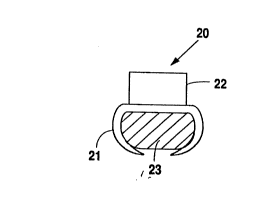
Referring to Figs. 1-2, the first embodiment of
the bone clamp according to the present invention
will be described. Bone clamp 20 is constructed of
nitinol and comprises bone hook 21 having a ~lncer-
type shape formed integrally with rod housing 22.
During surgery in order to mount bone clamp 20 to an
individual vertebra of the spine, bone clamp 20,
which originally is sized to securely fit the
vertebrae, is cooled and expanded to a size larger
than the vertebra (See Fig. 1). Bone hook 21 is then
placed about vertebra 23 and heated until it's
pincer's snugly encircle vertebra 23, thereby, firmly
attaching bone clamp 20 to vertebra 23, (See Fig. 2).
The above process is then repeated until the number
of bone clamps necessary to affix the rod to the
spine are connected to the bone in all locations.
Referring to Figs. 3-5, the second embodiment of
the bone clamp according to the present invention
will be described. Bone clamp 24 is constructed of
nitinol and comprises first and second members 25 and
26 which are coupled together during surgery to form
bone clamp 24 (See Fig. 3). Members 25 and 26 are
identical and comprise claws 27,28 formed integrally
with rod housings 29,30 wherein the inner face of the
rod housing edge integrally formed with claws 27,28
are provided with holes 31,32 on one side and
connector rods 33,34 having a hook at its end o the
other (See Fig. 4). To mount bone clamp 24 onto
vertebra 35 of a spine, first and second members 25
and 26 are cooled in order to straighten the hooks on
the end of each member 25,26 and expand claws 27,28
(See. Fig. 5). Members 25,26 are then placed in
2~46~38
-12-
opposed relation about vertebra 35. That is, claws
27,28 face each other and surround vertebra 35 while
connector rods 33,34 of each member 25,25 fit through
holes 31,32 provided in the opposite member. Next,
members 25,26 are heated which causes the hook at the
end of connector rods 33,34 to reform, thereby,
securing members 25,26 together and preventing their
uncoupling. In addition, claws 27,28 encircle
vertebra 35 to firmly connect bone clamp 24_ The
above process is then repeated until the number of
bone clamps necessary to affix the rod to the spine
are connected to the bone in all locations.
Although segmental affixation i5 essential to
the present invention so that the correction rod can
impart transverse and torsional corrective forces to
the spine, it is also essential that the correction
rod slide freely along the spine during some stages
of correction, while during others it must be rigidly
coupled to the spine. To permit the correction rod
to slide freely along the spine during some stages of
correction, yet be held completely rigid during
others, rod housing 22 of bone clamp 20 and rod
housings 29,30 of bone clamp 24 are fitted with a
blocker.
Referring to Figs. 6 and 7, the blockers, rod
housings, and affixation of the correction rod within
the housing will be described. Although the
affixation of the correction rod to the spine is
described with reference to a single rod housing and
blocker, it is to be understood that all the blockers
and rod housings operate similarly. Blocker 35 is
constructed of nitinol and comprises a tube which is
circularly-shaped such ~hat its edges overlap (See
Fig. 6). The original shape of blocker 36 is such
that its outer diameter is the same as the inner
diameter of rod housing 37, and its inner diameter is
the same as correction rod 38. Additionally, the
2 1 ~ 8 --
-13-
overlapping shape of blocker 36 is chosen because it
permits its inner diameter to be significantly
increased or decreased with only a small concurrent
change in its outer diameter. To mount blocker 36
within rod housing 37, blocker 36 is cooled to allow
its inner diameter to be expanded and its outer
diameter to shrink slightly which permits blocker 36
to securely fit within rod housing 37 ~hile
correction rod 38 easily fits within bloc~eæ 36.
However, although the inne~A it to return to its
original shape, thereby, increasing the frictional
forces between blocker 36 and rod 38 sufficiently to
clamp correction rod 38 firmly within rod housing 37.
After the correction rod is segmentally fixed to
the patient's spine, the surgical operation is
complete. Post-operatively, the rod will apply
corrective forces to the patient's spine if it is
heated above the shape transition temperature and
undergoes transformation to the parent phase crystal
configuration. The shape-memory effect is a local
phenomena. Thus, localized portions of the rod may
be heated selectively in order to produce localized
correctional forces applied only at selected
vertebral levels. Moreover, by controlling the
amount of heat transferred to the rod, the corrective
forces may be produced gradually in whatever
increments the physician deems appropriate This
minimizes the stress which must be borne by the
fixation points and hence the probability of failure
at the bone-metal interface. The incremental
application of correctional forces also allows the
physician to monitor the patient for any neural
dysfunction as the treatment progresses as well as
observe the spinal correction actually produced via
fluoroscopy.
The preferred method of heating is a radio
frequency induction heater. In such a heater, an
21~438
-14-
alternating current is passed through a coil antenna.
A time-varying magnetic field is thus produced which
induces eddy currents in the metal rod. The eddy
currents then produce heat owing to the electrical
resistance of the me~al. The frequency of the
driving current is selected to be low enough to not
produce dipole reversals in water molecules and thus
avoid any heating of surrounding tissues. This
occurs appreciably only when the electro~netic
waves emitted by the antenna are in the microwave
region. The preferred frequency, about 450 KH" is
well below that.
Although the invention has been described in
conjunction with the foregoing, many alternatives,
variation and modifications are apparent to ~hose of
ordinary skill in the art. Those alternatives,
variations and modifications are intended to fall
within the spirit and scope of the appended claims.