Note: Descriptions are shown in the official language in which they were submitted.
-
.
46 5~5
COMPOSITIONS FOR T~TM~NT OF INFLAMED TISSUES
Field of the Invention
The present invention relates to a liposomal composi-
tion suitable for use in concentrating therapeutics at
sites of inflammation in the body.
References
Allen, T.M., (1981) Biochem. Biophys. Acta 640:385-
397.
Ashwell, G., and Morell, A.G. (1974) Adv. Enzymology
41:99-128.
Bartlett, G.R. (1959) J. Biol. Chem. 234:466-468.
Czop, J.K. (1978) Proc. Natl. Acad. Sci. USA 75:3831.
Durocher, J.P., et al. (1975) Blood 45:11.
Ellens, H., et al. (1981) Biochim. Biophys. Acta
674:10-18.
Engelberg, I. and Kohn, J. (1991) Biomaterials 12:292-
304.
Gilman, A.G. et al., eds. (1990) The Pharmacological
Basis of Therapeutics (Eighth Edition) Pergamon Press, New
York.
Gabizon, A., Goren, D. and Barenholz, Y. (1988) Israel
J. Med. Sci. 24:512-517.
A
CA 02146~6~ 1998-0~-06
Gabizon, A., Huberty, J., Straubinger, R.M., Price,
D. C. and Papahadjopoulos, D. (1988-1989) J. Liposome
Res. 1:123-135.
Gabizon, A., Shiota, R. and Papahadjopoulos, D.
(1989) J. Natl. Cancer Inst. 81:1484-1488.
Greenberg, J.P., et al. (1979) Blood 53:916.
Gregoriadis, G., and Neerunjun, D. (1974) Eur. J.
Gregoriadis, G., and Senior, J. (1980) FEBS Lett.
119 .
Hakomori, S. (1981) Ann. Rev. Biochem. 50:733-764.
Haynes, Jr. R. C. (1990) in Gilman, A.G. et al.,
eds. (1990) The Pharmacological Basis of Therapeutics
(Eighth Edition) Pergamon Press, New York, pp 1431-1462.
Huang, S.K., et al. (1991) Biochim. Biophys. Acta
1069(1):117-121.
Hwang, K.J., et al. (1980) Proc. Natl. Acad. Sci.
USA 77:4030.
Jonah, M.M., et al. (1975) Biochem. Biophys. Acta
401:336-348.
Juliano, R.L., and Stamp, D. (1975) Biochem.
Biophys. Res. Commun. 63:651-658.
Kadison, P. et al. (1988) In: Inflammation: Basic
Principles and Clinical Correlates, ed. by Gallin, J.A.,
et al, Raven Press, NY.
Karlsson, K.A. (1982) In: Bioloqical Membranes, Vol.
4, D. Chapman (ed.) Academic Press, N.Y., pp. 1-74.
Kimelberg, H.K., et al. (1976) Cancer Res. 36:2949-
2957.
Kirby, C.J. and Gregoriadis (1984) In: Liposome
Technology, Vol. 3, G. Gregoriadis (ed.) CRC Press, Boca
Raton, FL., p. 19.
Lopez-Berestein, G., et al. (1984) Cancer Res.
44:375-378.
Love, W.G., et al. (1987) Ann.Rheum.Dis. 46:314-318.
Love, W.G., et al. (1989) Ann.Rheum.Dis. 48:143-148.
Love, W.G., et al. (1990) Ann.Rheum.Dis. 49:611-614.
~ ~ 094/07~6 2 ~ PCT/US93/09572
}
Martin, F.J. (1990) In: Specialized Druq Delivery
Systems - Manufacturinq and Production Technoloqy, P. Tyle
~ (ed.) Marcel Dekker, New York, pp. 267-316.
McDonald, D.M. (1988) J. Neurocytol. 17:583-603.
Niwa, Y. (1989) Dermatologica 179(supp . 1):101-106.
Okada, N. (1982) Nature 299:261.
Olson, F., et al., Eur. J. Cancer, 18:;67-176 (1982).
Papahadjopoulos, D., et al., "Sterically Stabilized
Liposomes: Improvements in Pharmacokinetics and Anti-Tumor
Therapeutic Efficacy," PNAS, 88:11460-11464.
Papenfuss, H.D., et al. (1979) Microvascular Res.
8:311-318.
Pincelli, C. et al. ( 1992) J. Invest. Dermatol.
98:421-427.
Poste, G., et al., in LiPosome Technoloqy Volume 3,
page 1 (Gregoriadis, G., et al, eds.), CRC Press, Boca
Raton (1984).
Richardson, V.J., et al. (1979) Br. J. Cancer 40:3543.
Schauer, R. (1982) Adv. Carbohydrate Chem. Biochem.
40:131.
Scherphof, T., et al. ~1978) Biochim. Biophys. Acta
542, 296-307.
Senior, J., and Gregoriadis, G. (1982) FEBS Lett.
145:109-114.
Senior, J., et al. (1985) Biochim. Biophys. Acta
839:1-8.
Sundberg, J.P., et al., J. Investigative Dermatology
95r5):615-635 (1990).
Szoka, F., Jr., and Papahadjopoulos, D., Proc. Natl.
Acad. Sci. USA 75(9):145-1497 (1978).
The PhYsician~s Desk Reference, (1992) Medical
Economics Company, Oradell, N.J.
Williams, B.D., et al. (1987) Ann. Rheum. Dis. 46:314-
318.
SUBSTITUTE SHEET
W094/07466 PCT/US93/09 ~
~146~
Woodle, M.C., et al., (1990). "Improved long- circu-
lating (Stealth~) Liposomes using synthetic liposomes,"
Proceed. Intern. Symp. Control . Rel . Bioact . Mater., 17:77.
Woodle, M.C., et al., (1991). "In Vivo Studies of Long
Circulating (Stealth~) Liposomes in Rats," Period Biol .,
93:349.
Woodle, M.C. et al. (1992) Pharm. Res. 9:260-265.
Woodruff, J.J., et al. (1969) ~. Exp. Med. 129:551.
Wu, N.Z., and Dewhirst, M.W. (1991) "Measuring tissue
uptake of intravenously injected macromolecules using flu-
orescence video microscopy," ~icrovascular Res., Louis-
ville, Kentucky.
Bac~ G~d of the Invention
The inflammatory process is a sequence of physiologi-
cal events which can be elicited by numerous stimuli, in-
cluding infectious agents, ischemia, antigen-antibody in-
teractions, and thermal or other injurious insults. Al-
though the sequence of events constituting an inflammatory
reaction may vary according to the nature and location of
the eliciting insult, there are certain events common to
most inflammatory reactions. These include, in the acute
phase, vasodilation, resulting in increased blood flow to
the inflamed region and increased capillary permeability.
This phase is followed by an increase in fluid in the
region (edema) and movement of blood leukocytes and, final-
ly, phagocytes from the blood vessels to the region.
It would be desirable, for treatment of inflamed tis-
sues or regions, to target therapeutic compounds selective-
ly to the region via the bloodstream. Site-specific tar-
geting would be particularly helpful in reducing toxic side
effects and in increasing the dose of drug which can safely
be delivered to an inflamed region.
Liposomes have been proposed as a drug carrier for
intravenously (IV) a~ ctered compounds, including both
SUBSTITUTE SHEET
094/07~6 ~ 6~ PCT/US93/09572
imaging and therapeutic compounds. However, the use of
liposomes for site-specific targeting via the bloodstream
has been severely restricted by the rapid clearance of con-
ventional liposomes by cells of the reticuloendothelial
system (RES). Typically, the RES will remove 80-95% of a
dose of IV injected liposomes within one hour, effectively
out-competing the selected target site for uptake of the
liposomes.
This phenomenon of RES uptake of liposomes has been
utilized to image inflamed arthritic joints using very
small conventional radiolabeled liposomes (20-60 nm) taken
up by phagocytes present in the synovial fluid (Love, 1987;
Williams). Such small size liposomes are known to have
relatively long circulation times in the blood, but are not
suited for drug delivery compositions, due their limited
capacity for drug entrapment. When larger size convention-
al liposomes were used in such targeting, much lower levels
ac~ ted in the synovial fluid (Love, 1989, 1990).
The failure of larger conventional liposomes to
effectively concentrate in inflamed regions may be due to
the combination of phagocytic uptake and their inability to
penetrate the continuous endothelial cell layer and
underlying basement membrane surrounding the vessels
supplying blood to the region. A characteristic of local
inflammation is a general, acute increase in permeability
of the vasculature to proteins in the region of the
inflammation, followed by migration of neutrophils out of
the bloodstream into the inflamed region. However, neither
of these events predicts the ability of liposomes to pass
through the epithelial cell barriers and adjacent basement
membrane, since proteins are generally much smaller than
liposomes, and neutrophils possess specific binding sites
and active ech~n; c~c for penetrating the blood vessels.
SUBSTITUTE SHEET
W094/07466 PCT/US93/0 ~
2i~
In fact, studies reported to date indicate that even
where the permeability of blood vessels increases, extrava-
sation of conventional liposomes through the vessels does
not increase significantly (Poste). Based on these find-
ings, it was concluded that although extravasation of lipo-
somes from capillaries compromised by disease may be occur-
ring on a limited scale below detection levels, its thera-
peutic potential would be ;n;~ (Poste).
Summ~ry of the Invention
The invention includes, in one aspect, the use of a
pharmaceutical composition comprising liposomes (i)
composed of vesicle-forming lipids including between 1-20
mole percent of an amphipathic vesicle-forming lipid
derivatized with polyethylene glycol and having a selected
mean particle diameter in the size range between about
0.07-0.20 microns, and (ii) contA;n;ng a therapeutic
compound in liposome-entrapped form, in the manufacture of
a medicament effective in concentrating the encapsulated
compound in an inflamed tissue region, when the liposomes
are a~; n; ~tered intravenously to the subject.
In a preferred embs~; -nt of the invention, a~m;n;s-
tration of the composition of the invention will result in
an amount of compound in the inflamed tissue region that is
at least several-fold higher than an amount of compound
which will be found in such a tissue, when the compound is
administered (i) in the absence of liposomes, or (ii) in
liposomes lacking derivatized polyethylene glycol. In
another preferred embodiment the polyethylene glycol has 2
30 molecular weight between about 300 and 5,000 daltons.
In other preferred embodiments, the liposome entrapped
compound is a steroidal anti-inflammatory compound.
Exemplary steroidal anti-inflammatory compounds suitable
for inclusion in the liposomal composition include, but are
not limited to prednisone, methylprednisolone, parametha-
SUBSTITUT~: SHEET
~ 094/07~ PCT/US93/09572
6 ~
zone, 11-fludrocortisol, triamcinolone, betamethasone and
dexamethasone. In yet another preferred embodiment, the
- liposome-encapsulated compound is a gold-containing
compound. In still another preferred embodiment, the
compound is cyclosporin.
In another aspect, the invention includes the use of
a pharmaceutical composition comprising liposomes (i)
composed of vesicle-forming lipids including between 1-20
mole percent of an amphipathic vesicle-forming lipid
derivatized with polyethylene glycol and having a selected
mean particle diameter in the size range between about
0.07-0.20 microns, and (ii) containing a therapeutic
compound in liposome-entrapped form, in the manufacture of
a medicament effective in concentrating the compound in an
inflamed dermal region, when the liposomes are administered
intravenously to the subject.
In a preferred embodiment of this aspect of the
invention, a~ ;n;~tration of the composition of the
invention will result in an amount of compound in the
inflamed dermal region that is at least several-fold higher
than an amount of compound which will be found in such a
tissue, when the compound is administered (i) in the
absence of liposomes, or (ii) in liposomes lacking deriva-
tized polyethylene glycol. In another preferred embodiment
the polyethylene glycol has a molecular weight between
about 300 and 5,000 daltons.
In another embodiment of this aspect of the invention,
the therapeutic compound is concentrated in a region of
psoriatic inflammation, and the liposome-entrapped compound
is selected from the group consisting of steroidal anti-
inflammatory agents, non-steroidal anti-inflammatory
agents, immunosuppressant agents, methotrexate, azaribine,
etretinate, anthralin, cyclosporin and psoralins. In a
further embodiment, the compound is a steroidal anti-
inflammatory agent selected from the group consisting of
STlT~TE S;~EET
W094/07466 PCT/US93/09
-~ 2~
prednisone, methylprednisolone, paramethazone, 11-fludro-
cortisol, triamcinolone, betamethasone and dexamethasone.
Brief Description of the Figures
Figure 1 illustrates a general reaction scheme for
derivatizing a vesicle-forming lipid amine with a polyal-
kylether;
Figure 2 is a reaction scheme for preparing phosphati-
dylethanolamine (PE) derivatized with polyethyleneglycol
via a cyanuric chloride linking agent;
Figure 3 illustrates a reaction scheme for preparing
phosphatidylethanolamine (PE) derivatized with polyethy-
leneglycol by means of a diimidazole activating reagent;
Figure 4 illustrates a reaction scheme for preparing
phosphatidylethanolamine (PE) derivatized with polyethy-
leneglycol by means of a trifluoromethane sulfonate
reagent;
Figure 5 illustrates a vesicle-forming lipid deriva-
tized with polyethyleneglycol through a peptide (5A), ester
(5B), and disulfide (5C) linkage;
Figure 6 illustrates a reaction scheme for preparing
phosphatidylethanolamine (PE) derivatized with polylactic
acid (PLA), polyglycolic acid (PGA), and copolymers of the
two;
Figure 7 is a plot of liposome residence times in the
blood, expressed in terms of percent injected dose as a
function of hours after IV injection, for PEG-PE liposomes
cont~;n;ng 4.7 (triangles) or 14 (circles) mole percent of
phosphatidylglycerol;
Figure 8A is a plot similar to that of Figure 7,
showing blood residence times of liposomes composed of
predo~;n~ntly unsaturated phospholipid components;
Figure 8B is a plot similar to that of Figure 7,
showing the blood residence times of PEG-coated liposomes
SUBSTl rUTE SHFET
~,V094/07~6 PCT/US93/09572
(solid triangles) and conventional, uncoated liposomes
(solid circles);
Figure 9 is a plot similar to that of Figure 7,
shcwing the blood residence times of liposcmes formulated
with less than 35 mole percent cholesterol and a hydrophil-
ic polymer (PEG-1900)-derivatized PE (solid circles), 50
moie percent cholesterol and PEG-1900-PE (solid squares),
with PEG having a molecular weight of 750 (7s~EG-PE; solid
triangles), with PEG having a molecular weight of 350
(35~EG-PE; solid diamonds) and in the absence of hydrophil-
ic polymeric coating ("conventional"; open circles);
Figure 10 is a plot similar to that of Figure 7, show-
ing the blood residence time of polylactic or polyglycolic
acid-coated liposomes (solid triangles; upper line) and
conventional uncoated liposomes (solid circles; lower
line);
Figure ll is a plot similar to that of Figure 7, show-
ing the blood residence time of polylactic or polyglycolic
acid-coated liposomes (upper line) and polyvinyl alcohol-
coated liposomes (lower line);
Figure 12A shows a time course plot of light emission(intensity) from fluorescently labeled albumin in the
specific regions within the blood vessels (as a reference)
(I*, open triangles), entire tissue area (I<total>, open
squares), total blood vessels (closed triangles), and total
interstitium (closed squares) in a rat skin flap window
model before and after application of bradykinin (Bradyki-
nin, open bar), monitored by video-enhanced fluorescence
microscopy;
Figure 12B shows the same data as in Figure 12A
plotted as fluorescence intensity (Y) vs. time (X) before
(solid squares) and after (solid triangles) application of
bradykinin;
Figure 13A shows a time course plot of light emission
(intensity) from fluorescently labeled liposomes in the
S~IBSTITUTE Sl-IEE~
W094/07466 PCT/US93/09.
specific regions within the blood vessels (as a reference)
(I*, open triangles), entire tissue area (I<total>, open
squares), total blood vessels (closed triangles), and
interstitium (closed squares) in a rat skin flap window
model before and after application of bradykinin (Bradyki-
nin, open bar);
Figure 13B shows the same data as in Figure 13A
plotted as fluorescence intensity (Y) vs. time (X) before
(solid squares) and after (solid triangles) application of
bradykinin;
Figures 14A-14E show micrographs of vasculature from
the dorsal flap window preparation in which images of the
microvasculature are shown using transmitted light (14A),
fluorescent emission ; ~ tely after intravenous injec-
tion of Rhodamine-labeled liposomes (14B), fluorescent
emission from rhodamine-labeled liposomes 30 minutes after
bradykinin treatment (14C), fluorescent image ;~;ately
after intravenous injection of fluorescein labeled-albumin
(14D), and 30 minutes after albumin injection (14E);
Figure 15 shows a FSN mouse with psoriasis gene
mutation exhibiting a psoriatic lesion characterized by a
nude patch with erythematous and large parakeratotic
crusts; and
Figures 16A-16C show micrographs of tissue sections of
a psoriatic lesion having liposomes concentrated therein.
Detailed Description of the Invention
I. Pre~aration of Derivatized Lipids
Figure 1 shows a general reaction scheme for preparing
a vesicle-forming lipid derivatized with a biocompatible,
hydrophilic polymer, as exemplified by polyethylene glycol
(PEG), polylactic acid (PLA), polyglycolic acid (PGA) and
polyvinyl alcohol (PVA). These polymers are readily water
soluble, can be coupled to vesicle-forming lipids, and are
tolerated in vivo without toxic effects. The hydrophilic
SUBSTITUTE SHEET
* W094/07~6 PCT/US93/09572
2t~6~
- polymer which is employed, e.g., PEG, is preferably capped
by a methoxy, ethoxy or other unreactive group at one end
or, alternatively, has a chemical group that is more highly
reactive at one end than the other. The polymer is acti-
vated at one of its ends by reaction with a suitable
activating agent, designated R* in the figure, such as
cyanuric acid, diimidazole, anhydride reagent, or the like,
as described below. The activated compound is then reacted
with a vesicle-forming lipid, such as a diacyl glycerol,
including diacyl phosphoglycerols, where the two hydro-
carbon rhA; n~ are typically between 14-22 carbon atoms in
length and have varying degrees of saturation, to produce
the derivatized lipid. Phosphatidylethanolamine (PE) is an
example of a phospholipid which is preferred for this pur-
pose since it contains a reactive amino group which is con-
venient for coupling to the activated polymers. Alterna-
tively, the lipid group may be activated for reaction with
the polymer, or the two groups may be joined in a concerted
coupling reaction, according to known coupling methods.
PEG capped at one end with a methoxy or ethoxy group can be
obtained rom~ercially in a variety of polymer sizes, e.g.,
500-20,000 dalton molecular weights.
The vesicle-forming lipid is preferably one having two
hydrocarbon rhA; ~, typically acyl chains, and a polar head
group. Included in this class are the phospholipids, such
as phosphatidylcholine (PC), PE, phosphatidic acid (PA),
phosphatidylinositol (PI), and sphingomyelin (SM), where
the two hydrocarbon ÇhA; n~ are typically between about 14-
22 carbon atoms in length, and have varying degrees of
unsaturation. Also included in this class are the glycoli-
pids, such as cerebrosides and gangliosides.
Another vesicle-forming lipid which may be employed is
cholesterol and related sterols. Tn general, cholesterol
may be less tightly anchored to a lipid bilayer membrane,
particularly when derivatized with a high molecular weight
5UBSTITUTE SHEET
W094/07466 PCT/US93/09 ~
;2~.g&5~
polymers, such as polyalkylether, and therefore be less
effective in promoting liposome evasion of the RES in the
bloodstream.
More generally, and as defined herein, "vesicle-form-
ing lipid" is intended to include any amphipathic lipidhaving hydrophobic and polar head group moieties, and which
(a) by itself can form spontaneously into bilayer vesicles
in water, as exemplified by phospholipids, or (b) is stably
incorporated into lipid bilayers in combination with bi-
layer forming phospholipids, with its hydrophobic moiety incontact with the interior, hydrophobic region of the bi-
layer membrane, and its polar head group moiety oriented
toward the exterior, polar surface of the membrane. An
example of a latter type of vesicle-forming lipid is chol-
esterol and cholesterol derivatives, such as cholesterolsulfate and cholesterol hemisuccinate.
According to one important feature of the invention,
the vesicle-forming lipid may be a relatively fluid lipid,
typically meaning that the lipid phase has a relatively low
liquid to liquid-crystalline melting temperature, e.g., at
or below room temperature, or relatively rigid lipid, mean-
ing that the lipid has a relatively high melting tempera-
ture, e.g., up to 60~C. As a ruie, the more rigid, i.e.,
saturated, lipids contribute to greater membrane rigidity
in a lipid bilayer structure and also contribute to greater
bilayer stability in serum. Other lipid components, such
as cholesterol, are also known to contribute to membrane
rigidity and stability in lipid bilayer structures. As
mentioned above, a long chain (e.g. C-18) saturated lipid
plus cholesterol is one preferred composition for deli-
vering therapeutics to inflamed sites, since these lipo-
somes do not tend to release the drugs into the plasma as
they circulate through the bloodstream and enter the in-
flamed region during the first 48 hours following injec-
tion. Phospholipids whose acyl chains have a variety of
5UBSTITUTE SHEET
CA 02146~6~ 1998-0~-06
degrees of saturation can be obtained commercially, or
prepared according to published methods.
According to another important feature of the inven-
tion, the vesicle-forming lipid includes an amphipathic
vesicle-forming lipid having a derivatized hydrophilic
biocompatible polymer. In experiments in support of the
invention, and as noted below, it has been found that the
presence of such polymers derivatized to vesicle-forming
lipids in liposomal compositions is effective to signifi-
cantly enhance liposome blood circulation time, in compa-
rison to liposomes formed from lipids in the absence of
such derivatized hydrophilic polymers.
It will be appreciated that the polymer-derivatized
lipids must be (a) safe for parenteral administration,
both in terms of toxicity, biodegradability, and tissue
compatibility, (b) compatible with stable lipid bilayer
formation and structure, and (c) amenable to liposome
preparation and processing steps. These requirements are
met by PEG polymers which have been approved for human
use in the U.S., and also by the thermoplastic polyester
polymers polylactic acid and polyglycolic acid (also
referred to as polylactide and polyglycolide), copolymers
of lactide and glycolide, such as poly(lactide-co-
glycolide), and polyvinyl alcohols. In particular, the
polyester polymers are safe to administer because they
biodegrade by undergoing random, nonenzymatic, hydrolytic
cleavage of their ester linkages to form lactic acid and
glycolic acid, which are normal metabolic compounds
(Engelberg).
Figure 2 shows a reaction scheme for producing a PE-
PEG lipid in which the PEG is derivatized to PE through
a cyanuric chloride group. Details of the reaction are
provided in Example 1. Briefly, methoxy-capped PEG is
activated with cyanuric chloride in the presence in
sodium carbonate under conditions which produced the
activated PEG compound shown in the figure. This mate-
rial is purified to
2 1 4 6 5 ~ ~ .
~ 14
remove unreacted cyanuric acid. The activated PEG compound
is reacted with PE in the presence of triethyl amine (TEA)
to produce the desired PE-PEG compound shown in the figure.
The yield is about 8-10~ with respect to initial quantities
of PEG.
The method just described may be applied to a variety
of lipid amines, including PE, cholesteryl amine, and glyco-
lipids with sugar-amine groups.
A second method of coupling a polyalkylether, such as
capped PEG to a lipid amine, is illustrated in Figure 3.
Here the capped PEG is activated with a carbonyl diimid-
azole (CDI) coupling reagent, to form the activated imid-
azole compound shown in Figure 3. Reaction with a lipid
amine, such as PE, leads to PEG coupling to the lipid
through an amide linkage, as illustrated in the PEG-PE
compound shown in the figure. Details of the reaction are
given in Example 2.
A third reaction method for coupling a capped poly-
alkylether to a lipid amine is shown in Figure 4. Here PEG
is first protected at its OH end by a trimethylsilane
group. The end-protection reaction is shown in the figure,
and involves the reaction of trimethylsilylchloride with
PEG in the presence of triethylamine (TEA). The protected
PEG is then reacted with the anhydride of trifluoromethyl
sulfonate to form the PEG compound activated with trifluoro-
methyl sulfonate. Reaction of the activated compound with
a lipid amine, such as PE, in the presence of triethyl-
amine, gives the desired derivatized lipid product, such as
the PEG-PE compound, in which the lipid amine group is
coupled to the polyether through the te~ ;n~l methylene
carbon in the polyether polymer. The trimethylsilyl
protective group can be released by acid treatment, as
indicated in the figure, or, alternatively, by reaction
with a quaternary amine fluoride salt, such as the fluoride
salt of tetrabutylamine.
~ 094/07466 ~ PCT/US93/09572
'~5
It will be appreciated that a variety of known coup-
ling reactions, in addition to those just described, are
~ suitable for preparing vesicle-forming lipids derivatized
with hydrophilic polymers such as PEG, polylactic acid,
polyglycolic acid, polylactic-polyglycolic copolymers and
polyvinyl alcohol. For example, the sulfonate anhydride
coupling reagent illustrated in Figure ~ can be used to
~oin an activated polyalkylether to the ~ydroxyl group of
an amphipathic lipid, such as the 5'-OH of cholesterol.
Other reactive lipid groups, such as an acid or ester lipid
group may also be used for coupling, according to known
coupling methods. For example, the acid group of phospha-
tidic acid can be activated to form an active lipid an-
hydride, by reaction with a suitable anhydride, such as
acetic anhydride, and the reactive lipid can then be joined
to a protected polyalkylamine by reaction in the presence
of an isothiocyanate reagent.
In another embodiment, the derivatized lipid compo-
nents are prepared to include a labile lipid-polymer link-
age, such as a peptide, ester, or disulfide linkage, whichcan be cleaved under selective physiological conditions,
such as in the presence of peptidase or esterase enzymes or
r2ducing agents such as glutathione present in the blood-
stream. Figure 5 shows exemplary lipids which are linked
through (5A) peptide, (5B), ester, and (5C), disulfide
cont~; n; ng linkages. The peptide-linked compound can be
prepared, for example, by first coupling a polyalkylether
with the N-terminal amine of the tripeptide shown, e.g.,
via the reaction shown in Figure 3. The peptide carboxyl
group can then be coupled to a lipid amine group through a
carbodiimide coupling reagent conventionally. The ester
linked compound can be prepared, for example, by coupling
a lipid acid, such as phosphatidic acid, to the terminal
alcohol group of a polyalkylether, using alcohol via an
anhydride coupling agent. Alternatively, a short linkage
SU~STITUTE SHEET
W094/07466 ; PCT/US93/09 ~
6 ~ ~ ~
fragment cont~; n; ng an internal ester bond and suitable end
groups, such as primary amine groups can be used to couple
the polyalkylether to the amphipathic lipid through amide
or carbamate linkages. Similarly, the linkage fragment may
contain an internal disulfide linkage, for use in forming
the compound shown in Figure 5C. Polymers coupled to phos-
pholipids via such reversible linkages are useful to pro-
vide high blood levels of liposomes which contain them for
the first few hours post injection. After this period,
plasma components cleave the reversible bonds releasing the
polymers and the "unprotected" liposomes are rapidly taken
up by the RES by the same mechanism as conventional lipo-
somes.
Figure 6 illustrates a method for derivatizing poly-
lactic acid, polyglycolic acid and polylactic-polyglycolic
acid copolymers with PE in an amide linkage. The polylac-
tic acid is reacted, in the presence of PE, with dicyclo-
hexylca L~ ;~e (DCCI), as detailed in Example 2. Similarly,
a vesicle-forming lipid derivatized with polyglycolic acid
may be formed by reaction of polyglycolic acid or glycolic
acid with PE in the presence of a suitable coupling agent,
such as DCCI, also as detailed in Example 2. Similar
chemistry may be used to form lipid derivatives of poly-
lactic-polyglycolic acid copolymers. Polyvinyl alcohol
(PVA) is similarly derivatized with PE to form a carbamate
linkage, as detailed in Example 2, by first reaching PE
with carbonyl diimidazole (CDI), followed by addition of a
low molecular weight fraction of PVA in the presence of
triethylamine. The vesicle-forming lipids derivatized with
either polylactic acid or polyglycolic acid and their co-
polymers or polyvinyl alcohol form part of the invention
herein. Also forming part of the invention are liposomes
cont~;n;ng these derivatized lipids.
- S~1BSTITUTE SHEET
094/07~ PCT/US93/09S72
II. PreParation of LiPosomes
A. Lipid Components
The lipid components used in forming the liposomes of
the invention may be selected from a variety of vesicle-
forming lipids, typically including phosphclipids, sphingo-
lipids and sterols. As will be seen, one requirement of
the liposomes of the present invention is long blood circu-
lation lifetime. It is therefore useful to establish a
s~n~rdized measure of blood lifetime which can be used
for evaluating the effect of lipid components on blood
halflife.
One method used for evaluating liposome circulation
time in vivo measures the distribution of IV injected lipo-
somes in the bloodstream and the primary organs of the RES
at selected times after injection. In the standardized
model which is used herein, RES uptake is measured by the
ratio of total liposomes in the bloodstream to total lipo-
somes in the liver and spleen, the principal organs of the
RES. It should be noted that although uptake in such
tissues is specifically into RES cells, the fixed macropha-
ges of the liver and spleen, evaluation of RES uptake is
conventionally carried out by measuring total uptake by the
whole tissues. Thus, when stated herein that RES uptake
was measured in the liver and spleen, it is understood that
such uptake was primarily by the fixed macrophages of the
liver and spleen. In practice, age and sex matched rats or
mice are injected IV through the tail vein with a radiola-
beled liposome composition, and each time point is deter-
mined by measuring total blood and combined liver and
spleen radiolabel counts, as detailed in Example 5.
Since the liver and spleen, and specifically, the
fixed macrophages in the liver and spleen, account for
nearly 100~ of the initial uptake of liposomes by the RES,
the blood/RES ratio just described provides a good approxi-
mation of the extent of uptake from the blood to the RES in
sua~ ", UTE SHEET
W094/07~6 ~ PCT/US93/09
18
vivo. For example, a ratio of about 1 or greater indicatesa predominance of injected liposomes remaining in the
bloodstream, and a ratio below about 1, a predom;n~nce of
liposomes in the RES. For most of the lipid compositions
of interest, blood/RES ratios were calculated at 1, 2, 3,
4, and 24 hours post injection.
The liposomes of the present invention include a
vesicle-forming lipid derivatized with a hydrophilic poly-
mer, described in Section I. According to one aspect of
the invention, it has been discovered that blood circula-
tion half-lives in these liposomes are largely independent
of the degree of saturation of the phospholipid components
making up the liposomes. That is, the phospholipid compo-
nents may be composed predominantly of fluidic, relatively
unsaturated, acyl chains, or of more saturated, rigidifying
acyl chain components. This feature of the invention is
seen in Example 6, which r~;nes blood/RES ratios in lipo-
somes formed with PEG-PE, cholesterol, and PC having vary-
ing degrees of saturation (Table 4). As seen from the data
in Table 5 in the example, high blood/RES ratios were
achieved in substantially all of the liposome formulations,
independent of the extent of lipid unsaturation in the bulk
PC phospholipid, and nc systematic trend, as a function of
degree of lipid saturation, was observed.
Accordingly, the vesicle-forming lipids may be selec-
ted to achieve a selected degree of fluidity or rigidity,
to control the stability of the liposomes in serum and the
rate of release of entrapped drug from the liposomes in the
bloodstream and/or inflamed region. The vesicle-forming
lipids may also be selected, in lipid saturation character-
istics, to achieve desired liposome preparation properties.
It is generally the case, for example, that more fluidic
lipids are easier to formulate and down-size by extrusion
and homogenization methods than more rigid lipid composi-
tions.
SUGSTITUTE S~EFT
~ 2 1 ~ 6 5 6 ~
19
Similarly, it has been found that the percentage of
cholesterol in the liposomes may be varied over a wide
range without significant effect on observed blood/RES
ratios. The studies presented in Example 7A, with refe-
rence to Table 6 therein, show virtually no change inblood/RES ratios in the range of cholesterol between 0-30
mole percent. On the other hand, cholesterol content may
affect the kinetics of drug distribution from the lipo-
somes. For example, delayed release of corticosteroids has
been demonstrated in liposomal compositions having high
(greater than 50 mole percent) steroidal components, in-
cluding cholesterol and cholesterol sulfate, as described
in co-owned, U.S. Patent No. 5,192,528. Example 7C
describes experiments in which hydrophilic polymer (methoxy
PEG-1900-DSPE)-containing liposomes were prepared using
high cholesterol concentrations (50 mole percent). The
blood/RES distribution of these liposomes was determined
(solid circles) in comparison to PEG-cont~;n;ng liposomes
having less than 35 mole % cholesterol (solid squares) and
"conventional" liposomes lacking derivatized hydrophilic
polymers (Figure 9). It is apparent that the presence of
a high mole percent of cholesterol is not detrimental to
the ability of PEG-containing liposomes to prolong blood
circulation time, and may actually enhance this effect.
It has also been found, in studies conducted in sup-
port of the invention, that blood/RES ratios are also
relatively unaffected by the presence of charged lipid com-
ponents, such as phosphatidylglycerol (PG). This can be
seen from Figure 7, which plots percent loss of encapsula-
ted marker for PEG-PE liposomes cont~;n;ng either 4.7 mole
percent PG (triangles) or 14 mole percent PG (circles).
Virtually no difference in liposome retention in the blood-
stream over a 24 hour period was observed. The option of
including negative charge in the liposome without aggrava-
'''~4'''~
CA 02146~6~ 1998-0~-06
ting RES uptake provides a number of potential advantag-
es. Liposome suspensions which contain negative charge
tend to be less sensitive to aggregation in high ionic
strength buffers and hence physical stability is en-
hanced. Also, negative charge present in the liposomemembrane can be used as a formulation tool to effectively
bind high amounts of cationic drugs.
The vesicle-forming lipid derivatized with a biocom-
patible hydrophilic polymer is also present in the lipo-
somal composition. The amount of such derivatized hydro-
philic polymer is preferably between about 1-20 mole
percent, on the basis of moles of derivatized lipid as a
percentage of total moles of vesicle-forming lipids.
These preferred mole ratios are applicable particularly
to lipids derivatized with PEG having molecular weights
between about 1,000-5,000 daltons. It will be appreciat-
ed that a lower mole ratio, such as less than one mole
percent, may be appropriate for a lipid derivative with
a large molecular weight polymer, and that such a
composition may be effective in achieving significantly
enhanced liposome blood-circulation times when the
hydrophilic polymer, e.g., PEG, has a relatively high
molecular weight, e.g, greater than about 1,000-5,000
daltons. Conversely, a higher mole ratio will be
effective for a lipid derivative having a low molecular
weight polymer, such as PEG having a molecular weight of
350 daltons. Such a composition may also be effective in
achieving significantly enhanced liposome blood-circula-
tion times. This is illustrated in Figure 9, which shows
the blood-circulation time for liposomes composed of PEG
having molecular weights of 750 (750PEG-PE; solid
triangles) and 350 (350PEG-PE; solid diamonds) were used
at 33~ molar ratios in liposome compositions. As seen,
both compositions exhibited extended blood circulation
times characteristic of the present invention. Specifi-
cally, as illustrated in Figure 9, such compositions
extend blood circulation
~ 21 ~4~65
time as measured 24 hours after injection of the liposomes,
at least severalfold over that achievable by liposomes
lacking derivatized hydrophilic polymers ("conventional"
liposomes, open circles).
As noted in Section I, the hydrophilic polymer in the
derivatized lipid preferably has a molecular weight between
about 200-20,000 daltons, and more preferably between about
300-5,000 daltons. Example 7B, which examines the effect
of very short ethoxy ether moieties on blood/RES ratios
indicates that polyether moieties of greater than about 5
carbon ethers are required to achieve significant enhance-
ment of blood/RES ratios.
B. Preparing the Liposome ComPosition
The liposomes may be prepared by a variety of techni-
ques, such as those detailed in Szoka et al, 1980. One
method for preparing drug-cont~;n;ng liposomes is the
reverse phase evaporation method described by Szoka et al.
and in U.S. Patent No. 4,235,871. The reverse phase
evaporation vesicles (REVs) have typical average sizes
between about 2-4 microns and are predominantly oligolamel-
lar, that is, contain one or a few lipid bilayer shells.
The method is detailed in Example 4A.
Multilamellar vesicles (MLVs) can be formed by simple
lipid-film hydration techniques. In this procedure, a
mixture of liposome-forming lipids of the type detailed
above dissolved in a suitable organic solvent is evaporated
in a vessel to form a thin film, which is then covered by
an aqueous medium, as detailed in Example 4B. The lipid
film hydrates to form MLVs, typically with sizes between
about 0.1 to 10 microns.
In accordance with one important aspect of the inven-
tion, the liposomes are prepared to have substantially
homogeneous sizes in a selected size range between about
~ 22 7 1 4~5~5
0.07 and 0.2 microns. In particular, it has been discov-
ered that liposomes in this size range are readily able to
extravasate into inflamed regions, as discussed in Section
III below, and at the same time, are capable of carrying a
substantial drug load to an inflamed region and are capable
of being filter sterilized. Although small unilamellar
vesicles of less than 0.07 ~ can easily extravasate into
inflamed regions, they are severely restricted in drug-
loading capacity. The upper end of this preferred size
range, 0.2 microns, is not necessarily the largest size
liposome capable of extravasation, rather it is approxi-
mately the upper limit of size of liposome which can be
conventionally filter sterilized prior to administration.
It can be appreciated that, allowing for adjustments in
pharmaceutical formulation procedures, liposomal composi-
tions above or below this size range may be effective in
delivering drugs to an inflamed region.
One effective sizing method for REVs and MLVs involves
extruding an aqueous suspension of the liposomes through a
series of polycarbonate membranes having a selected uniform
pore size in the range of 0.03 to 0.2 micron, typically
0.05, 0.08, 0.1, or 0.2 microns. The pore size of the
membrane corresponds roughly to the largest sizes of lipo-
somes produced by extrusion through that membrane, particu-
larly where the preparation is extruded two or more timesthrough the same membrane. This method of liposome sizing
is used in preparing homogeneous-size reverse evaporation
vesicle (REV) and multilamellar vesicle (MLV) compositions
described in the examples below. A more recent method
involves extrusion through an asymmetric ceramic filter.
The method is detailed in U.S. patent No. 4,737,323 for
Liposome Extrusion issued April 12, 1988. Homogenization
methods are also useful for down-sizing liposomes to sizes
of 100 nm or less (Martin).
A
~ 23 ~ ~ 4B5B5
Other methods of reducing particle size include appli-
cation of high pressures to the liposomes, as in a French
Press, and homogenization of the liposomes. In experiments
carried out in support of the present invention, high
pressure extrusion was generally used to control particle
size.
C. Compound Loadinq
Incorporation of compound into liposomes can be
achieved by one or more of a variety of active and passive
methods. These methods and characteristics of exemplary
compounds for use with these methods are described in
detail in co-owned U.S. Patent No. 5,213,804. Alternative-
ly or in addition, certain anti-inflammatory compounds can
be carried by liposomes in pro-drug form, wherein the com-
pounds are covalently attached to one or more of the lipids
forming the liposome and are releasable by enzymes present
in the blood or tissue. One suitable lipid-drug composi-
tion for incorporation into liposomal compositions of the
present invention is a lipid covalently attached to a sali-
cylate or to a non-steroidal anti-inflammatory compound and
is described in PCT application WO 91/16920, incorporated
herein by reference.
Passive loading by entrapment is employed for certain
markers, as described in Example 4, and for certain anti-
inflammatory agents, particularly those which are therapeu-
tically active at relatively low drug doses, and/or which
are highly soluble in aqueous solutions. Here the drug is
either dissolved in the aqueous phase used to hydrate the
lipid or included with the lipids in liposome formation
process, depending on the solubility of the compound.
Where the antiinflammatory composition includes a peptide
or protein drug, the liposomes are preferably prepared by
the reverse phase method, by a solvent injection system,
A~
~_ 21~6565
24
such as described in U.S. Patent No. 4,752,425, or by
rehydrating a freeze-dried mixture of the protein and a
suspension of small unilamellar vesicles with water. Both
methods combine passive loading with relatively high encap-
sulation efficiency, e.g., up to 50% efficiency. Nonencap-
sulated material can be readily removed from the liposome
suspension, e.g., by dialysis, diafiltration or exclusion
chromatography.
One class of antiinflammatory agents useful in the
invention described herein, the antiinflammatory cortico-
steroids, are characterized by a high degree of lipophili-
city. Another useful antiinflammatory agent, cyclosporin,
similarly is highly lipophilic. The concentration of
hydrophobic drug which can be accommodated in the liposomes
will depend on drug/lipid interactions in the membrane, but
is generally limited to a drug concentration of less than
about 20 ~g drug/mg lipid. It has been found that for
certain hydrophobic drugs, the highest concentration of
encapsulated material which can be achieved by passive
loading is limited by their low intrinsic water solubility.
It has also been found that liposomal trapping and delivery
of certain antiinflammatory steroids is enhanced by
inclusion of a relatively high concentration (greater than
50 mole percent) of cholesterol in the liposome composi-
tion, as described in co-owned U.S. Patents 5,049,389,
5,192,528 and 5,043,165.
Example 12 describes a method of preparing a steroidal
liposomal composition. In this procedure, a mixture of
liposome-forming components along with the selected
corticosteroid drug, are dissolved in a suitable solvent,
and the lipid solution is evaporated in a vessel to form a
thin film, which is then covered by an aqueous medium.
With addition of a suitable aqueous medium, such as
.~ ''
2 1 4~ 5 65
phosphate buffered saline medium, the lipid film hydrates
to form MLVs, typically with sizes between about 0.1 to 10
microns. Aqueous medium is preferably added to a final
lipid concentration of between about 10-100 ~mole-ml, and
preferably about 40 ~mole/ml. Liposomes are then sized by
extrusion, as described in Example 4.
In the case of amphipathic drugs having a positive
charge, it has been found that inclusion of 20-30 mole
percent of an anionic phospholipid such as PG is in the
liposomal membrane results in increasing the loading factor
significantly through formation of an "ion pair" complex
with the negatively charged PG at the membrane interface.
However, such charged complexed formulations may have
limited utility in the context of the present invention
because the drugs tend to be rapidly released from the
liposome membrane when introduced into plasma.
In some cases, in order to entrap high concentrations
of drugs in liposomes, it has been found to be useful to
use active loading methods. Methods for active loading of
amphipathic drugs into liposomes are described in co-owned
U.S. patent 5,192,549. In one such method, liposomes are
prepared in the presence of a relatively high concentration
of ammonium ion sulfate. After sizing the liposomes to a
desired size, the liposome suspension is treated to create
an inside-to-outside ammonium ion gradient across the lipo-
somal membranes. The gradient may be created by dialysis
or diafiltration against a non-ammonium cont~;n;ng medium,
such as an isotonic glucose medium, or by gel filtration,
such as on a Sephadex1 G-50 column equilibrated with 0.15M
NaCl or KCl, effectively replacing ammonium sulfate in the
exterior phase with sodium or potassium ions or a nonelec-
trolyte species. Alternatively, the liposome suspension
may be diluted with a non-ammonium solution, thereby
reducing the exterior-phase concentration of ammonium ions.
1TrP~ rk
~,~
~ t 4 ~ 5 ~ 5
26
The ammonium sulfate concentration inside the liposomes is
preferably at least 10 times, and more preferably at least
100 to 1000 times that in the external liposome phase.
The ammonium sulfate gradient across the liposomes in
turn creates a chemical gradient which permits capturing of
unionized amines as they pass through the membrane, as
ammonia is released across the liposome membrane, and the
drugs are protonated and trapped in the internal aqueous
phase of the liposome. To load liposomes with the selected
drug a suspension of the liposomes, e.g., about 20-200
mg/ml lipid, is mixed with an aqueous solution of the drug,
and the mixture is allowed to equilibrate over an period of
time, e.g., several hours, at temperatures ranging from
room temperature to 60~C - depending on the phase transi-
tion temperature of the lipids used to form the liposome.
In one typical method, a suspension of liposomes having a
lipid concentration of 50 ~moles/ml is mixed with an equal
volume of amphipathic drug at a concentration of about 5-8
mg/ml. At the end of the incubation period, the suspension
is treated to remove free (unbound) drug. One preferred
method of drug removal for drugs is by passage over an ion
exchange resin, such as Dowex1 50 WX-4, which is capable of
binding unencapsulated drug, but not liposomes cont~;n;ng
the drug. After liposome formation, loading and sizing,
free (unbound) drug is usually removed by ion exchange or
gel exclusion chromatographic methods known in the art or
by dialysis or diafiltration. Ion exchange resins are
selected for use, according to the chemical properties, and
particularly the charge, of the free drug in solution. An
example of an ion exchange resin which is generally useful
in removal of unbound cationic drugs is Dowexl AG50W. Such
a resin can be used in a batch or a column mode, using
standard methods known in the art. In order to complete
removal of free drug using a batch method, two treatments
with the ion exchange resin may be required. An
lT. ~
;. ,
~ 1 4 6 5 ~ ~
27
example of a gel exclusion chromatography column used for
removal of free drug is a Sephadexl G-50 sizing column.
Following drug removal using this method, concentration of
liposomes can be achieved by one or more techniques
st~n~rd to concentration of macromolecules. One particu-
larly useful t~chn;que includes repeated centrifugation in
a Centriprep 10 centrifuge concentrator (Aminco, Danvers
MA) following manufacturers instructions.
D. Characterization of Dru~-Entrapped Liposomal
Compositions
Liposomal drug formulations are characterized by mea-
surements of particle size, lipid concentration, and pH by
standard methods as described above. Drug incorporation
into the composition can be determined by inclusion of
radiolabled tracer, such as ~ labeled drug tracer, into
test compositions. The amount of liposome-entrapped drug
is then determined by gel permeation chromatography using
BioRad A-15M resin. Liposomal drug fraction is calculated
from the amount of radiolabel present in the void volume
of the column. The percentage of liposomal drug is then
determined from the ratio of the label eluting in the void
volume to the remaining label eluting from the column.
III. Liposome Localization in Inflamed Regions
A. Extended Bloodstream Halflife
One of the requirements for liposome localization in
a target inflamed tissue, in accordance with the invention,
is an extended liposome lifetime in the bloodstream
following parenteral liposome a~;n;~tration. One measure
of liposome lifetime in the bloodstream is the blood/RES
ratio determined at a selected time after liposome a~;n;~-
tration, as discussed above. Blood/RES ratios for a
variety of liposome compositions are given in Table 3 of
Example 5. In the absence of PEG-derivatized lipids,
lTTs~ ms.rk
CA 02146~6~ 1998-0~-06
28
blood/RES ratios were 0.03 or less. In the presence of
PEG-derivatized lipids, the blood/RES ratio ranged from
0.2, for low-molecular weight PEG, to between 1.7-4 for
several of the formulations, one of which lacks choles-
terol, and three of which lack an added charged phospho-
lipid (e.g., PG).
The data presented in Table 5 in Example 6 show
blood/RES ratios (excluding two points with low percent
recovery) between about 1.26 and 3.27, consistent with
the data given in Table 3. As noted in Section II above,
the blood lifetime values are substantially independent
of degree of saturation of the liposome lipids, presence
of cholesterol and presence of charged lipids.
The blood/RES values reported above can be compared
with blood/RES values reported in co-owned U.S. Patent
No. 4,920,016, which used blood/RES measurement methods
similar to those used in generating the data presented in
Tables 3 and 5. The best 24-hour blood/RES ratios which
were reported in the above-noted patent was 0.9, for a
formulation composed of GM1, saturated PC, and choleste-
rol. The next best formulations gave 24-hour blood/RES
values of about 0.5. Thus, typical 24-hour blood/RES
ratios obtained in a number of the current formulations
were more than twice as high as the best formulations
which have been reported using liposomes lacking
derivatized hydrophilic polymers. Further, ability to
achieve high blood/RES with GM1 or HPI lipids was depen-
dent on the presence of predominantly saturated lipids
and cholesterol in the liposomes.
Plasma pharmacokinetics of a liposomal marker in the
bloodstream can provide another measure of the enhanced
liposome lifetime which is achieved by the liposome
formulations of the present invention. Figures 7 and 8A
discussed above show the slow loss of liposomal marker
from the bloodstream over a 24 hour period in typical
PEG-liposome formulations, substantially independent of
whether the
~ 29 ~ 1 4 ~ 5 6 ~ !
marker is a lipid or an encapsulated water-soluble compound
(Figure 8A). In both plots, the amount of liposomal marker
present 24 hours after liposome injection is greater than
10% of the originally injected material.
Figure 8B shows the kinetics of liposome loss from the
bloodstream for a typical PEG-liposome formulation and the
same liposomes in the absence of a PEG-derivatized lipid.
After 24 hours, the percentage of PEG-liposomes remaining
in the blood was greater than about 20%, whereas the
conventional liposomes showed less than 5% retention in the
blood after 3 hours, and virtually no detectable marker at
24 hours. Figure 9 shows the kinetics of liposome loss
from the bloodstream for low molecular weight PEG liposome
formulations using DSPE derivatized to PEG having a
15 molecular weight of 350 (350 PEG, open triangles) or 750
(750 PEG, closed triangles). These formulations showed
blood retention times similar to those observed with
liposomes formulated using higher molecular weight PEG
(typically 1000-5000 daltons).
Similarly, liposomes cont~;n;ng PLA- or PGA-deriva-
tized PE or PVA-derivatized DSPE show plasma kinetics
which are superior to conventional liposomes consisting of
PG, PC and cholesterol (Figures 10 and 11), in that the
liposomes contA;n;ng the derivatized PE or DSPE are cleared
from the bloodstream at a rate which is severalfold slower
than the formulations without the derivatized PE or DSPE.
The results seen in Figures 7-11 are consistent with
24 hour blood liposome values measured for a variety of
liposome formulations, and reported in Tables 3 and 5-7 in
Examples 5-8 below. As seen in Table 3 in Example 5, the
percent dose r~mA;n;ng at 24 hours was less than 1% for
conventional liposomes, versus at least 5% for the PEG-
liposomes. In the best formulations, values between about
20-40% were obtained. Similarly in Table 5 from Example 6,
liposome levels in the blood after 24 hours (again neglect-
A
~ 2 1 4~5~
ing two entries with low recovery values) were between 12and about 25 percent of total dose given. Similar results
are reported in Tables 6 and 7 of Example 7.
For both blood/RES ratios, and liposome retention time
in the bloodstream, the data obtained from a model ~n;m~l
system can be reasonably extrapolated to humans and veteri-
nary animals of interest. This is because uptake of lipo-
somes by the fixed macrophages of liver and spleen has been
found to occur at similar rates in several mammalian spe-
cies, including mouse, rat, monkey, and human (Gregoriadis,1974; Jonah; Kimelberg, 1976; Juliano; Richardson; Lopez-
Berestein). This result likely reflects the fact that the
biochemical factors which appear to be most important in
liposome uptake by the RES -- including opsonization by
serum lipoproteins, size-dependent uptake effects, and cell
shielding by surface moieties -- are common features of all
m~mm~l ian species which have been examined.
In studies carried out in support of the present
invention, in a set of healthy control animals, liposome
accumulation in the liver and spleen were less than 20~ and
10~, respectively, of the injected dose over a monitoring
period of 40 hours. These levels are well below the levels
maintained in the blood during most of the monitoring
period, and significantly, during the first 24 hours
following injection.
Comparison of PEG-containing liposomes of the inven-
tion with conventional liposome preparations reveals that
a desirable feature of anti-inflammatory therapy - prolon-
gation of blood levels of drug-carrying liposomes for two
or more days in order to give increased and sustained
accumulation in target sites- was not achieved by conven-
tional liposomal preparations, but is achieved with the
hydrophilic polymer-derivatized liposomal formulation of
the invention, as exemplified by PEG-, PGA-, PLA-, and PVA-
A
~ 094/07466 PCT/US93/09572
s
derivatized liposomal formulations (Figures 8B, 9, 10 and
11) .
B. Extravasation into Inflamed Tissues
Another required feature for high-activity liposome
targeting to an inflamed region, in accordance with the
invention, is liposome extravasation into the region from
the bloodstream through the endothelial cell barrier and
underlying basement membrane separating a capillary from
the tissue cells supplied by the capillary. Liposomes with
sizes between about 0.07 and 0.2 microns exhibit this
ability to extravasate into inflamed regions. Although
liposomes with sizes of less than 0.07 microns would also
be expected to extravasate, a limited drug-carrying
capacity of these small liposomes render them less effec-
tive as drug carriers for the present system. Similarly,
liposomal sizes greater than 0.2 microns may also extrava-
sate to inflamed regions; however, as stated in Section II,
such liposomes cannot easily be filter sterilized, follow-
ing hydration, in conventional pharmaceutical production.
For the purposes of the present invention, then, the
optimal size range for liposomes would strike a balance
between ability to extravasate, drug-carrying capacity, and
feasibility of sterile filtration, that is, between about
25 0.07 and 0.2 microns in diameter.
In experiments carried out in support of the present
invention, detailed in Example 10, fluorescently labeled
PEG-cont~;n;ng liposomes and a fluorescently labeled
protein (bovine serum albumin) were employed to ~m; ne
extravasation characteristics in a model of bradykinin-
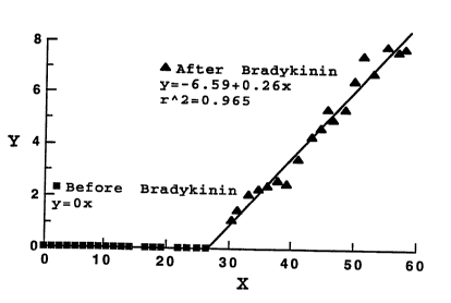
induced inflammation- Figure 12A and Figure 13A show plots
of the appearance of the fluorescently labeled bovine serum
albumin (BSA; Figure 12A) and PEG-containing liposomes
(Figure 13A) in the vascular (solid triangles) and inter-
stitial regions (solid sguares) before and after bradykinin
SUBSTIT~JTE S~EET
W094/07466 PCT/US93~09 ~
2 ~
application to the region. Sharp increases in fluorescence
attributable to BSA-associated label and to liposome-
associated label were observed in the interstitial region
just after application of bradykinin, indicating extravasa-
tion to the interstitium. Visual assessment of the regionsconfirmed the accumulation of protein and liposomes in the
interstitial region, by the presence of bright fluorescent
spots following bradykinin treatment (Figures 14C and 14E).
Such visually apparent fluorescence was not observed prior
to the application of bradykinin to the region.
These data were converted to a plot of averaged perme-
ability constants (Figure 12B and Figure 13B), calculated
according to the permeability equation (Wu, 1991) and shown
as fluorescence intensity (Y) in the figures, as a function
of time (X). As plotted, permeability is proportional to
the slope, ~, of the plot of Y vs. X. Using these calcula-
tions, a 10-fold increase in vascular permeability to
albumin was measured following bradykinin treatment. Prior
to bradykinin application, permeability to liposomes was
essentially zero, so that a fold-comparison cannot be made.
However, permeability of 90 nm liposomes was about 1/3 that
of albumin, just following bradykinin treatment, and about
4 times that of albumin measured subsequent to bradykinin
treatment.
In further experiments carried out in support of the
present invention, concentration of liposomes in an area of
inflammation in a mouse model of psoriasis was ~Y~;ned, as
detailed in Example 11. Briefly, strain FSN mice having a
single gene immunologic mutation which results in develop-
ment of dermal lesions resembling human psoriasis
(Sundberg, et al.), were used. A mouse with such a lesion
is shown in Figure 15. Erythematous and larg~ parakeratot-
ic crusts were characteristic of the psoriatic lesions.
Histologic ~Y~;n~tion of the lesions revealed squirting
papilla with focal paraketosis and proliferated mast cells
SUBSTITUTE S~FET
O9~/~7J66 ~ 1 4 6 ~ ~ ~ PCT/US93/09572
in the region between dermis and epidermis, as shown in
Figure 16A. Colloidal gold-containing, PEG-cont~;n;ng
liposomes were injected into these mice. The mice were
sacrificed 24 hours after liposome injection, tissues were
collected following fixation, and tissue sections were
prepared for silver enhancement of gold deposition, as
detailed in Example ll.
Figures 16A-16C show micrographs of tissue sections
from experiments in which gold-containing, PEG-containing
liposomes were injected into psoriatic mice. Figure 16C
shows a low power (x400) micrograph of a section through a
psoriatic lesion in which is apparent a concentration of
silver-enhanced gold particles in the lesions around the
psoriasis lesions between epidermis and dermis.
Figures 16A and 16B show higher power mic~ Gy r aphs of
the region, in which is apparent accumulation of silver-
~nh~nC~ gold particles pred~~ in~ntly in the boundary of
dermis region close to epidermis, and concentrated in the
tip of papillae (Figure 16A). In some early and developed
lesions, inflammatory foci were highly proliferated with
macrophages, polymorphonuclear leukocytes, and mast cells.
Silver-enhanced colloidal gold was found scattered around
these inflammatory cells. This can be seen in Figure 16B.
In addition, silver-enhanced gold particles were observed
in regions surrounding hair follicles.
IV. Treatment of Inflamed Reqions
As described above, liposomes of the invention are
effective to localize and concentrate an entrapped thera-
peutic agent specifically in an inflamed region. Inaccordance with the present invention, such treatment is
particularly effective for acute inflammations (i.e.,
inflammations that have existed for less than about a
week).
SUBSTITILITE SHEET
W094/07466 ~; PCT/US93/09 ~
2 1 ~
34
Liposome compositions of the invention preferably have
a relatively high drug carrying capacity and minimal lea-
kage of the entrapped drug during the time required for the
liposomes to distribute to and enter the inflamed region
(the first 24-48 hours following injection in mice or
rats). The PEG-cont~;n;ng liposomes thus provide an
effective method for concentrating the liposome-entrapped
therapeutic co~.~ou,.d in an inflamed region. In the context
of the present invention, "concentrating" of a compound in
a tissue is achieved, when the compound is present in the
tissue in an amount or at a concentration (mole/g tissue)
that is higher than an amount or concentration in the
tissue that is achieved subsequent to administration of a
similar dose of either the free compound or of the compound
entrapped in conventional liposomes (liposomes having the
same lipid composition but lacking PEG or hydrophilic
polymer coating). Preferably, such concentrating by PEG-
containing liposomes will result in a concentration of
compound in the inflamed region that is at least several-
fold higher than a concentration achieved subsequent toa~ ;n;~tration of free drug or of compound in conventional
liposomes.
In accordance with the invention, the therapeutic
compound is entrapped by such PEG-cont~;n;ng liposomes and
the liposomal formulation is administered parenterally to
a subject, preferably directly into the bloodstream, as by
intravenous injection.
In the context of the present invention, an inflamed
site or region is generally a region anatomically at a site
outside the bloodstream that is accessible from and
adjacent a capillary bed. Inflamed regions which are most
~n~hle to treatment by the method of the invention are
characterized by an acute increase in permeability of the
vasculature in the region of inflammation. In this case,
for an IV injected liposome-entrapped therapeutic composi-
8UBSTITUTE S~EET
~ 094/07~6 PCT/US93/09572
5~,~
tion to reach the inflamed site, it must leave the blood-
stream and enter the inflamed region. In one embodiment,
the method of the invention is used to treat inflammation
by concentrating an anti-inflammatory agent selectively in
the inflamed region.
Therapeutic agents useful in the treatment of local-
ized inflammation vary, according to the cause and site of
the inflammation. Agents directed against the primary
cause of the inflammation, such as infection, can be used
in the treatment method. Commonly, general anti-inflamma-
tory agents such as steroidal or non-steroidal anti-
inflammatory agents will be used. More generally, the
method of the invention includes a liposomal composition
having an agent known to be useful in the treatment of a
specific inflammatory state. By selectively localizing
such therapeutic agents to inflamed regions, the method of
the invention has the advantage over conventional drug
regimens of decreased exposure of unaffected tissues to
high doses of drug. This is expected to lower unwanted
side effects of drug therapy.
The following discussion of exemplary inflammatory
states and therapeutic agents useful in their treatment is
intended to represent some of the types of inflammation
which can be treated, using the treatment method described
herein. This discussion is not intended to limit the scope
of the invention, but is provided as a guide to the general
applicability of the treatment metnod to states of inflam-
mation. In general, a treatment regimen using liposomal
preparations of the invention can be determined on the
basis of knowledge of conventional therapeutic drugs and
their effective concentration ranges for a particular
disorder. Such information is available in standard
medical reference guides such as Goodman (1990) or The
Phvsician's Desk Reference. According to the present
invention, the amount of drug entrapped in a particular
SUB5Ti~3TE SH~ET
W094/07466 PCT/US93/09 ~
2~
36
liposomal preparation is determined according to the me-
thods described in Section II. The percentage of such a
preparation which will be delivered to a particular site of
inflammation will depend on the size and vascularization of
the region. Such a percentage can be estimated, in accord-
ance with the working examples presented herein. Such data
provide basis for determination of an appropriate dose or
dose range for treatment of ir.flammation in an individual.
General antiinflammatory agents include, as noted
above, steroids and non-steroids. Steroids commonly used
as antiinflammatory agents include those corticosteroids
having antiinflammatory effects greater than or equal to
that of naturally occurring human cortisol (Haynes).
- Examples of antiinflammatory steroids given systemically
include prednisone, methylprednisolone, paramethazone, 11-
flurocortisol, triamcinolone, betamethasone and dexametha-
sone. Additionally, it is anticipated that certain
antiinflammatory steroids, such as beclomethasone, which
are conventionally a~; n; stered only topically, due to
their toxicity and/or high lipophilicity, will become
available for systemic a~m;n;ctration in such liposomal
scitions.
A number of inflammatory diseases and allergic
reactions may be treated systemically with steroidal
antiinflammatory agents; however, due to the undesirable
side effects of such agents, prolonged sytemic treatment is
generally reserved for particularly severe afflictions.
A~m; n;ctration of systemic steroids is indicated for
urticaria resulting from an undesirable immune reaction,
multiple sclerosis, and organ implant. It is appreciated
that these states can be advantageously be treated with
liposome-entrapped steroidal compounds, and that such
treatment is anticipated to reduce overall the dose of drug
a~m; n; -ctered to the whole body and thereby to reduce
unwanted side effects attributable to such drugs. Addi-
SUBSrITUTE S~EET
094/07466 ~1 ~ PCT/US93/09572
tionally, as discussed below, by reducing such systemic
side effects, the method of the invention makes feasible
~ treatment of diseases or conditions in which use of
steroids was previously considered unwarranted or unadvis-
able, due to the relative lack of severity of the disease
state, relative to the side effects, and/or the length of
treatment required. For example, long term use of adreno-
corticosteroids for treatment of less severe cases of
inflammation, such as for eczematous dermatitis, although
considered beneficial, is not generally recommended, due to
the side effects inherent to systemic treatment with
steroids. Side effects associated with long term systemic
adrenocorticosteroid usage include suppression of the
hypoth~lA~;c-pituitary-adrenal axis (Cushing's syndrome),
fluid and electrolyte disturbances, hypertension, peptic
ulceration, osteoporosis, and myopathy.
Other agents generally useful in the treatment of
inflammation include, but are not limited to, free radical
scavenging agents such as superoxide dismutase and non-
steroidal antiinflammatory drugs (NSAIDs), including, butnot limited to salicylates (exemplified by aspirin),
pyrazolon derivatives (exemplified by phenylbutazone),
indomethacin, sulindac, tolmetin, fenamates (exemplified by
meclofenamate), proprionic acid derivatives (exemplified by
ibuprofen), oxicam derivatives (exemplified by piroxicam),
phenylacetic acid derivatives (exemplified by diclofenac),
etodolac, and nabumetone. Generally, although many of
these drugs possess excellent antiinflammatory properties,
side effects limit their use at doses effective to provide
effective antiinflammatory treatment. In accordance with
the invention, formulations of such drugs in liposomes
having enhanced circulation times are contemplated to pro-
vide selective relief of inflammation in subjects requiring
such treatment. Other exemplary antiinflammatory agents
are discussed with respect to specific indications, below.
SUE~ST~TUTÇ~ 5~EET
W094/07466 PCT/US93/0
38
Rheumatoid arthritis is an inflammatory condition in
which steroid, as well as non-steroid therapeutics are
useful treatments. NSAIDs, as described above, are also
indicated in providing antiinflammatory relief in patients
having arthritis (rheumatoid and osteoarthritis) and anky-
losing spondylitis. Doses of NSAIDS required to provide
pain re7ief are generally quite high, and are associated
with significant side effects, including ulceration of the
stomach ~nd duodenum. Treatment with cyclosporin has also
been found to be beneficial to sufferers of rheumatoid
arthritis. Liposomal delivery of such drugs to inflamed
regions, particularly joints, would be expected to reduce
exposure of such susceptible regions. Other drugs useful
in the treatment of arthritis include methotrexate,
sulfalazine, D-penicillamine, and nambumetone. Gold-
con~;n;ng compounds, such as aurothioglucose and aurano-
fin, may also, in addition to reducing inflammation, reduce
progression of the disease. Because they are bound by
plasma proteins and sequestered by macrophages in a number
of tissues, relatively high doses of such agents must be
given to achieve therapeutic concentrations in affected
regions (synovial fluid of joints). Such doses are associ-
ated with side effects, including blood dyscrasias, lesions
of the mucous membranes, and chrysiasis. Such high doses
are also relatively expensive. When given in liposome
entrapped form, it is contemplated that lower doses of
compound will be required, to achieve therapeutic concen-
trations in inflamed regions.
Gout, in its acute phase, is characterized as an in-
flammatory reaction to urate crystals present in joints,
and includes local infiltrations of granulocytes. Acute
phase symptoms of gout can be relieved by oral or intrave-
nous a~;n;stration of colchicine. Liposomal entrapment of
this compound is expected to reduce serious side effects,
5~B!~;~IT~JTE ~I-IEET
~ 094/07~6 PCT/US93/09S72
6~
such as agranulocytosis, associated with long-term treat-
ment with the compound.
Neurogenic inflammation refers to a local tissue re-
sponse elicited by stimulation of sensory nerves in a
number of tissues. Commonly, susceptible organs include
the eye, skin, joints and respiratory tract. In ~ni~l
models of the respiratory tract, neurogenic inflammation is
characterized by increased permeability of postcapillary
venules and collecting venules in specific regions of the
respiratory tract (McDonald). Systemic antiinflammatory
therapy using the liposomal preparations of the invention
is therefore expected to be useful in the delivery of
therapeutic agents useful in the treatment of neurogenic
inflammation.
Necrotizing vasculitides are diseases of the vascular
system characterized by inflammation of the vascular
system. These diseases, which may involve a variety of
etiologies, are thought to have in common inappropriate
immunologic reactions, as evidenced by the deposition of
immune complexes in and around vessel walls.
Examples of diseases which fall within the category of
vasculitis include, but are not limited to, polyarteritis
nodosa, serum sickness, Wegener's granulomatosis, and
Kawasaki's syndrome (Kadison). Antiinflammatory steroids
such as prednisone are indicated in many of these diseases.
Therapy using the method of the invention is contemplated
to be beneficial, as described above.
one class of inflammatory disease that is particularly
amenable to treatment with compositions of the invention is
the class comprising dermatological lesions. These inflam-
mations may be caused by a number of local as well as sys-
temic events, such as infections. Systemic administration
of high doses of adrenocorticosteroids is currently consi-
dered appropriate therapy only for particularly severe
chronic dermatoses, including pemphigus vulgaris, as well
~;~IBSTIT~TE SHEET
W094/07466 PCT/US93/09 ~
2~6~6~
as in more acute severe dermatoses, such as allergic con-
tact dermatitis. However, lesser disorders such as pso-
riasis, for which intralesional a~m;n;stration of corti-
costeroids is currently indicated, may also benefit from
systemic administration of liposome-entrapped cortico-
steroids, as wel~ as from liposomal therapies using anti-
proliferative drugs, immunosuppressive compounds such as
cyclosporin A ~nd selection (e.g., ELAM-1, GMP-140)-
directed therapeutics, retinoids such as isotretinoin, and
5-f 1UG1 OU1 dCil . Likewise, urticaria, and particularly the
pruritis associated with urticaria, is conventionally
treated with antihistamines having both H~ and H2 specifici-
ty, such as diphenhydramine and famotidine, respectively,
as well as with cromolyn sodium. Encapsulation of such
compounds in liposomes will reduce dosages required and
reduce side effects as a conseguence.
The use of cyclosporin A in such dermatological in-
~lammatory diseases as resistant psoriasis and atopic
dermatitis is currently gaining favor. Poor absorption of
cyclosporin topically necessitates systemic a~ ;n;stration
of the compound. In accordance with the present invention,
liposomal formulations of cyclosporin are anticipated for
such uses. Example 13 describes a method of producing sud~
a cyclosporin liposomal formulation. Such formulations
will also find use in other known clinical uses of cyclo-
sporin, such as in organ transplant and graft vs. host
disease, and rheumatoid arthritis, as mentioned above.
Certain dermatological lesions associated with cancerous
conditions, including ~aposi's sarcoma and T-cell lymphoma,
may be treated with antiproliferative agents including
vincristine and etoposide.
As described above, the liposomal compositions and
treatment methods of the invention can also be used to
concentrate compounds psoriatic lesions. In humans, pso-
riasis is a chronic condition which, although not life-
SUBSTITUTE SHFET
~ 094/07~6 PCT/US93/09572
5~$
41
threatening, can be debilitating. The precise cause of thedisorder is unknown, although it has been suggested that
neurogenic factors, including neuropeptides, are involved
in its etiology (Pincelli). A number of therapeutic
agents, including steroidal and non-steroiaal antiinflam-
matory agents, antiproliferative agents (methotrexate,
azaribine), immunosuppressants such as cyclosporin, and
miscellaneous agents, such as etretinate, anthralin,
psoralins and coal tar, are currently used in treatment of
psoriasis. As described in Section III, above, and
detailed in Example 11, liposomal compositions made in
accordance with the invention are effective to localize and
concentrate in psoriatic lesions. It is contemplated that
similar liposomes, having entrapped antipsoriasis agents
will be useful in treatment of psoriasis and other dermato-
logical lesions. A particularly advantageous composition
for treating psoriasis is a psoralen-containing liposomal
composition of the invention. Such a composition will
concentrate in psoriatic lesions, which are then irradiated
with a W -A light source, in analogy to "P WA" therapy
(combined systemic psoralen plus W -A light).
In summary, the present invention contemplates treat-
mer.t of a variety of disorders which are e ther primarily
inflammatory in presentation or which have as a component
of their presentation, an inflammatory phase. Examples of
disorders having inflammatory phases include adult respira-
tory distress syndrome, reperfusion injury following
m ocardial infarction and/or thrombolysis, septic shock,
organ transplantation and diabetes.
The following examples illustrate, but in no way are
intended to limit the present invention.
SUeSTlTUTE SHEET
W094/07466 ~ PCT/US93/09
42
Materials and Methods
Materials
Cholesterol (Chol) was obtained from Sigma (St.
Louis, MO) or from Calbiochem (La Jolla, CA). Sphingomy-
elin (SM), egg phosphatidylcholine (lecithin or PC),partially hydrogenated PC having the composition IV40,
IV30, IV20, IV10, and IV1, phosphatidylglycerol (PG),
phosphatidylethanolamine (PE), dipalmitoyl-phosphatidyl
glycerol (DPPG), dipalmitoyl PC (DPPC), dioleyl PC (DOPC),
distearoyl PC (DSPC) and egg PC were obtained from Avanti
Polar Lipids (Alabaster, AL) or Austin Chemical Company
(Chicago, IL). Distearyl phosphatidyl ethanolamine was
obtained ~rom Calbiochem (La Jolla, CA).
tl~I]-tyraminyl-inulin was made according to published
procedures. ~Gallium-citrate was supplied by NEN Neoscan
(Boston, MA). ~I3gentamicin was from NE~ (Cambridge,
MA). Doxorubicin Hcl and Epirubicin HCL were obtained from
Adria Laboratories (Columbus, OH) or Farmitalia Carlo Erba
(Milan, Italy).
Polyvinyl alcohol (PVA), glycolic acid, and lactic
acid were from Aldrich (St. Louis, MO). Polylactic acid
was supplied by ICN (Cleveland, OH).
All organic solvents used were reagent grade or high
pressure liquid chromatography grade.
Example
PreParation of PEG-PE Linked bY
CYanUric ChlGride
A. Preparation of Activated PEG
2-0-Methoxypolyethyleneglycol1900-4,6-dichloro-1,3,5
triazine previously called activated PEG was prepared as
described in J. Biol . Chem., 252:3582 (1977) with the fol-
lowing modifications.
Cyanuric chloride (5.5 g; 0.03 mol) was dissolved in
400 ml of anhydrous benzene cont~in;ng 10 g o~ anhydrous
8l~B~ 11' l UTE S~ EET
( -
~ 43 2~465~9 ;~'
sodium carbonate, and PEG-1900 (19 g; 0.01 mol) was added
and the mixture was stirred overnight at room temperature.
The solution was filtered, and 600 ml of petroleum ether
(boiling range, 35-60~) was added slowly with stirring.
The finely divided precipitate was collected on a filter
and redissolved in 400 ml of benzene. The precipitation
and filtration process was repeated several times until the
petroleum ether was free of residual cyanuric chloride as
determined by high pressure liquid chromatography on a
column (250 x 3.2 mm) of 5-m "LiChrosorb1" (E. Merck),
developed with hexane, and detected with an ultraviolet
detector. Titration of activated PEG-1900 with silver
nitrate after overnight hydrolysis in aqueous buffer at pH
10.0, room temperature, gave a value of 1.7 mol of chloride
liberated/mol of PEG.
TLC analysis of the product was carried out on TLC
reversed-phase plates obtained from Baker using methanol/-
water, 4:1; v/v, as developer and exposure to iodine vapor
for visualization. Under these conditions, the starting
methoxy polyglycol 1900 appeared at R~0.54 to 0.60. The
activated PEG appeared at R=0.41. Unreacted cyanuric
chloride appeared at R~0.88 and was removed.
The activated PEG was analyzed for nitrogen and an
appropriate correction was applied in selecting the
quantity of reactant to use in further synthetic steps.
Thus, when the product contained only 20% of the theore-
tical amount of nitrogen, the quantity of material used in
the next synthetic step was increased by 100/20, or 5-fold.
When the product contained 50% of the theoretical amount of
nitrogen, only 100/50 or a 2-fold increase was needed.
B. Pre~aration of N-(4-Chloro-~olyethylene-qlycol
1900)-1 3 5-triazinYl Eqq Phosphatidylethanol-
amine
In a screw-capped test tube, 0.74 ml of a 100 mg/ml
(0.100 mmole) stock solution of egg phosphatidylethanol-
lTrademark
_ 44
amine in chloroform was evaporated to dryness under astream of nitrogen and was added to the residue of the
activated PEG described in section A, in the amount to
provide 205 mg (0.100 mmole). To this mixture, 5 ml an-
hydrous dimethyl formamide was added. 27 microliters(0.200 mmole) triethylamine was added to the mixture, and
the air was displaced with nitrogen gas. The mixture was
heated overnight in a sand bath maintained at 110~C.
The mixture was then evaporated to dryness under
vacuum and a pasty mass of crystalline solid was obtained.
This solid was dissolved in 5 ml of a mixture of 4 volumes
of acetone and 1 volume of acetic acid. The resulting
mixture was placed at the top of a 21 mm x 240 mm chromato-
graphic absorption column packed with silica gel (Merck
Kieselgel1 60, 70-230 mesh) which had first been moistened
with a solvent composed of acetone acetic acid, 80/20; v/v.
The column chromatography was developed with the same
solvent mixture, and separate 20 to 50 ml aliquots of ef-
fluent were collected. Each portion of effluent wasassayed by TLC on silica gel coated plates, using 2-buta-
none/acetic acid/water; 40/25/5; v/v/v as developer and
iodine vapor exposure for visualization. Fractions
containing only material of R=about 0.79 were combined and
evaporated to dryness under vacuum. Drying to constant
weight under high vacuum afforded 86 mg (31.2 micromoles)
of nearly colorless solid N-(4-chloro-polyglycol
1900)-1,3,5-triazinyl egg phosphatidylethanolamine con-
taining phosphorous.
The solid compound was taken up in 24 ml of ethanol/-
chloroform; 50/50 chloroform and centrifuged to remove
insoluble material. Evaporation of the clarified solution
to dryness under vacuum afforded 21 mg (7.62 micromoles) of
colorless solid.
lTrademark
~A
CA 02l46~6~ l998-0~-06
Example 2
Preparation of Carbamate and Amide Linked
Hydrophilic Polymers with PE
A. Preparation of the Imidazole Carbamate of
Polyethylene Glycol Methyl Ether 1900.
9.5 grams (5 mmoles) of polyethylene glycol methyl
ether 1900 obtained from Aldrich Chemical Co. was dis-
solved in 45 ml benzene which has been dried over molecu-
lar sieves. O. 89 grams (5.5 mmoles) of pure carbonyl
diimidazole was added. The purity was checked by an
infra-red spectrum. The air in the reaction vessel was
displaced with nitrogen. Vessel was enclosed and heated
in a sand bath at 75~C for 16 hours.
The reaction mixture was cooled and the clear solu-
tion formed at room temperature. The solution was dilu-
ted to 50.0 ml with dry benzene and stored in the refri-
gerator as a 100 micromole/ml stock solution of the
imidazole carbamate of PEG ether 1900.
B. Preparation of the Phosphatidylethanolamine
Carbamate of Pol~ethylene Glycol Methyl Ether
1900 .
10.0 ml (1 mmol) of the 100 mmol/ml stock solution
of the imidazole carbamate of polyethylene glycol methyl
ether 1900 was pipetted into a 10 ml pear-shaped flask.
The solvent was removed under vacuum. 3. 7 ml of a 100
mg/ml solution of egg phosphatidyl ethanolamine in chlo-
roform (O. 5 mmol) was added. The solvent was evaporated
under vacuum. 2 ml of 1,1,2,2-tetrachloroethylene and
139 microliters (1.0 mmol) of triethylamine VI was added.
The vessel was closed and heated in a sand bath main-
tained at 95~C for 6 hours. At this time, thin-layer
chromatography was performed with fractions of the above
mixture to determine an extent of conjugation on SiO2
35 coated TLC plates, using butanone/acetic acid/water;
40/5/5; V/V/V; was performed as developer. Iodine vapor
visualization revealed that most of the free phosphatidyl
ethanolamine of Rf=O. 68,
2'1 4656~;
46
had reacted, and was replaced by a phosphorous-containing
lipid at R~0.78 to 0.80.
The solvent from the remaining reaction mixture was
evaporated under vacuum. The residue was taken up in 10 ml
methylene chloride and placed at the top of a 21 mm x 270
mm chromatographic absorption column packed with Merck
Kieselgell 60 (70-230 mesh silica gel), which has been
first rinsed with methylene chloride. The mixture was
passed through the column, in sequence, using the following
solvents.
... .... . .. ... . ........ .. . ... ....... ...... ...
db.e
,, .
15 'ml
100 100% 0%
200 95% 5%
200 90% 10%
200 85% 15%
20 200 60% 40%
50 ml portions of effluent were collected and each
portion was assayed by TLC on SiO2 - coated plates, using
12 vapor absorption for visualization after development
with chloroform/methanol/water/concentrated ammonium
hydroxide; 130/70/8/0.5%; v/v/v/v. Most of the phosphates
were found in fractions 11, 12, 13 and 14.
These fractions were combined, evaporated to dryness
under vacuum and dried in high vacuum to constant weight.
They yielded 669 mg of colorless wax of phosphatidyl etha-
nolamine carbamate of polyethylene glycol methyl ether.
This represented 263 micromoles and a yield of 52.6% based
on the phosphatidyl ethanolamine.
ITrademark
~; ~
~ ~ 4 ~ 5 ~ S
~ 47
An NMR spectrum of the product dissolved in deutero-
chloroform showed peaks corresponding to the spectrum for
egg PE, together with a strong singlet due to the methylene
groups of the ethylene oxide chain at Delta = 3.4 ppm. The
ratio of methylene protons from the ethylene oxide to the
terminal methyl protons of the PE acyl groups was large
enough to confirm a molecular weight of about ZOOO for the
polyethylene oxide portion of the molecule of the desired
product polyethylene glycol conjugated phosphatidyletha-
nolamine carbamate, M.W. 2,654.
C. Preparation of Polylactic Acid Amide of Phospha-
tidylethanolamine
200 mg (0.1 mmoles) poly (lactic acid), mol. wt. =
2,000 (ICN, Cleveland, Ohio) was dissolved in 2.0 ml
dimethyl sulfoxide by heating while stirring to dissolve
the material completely. Then the solution was cooled im-
mediately to 65~C and poured onto a mixture of 75 mg
(0.1 mmoles) ofdistearylphosphatidyl-ethanolamine (CalBio-
chem, La Jolla) and 41 mg (0.2 mmoles) dicyclohexylcarbodi-
imide. Then 28 ml (0.2 mmoles) of triethylamine was added,
the air swept out of the tube with nitrogen gas, the tube
capped, and heated at 65~C for 48 hours.
After this time, the tube was cooled to room tempera-
ture, and 6 ml of chloroform added. The chloroformsolution was washed with three successive 6 ml volumes of
water, centrifuged after each wash, and the phases sepa-
rated with a Pasteur pipette. The remaining chloroform
phase was filtered with suction to remove suspended
distearylphosphatidyl ethanolamine. The filtrate was dried
under vacuum to obtain 212 mg of semi-crystalline solid.
This solid was dissolved in 15 ml of a mixture of 4
volumes ethanol with 1 volume water and passed through a 50
mm deep and 21 mm diameter bed of H+ Dowexl 50 cation
exchange resin, and washed with 100 ml of the same solvent.
lTrademark
CA 02146~6~ 1998-0~-06
48
The filtrate was evaporated to dryness to obtain 131 mg
colorless wax.
291 mg of such wax was dissolved in 2.5 ml chloro-
form and transferred to the top of a 21 mm x 280 mm
column of silica gel wetted with chloroform. The
chromatogram was developed by passing through the column,
in sequence, 100 ml each of:
100% chloroform, 0% (1% NH4OH in methanol);
90% chloroform, 10~ (1% NH40H in methanol);
85% chloroform, 15% (1% NH40H in methanol);
80% chloroform, 20% (1% NH40H in methanol);
70% chloroform, 30% (1% NH40H in methanol).
Individual 25 ml portions of effluent were saved and
assayed by TLC on SF02-coated plates, using CHCl3, CH30H,
H20, con. NH40H, 130, 70, 8, 0.5 v/v as developer and I2
vapor absorption for visualization.
The 275-325 ml portions of column effluent contained
a single material, PO4 +, of Rf = 0.89. When combined
and evaporated to dryness, these afforded 319 mg color-
less wax. Phosphate analysis of the substance confirmeda molecular weight of about 115,000. This material was
used to produce polylactic acid-PE containing liposomes
used in experiments summarized in Figure 10.
In this preparation, it appeared that the polymer-
ization of the poly (lactic acid) occurred at a rate
comparable to that at which it reacted with
phosphatidylethanolamine. Minimization of this side-
reaction could be achieved by using more dilute solutions
of the reactants.
D. Preparation of Polyqlycolic Acid Amide of DSPE
A mixture of 266 mg. (3.50 mmoles) glycolic acid,
745 mg (3.60 mmoles) dicyclohexyl carbodiimide, 75 mg.
(0.10 mmoles) distearoyl phosphatidyl ethanolamine, 32
microliters (0.23 mmoles triethyl amine, and 5.0 ml dry
dimethyl sulfoxide was heated at 75~C, under a nitrogen
atmosphere,
~ 2 1~65~
49
cooled to room temperature, then diluted with an equal
volume of chloroform, and then washed with three successive
equal volumes of water to remove dimethyl sulfoxide.
Phases were centrifuged and separated with a Pasteur
pipette each time.
The chloroform phase was filtered under reduced
pressure to remove a small amount of suspended material.
The filtrate was then evaporated under vacuum to dryness to
obtain 572 mg. pale amber wax. This material was re-
dissolved in 2.5 ml chloroform and transferred to the top
of a 21 mm x 270 mm column of silica gel (Merck Kieselgel
60) previously wetted with chloroform.
The chromatogram was developed by passing through the
column, in sequence, 100 ml each of:
100% chloroform, 0 % (1% NH40H in methanol);
90% chloroform, 10% (1% NH40H in methanol);
85% chloroform, 15% (1% NH40H in methanol);
80% chloroform, 20% (1% NH40H in methanol);
70% chloroform, 30% (1% NH40H in methanol).
Individual 25 ml portions of effluent were collected
and assayed by TLC on Si)2-coated plates, using CH Cl3, CH3
OH, H2O, con-NH4OH; 130, 70, 8, 0.5 v/v as developer.
Almost all of the PO4-positive material was found in the
275-300 ml portion of effluent. Evaporation of this
portion to dryness under vacuum, followed by high-vacuum
drying, afforded 281 mg of colorless wax. Phosphate
analysis of the wax confirmed a molecular weight of
924,000. This material was used to produce polyglycolic
acid-PE-containing liposomes used in experiments summarized
i~ Fig~re~lC.
Manipulation of solvent volume during reaction and
molar ratios of glycolic acid and dicyclohexyl carbodiimide
produces compounds having different molecular weights.
lTrs~-sm~rk
.~ ~
CA 02l46~6~ l998-0~-06
E. Preparation of Polyglycolic/Polylactic Acid
Amide of PE
The same synthetic approach detailed above can be
applied to the preparation of random polylactic/polygly-
5 colic copolymers chemically linked to PE by an amidebond. In this case, equimolar quantities of distearoyl
phosphatidyl ethanolamine and a l-to-l mixture of poly-
glycolic acid, polylactic acid are mixed with a three-
fold molar excess of dicyclohexyl carbodiimide and a two-
fold molar excess of triethylamine in a sufficient volumeof dimethyl sulfoxide to dissolve all components at 75~C.
The reaction is allowed to proceed 48 hours under an
inert atmosphere. The product is purified by column
chromatography as described above for the polylactic and
15 polyglycolic amides of PE.
F. Preparation of Polyvinyl Alcohol Carbamate of
PE
Ten grams of 50,000 molecular weight polyvinyl
20 alcohol (PVA) were dissolved in 200 ml water by heating.
The solution was cooled to 45~C and filtered. The
solution was then mixed with an equal volume of acetone,
and the resulting solid precipitate removed by filtration
(Whatman #1). A low molecular weight (MW) PVA fraction
25 was recovered from the filtrate by evaporation of the
solvent. A yield of 920 mg was obtained.
PE was reacted with carbonyl diimidazole (CDI) in
the presence of triethylamine (TEA) at a molar ratio of
1:1.1:1 (PE:CDI:TEA) in benzene at 75~C for 4 hours. A
30 low molecular weight fraction of PVA, prepared as
detailed above, was then added (0.1 mole per mole of PE)
and the reaction was continued at 75~C for 24 hours. The
resulting PVA-PE was used to prepare liposomes which were
used in experiments summarized in Figure 11.
CA 02l46~6~ l998-0~-06
Example 3
Preparation of Ethylene-Linked PEG-PE
A. P r e p a r a t i o n o f I - t r i m e t h y -
lsilyloxy-polyethYlene Glycol
Preparation of I-trimethylsilyloxy-polyethylene
glycol is illustrated in the reaction scheme shown in
Figure 3.
15.0 gm (10 mmoles) of polyethylene glycol M.Wt.
1500, (Aldrich Chemical) was dissolved in 80 ml benzene.
1.40 ml (11 mmoles) of chlorotrimethyl silane (Aldrich
Chemical Co.) and 1. 53 ml (lmmoles) of triethylamine was
added. The mixture was stirred at room temperature under
an inert atmosphere for 5 hours.
The mixture was filtered with suction to separate
15 crystals of triethylammonium chloride and the crystals
were washed with 5 ml benzene. Filtrate and benzene wash
liquids were combined. This solution was evaporated to
dryness under vacuum to provide 15.83 grams of colorless
oil which solidified on standing.
TLC of the product on Si-C18 reversed-phase plates
using a mixture of 4 volumes of ethanol with 1 volume of
water as developer, and iodine vapor visualization,
revealed that all the polyglycol 1500 (Rf=0. 93) was con-
sumed, and was replaced by a material of Rf=0. 82. An
25 infra-red spectrum revealed absorption peaks characteris-
tic only of polyglycols.
Yieldof I-trimethylsilyoxypolyethylene glycol, M.W.
1500 was nearly quantitative.
B. Preparation of Trifluoromethane Sulfonyl Ester
of I-Trimethylsilyloxy-Polyethylene Glycol
15. 74 grams (10 mmol) of the crystalline
I-trimethyl-silyloxy polyethylene glycol obtained above
was dissolved in 40 ml anhydrous benzene and cooled in a
bath of crushed ice. 1. 53 ml (11 mmol) triethylamine and
1.85 ml (11 mmol) of trifluoromethanesulfonic anhydride
obtained from Aldrich
~ . ~ ~
4656g ~:
52
Chemical Co. were added and the mixture was stirred over
night under an inert atmosphere until the reaction mixture
changed to a brown color.
The solvent was then evaporated under reduced pressure
and the residual syrupy paste was diluted to 100.0 ml with
methylene chloride. Because of the great reactivity of
trifluoromethane sulfonic esters, no further purification
of the trifluoromethane sulfonyl ester of I-trimethylsilyl-
oxy polyethylene glycol was done.
C. PreparationofN-1-Trimethylsilyloxy PolYethylene
Glycol 1500 PE
10 ml of the methylene chloride stock solution of the
trifluoromethane sulfonyl ester of 1-trimethylsilyloxy
polyethylene glycol was evaporated to dryness under vacuum
to obtain about 1.2 grams of residue (approximately 0.7
mmoles). To this residue, 3.72 ml of a chloroform solution
cont~;n;ng 372 mg (0.5 mmoles) egg PE was added. To the
resulting solution, 139 microliters (1.0 mmole) of trieth-
ylamine was added and the solvent was evaporated under
vacuum. To the obtained residue, 5 ml dry dimethyl form-
amide and 70 microliters (0.50 mmoles) triethylamine (VI)
was added. Air from the reaction vessel was displaced with
nitrogen. The vessel was closed and heated in a sand bath
a 110~C for 22 hours. The solvent was evaporated under
vacuum to obtain 1.58 grams of brownish colored oil.
A 21 x 260 mm chromatographic absorption column filled
with Kieselgel1 60 silica 70-230 mesh, was prepared and
rinsed with a solvent composed of 40 volumes of butanone,
25 volumes acetic acid and 5 volumes of water. The crude
product was dissolved in 3 ml of the same solvent and
transferred to the top of the chromatography column. The
chromatogram was developed with the same solvent and
sequential 30 ml portions of effluent were assayed each by
TLC.
.. lTrademark
-A
~ W094/07~6 ~ 6 ~ ~ PCT/US93/09S72
The TLC assay system used silica gel coated glass
plates, with solvent combination butanone/acetic acid/wa-
ter; 40/25/5; v/v/v. Iodine vapor absorption served for
visualization. In this solvent system, the N-1-trimeth-
5ylsilyloxy polyethylene glycol 1500 PE appeared at Rf-0.78.
Unchanged PE appeared at Rf=0.68.
The desired N-1-trimethylsilyloxy polyethylene glycol
1500 PE was a chief constituent of the 1?0-300 ml portions
of column effluent. When evaporated to dryness under
vacuum these portions afforded 111 mg of pale yellow oil of
compound.
D. Pre~aration of N-~olvethvlene g1YCY1 1500:
Phosphatidylethanolamine Acetic Acid DeDro-
15tection
Once-chromatographed, PE compound was dissolved in 2
ml of tetrahydrofuran. To this, 6 ml acetic acid and 2 ml
water was added. The resulting solution was let to stand
for 3 days at 23~C. The solvent from the reaction mixture
was evaporated under vacuum and dried to constant weight to
obtain 75 mg of pale yellow wax. TLC on Si-C18 reversed--
phase plates, developed with a mixture of 4 volumes
ethanol, 1 volume water, indicated that some free PE and
some polyglycol-like material formed during the hydrolysis.
The residue was dissolved in 0.5 ml tetrahydrofuran
and diluted with 3 ml of a solution of ethan~l water,
80:20; v:v. The mixture was applied to the top of a 10 mm
x 250 mm chromatographic absorption column packed with
octadecyl bonded phase silica gel and column was developed
with ethanol water 80:20% by volume, collecting sequential
20 ml portions of effluent. The effluent was assayed by
reversed phase TLC. Fractions containing only product of
Rf=0.08 to 0.15 were combined. This was typically the
3520-100 ml portion of effluent. When evaporated to dryness,
SUBSTITUTE SHE~T
W094/07466 PCT/US93/09 ~
~4~
under vacuum, these portions afforded 33 mg of colorless
wax PEG-PE corresponding to a yield of only 3~, based on
the starting phosphatidyl ethanolamine.
NMR analysis indicated that the product incorporated
both PE residues and polyethylene glycol residues, but that
in spite of the favorable-appearing elemental analysis, the
chain length of the polyglycol chain has been reduced to
about three to four ethylene oxide residues. The product
prepared was used for a preparation of PEG-PE liposomes.
E. Preparation of N-PolvethYlene GlYcol 1500 P.E. by
Fluoride De~rotection.
500 mg of crude N-l-trimethylsilyloxy polyethylene
glycol PE was dissolved in 5 ml tetrahydrofuran and 189 mg
(0.600 millimoles) of tetrabutyl ammonium fluoride was
added and agitated until dissolved. The reactants were let
to stand over night at room temperature (20~C).
The solvent was evaporated under reduced pressure and
the residue was dissolved in 10 ml chloroform, washed with
two successive 10 ml portions of water, and centrifuged to
separate chloroform and water phases. The chloroform phase
was evaporated under vacuum to obtain 390 mg of
orange-brown wax, which was determined to be impure
N-polyethylene glycol 1500 PE compound.
The wax was re-dissolved in 5 ml chloroform and trans-
ferred to the top of a 21 x 270 mm column of silica gel
moistened with chloroform. The column was developed by
passing 100 ml of solvent through the column. The Table 2
solvents were used in sequence.
SUB~ 11 1 ~TE SHEET
CA 02l46~6~ l998-0~-06
'a~ e
~ J~ o (c - ~- c~
Vo~ ur~ lC-,
~chlc-c-G: -m~ y~
100~ 0%
95~ 5
90~ 10
85~ 15
80~ 20
70~6 30
60% 40
50~ 50
0~ 100
Separated 50 ml fractions of column effluent were
saved. The fractions of the column were separated by TLC
on Si-C18 reversed-phase plates. TLC plates were deve-
loped with 4 volumes of ethanol mixed with 1 volume of
20 water. Visualization was done by exposure to iodine
vapor.
Only those fractions containing an iodine-absorbing
lipid of Rf about 0. 20 were combined and evaporated to
dryness under vacuum and dried in high vacuum to constant
25 weight. In this way 94 mg of waxy crystalline solid was
obtained of M.W. 2226. The proton NMR spectrum of this
material dissolved in deuterochloroform showed the ex-
pected peaks due to the phosphatidyl ethanolamine portion
of the molecule, together with a few methylene protons
30 attributable to polyethylene glycol. (Delta = 3 . 7) .
-
56 ~465~z3
ExamPle 4
Preparation of REVs and MLVs
A. Sized REVs
A total of 15 ~moles of the selected lipid components,
in the mole ratios indicated in the examples below, were
dissolved in chloroform and dried as a thin film by rotary
evaporation. ~h}s lipid fill-l was dissolved in 1 ml of
diethyl ether washed with distilled water. To this lipid
solution was added 0.34 ml of an aqueous buffer solution
containing 5 Mm Tris, 100 Mm NaCl, 0.1 Mm EDTA, pH 7.4, and
the mixture was emulsified by sonication for 1 minute,
maintaining the temperature of the solution at or below
room temperature. Where the liposomes were prepared to
contain encapsulated [125I] tyraminyl-inulin, the compound
was included in the phosphate buffer at a concentration of
about 4 ~Ci/ml buffer.
The ether solvent was removed under reduced pressure
at room temperature, and the resulting gel was taken up in
0.1 ml of the above buffer, and shaken vigorously. The
resulting REV suspension had particle sizes, as determined
by microscopic ~;n~tion, of between about 0.1 to 20
microns, and was composed predominantly of relatively large
(greater than 1 micron) vesicles having one or only a few
bilayer lamellae.
The liposomes were extruded twice through a polycar-
bonate filter (Szoka, 1978), having a selected pore size of
0.4 microns or 0.2 microns. Liposomes extruded through the
0.4 micron filter averaged 0.17+(0.05) micron diameters,
and through the 0.2 micron filter, 0.16+(0.05) micron dia-
meters. Non-encapsulated [l25I] tyraminyl-inulin was re-
moved by passing the extruded liposomes through Sephadex
G-50 (Pharmacia).
lTrademark
094/07~6 ~ PCT/US93/09~72
B. Sized MLVs
Multilamellar vesicle (MLV) liposomes were prepared
according to standard procedures by dissolving a mixture of
lipids in an organic solvent containing primarily CHCl3 and
drying the lipids as a thin film by rotation under reduced
pressure. In some cases a radioactive label for the lipid
phase was added to the lipid solution before drying. The
lipid film was hydrated by addition of the desired aqueous
phase and 3 mm glass beads followed by agitation with a
vortex and shaking above the phase transition temperature
of the phospholipid component for at least 1 hour. In some
cases a radioactive label for the a~ueous phase was
included in the buffer. In some cases the hydrated lipid
was repeatedly frozen and thawed three times to provide for
ease of the following extrusion step.
MLVs having specific compositions were produced by the
method described below.
1. MLV Method 1. Multilamellar vesicles were
prepared by hydrating either of two solid lipid mixture
forms: thin film or lyophilized tertbutanol solution.
Lipid mixtures were prepared with one or more of the
following: partially hydrogenated egg phosphatidvlcholine
(PHEPC) with an iodine value of 40 (Asahi Chemical, Japan)
hydrogenated soy phosphatidylcholine (HSPC) (Avanti Polar
Lipids, Alabaster, AL), USP grade cholesterol (C) (Croda),
N-carbamyl-poly(ethylene glycol methyl ether)-1,2-diste-
aryl-sn-glycero-3-phospho-ethanolamine, sodium salt (MPEG-
1900-DSPE) (Chemsyn, Lenexa, KS). Thin films of lipids
were hydrated by ~h~k;ng with the component. The resulting
liposomes dispersions were frozen and thawed three times
before further processing. Lyophilized lipid mixtures were
hydrated by ~h~k;ng with the aqueous phase as above.
Extrusion was performed under high pressure in a stainless
steel cell (MICO, Middleton, WI) through successively
SUBSTITIJTE S~EET
W094/07466 PCT/US93/09 ~
5 ~
58
smaller defined pore filters until a pore size of 0.05 ~m
diameter was reached (Nucleopore, Pleasanton, CA) or a mean
particle diameter of less than or equal to 100 nm. The
particle size distribution was determined by dynamic light
scattering (Coulter N4SD). Phospholipid concentrations
were measured by phosphorus determination (Bartlett, 1959).
In some cases, the lipids were hydrated by slowly pouring
ethanol lipid solutions into an aqueous solution above the
phase transition temperature of the phospholipid component
and shaking for 60 min. These dispersions were homogenized
with a Rannie Minilab-8 (St. Paul, Minnesota) above the
phase transition temperature of the phospholipid component
at pressures sufficient to give a mean particle diameter of
less than or equal to 100 nm. In one case, the homogeniza-
tion pressure was reduced to yield a sample with a meanparticle diameter of 150 nm.
PEG-cont~;n;ng liposomes typically had a composition
comprising PEG-DSPE:HSPC:Cho = 0.15:1.85:1, representing a
PEG-DSPE content of 5 mole percent, unless otherwise
indicated.
C. Loadinq of Labeled Compounds into LiPosomes
1. 67Ga-DF-labeled Li~osomes. The protocol for
preparation of 67Ga-DF labeled liposomes as adapted from
known procedures (Gabizon, 1988-1989). Briefly, REV or MLV
liposomes were prepared as described above. The ion
chelator desferal mesylate (DF) was encapsulated in the
internal aqueous phase of the liposomes and used to
irreversibly trap 67Ga-DF in the liposome.
67Ga-citrate for injection (Neoscan, NEN Cambridge
Mass.) is supplied as a 2 mCi/ml solution. Conversion of
the citrate chelate to a bilayer permeable oxide chelate
(hydroxyquinoline) was performed by diluting the Ga-citrate
stock 1:10 with 5 mg/ml hydroxyquinoline sulfate (Sigma
Chemical Co.) in 0.9% saline for injection and heating to
SUBSTITUTE SHEET
CA 02146~6~ 1998-0~-06
50~C for 1 hr. The heating step was performed on a 1-2
ml solution in a capped and sealed 15 ml conical test
tube in a lead shipping container placed on a hot plate
and filled with about 2 ml of water. After heating, the
Ga-oxide stock solution was allowed to cool and stored at
room temperature in a lead shipping container.
For 67Ga-DF loading of liposomes, samples were
hydrated with desferoxamine mesylate (DF, Sigma Chemical,
St. Louis, Mo) in the buffer. Unentrapped DF was removed
by either dialysis or gel permeation chromatography. Gel
permeation chromatography was performed on columns pre-
equilibrated with the buffer but lacking DF. The samples
were then mixed 10:1 with the Ga-oxide solution and then
capped, mixed, and incubated at 4~C. Loading with 0.1-3
~Ci/~M lipid gave good results. Unentrapped Ga label was
removed by either dialysis or gel chromatography.
2. Colloidal Gold Liposomes. A solution of
citric acid (120 Mm) and K2CO3 (30 Mm) was freshly
prepared and mixed with gold tetrachloride (HAuCL4; 12.72
Mm) in a ratio of 1:1, Ph 3.4. Liposomes composed of
either PC/C/GMl (molar ratio 10:5:1), or PC/C/PEG-DSPE
(molar ratio 10:5:1) were prepared by reverse-phase
evaporation (Szoka) with gold chloride/citrate in the
aqueous phase (Huang). A thin lipid film (10 ~mol
phospholipid) was dissolved in 1 ml of diethyl ether, and
mixed with 0.5 ml gold chloride/citrate solution. The
mixture was emulsified for 3 minutes in a bath sonicator,
and diethyl ether was removed under vacuum at room
temperature. The liposomes underwent three cycles of
freezing and thawing, and then were extruded under
pressure (Olson) through Nucleopore membranes
(Pleasanton, CA), twice through pore-size 0.1 ~m and five
times through 0.05 ~m. Immediately after final extru-
sion, the pH of the liposome suspension was raised to 6by adding NaOH. It was then incubated at 55~C for 30
minutes. The color of the
~ ~ ~ 4656~
liposome suspension turned pink-purple, which indicated an
appropriate particle size. After gold particles had
formed, unencapsulated free gold and excess citrate were
removed by passing the liposome suspension through a column
(lxlS cm) of Sephacryll S-500 (Pharmacia, Piscataway, NJ).
The average size of the liposomes was 80-100 nm diameter
determined by electron microscopy. The percentage of
liposomes containing gold as determined by negative stain
electron microscopy (Huang) was between 60-90, varying
among preparations. Most of them contain more than one
gold particle. The gold-liposomes were stable during 2
weeks storage under argon at 4 ~ C . In vivo, gold-cont~;n;ng
liposomes remain intact in the blood stream, the relative
ratio of gold-containing and plain liposomes recovered in
plasma at 24 h after i.v. injection in mice was almost the
same as before injection (Huang).
3. Rhodamine-Labeled Liposomes. Sized REVs were
prepared as described in Example 4A, above. The liposome
composition was PC/Chol/PEG-DSPE/Rho-PE in molar ratio of
10:5:0.8:0.1. Rhodamine-PE was incorporated as one of the
lipids in the composition.
D. Determination of Liposome Particle Size Distribu-
tion by DYnamic Light Scattering
Liposome particle size distribution measurements were
obtained by DLS using a NICOMP Model 200 with a Brookhaven
Instruments BI-2030AT autocorrelator attached, or with a
Coulter N4SD. The instruments were operated according to
the manufacturer's instructions. The NICOMP results were
expressed as the mean diameter and standard deviation of a
Gaussian distribution of vesicles by relative volume.
lTrademark
W094/074~ PCT/US93/09572
2t ~6~i65
Example 5
Li~osome Blood Lifetime Measurements
A. Measurinq Blood Circulation lime and Blood/RES
Ratios
In vivo studies of liposomes were performed in two
different animal models: Swiss-Webster mice at 25 g each
and laboratory rats at 200-300 g each. The studies in mice
involved tail vein injection of liposome samples at 1 ~M
phospholipid/mouse followed by animal sacrifice after a
defined time and tissue removal for label quantitation in
a scintillation counter. The weight and percent of the
injected dose in each tissue were determined. The studies
in rats involved establishment of a chronic catheter in a
femoral artery for removal of blood samples at defined
times after injection of liposome samples in a catheter in
the other femoral vein at 3-4 ~M phospholipid/rat.
Alternatively, liposome samples were administered to the
tail vein and bold samples obtained by retro-orbital
bleeding. In general, rat studies were carried out using
67Ga-DE loaded liposomes and radioactivity was measured
using a gamma counter. The percent of the injected dose
remaining in the blood at several time points up to 24
hours, and in selected tissues at 24 hours, was determined.
B. Time Course of Liposome Retention in the Blood-
stream
PEG-PE composed of methoxy PEG, molecular weight 1900
and PEG-DSPE was prepared as in Example 2. The PEG-DSPE
lipid was combined with and partially hydrogenated egg PC
(PHEPC) in a lipid:lipid mole ratio of about 0.1:2, and the
lipid mixture was hydrated and extruded through a 0.1
micron polycarbonate membrane, as described in Example 4,
to produce MLV's with average size about 0.1 micron. The
MLV lipids included à small amount of radiolabeled lipid
SUB~ 11 1 UTE SHEE r
W094/07 ~ PCT/USg3/09
~46s65
62
marker l4C-cholesteryl oleate, and the encapsulated marker,
3H-inulin or 67Ga-DF, as described in Example 4.
The liposome composition was injected and the percent
initial injected dose in mice was determined at 1, 2, 3, 4,
and 24 after injection. The time course of loss of
radiolabeled material is seen in Figure 7 which is a plot
of percent injected dose for encapsulated inulin (solid
circles), inulin marker correc~ed to the initial injection
point of 100% (open circles), and lipid marker (closed
triangles), over a 24-hour period post injection. As seen,
both lipid and encapsulated markers showed greater than 10%
of original injected dose after 24 hours.
C. 24 Hour Blood Liposome Levels
Studies to determine percent injected dose in the
blood, and blood/RES ratios of a liposomal marker, 24 hours
after intravenous liposome injection, were carried out as
described above. Liposome formulations having the composi-
tions shown at the left in Table 3 below were prepared as
described above. Unless otherwise noted, the lipid-
derivatized PEG was PEG-1900, and the liposome size was 0.1
micron. The percent dose remaining in the blood 24 hours
after intravenous administration, and 24-hour hlood/RES
ratios which were measured are shown in the center and
right columns in the table, respectively.
SUBSTITUTE SHEET
- ~ 094J07466 PCT/US93/09572
63
Table 3
24 Hours after IV
Dose
Lipid Composition* % Injected
Dose in B/RES
Blood
PG:PC:Chol (.75:9.25:5) 0.2 0.01
PC:Chol (10:5) 0.8 0.03
PEG-DSPE:PC:Chol 23.0 3.0
PEG-DSPE:PC:Chol (250 nm) 9.0 0.5
PEGs~-DSPE:PC:Chol 21.0 2.2
PEG~sO-DSPE:PC:Chol 3.2 0.3
PEGl?0-DSPE:PC:Chol 5.0 0.2
PEG-DSPE:PC (0.75:9.25)22.0 1.7
PEG-DSPE:PG:PC:Chol 40.0 4.0
(0.75:2.25:7:5)
PEG-DSPE:NaCholS04:PC:Chol
(0.75:0.75:9.25:4.25)25.0 2.5
*All formulations contain 33% cholesterol and 7.5%
charged component and were 100 nm mean diameter except
as noted. PEG-DSPE consisted of PEG l~ except as
noted. Liposome distribution and klnetics were
followed using encapsulated 67Ga-DF as a label. Rates
were injected IV as described in Example 4.
As seen, percent dose remaining in the blood 24 hours
after injection ranged between 5-40% for liposomes contain-
ing PEG-derivatized lipids. By contrast, in both liposome
formulations lacking PEG-derivatized lipids, less than 1%
of liposome marker remained after 24 hours. Also as seen
in Table 3, blood/RES ratios increased from 0.01-0.03 in
control liposomes to at least 0.2, and as high as 4.0 in
liposomes containing PEG-derivatized liposomes.
SU13ST~T~J~E SHEET
W094/07466 PCT/US93/09 ~-
2~4~
.. ..
64
D. Blood Lifetime Measurements with Polvlactic Acid
Derivatized PE.
Studies to determine percent injected dose in the
blood at several times after intravenous liposome injection
were carried out as described above. Typical results with
extruded MLV liposome formulation having the composition
Polylactic Acid-PE:HSPC:Chol at either 2:3.5:1 or 1:3.5:1
weight% is shown in Figure 10 (solid squares). The percent
dose remaining normalized at 15 min. is shown over 24
hours.
These data indicate that the clearance of the poly-
lactic acid-coated liposomes is severalfold slower than
similar formulations without polylactic acid derivatized
PE.
E. Blood Lifetime Measurements with Pol~qlvcolic
Acid Derivatized PE.
Studies to determine percent injected dose in the
blood at several times after intravenous liposome injection
were carried out as described above. Typical results with
extruded MLV liposome formulation having the composition
Polyglycolic Acid-PE:HSPC:Chol at 2:3.5:1 weight~ are shown
in Figure 10 (open triangles). The percent dose remaining
normalized at 15 min. is shown over 24 hours.
These data indicate that the clearance of the poly-
glycolic acid-coated liposomes is severalfold slower than
similar formulations without polyglycolic acid derivatized
PE.
F. Blood Lifetime Measurements with PolYvin~l
Alcohol PE
Studies to determine percent injected dose in the
blood at several times after intravenous liposome injection
were carried out as described above. Typical results with
extruded MLV liposome formulation having the composition
Polyvinyl alcohol-HSPE:PC:Chol at either 2:3.5:1 or 1:3.5:~
SUBSTITUTE SHEET
~ 094/07~6 PCT/US93/09S72
~ ~ 6$~
weight% is shown in Figure 11 in comparison to the results
of polylactic acid- and polyglycolic acid-derivatized lipo-
somes described in Sections E and F, above. The percent
dose remaining normalized at 15 min. is shown over 24
hours. The results for blood residence time of polyvinyl
alcohol-derivatized liposomes are similar to results ob-
tained for polylactic acid- and polyglycolic acid-deriva-
tized species, as described above. These data indicate
that the clearance of the polyvinyl alcohol-coated lipo-
somes is severalfold slower than similar formulationswithout polyvinyl alcohol derivatized PE.
Exam~le 6
Effect of Phos~holi~id Acyl-Chain .Saturation on
Blood/RES Ratios in PEG-PE LiPosomes
PEG-PE composed of methoxy PEG, molecular weight 1900
and distearyl PE (DSPE) was prepared as in Example 2. The
PEG-PE lipids were formulated with selected lipids from
among sphingomyelin (SM), fully hydrogenated soy PC (PC),
cholesterol (Chol), partially hydrogenated soy PC (PHSPC),
and partially hydrogenated PC lipids identified as PC IVl,
IV10, IV20, IV30, and IV40 in Table 4. The lipid compo-
nents were mixed in the molar ratios shown at the left in
Table 5, and used to form MLV's sized to 0.1 micron as
described in Example 4.
Table 4
.Phase Mole % Fatty Acid Comp.
Egg PC Transition
- Form Temperature 18:018:118:'220:0 20:1-4 22:1-6
Ranqe ~13C
Native <0 .23~ 15 0 05
IV ~0 <0 _43' 4 0 04
IV ;0 <20-30 .03 0 ~ 3
IV ,0 23-45 '01~ 0 . _ 3
IV 0 37-_0 ~24 G ~ : 4~
IV 1 49- 4 560 0 6
SUBSTOTUTE SHEET
W094/07466 PCT/US93/09 ~
6~
Table 5'
% Remain-
Blood RES B~RES ing
PEG-PE:SM:PC:Chol
0.2:1:1:1 19.23 6.58 2.92 49.23
PEG-PE:PHSPC:Chol
0.15:1.85:1 20.54 7.17 2.86 55.14
PEG-PE:PC IVl:Chol
0.15:1.85:1 17.24 13.71 1.26 60.44
PEG-PE:PC IVl:Chol
(two ~n;~lc)
0.15:1.85:1 19.16 10.07 1.90 61.87
PEG-PE:PC IVlO:Chol
(two ~n; -l~)
0.15:1.85:1 12.19 7.31 1.67 40.73
PEG-PE:PC IVlO:Chol
0.15:1.85:1 2.4 3.5 0.69 12.85
PEG-PE:PC IV20:Chol
0.15:1.85:1 24.56 7.52 3.27 62.75
PEG-PE:PC IV40:Chol
0.15:1.85:1 5.2 5.7 0.91 22.1
PEG-PE:PC IV40:Chol
0.15:1.85:1 19.44 8.87 2.19 53.88
PEG-PE:PC IV:Chol
0.15:1.85:0.5 20.3 8.8 2.31 45.5
PEG-PE:EPC:Chol
0.15:1.85:1 15.3 9.6 l.S9 45.9
of at least 3 m~ce were used per experiment
except where otherwise noted and ~Ga-DF was used to
follow the liposomes.
~alues with low recoveries (i.e., <40~) are consid-
ered unreliable.
Twenty-four hours after injection, the percent
material injected (as measured by percent of ~Ga-DF)
r~;n;ng in the blood and in the liver (L) and spleen (S)
were determined, and these values are shown in the two data
columns at the left in Table 5. The blood and L+S (RES)
values were used to calculate a blood/RES value for each
composition. The column at the right in Table 5 shows
total amount of radioactivity recovered. The two low total
SUBSTITUTE SHEET
094/07~6 PCT/US93/09572
recovery values in the table indicate anomalous clearance
behavior.
The results from the table demonstrate that the
blood/RES ratios are largely independent of the fluidity,
or degree of saturation of the phospholipid components
forming the liposomes. In particular, there was no
systematic change in blood/RES ratio observed among lipo-
somes cont~;n;ng largely saturated PC components (e.g., IV1
and IV10 PC's), largely unsaturated PC components (IV40),
and intermediate-saturation components (e.g., IV20).
In addition, a comp~rison of blood/RES ratios obtained
using the relatively saturated and relatively compounds
(Example 5) indicates that the degree of saturation of the
derivatized lipid is itself not critical to the ability of
the liposomes to evade uptake by the RES.
Example 7
Effect of Cholesterol and EthoxYlated
20Cholesterol on Blood/RES
Ratios in PEG-PE Li~osomes
A. Effect of Added Cholesterol
Methoxy PEG, molecular weight 1900 and was derivatized
with DSPE as described in Example 2. The PEG-PE lipids
were formulated with selected lipids from among sphingomy-
elin (SM), fully hydrogenated soy PC (PC), and cholesterol
(Chol), as indicated in the column at the left in Table 6
below. The three formulations shown in the table contain
about 30, 15, and 0 mole percent cholesterol. Both REV's
(0.3 micron size) and MLV's (0.1 micron size) were pre-
pared, substantially as in Example 4, with encapsulated
tritium-labeled inulin.
The percent encapsulated inulin remaining in the blood
2 and 24 hours after administration, given at the left in
SUB~i ri r~ ;HE~T
W094/07466 PCT/US93/09
2 ~ 0 ~
68
Table 6 below, show no measurable effect of cholesterol, in
the range 0-30 mole percent.
Table 6
Injected Dose in Blood
2 hr. l24 hr. 2 hr.l24 h.
3H-I~ulin ~H Aqucous l4C-Llpid
Label (Leakage) Label
SM:PC:Chol:PEG-DSPE
1: 1: 1: 0.2
100 nm MLV ¦ 19 ' 5 ¦ 48 ¦ 24
300 nm REV ¦ 23 ¦15 ¦ 67 ¦ 20
SM:PC:Chol:PEG-DSPE
1: 1: 0.5: 0.2
300 nm REV ¦ 23 ¦ 15 ¦ 71 ¦ 17
SM:PC:PEG-DSPE
1: 1: 0.2
100 nm MLV I 19 1 6 1 58 1 24
300 nm REV ¦ 32 ¦ 23 ¦ 76 ¦ 43
B. Effect of Ethoxylated Cholesterol
Methoxy-ethoxy-cholesterol was prepared by coupling
methoxy ethanol to cholesterol via the trifluorosulfonate
coupling method described in Section I. PEG-PE composed of
methoxy PEG, molecular weight 900 and was derivatized DSPE
as described in Example 2. The PEG-PE lipids were formula-
ted with selected lipids from among distearyl-PC tDSPC),
partially hydrogenated soy PC (HSPC), cholesterol, and
ethoxylated cholesterol, as indicated at the left in Table
7. The data show that (a) ethoxylated cholesterol, in
combination with PEG-PE, gives about the same degree of
enhancement of liposome lifetime in the blood as PEG-PE
alone. By itself, the ethoxylated cholesterol provides a
moderate degree of enhancement of liposome lifetime, but
substantially less than that provided by PEG-PE.
SVE~STITUTE SHEET
CA 02146~6~ 1998-0~-06
69
~ ~ ~a'; e '~
Fo-n~l_t~ c ~ c
~SPC:Cho_:PEG-DSPE
1.85:1:0.15 55 9
HSPC:Chol:PEG-
DSPE:PEG5-Chol 57 9
1.85:0.85:0.15:0.15
HSPC:Chol:HPG:PEG5-Chol
1.85:0.85:0.15:0.15 15 2
HSPC:Chol:HPG
1.85:1:0.15 4
C. Effect of Hiqh Cholesterol Content on
Blood/RES Ratios in PEG-PE Liposomes
Methoxy PEG, molecular weight 1900 was derivatized
with DSPE as described in Example 2. The PEG-PE lipids
were formulated with lipids selected from among
distearoyl PC (DSPC) and cholesterol (Chol), prepared as
MLV's, sized to 0.1 micron as in Example 4. The
liposomal formulations were used in experiments summa-
rized in Figure 9, where liposomes containing about 5
mole percent cholesterol (solid circles) or 50 mole
percent cholesterol (solid squares) were compared to
conventional liposomes lacking PEG (open circles).
The percent of injected liposome dose present 0.25,
1, 2, 4, and 24 hours after injection are plotted for all
formulations in Figure 9. As seen, the percent choles-
terol in the composition had little or no effect on lipo-
some retention in the bloodstream. The slight increase
in liposome retention observed for the high (50 mole
percent) cholesterol liposomes may be attributable to a
slower rate of loss of encapsulated marker.
-
~ f~
Example 8
Effect of Charged Lipid comPonents on
Blood/RES Ratios in PEG-PE Liposomes
Methoxy PEG, molecular weight 1900 was derivatized
with DSPE as described in Example 2. The PEG-PE lipids
were formulated with lipids selected from among egg PG
(PG), partially hydrogenated egg PC (PHEPC), and choleste-
rol (Chol), prepared as MLV's, sized to 0.1 micron as in
Example 4. The two liposomal formulations were used in
experiments summarized in Figure 7, where liposomes
containing about 4. 7 mole percent (triangles) or 14 mole
percent (circles) were compared.
The percent of injected liposome dose present 0.25, 1,
2, 4, and 24 hours after injection are plotted for both
formulations in Figure 7. As seen, the percent PG in the
composition had little or no effect on liposome retention
in the bloodstream. The rate of loss of encapsulated
marker seen is also similar to that observed for similarly
prepared liposomes containing no PG.
ExamPle 9
Plasma Kinetics of PEG-Coated
and Uncoated Liposomes
Methoxy PEG, molecular weight 1900 and distearyl-PE
(DSPE) were prepared as in Example 2. The PEG-PE lipids
were formulated with PHEPC, and cholesterol, in a mole
ratio of 0.15:1.85:1. A second lipid mixture contained the
same lipids, but without PEG-PE. Liposomes were prepared
from the two lipid mixtures as described in Example 5, by
lipid hydration in the presence of desferal mesylate,
followed by sizing to 0.1 micron, and removal of non-en-
trapped desferal by gel filtration with subsequent loading
of 67Ga-oxide into the liposomes. The unencapsulated 67Ga
was removed during passage through a Sephadex1 G-50 gel
IT~
~ ~094/07~6 PCT/US93/09S72
&~
exclusion column. Both compositions contained 10 ~moles/ml
in 0.15 M NaCl, 0.5 Mm desferal.
The two liposome compositions (0.4 ml) were in~ected
IV in ~n; ~1~, as described in Example 6. At time 0.25, 1,
3 or 5 and 24 hours after injection, blood samples were
removed and assayed for amount inulin remaining in the
blood, expressed as a percentage of the amount measured
; o~;ately after injection. The results are shown in
Figure 8B. As seen, the PEG-coated liposomes have a blood
halflife of about 11 hours, and nearly 30% of the injected
material is present in the blood after 24 hours. By
contrast, uncoated liposomes showed a halflife in the blood
of less than 1 hour. At 24 hours, the amount of injected
material was undetectable (Figure 8B).
Example 10
Extravasation of Liposomes into Sites of
BradYkinin-Induced Inflammation
A. Rat Skin Fla~ Model
Rats were prepared having a dorsal skin flap window
chamber for induction of inflammation by topical applica-
tion of bradykinin (Papenfuss, et al., 1979).
Albumin (bovine serum; BSA) conjugated to fluorescein
isothiocyanate (FITC) was obtained from Molecular Probes,
Eugene, OR. Unconjugated BSA was from Sigma (St. Louis,
MO). Fluorescently labeled (rhodamine) liposomes were
composed of PC/Chol/PEG-DSPE/Rho-PE in molar ratio of
10:5:0.8:0.1. They were prepared as described in Example
4. The liposomes used in these experiments were approxi-
mately 80-95 nm in diameter.
Fluorescently tagged (FITC) BSA (dose: 40 mg/ml, 0.15
cc) and rhodamine-liposomes (dose: 10 ~mole phospholip-
id/ml; 0.1 ml) were co-injected into a rat having a skin
flap window, as described above. Light intensity of
emission fluorescence was measured from the interstitial
SIVBSTITUTE SHEET
W094/07466 ~ PCT/US93/09 ~
and vascular spaces in the region of the skin flap window.
Excitation and emission frequencies used were as follows:
494 and 520 nm for FITC-BSA; 546 and 590 nm for Rho-
liposomes, respectively. Bradykinin solution (1 ~M) was
applied directly into the window chamber of the rat skin
flap model following removal of one side of glass, and
light emission was monitored continuously, as shown in
Figure 12A (using approximately 5 Nm BSA) and Figure 13A
(using approximately 90 nm liposomes), in which emission
light intensity in vascular (solid triangles) and intersti-
tial regions (solid squares) was measured before and after
bradykinin application. Sharp increases in fluorescence
attributable to BSA-associated label and to liposome-
associated label were observed in the interstitial region
just after application of bradykinin, as indicated. Visual
ass~ nt of the regions confirmed the aCc~ Ation of
liposomes in the interstitial region, by the presence of
bright spots therein following bradykinin treatment. Such
visually apparent fluors were not observed prior to the
application of bradykinin to the region.
These data were converted to a plot of averaged
permeability constants (Figure 12B and Figure 13B),
calculated according to the permeability equation (Wu,
1991) and shown as fluorescence intensity Y in the figures,
as a function of time (X). As plotted, permeability is
proportional to the slope, ~, of the plot of Y vs. X. A
10-fold increase in vascular permeability to albumin was
observed following bradykinin treatment. Permeability to
liposomes was essentially zero, prior to bradykinin
application. Permeability of -90 nm liposomes was about 1h
that of approximately 5 Nm albumin following bradykinin
treatment and about 4 times that of albumin measured
subsequent to bradykinin treatment.
Micrographs of vasculature from the dorsal flap window
preparation before and after treatment with bradykinin are
SUBSTITUTE SHEET
~ ~ ~ 46~6~
73
shown in Figures 14A-E. Figure 14A shows the microvascula-
ture in transmitted light. Figure 14B shows fluorescence
image immediately after intravenous injection of Rhodamine
labeled-liposomes. Figure 14C is a fluorescence image of
rhodamine-labeled liposomes in the region obtained 30
minutes after bradykinin treatment. Several leakage sites
at vessels are shown (bright areas) by extravasation of
Rhodamine labeled-liposomes. Extrusion of FITC-labeled
albumin is also seen after bradykinin treatment (comparing
Figure 14D to Figure 14E).
Example 11
Extravasation of Colloidal Gold-Labeled
LiPosomes into Psoriatic Lesions
A. Animal Models
Mice, strain FSN, with single gene immunologic mutants
were obtained from Dr. Leonard D. Shultz, the Jackson
Laboratory. They developed dermal lesions resembling human
psoriasis (Sundberg, et al.), as shown in Figure 15. Some
of the mice showed visible nude patches on the skin.
B. Injection of Gold-labeled Liposomes and Tissue
Processing
Colloidal gold-containing liposomes (0.25 ml, 2 ~mol
phospholipid) were injected into mice having the psoriatic
lesions described above via the tail vein. The mice were
sacrificed 24 hours after liposome injection. Tissues were
collected following perfusion with heparinized PBS and
fixative (1.5% glutaraldehyde, 0.1 M Na cacodylate, 1%
sucrose, pH 7.4).
The tissue specimens were embedded in water-soluble
JB-4 resin, from Polysciences, Inc., Warrington, PA. All
procedures involving tissue handling were performed at 4~C.
Sections were cut from embedded specimens with a Sorvall
JB-4 microtome at a thickness of 2.5 ~m.
''~ ' '~L
CA 02146~6~ 1998-0~-06
Reagents A (enhancer) and B (initiator) for silver
enhancement were purchased from Amersham (Arlington
Heights, IL). The sample area on the slide was covered
with mixture (A and B) for 15 minutes at 22~C. The thin
sections were stained with hematoxylin for 1 minute and
Eosin Y (1~) for 15 minutes. Sections were examined by
light microscopy. Figures 16A-16C show micrographs from
these experiments.
Example 12
Preparation of Steroidal Liposome Suspension
A lipid mixture containing PEG-DSPE (4-6 mole ~),
cholesterol sulfate (30-60 mole %), cholesterol (20-50
mole ~), beclomethasone dipropionate (10 mole ~) in
amounts of 40 ~mole/ml per liposomal formulation is
dissolved in 10 ml methanol:chloroform (2:1), added to a
screw-cap test tube and dried under nitrogen. The
procedure is repeated three times and the dried film is
lyophilized for half an hour at room temperature.
Depending on the liposomal volume needed, the residue is
resuspended in about 2 to 5 ml of phosphate buffered
saline and sonicated with a bath sonicator (Model
G112SPlT, 600 volts, 80 KC, .05 Amps) for half an hour to
prepare multilamellar vesicles (MLVs) as detailed in
Example 4. An aliquot of the sonicated, per-extruded
MLVs sample is saved and volume of preparation recorded
by determination of baseline values. The suspension is
extruded with a succession of Nucleopore polycarbonate
membranes to produce liposomes in the size range of 0.07-
0.2 ~m in diameter, as described in Example 4.
A post-extrusion sample is saved to determine the
amount of drug or lipid lost in the sizing process.
Post-extrusion volume is noted. Free drug, if any, is
removed by repeated washing with phosphate buffered
saline and centrifugation. Liposomes are centrifuged
three times on the Beckman L8-70M Ultracentrifuge at a
temperature of 4~C,
094/07~6 ~ PCT/US93/09572
at 47,600 rpm, for 1 hour, using 50 Ti rotor. The superna-
tant is discarded and the pellet resuspended in a volume
equal to the post-extrusion volume after each centrifuga-
tion.
ExamPle 13
Preparation of Cyclosporin-Containinq LiPosomes
Liposomes were prepared usirg the conventional
film method (Example 4) with the drug included in the lipid
film. PEG-DSPE (4-6 mole %), PC or negative charged lipid
such as PG (40-60 mole ~), Cholesterol or cholesterol
sulfate (30-40 mole %) were mixed with cyclosporin A (CsA)
powder (10-25 mole %) in a round-bottom flask and dissolved
in a ethanol solution. After evaporating to dryness under
vacuum at room temperature followed by flushing with N2 gas,
liposomes were prepared by shaking the dried lipid film
vigorously with 0.15M NaCl-O.OlM Tris/Hcl (pH 7.4) at room
temperature for 30 minutes under Nz. The liposome suspen-
sion was extruded under pressure (3000 psi) by French
Press. Resulting liposomes were unilamellar approximately
80-100 nm in diameter. Gradient centrifugation with 0%,
15%, 30% metrizemide was used for separate liposomal
cyclosporin with free cyclosporin (or cyclosporin-lipid
particles). Free drug, if any, was removed by the gradient
centrifugation in a Beckman L8-70M Ultracentrifuge at
45,000 rpm, 4~C for 1 hour, using a 50 Ti rotor. The
final liposomal and cyclosporin concentration were deter-
mined by phosphate assay and HPLC respectively.
Table 8 shows encapsulation of cyclosporin A in
various formulations of PEG-containing liposomes:
- SUBSTITUTE SHEET
W094/07466 PCT/US93/09 ~
2 1 ~
Table 8
Encapsulation Percellt
Composition Ratio (MJM) Ratio Enc~p-
PL:CsA (N/W) sulation
... ... ~%)
eggPC/DMPG/Chol.
sulph./MPEG-7:2:3:0.6:1 15:1 36%
DSPE/CsA
eggPC/DMPG/Chol.
sulph./MPEG-7:2:3:0.6:2 10:1 27
DSPE/CsA
eggPC/Chol.
sulph./MPEG-13:5:0.6:2 21:1 21
DSPE/CsA
While the invention has been described with reference
to specific methods and embodiments, it will be appreciated
that various modifications and changes may be made without
departing from the invention.
SURSTITUTE Sl-IEET