Note: Descriptions are shown in the official language in which they were submitted.
wO 94/08627 2146783 PC'r/GB93/02091
PREPARATION OF FURTHER DIAGNOSTIC AGENTS
The present invention relates to the preparation of diagnostic agents
comprising
hollow microcapsules used to enhance ultrasound imaging.
The fact that air bubbles in the body can be used for echocardiography has
been
known for some time. Bubble-containing liquids can be injected into the
bloodstream for this purpose (see Ophir et al (1980) "Ultrasonic Imaging" 2,
67-77, who stabilised btibbles in a collagen membrane, US-A-4 446 442
(Schering) and EP-A-131 540 (Schering)) and US-A-4 718 433, US-A-4 774
958 and US-A-4 844 882 disclose the tise of btibbles prepared by sonicating an
albumin solution. However, the size distribution of the bubbles is apparently
uncontrollable and the bubbles disappear when subjected to pressure
experienced in the left ventricle (Shapiro et al. (1990) J. Am. Coll.
Cardiology,
16(7), 1603-1607).
EP-A-52575 discloses, for the same ptirpose, solid particles which have gas
entrained in them, the gas being released from the particles in the
bloodstream.
EP 458 745 (Sintetica) discloses a process of preparing air- or gas-filled
microballoons by interfacial polymerisation of synthetic polymers such as
polylactides and polyglycolides. WO 91/12823 (Delta Biotechnology) discloses
a similar process using albtimin. Wheatley et al (1990) Biomaterials 11, 713-
717 discloses ionotropic gelation of alginate to form microbtibbles of over 30
m diameter. WO 91/09629 discloses liposomes for tise as ultrasound contrast
agents. Our co-pending patent application PCT/GB92/00643 (published since
the priority date of this application as WO 92/18164) discloses a spray-drying
method which leads to partictilarly advantageous microspheres having the
required strength and tightly controlled size distribution. Other spray-drying
processes, for different purposes, were disclosed in Przyborowski et al (1982
WO 94/08627 PCT/GB93/0J*
,2 Ex461$3
2
Eur. J. Nucl. Med. 7, 71-72), namely the preparation of human serum albumin
(HSA) microspheres for radiolabelling and subsequent use in scintigraphic
imaging of the lung.
The Przyborowski et al article refers to two earlier disclosures of methods of
obtaining albumin particles for lung scintigraphy. Aldrich & Johnston (1974)
Int. J. Appl. Rad. Isot. 25, 15-18 disclosed the use of a spinning disc to
generate 3-70 m diaineter particles which are then denatured in hot oil. The
oil is removed and the particles labelled with radioisotopes. Raju et al
(1978)
Isotopenpraxis 14(2), 57-61 used the same spinning disc technique but
denatured the albumin by simply heating the particles. In neither case were
hollow microspheres mentioned and the particles prepared were not suitable for
echocardiography.
We have now developed our previous spray-drying process (WO 92/18164) and
adapted it to produce further advantageous products.
One aspect of the present invention provides a process comprising a first step
of atomising a solution or dispersion of a wall-forming material in order to
obtain (i) hollow microspheres of 15-20 gm diameter, (ii) hollow microspheres
having a prolonged half-life in the human bloodstream or (iii) hollow
microspheres which are adapted for selective targeting to an area of the human
or animal body.
These three microsphere products will be termed herein "the large
microspheres", "the long life microspheres" and "the targeted microspheres",
respectively.
Preferably, the product obtained in the said process is subjected to a second
step of reducing the water-solubility of at least the outside of the said
owO 94/08627 PCT/GB93/02091
146783
3
microspheres.
The said two steps may be carried out as a single process or the intermediate
product of the first step may be collected and separately treated in the
second
step. These two possibilities are referred to hereinafter as the one step and
two
step processes.
The wall-forming material and process conditions should be so chosen that the
product is sufficiently non-toxic and non-immunogenic in the conditions of
use,
which will clearly depend on the dose adininistered and duration of treatment.
The wall-forming material may bea starch derivative, a synthetic polymer such
as tert-butyloxycarbonylinethyl polyglutamate (US Patent No 4 888 398) or a
polysaccharide such as polydextrose or starch.
Generally, the wall-forming material cagi be selected from most hydrophilic,
biodegradable physiologically compatible polyiners. Among such polymers one
can cite polysaccharides of low water solubility, polylactides and
polyglycolides
and their copolymers, copolyiners of lactides and lactones such as E-
caprolactone, 6-valerolactone, polypeptides, and proteins such as gelatin,
collagen, globulins and albumins. Other suitable polymers include poly-
(ortho)esters (see for instance US-A-4,093,709; US-A-4,131,648; US-A-
4,138,344; US-A-4,180,646; polylactic and polyglycolic acid and their
copolymers, for instance DEXON (see J. Heller (1980) Biomaterials 1, 51;
poly(DL-lactide-co-S-caprolactone), poly(DL-lactide-co-S-valerolactone),
poly(DL-lactide-co-g-butyrolactone), polyalkylcyanoacrylates; polyamides,
polyhydroxybutyrate; polydioxanone; poly-0-aminoketones (Polymer23 (1982),
1693); polyphosphazenes (Science 193 (1976), 1214); and polyanhydrides.
References on biodegradable polymers can be found in R. Langer et al (1983)
Macromol. Chem. Phys. C23, 61-125. Polyamino-acids such as polyglutamic
and polyaspartic acids can also be used as well as their derivatives, ie
partial
CA 02146783 2003-11-05
WO 94/08627 PCT/GB93/02091
=
4
esters with lower alcohols or glycols. One useful exainple of such polymers
is poly-(t,butyl-glutamate). Copolyiners with other amino-acids such as
methionine, leucine, valine, proline, glycine, alanine, etc are also possible.
Recently some novel derivatives of polyglutamic and polyaspartic acid with
controlled biodegradability have been reported (see WO 87/03891; US
4,888,398 and EP 130 935. These polymers (and copolymers with other
amino-acids) have the formulae of the following type:
-(NH-CHA-CO)X(NH-CHX-CO)y
where X designates the side chain of an amino-acid residue and A is a group
of formula -(CH)oCOOR'R2OCOR(1I), with R' and R= being H or lower alkyls,
and R being alkyl or aryl; or R and R' are connected together by a substituted
or unsubstituted linking meinber to provide 5- or 6-membered rings.
A can also represent groups of forinulae:
-(CH,).COO-CHR'COOR (I)
and
-(CH2)oCO(NH-CHX-CO),,,NH-CH(COOH)-(CH,)rCOOH (III)
and corresponding anhydrides. In all these formulae n, m and p are lower
integers (not exceeding 5) and x and y are also integers selected for having
molecular weights not below 5000.
The aforementioned polymers are suitable for making the micrbspheres
according to the invention and, depending on the nattire of substituents R,
R',
R2 and X, the properties of the wall can be controlled, for instance,
strength,
elasticity and biodegradability. For instance X can be methyl (alanine),
isopropyl (valine), isobutyl (leucine and isoleucine) or benzyl
(phenylalanine).
Preferably, the wall-forming material is proteinaceous. For example, it may
be collagen, gelatin or (sertim) albumin, in each case preferably of human
origin (ie derived from humans or corresponding in structure to the human
CA 02146783 2003-11-05
WO 94/08627 PCT/GB93/02091
=
protei_n). Most preferably, it is htiman serum albumin (HA) derived from blood
donations or from the fermentation of microorganisins (including cell lines)
which have been transformed or transfected to express HA.
5
Techniques for expressing HA (which term includes analogues
and fragments of human albumin, for example those of EP-A-
322094, and polymers of monomeric albumin) are disclosed in, for
example, EP-A-201239 and EP-A-286424. "Analogues and frag-
ments" of HA include all polypeptides (i) which are capable of forming a
microsphere in the process of the invention and (ii) of which a continuous
region of at least 50 %(preferably at least 75%, 80%, 90% or 95%) of the
amino acid sequence has at least 80% sequence identity (preferably at least
90%, 95% or 99% identity) with a continuous region of at least 50%
(preferably 75%, 80%, 90% or 95%) of human albumin. HA which is
produced by recombinant DNA techniques is particularly preferred. Thus, the
HA may be produced by expressing an HA-encoding nucleotide sequence in
yeast or in another microorganism and purifying the product, as is known in
the art.
In the following description of preferred embodiments, the term "protein" is
used since this is what we prefer but it is to be understood that other
biocompatible wall-forming materials can be used, as discussed above.
The protein solution or dispersion is preferably 0.1 to 50% w/v, more
preferably about 5.0 - 25.0% protein, particularly when the protein is
albumin.
About 20% is optimal. Mixtures of wall-forming materials may be used, in
which case the percentages in the last two sentences refer to the total
content
of wall-forming material.
The preparation to be sprayed may contain substances other than the wall-
WO 94/08627 ' - " PCT/GB93/&l
= . +..~.
6
forming material and solvent or carrier liquid. Thus, the aqueous phase may
contain 1-20% by weight of water-soluble hydrophilic compounds like sugars
and polymers as stabilizers, eg polyvinyl alcohol (PVA), polyvinyl pyrrolidone
(PVP), polyethylene glycol (PEG), gelatin, polyglutamic acid and
polysaccharides such as starch, dextran, agar, xanthan and the like. Similar
aqueous phases can be used as the carrier liquid in which the final
microsphere
product is suspended before use. Emulsifiers may be used (0.1-5 % by weight)
including most physiologically acceptable emulsifiers, for instance egg
lecithin
or soya bean lecithin, or synthetic lecithins such as saturated synthetic
lecithins,
for example, dimyristoyl phosphatidyl choline, dipalmitoyl phosphatidyl
choline
or distearoyl phosphatidyl choline or unsaturated synthetic lecithins, such as
dioleyl phosphatidyl choline or dilinoleyl phosphatidyl choline. Emulsifiers
also include surfactants such as free fatty acids, esters of fatty acids with
polyoxyalkylene compounds like polyoxypropylene glycol and polyoxyethylene
glycol; ethers of fatty alcohols with polyoxyalkylene glycols; esters of fatty
acids with polyoxyalkylated sorbitan; soaps; glycerol-polyalkylene stearate;
glycerol-polyoxyethylene ricinoleate; homo- and copolymers of polyalkylene
glycols; polyethoxylated soya-oil and castor oil as well as hydrogenated
derivatives; ethers and esters of sucrose or other carbohydrates with fatty
acids,
fatty alcohols, these being optionally polyoxyalkylated; mono-, di- and
triglycerides of saturated or unsaturated fatty acids, glycerides or soya-oil
and
sucrose.
Additives can be incorporated into the wall of the microspheres to modify the
physical properties such as dispersibility, elasticity and water permeability.
Among the useful additives, one may cite compounds which can
"hydrophobize" the wall in order to decrease water permeability, such as fats,
waxes and high molecular-weight hydrocarbons. Additives which improve
dispersibility of the microspheres in the injectable Iiquid-carrier are
amphipathic
CA 02146783 2003-11-05
WO 94/08627 PCT/GB93/02091
=
7
compounds like the phospholipids; they also increase water permeability and
rate of biodegradability.
Additives which increase wall elasticity are the plasticizers like isopropyl
myristate and the Iike. Also, very useful additives are constituted by
polymers
akin to that of the wall itself but with relatively low molecular weight. For
instance when using copolymers of polylactic/polyglycolic type as the wall-
forming material, the properties of the wall can be modified advantageously
(enhanced softness and biodegradability) by incorporating, as additives, low
molecular weight (1000 to 15,000 Dalton) polyglycolides or polylactides. Also
polyethylene glycol of moderate to low MW (eg PEG 2000) is a useful
softening additive.
The quantity of additives to be incorporated in the wall is extremely variable
and depends on the needs. In some cases no additive is used at all;,in other
cases amounts of additives which inay reach about 20% by weight of the wall
are possible.
The protein solution or dispersion (preferably solution), referred to
hereinafter
as the "protein preparation", is atomised and spray-dried by any suitable
technique which results in discrete microspheres of 1.00 - 50.0 m diameter.
These figures refer to at least 90% of the population of microspheres, the
diameter being measured with a Coulter Master Sizer IIT'", The term
"microspheres" means hollow particles enclosing a space, which space is filled
with a gas or vapour but not with any solid materials. Honeycombed particles
resembling the confectionery sold in the UK as "Maltesers" (Regd TM) are not
formed. It is not necessary for the space to be totally enclosed (although
this
is preferred) and it is not necessary for the microspheres to be precisely
spherical, although they are generally spherical. If the microspheres are not
spherical, then the diaineters referred to above relate to the diameter of a
WO 94/08627 PC'T/GB93/CJ*1
8
corresponding spherical inicrosphere having the same mass and enclosing the
same volume of hollow space as the non-spherical microsphere.
The atomising comprises forining an aerosol of the protein preparation by, for
example, forcing the preparation through at least one orifice under pressure
into, or by using a centrifugal atomizer in, a chamber of warm air or other
inert gas. The chamber should ideally be big enough for the largest ejected
drops not to strike the walls before drying. The gas or vapour in the chamber
is clean (ie preferably sterile and pyrogen-free) and non-toxic when
administered into the bloodstream in the amounts concomitant with
administration of the inicrospheres in echocardiography. The rate of
evaporation of the liquid from the protein preparation should be sufficiently
high to form hollow microspheres but not so high as to burst the microspheres.
The rate of evaporation inay be controlled by varying the gas flow rate,
concentration of protein in the protein preparation, nattire of liquid
carrier, feed
rate of the solution and, most importantly, the temperature of the gas
encountered by the aerosol. With an albuinin concentration of 15-25% in
water, an inlet gas temperature of at least about 100 C, preferably at least
110 C, is generally sufficient to ensure hollowness and the temperature may
be as high as 250 C without the capsules bursting. About 180-240 C,
preferably about 210-230 C and most preferably about 220 C, is optimal, at
least for albumin. The temperature may, in the one step version of the process
of the invention, be sufficient to insolubilise at least part (usually the
outside)
of the wall-forming material and frequently substantially all of the wall-
forming
material. Since the temperature of the gas encountered by the aerosol will
depend also on the rate at which the aerosol is delivered and on the liquid
content of the protein preparation, the outlet temperature may be monitored to
ensure an adequate temperature in the chamber. An outlet temperature of 40-
150 C has been found to be suitable. Apart from this factor, however,
controlling the flow rate has not been found to be as useful as controlling
the
IWO 94/08627 2146783, PCT/GB93/02091
9
other parameters.
In the two step process, the intermediate microspheres comprise typically 96-
98% monomeric HA and have a limited in vivo life time for ultrasound
imaging. They may, however, be used for ultrasound imaging (at least in some
uses of the microspheres of the invention), or they may be stored and
transported before the second step of the two step process is carried out.
They
therefore form a further aspect of the invention.
In the second step of the process, the intermediate inicrospheres prepared in
the
first step are fixed and rendered less water-soluble so that they persist for
longer whilst not being so insoluble and inert that they are not
biodegradable.
This step also strengthens the microspheres so that they are better able to
withstand the rigours of adininistration, vascular shear and ventricular
pressure.
If the microspheres burst, they becoine less echogenic. Schneider et al (1992)
Invest. Radiol: 27, 134-139 showed that prior art sonicated albumin
microbubbles do not have this strength and rapidly lose their echogenicity
when
subjected to pressures typical of the left ventricle. The second step of the
process may employ heat (for example inicrowave heat, radiant heat or hot air,
for example in a conventional oven), ionising irradiation (with, for example,
a 10.0-100.0 kGy dose of gamma rays) or cheinical cross-linking using, for
example, formaldehyde, glutaraldehyde, ethylene oxide or other agents for
cross-linking proteins and is preferably carried out on the substantially
dry
intermediate microspheres formed in the first step, or on a suspension of such
microspheres in a liquid in which the inicrospheres are insoluble, for example
a suitable solvent. In the one step version of the process, a cross-linking
agent
such as glutaraldehyde may be sprayed into the spray-drying chamber or may
be introduced into the protein preparation just upstream of the spraying
means.
Alternatively, the temperature in the chamber may be high enough to
insolubilise the microspheres.
CA 02146783 2003-11-05
The "long life microspheres" and the "targeted microspheres" may, if one
wishes, consist of microspheres having a diameter of 0.05 to 50.0 m
(measured in the same way as the intermediate microspheres), but ranges of 0.
1
to 20.0 m and especially 1.0 to 8.0 m are obtainable with the process of the
5 invention and are preferred for echocardiography. We have found that a range
of about 0.5 to 3.0 m may be especially suitable for the production of a low
contrast image and for use in colour Doppler imaging, whereas a range of
about 4.0 to 6.0 m may be better for the production of sharp images. One
needs to take into account the fact that the second step may alter the size of
the
10 microspheres in determining the size produced in the first step.
It has been found that the process of the invention can be controlled in order
to obtain microspheres with desired characteristics. Thus, the pressure at
which the protein solution is supplied to the spray nozzle may be varied, for
example from 1.0-10.0 x 10-` Pa, preferably 2.0-6.0 x 10⁵ Pa and most
preferably about 5 x 10⁵ Pa. Other paraineters inay be varied as disclosed
above and below. In this way, novel microspheres may be obtained.
A further aspect of the invention provides large, long life or targeted hollow
microspheres in which more than 30%, preferably more than 40%, 50%, or
60%, of the microspheres have a diameter within a 2 m range and, in the case
of the long life or targeted microspheres, at least 90%, preferably at least
95%
or 99%, have a diameter within the range 1.0-8.0 m. In the case of the large
microspheres, the corresponding diameter range is 12-25 in.
Thus, the interquartile range inay be 2 m, with a median diameter (for the
long life or targeted microspheres) of 3.5, 4.0, 4.5, 5.0, 5.5, 6.0 or 6.5 m.
In other aspects of the invention, the interquartile range of diameters is 2
m or
less and the median diameter is between 10.1 m and 19.9 m or between 2.0
m and 8.0 m.
Thus, at least 30%, 40%, 50% or 60% of the long life or targeted
microspheres may have diameters within the range 1.5-3.5 m, 2.0-4.0 tim,
OWO 94/08627 2146783 PC.'I'/GB93/02091
11
3.0-5.0 m, 4.0-6.0 m, 5.0-7.0 m or 6.0-8.0 m. Preferably a said
percentage of the said inicrospheres have diameters within a 1.0 m range,
such as 1.5-2.5 m, 2.0-3.0 m, 3.0-4.0 m, 4.0-5.0 m, 5.0-6.0 m, 6.0-7.0
m or 7.0-8.0 m.
A further aspect of the invention provides large, long life or targeted hollow
microspheres with proteinaceous walls in which at least 90%, preferably at
least 95% or 99%, of the microspheres have a diameter in the range 1.0-8.0
m (or, in the case of the large microspheres, 12-25 m); at least 90%,
preferably at least 95% or 99 %, of the microspheres have a wall thickness of
40-500 nm, preferably 100-500 nin; and at least 50% of the protein in the
walls
of the microspheres is cross-linked.
Scanning electron microscopy of the inicrocapsules shows that they are hollow
spheres with no solid inatter other than in the wall. Hence, the wall
thickness
can either be measured inicroscopically or can be calculated as follows. The
mass of wall-forming inaterial in each of the sprayed droplets is given by
(I) Mass =(volume of droplet) x (concentration of wall-forming
material in solution sprayed)
4 3
= 37rr,c
where r, is the radius of the droplet and c is the said concentration.
Our studies have shown that the external diinension of the droplet is
essentially
unchanged whilst the solvent is evaporated off. The mass of wall-forming
material in the dried microcapsule is therefore given by
WO 94/08627 PCT/GB93/00
~ õ.
21~6783 12
(II) mass = 3n(r3-rt)p
where r. is the external radius of the inicrocapsule (saine as that of the
droplet),
r; is the internal radius of the inicrocapsule and p is the density of the
wall-
forming material. The wall thickness is then represented by rC r;. The
quantity
r, is known from straightforward ineasurement of the microcapsules using a
Coulter Counter, and r; is obtained by
(III)
3 3
3 rc C
r= = re - -
P
Hence, for an external diameter of 5 ni (external radius of 2.5 m), a
concentration in the solution sprayed of 0.2 g/ml (20%) and a wall density of
1.31 g/cm' (determinable by heliuin pycnometry), the wall thickness can be
calculated to be 134 nin.
Preferably, at least 75 %, 90 %, 95 %, 98.0 %, 98.5 % or 99 % of the protein
in
any of the three kinds of inicrospheres of the invention is sufficiently cross-
linked to be resistant to extraction with a I % HCl solution for 2 minutes.
Extracted protein is detected using the Coomassie Blue protein assay,
Bradford.
The protein content in the washings is expressed as a percentage of the
original
mass of microcapsules.
The degree of cross-linking is controlled by varying the heating, irradiation
or
chemical treatinent of the protein. During the cross-linking process, protein
monotner is cross-linked and quickly becomes unavailable in a simple
dissolution process, as detected by gel pertneation HPLC or gel
electrophoresis,
as is shown in Example 8 below. Continued treatinent leads to further cross-
linking of already cross-linked material such that it becomes unavailable in
the
HCl extraction described above. During heating at 175 C, rHA microspheres
CA 02146783 2003-11-05
WO 94/08627 PCT/GB93/02091
=
13
in accordance with the invention lose about 99% of HCI-extractable protein
over the course of 20 minutes, whereas, at 150 C, 20 minutes' heating removes
only about 5% HCI-extractable protein, 30 mins reinoves 47.5%, 40 mins
83%, 60 mins 93%, 80 mins 97% and 100 mins removes 97.8% of the HCl-
extractable protein. To achieve good levels of cross-linking therefore, the
microspheres may be heated at 175 C for at least 17 (preferably 20-40 mins,
most preferably 35-40 mins) mins, at 150 C for at least 80 mins and at other
temperatures for correspondingly longer or shorter times. We have found that
serum-derived albumin needs less time to cross-link than rHA.
The injectable microspheres of the present invention can be stored dry in the
presence or in the absence of additives to improve conservation and prevent
coalescence. As additives, one inay select froin 0.1 to 25% by weight of
water-soluble physiologically acceptable compotinds such as mannitol,
galactose, lactose or sucrose or hydrophilic polymers like dextran, xanthan,
agar, starch, PVP, polygltitamic acid, polyvinylalcohol (PVA) and gelatin.
In order to minimise any agglomeration of the microspheres, the microspheres
can be milled with a sttitable inert excipient using a FritschT"' centrifugal
pin mill
equipped with a 0.5 min screen, or a Glen CrestonT'`" air impact jet mill.
Suitable
excipients are finely milled powders which are inert and suitable for
intravenous use, such as lactose, glticose, mannitol, sorbitol, galactose,
maltose
or sodium chloride. Once milled, the microspheres/excipient mixture can be
suspended in aqueous tneditim to facilitate reinoval of non-
fttnctional/defective
microspheres. Upon reconstittition in the aqtieotis phase, it is desirable to
include a trace amotint of surfactant to prevent agglotneration. Anionic,
cationic and non-ionic surfactants stiitabie for this purpose include
poloxamers,
sorbitan esters, polysorbates and lecithin.
The microsphere stispension may then be allowed to float, or may be
WO 94/08627 PCr/GB93/601
14
centrifuged to sediment any defective particles which have surface defects
which would, in use, cause them to fill with liquid and be no longer
echogenic.
The microsphere suspension may then be remixed to ensure even particle
distribution, washed and reconstituted in a buffer suitable for intravenous
injection such as 0.15M NaCl 0.01 mM Tris pH 7Ø The suspension may be
aliquoted for freeze drying and subsequent sterilisation by, for example,
gamma
irradiation, dry heating or ethylene oxide.
An alternative method for deagglomeration of the insolubilised or fixed
microspheres is to suspend thein directly in an aqueous medium containing a
surfactant chosen from poloxamers, sorbitan esters, polysorbates and lecithin.
Deagglomeration may then be achieved using a suitable homogeniser.
The microsphere suspension inay then be allowed to float or may be centrifuged
to sediment the-defective particles, as above, and itirther treated as above.
Although the microspheres of this invention can be marketed in the dry state,
more particularly when they are designed with a limited life time after
injection, it may be desirable to also sell ready-made preparations, ie
suspensions of microspheres in an aqueous liquid carrier ready for injection.
The product is generally, however, supplied and stored as a dry powder and is
suspended in a suitable sterile, non-pyrogenic liquid just before
administration.
A further aspect of the invention provides large, long life or targeted hollow
microspheres, at least 10% of the microspheres, when suspended in water,
being capable of surviving a 0.25 s application of a pressure of 2.66 x 10⁴ Pa
without bursting, collapsing or filling with water. The transient maximum
pressure in the human left ventricle is about 200 ininHg (2.66 x 10⁴ Pa).
OVO 94/08627 2 ~ 49793 PC'T/GB93/02091
Preferably 50%, 75%, 90% or 100% stirvive the said 0.25 s application of
2.66 x 10⁴ Pa when tested as above, ie remain echogenic. In vivo, preferably
the same percentages will remain echogenic during one passage through both
ventricles of the heart.
5
The "large" microspheres of the invention are characterised by the fact that
at
least 90%, preferably at least 95% or 99%, of the microspheres have a
diameter within the range 10.1-19.9 m, preferably 13-18 m.
10 It should be noted that these microspheres are "large" only in relation to
the
preferred microspheres of otir earlier patent application WO 92/18164 and in
relation to the preferred sizes of long life and targeted microspheres
disclosed
herein; prior art microspheres were freqtiently larger than 25 m.
15 The large microspheres of the invention may be produced by controlling the
parameters of the spray-drying process. The concentration of the wall-forming
material in the liquid to be sprayed may be the same as for the smaller
microspheres described above, namely 0.1-50.0% w/v (preferably about 5.0-
25.0%, especially when the wall-forining inaterial is albumin), as may the
temperature in the warm chamber (100-250 C, preferably 200-250 C) and the
second step of the process, btit the spraying presstire is reduced to less
than 2
bar (2 x 10⁵ Pa) and is preferably no more than 1.8 x 10⁵ Pa, 1.5 x 10⁵ Pa or
f.3 x 10S Pa. A minimum presstire of I x 105 Pa is preferred.
The large microspheres of the invention are stiitable for use as a deposit
echocontrast agent to delineate tinder-perfiised areas of microcirculation. We
have found that microspheres of mean size 15.0 m have echogenicities some
4.6 x 10⁴ fold higher than similar microspheres of mean size 5.0 m. Hence,
a relatively low dose can be used to image regions deep inside the body which
are inaccessible to norinal tiltrasotind techniqties. The microspheres can be
WO 94/08627 PC'I'/GEt93/(O1
2146783
16
delivered by known techniques using a catheter to deliver the microspheres to,
for example, the capillaries of the liver, kidney or coronary blood vessels.
An
advantage, compared to classical radiolabelled microsphere studies, is that,
following arterial administration, catheter withdrawal and patient
stabilisation,
multiple plane images may be taken to build a 3D perfusion map of the
myocardium or similar capillary bed. Regional myocardial blood flow can be
qualitatively assessed in patients with coronary artery disease at the time of
angiography by imaging the heart following the direct intracoronary injection
of the microspheres. These microspheres are trapped in the microvasculature
of the heart during the initial transmit throttgh the coronary circtilation.
Since
only a very small fraction of the capillaries or arterioles is embolized, no
detectable adverse haemodynainic or electrophysiological effects are expected.
When nutrient blood flow to a segment of the left ventricular myocardium is
diminished, as in a region of myocardial scar or in a region supplied by an
occluded or severely stenotic coronary artery, the number of microspheres
delivered to these segments is redticed. This is appreciated as a focal
reduction
in activity secondary to regional underperfusion. Because the microspheres are
introduced into the arteries, reinoval of the microspheres in the capillaries
of
the lung is avoided.
In the context of angiography, a catheter is placed within the left ventricle
via
insertion in the femoral artery. X-ray opaque dyes are injected both in the
left
ventricle and within the coronary arteries themselves. Injection of such
agents
enables the visualisation of vessels to the 100 Ecm diameter level by
projecting
the 3D information onto a 2D plane. Currently angiography enables stenosis
of the major coronary arteries to be identified.
The use of the large microspheres of the invention with ultrasound technology
may enable the generation of mtiltiple tomographic images and also 3D
reconstruction of images. With the inicrospheres depositing for sufficient
time
OVVO 94/08627 2146783 PCT/GB93/02091
17
to enable tomographic images or 3D image reconstruction of the vascular bed,
perfusion beds may be delineated. Therefore, as an adjunct to angiography to
identify the major causative lesion, a deposit echocontrast agent constituted
by
the large microspheres of the invention may enable 3D perfusion territories to
be identified.
Due to the pressure stability of the preferred microspheres, they retain air
and
hence echogenicity for a substantial period of tiine. The microspheres may
deposit in the vasculature following catheter adininistration in a manner
similar
to classical microsphere studies, reflecting the amount of flow to any given
perfusion territory. Iinaging of the territory may then be made after catheter
withdrawal and patient stabilisation, to enable more optimal images in
multiple
planes to be gathered. Coinparison with a baseline unenhanced image thus
enables the perfusion, following a corrective procedure, to be assessed.
The microspheres may be tailored for intracoronary use not only by
manipulation of their size and pressure stability but also by their rate of
biodegradation.
For intracoronary use, it is preferable to crosslink the large (10-20 ftm)
microcapsules at 175 C for a period of 18-60 minutes, more preferably 20-40
minutes and most preferably 35-40 ininutes. This yields microcapsules that are
pressure resistant but have a shortened tissue half life compared to the
microcapsules of WO 92/18164 and therefore are inore applicable to use in the
microcirculation of the inyocardium. The tissue half-life can be measured by
labelling the microcapsules with ''-SI by the Chloramine T method and
assessing
the organ content of inicrocapsules by necropsy or the release of "I into the
urine and faeces.
The "targeted" microspheres of the invention are characterised by having in or
WO 94/08627 2146783 PCT/GB93/001
18
on their walls a material to direct or target the microspheres to a desired
location in the body.
The "targeted" microspheres of the invention may be prepared by including in
or on the wall of the microsphere material which alters the electrical charge
of
the microsphere.
Thus, a positive or negative charge can be imparted by applying a positively
or negatively charged material, respectively, or existing positive or negative
charges can be reduced or eliininated. These effects can be achieved in a
variety of ways. The final product (ie pressure resistant) microspheres
produced by the basic one or two step process described above may be milled
as described above and resuspended at a microsphere concentration of 1.0-250
x 106/ml in: a 0.5-20.0% w/v solution (preferably 1.0-10.0% w/v, for
example about 5%) of a positively or negatively charged material (if polymeric
of 1-30 kD, preferably 5-15 kD) and incubated for 5-60 hours (preferably about
8-24 hours) at 5-30 C (preferably about 20 C). Positively charged polyamino
acids include polylysine, polyaspartamide, polyarginate and polyhistidine.
Negatively charged polyamino acids include polyglutamate and polyaspartate.
Other negatively charged polymers include phospholipids, hyaluronic acid and
polygluconic acid. An advantage of such coated echocontrast agents is to
increase the echogenicity of the blood pool to enable signal enhancement of
doppler signals.
Alternatively, and more preferably, positive or negative charges on
microspheres may be increased by incorporating the material in the spraydrying
feedstock in the range of 1-30%, preferably 2-10% w/v. This latter method is
particularly preferred for polyglutamate, and for negatively charged additives
generally.
CA 02146783 2003-11-05
N'1'O 94/08627 PCf/6B93/02091
~
19
Other materials which can be used in the same way to impart a negative charge
include anhydrides and chlorides of C,.,o organic acids, such as acetic,
fumaric
and succinic acids. A final concentration of the chloride or anhydride of 5-
1000 mg/mi is generally suitable, in a non-polar solvent such as
dimethylformamide or tetrahydrofuran. An incubation time of 0.5-5 hours,
preferably about 1 hour, at 5-30 C, preferably about 20 C, is suitable,
followed by washing with excess water.
Existing negative charges on the inicrospheres prepared by the basic spray-
drying process may be reinoved by exposing the microspheres to a
carbodiimide agent such as N-ethyl-N'-(3-dimethylaminopropyl)carbodiimide
hydrochloride (EDC), at a concentration of about 5-1000 mg/mi for a period
of about 5-30 hours (preferably about 16 hours) at 5-30 C (preferably about
C). Excess reagent is then quenched with, for example, ethanolamine to
15 an equivalent concentration during a further such incubation before the
microspheres are washed.
The electrophoretic mobility of the microspheres inay be assessed in a Malvern
Zeta sizerTM or in a Pen Kem SystemTM 3000 (USA) minielectrophoresis cell, for
20 example for 20 particles in buffers of pH4-l0. Preferably, the
electrophoretic
mobility is in one of the ranges plus or minus 0.001-5.0 x 10' m/sec/v/cm.
In these ranges the charge upon the inicrospheres alters their circulatory
behaviour. More preferably, the mobility is in one of the ranges plus or minus
0.01 to 0.5 x 10' m/sec/v/cm, suitably in one of the ranges plus or minus 0.1
to 0.5 x 10"8 m/sec/v/cm.
In all of these methods of altering the charge on the microspheres, the
resulting
microspheres may finally be formulated for storage as described above, for
example suspending them in a mannitol/Pluronic F68T1" solution, flash freezing
and freeze-drying.
WO 94/08627 PCT/GB93/0-'Oh't
,.. ,
2 14
The surface charge of microcapsules can affect the iinaging properties of the
product through its influence on the in vivo fate of particles. For example,
it
is known that after intravenous injection negatively charged polystyrene
particles are taken up at high efficiency by the liver, whereas particles with
a
5 positive charge accumulate initially in the lung. Additionally, it is known
that
the endothelial cell surface is coated with a glycocalyx carrying a net
negative
charge at physiological pH values. The inner surface of endothelium may
therefore be stained with collodial iron particles carrying a net positive
charge.
Therefore, in areas of slow or sluggish flow, such as that experienced in the
10 capillary beds of the peripheral vasculature, liver, kidney and myocardium,
increasing the net positive charge on the inicrocapsule shell and endothelial
lining may lead to hindered transit through the microcirculation. This creates
the possibility of extended iinaging windows or even deposit echocontrast
agents for analysis of the microvasculature following intravenous
15 administration.
The "long-life" microspheres have an increased circulation time in the body,
such that serum t,,, is at least 5 ininutes, preferably at least 10 minutes
and most
preferably at least 15 minutes. Such increased circulation times may be
20 achieved by coating the microspheres with a inaterial which directs the
microspheres away from the reticul-endothelial system.
In vivo t'/i may be assessed by labelling the microcapsules with ''-SI using
the
well known Chloramine T method, and administering them into the ear vein of
a male adult New Zealand rabbit as is generally described in Specific Example
10 below. The serum level of ''-SI is measured by gamma counting.
For example, the said material may be one which reduces or substantially
prevents "opsonization", the deposition of proteinaceous material (such as
fibrinogen) on the microspheres, thus directing the inicrospheres away from
the
t~ WO 94/08627 PC'T/GB93/02091
21
liver and spleen. Suitable materials with which to coat the inicrospheres
include block copolymers of the poloxamer series (ie polyethylene
glycol/polyethylene oxide copolyiners), such as poloxamer 338, poloxamer 407
and poloxamer 908.
By prolonging the circulatory half-life of highly pressure resistant air-
containing
microcapsules, areas of very low flow such as fotind in the capillary beds are
detectable beyond enhanced doppler studies. Abnormal blood flow associated
with hepatocellular carcinoinas, renal carcinoinas, and breast ttimours can be
detected with tise of Doppler techniqties. In general, larger inalignant
tumours
show the greatest signal changes, and the abnormal Doppler signals become
more difficult to detect in sinaller ttimotirs. With inalignant breast
tumours, for
instance, the low signal strength from moving scatterers whose echo is
"diluted" by that of stationary solid tisstie is one limiting factor in the
detection
of small tumotirs. One criterion for the Doppler detection of ttimotir flow is
the inhomogeneity of the spatial distribtition of vessels after
neovascularization.
Contrast enhancement allows the display of sinaller vessels and hence increase
the utility of this criterion in colotir Doppler studies. The agent may
enhance
backscatter in both tumotir and normal vessels. Enhanced blood reflectivity
improves detection and differentiation of sinall ttiinotirs in such organs as
the
breast, liver, kidneys, pancreas and ovaries.
Also, the ultrasound contrast agent may help differentiate areas of normal
vascularity from areas of reduced or absent flow due to the presence of tumour
or necrosis. The demonstration of norinal parenchymal arterial flow within
areas that were considered abnormal inay help to distinguish normal
parenchyma from psetidottimotirs (focal fatty infiltration of the liver or
renal
columns of Bertin). Ultrasound contrast agents al so inay enhance echoes from
arterial blood for the detection of ischemia or occlusion. In cases of partial
occlusion, the flow is often fast enough for Doppler detection, but the
quantity
WO 94/08627 PC'T/GB93/0
22
of blood (which, with tissue attenuation, determines the signal strength)
passing
through the narrowing may not be great enough to be detected with current
Doppler equipment. Under certain circumstances, the introduction of more
reflectors can aid delineation of the site of narrowing. A contrast agent may
also aid the visualization of collaterals caused by occlusion or severe
stenosis.
The long-life microspheres are prepared in the same way as the targeted
microspheres described above, in other words the coating material may be
applied to a suspension of the spray-dried inicrospheres before they are
freeze-
dried or included in the spray feedstock.
A suspension of the microspheres of the invention is generally administered by
injection of about 1.0-10.0 nil into a suitable vein such as the cubital vein
or
other bloodvessel. A microsphere concentration of about 1.0 x 10⁵ to 1.0 x
1012 particles/ml is suitable, preferably about 5.0 x 10' to 5.0 x 10⁹.
Although ultrasonic imaging is applicable to various animal and human body
organ systems, one of its inain applications is in obtaining images of
myocardial
tissue and perfusion or blood flow patterns.
The techniques use ultrasonic scanning equipment consisting of a scanner and
imaging apparatus. The equipment produces visual images of a predetermined
area, in this case the heart region of a human body. Typically, the transducer
is placed directly on the skin over the area to be imaged. The scanner houses
various electronic components including ultrasonic transducers. The transducer
produces ultrasonic waves which perform a sector scan of the heart region.
The ultrasonic waves are reflected by the various portions of the heart region
and are received by the receiving transducer and processed in accordance with
pulse-echo methods known in the art. After processing, signals are sent to the
imaging apparatus (also well known in the art) for viewing.
CA 02146783 2003-11-05
WO 94/08627 PCT/GB93/02091
=
23
In the method of the present invention, after the patient is "prepped" and the
scanner is in place, the microsphere suspension is injected, for example
through
an arm vein. The contrast agent flows through the vein to the right venous
side
of the heart, through the inain pulmonary artery leading to the lungs, across
the
lungs, through the capillaries, into the pulmonary vein and finally into the
left
atrium and the left ventricular cavity of the heart.
With the microspheres of this invention, observations and diagnoses can be
made with respect to the amount of time reqtiired for the blood to pass
through
the lungs, blood flow patterns, the size of the left atriuin, the competence
of the
mitral valve (which separates the left atriuin and left ventricle), chamber
dimensions in the left ventricular cavity and wall motion abnormalities. Upon
ejection of the contrast agent froin the left ventricle, the competence of the
aortic valve also may be analyzed, as well as the ejection fraction or
percentage
of volume ejected from the left ventricle. Finally, the contrast patterns in
the
tissue will indicate which areas, if any, are not being adequately perfused.
In summary, such a pattern of images will help diagnose unusual blood flow
characteristics within the heart, valvular competence, chamber sizes and wall
motion, and will provide a potential indicator of myocardial perfusion.
The microspheres inay perinit left heart imaging froin intravenous injections.
The albumin microspheres, when injected into a peripheral vein, may be
capable of transpulmonary passage. This results in echocardiographic
opacification of the left ventricle (LV) cavity as well as myocardial tissue.
Besides the scanner briefly described above, there exist other ultrasonic
scanners, examples of which are disclosed in US Patents Nos. 4,134,554 and
4,315,435. Basically, these patents relate to various techniques including
dynamic cross-
WO 94/08627 PCT/GB93/030
24
sectional echography (DCE) for producing sequential two-dimensional images
of cross-sectional slices of aniinal or human anatomy by means of ultrasound
energy at a fraine rate sufficient to enable dynamic visualisation of moving
organs. Types of apparatus utilised in DCE are generally called DCE scanners
and transmit and receive short, sonic pulses in the form of narrow beams or
lines. The reflected signals' strength is a function of time, which is
converted
to a position using a nominal sound speed, and is displayed on a cathode ray
tube or other suitable devices in a manner somewhat analogous to radar or
sonar displays. While DCE can be used to produce images of many organ
systems including the liver, gall bladder, pancreas and kidney, it is
frequently
used for visualisation of tissue and inajor blood vessels of the heart.
The microspheres may be used for iinaging a wide variety of areas, even when
injected at a peripheral venous site. Those areas inciude (without
limitation):
(1) the venous drainage system to the heart; (2) the myocardial tissue and
perfusion characteristics during an exercise treadinill test or the like; and
(3)
myocardial tissue after an oral ingestion or intravenous injection of drugs
designed to increase blood flow to the tissue. Additionally, the microspheres
may be useful in delineating changes in the myocardial tissue perfusion due to
interventions such as (1) coronary artery vein grafting; (2) coronary artery
angioplasty (balloon dilation of a narrowed artery); (3) use of thrombolytic
agents (such as streptokinase) to dissolve clots in coronary arteries; or (4)
perfusion defects or changes due to a recent heart attack.
Furthermore, at the time of a coronary angiogram (or a digital subtraction
angiogram) an injection of the inicrospheres may provide data with respect to
tissue perfusion characteristics that would augment and complement the data
obtained from the angiogram procedure, which identifies only the anatomy of
the blood vessels.
OWO 94/08627 2146783 PC.T/GB93/02091
Through the use of the microspheres of the present invention, other non-
cardiac
organ systems including the liver, spleen and kidney that are presently imaged
by ultrasonic techniques may be suitable for enhancement of such currently
obtainable images, and/or the generation of new iinages showing perfusion and
5 flow characteristics that had not previously been susceptible to iinaging
using
prior art ultrasonic imaging techniques.
Preferred aspects of the present invention will now be described by way of
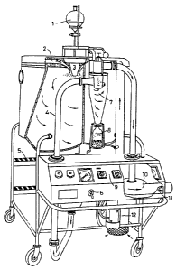
example and with reference to
Figure 1, which is a partly cut away perspective view from the front and one
side of suitable spray-drying apparatus for the first stage of the process of
the
invention,
Figure 2, which is a graph showing how the degree of fixation of the
microsphere walls (in this case albumin) may be controlled by varying the
temperature and the heating tiine in the second step of the process,
Figure 3, which is a graph showing how the pressure resistivity of the
microspheres may be varied by altering the length of the heating time in the
second step of the process,
Figure 4 is a graph showing how the in vitro biodegradation rate may be varied
by varying the length of heating time in the second step of the process,
assessed
by a turbidimetric measurement to measure disappearance of microcapsules,
and
Figures 5a and 5b are respective still copies from video tape showing the
appearance of pig myocardium before and after injection of 4 million of the
large microcapsules of the invention into the left ventricle.
WO 94/08627 PCT/GB93/0*
21~67.83 26
GENERAL PREPARATIVE EXAMPLE 1
A suitable spray dryer (Figure 1) is available from A/S Niro Atomizer,
Soeborg, Denmark under the trade designation "Mobile Minor". Details of its
construction are given iininediately before the claiins herein. It coinprises
a
centrifugal atomizer (Type M-02/B Minor), driven by an air turbine at an air
pressure of min 4 bar and up to max 6 bar. At 6 bar an atomizer wheel speed
of approx 33,000 rpm is reached. Turning on and off the compressed air to the
atomizer is done by means of a valve placed in the instrument panel. The
maximum consumption of compressed air to the atomizer is 17 Nm3/h at a
pressure of 6 bar. All parts coining into contact with the liquid feed and
powder are made of stainless steel AISI 316, except for the pump feed tube and
the atomizer wheel, which is inade of stainless steel AISI 329, made to resist
high centrifugal force. The stainless steel interconnecting pipe system 4 can
easily be stripped down for cleaning.
The drying chamber has an inside made of stainless steel AISI 316, well
insulated with Rockwool, and covered outside with a mild steel sheeting. The
drying chamber is provided with a side light and observation pane for
inspection during the operation and steps 5 for access to the chamber top. The
roof of the drying chamber is inade inside of stainless steel AISI 316 and
outside of stainless steel AISI 304. There is a switch 6 for an air valve for
activation of the pneumatic lifting device when raising the chamber lid.
An air disperser 2 made of stainless steel AISI 304 is used for distribution
of
the air in the drying chamber in order to achieve the best possible drying
effect.
Swirling air is directed around the vaned disc atomiser. An air duct, made of
stainless steel AISI 316, provides lateral transportation of the exhaust air
and
the powder to the cyclone 7, which is made of stainless steel AISI 316 and
designed to separate the powder and air.
*WO 94/08627 2146793 PCr/GB93/02091
27
A closing valve of the butterfly valve type, also made of stainless steel AISI
316 and having a gasket of silicone rubber, is used for powder discharge under
the cyclone into a powder collecting glass jar 8 tightly placed under the
cyclone
by means of a spring device.
A centrifugal exhaust fan 10 made of silumin, coinplete with 3-phase squirrel-
cage motor, 0.25 kW, and V-belt drive with belt-guard, draws air and powder
through the drying chamber and cyclone. There is a switch 11 for air flow
control via a damper.
An air heater 12 heats the drying air by means of electricity (total
consumption
7.5 kWh/h, infinitely variable) and can give inlet air temperatures of up to
about 350 C, although this is generally too high for preparing the
microspheres
of the invention.
The evaporative capacity is as follows:
Evaporative capacity
Drying Air Inlet Air Outlet Air Evaporative
Temperature Temperature Capacity
85 kg/h 150 C 80 C 1.3 kg/h
85 kg/h 170 C 85 C 1.7 kg/h
80 kg/h 2000C 900C 2.5 kg/h
80 kg/h 240 C 90 C 3.4 kg/h
75 kg/h 350 C 90 C 7.0 kg/h
Equipment for two-fluid nozzle atomization may be added, which is made of
stainless steel AISI 316, consisting of entrance pipe with nozzle holder and
nozzle, to be placed in the ceiling of the drying chamber. The equipment
WO 94/08627 2146PCT/GB93/0~
'~~~
28
includes an oil/water separator, reduction valve and pressure gauge for
compressed air to the two-fluid nozzle. Consumption of coinpressed air: 8-15
kg/h at a pressure of 0.5-2.0 bar (0.5-2.0 x 10⁵ Pa).
A suitable feed pump for transport of wall-forming preparation feed from a
reservoir 1 to the atoinizer nozzle 3 is a peristaltic pump. The pump is
provided with a motor (1 x 220V, 50 Hz, 0.18 kW) and a continuously variable
gear for manual adjustment. A feed pipe made of silicone hose leads from a
feed tank (local supply) I through the feed pump to the rotary or nozzle
atomization device 3.
An absolute air filter, consisting of prefilter, filter body in stainless
steel and
absolute air filter, is used for the treatinent of the ingoing drying air to
render
it completely clean. The whole apparatus is controlled via an instrument panel
9.
A 20% solution of sterile, pyrogen-free rHA in pyrogen-free water (suitable
for
injection) was pumped to the nozzle of a two fluid nozzle atomiser mounted in
the commercial spray drying unit described above. The peristaltic pump speed
was maintained at a rate of approxiinately 10 ml/ininute such that with an
inlet
air temperature of 220 C the outlet air teinperature was maintained at 95 C.
Compressed air was supplied to the two fluid atoinising nozzle at 2.0-6.0 Bar
(2.0-6.0 x lOSPa). In this range inicrospheres with a mean size of 4.25-6.2
Fcm
are obtained.
Typically an increase in inean particle size (by reduced atomisation pressure)
led to an increase in the amount of microspheres over 10 m in size (see Table
1).
CA 02146783 2003-11-05
WO 94/08627 PCT/GB93/02091
.
29
TABLE I
EFFECTS OF ATOMISATION PRESSURE ON FREOUENCY OF
MICROSPHERES OVER 10 M IN DIAMETER
Atomisation Pressure % Frequency over 10 m
(x 105 Pa)
6.0 0.8
5.0 3.0
3.5 6.6
2.5 8.6
2.0 13.1
In the second step of the process, 5 g of inicrospheres were heated in a glass
beaker using a GallemkampTM fan oven. A temperature of 175 C for 1 hour was
sufficient to yield microspheres with 100% fixation as determined by HPLC.
The effect of this heat fixation was to increase the in vitro echogenic half
life
from a few seconds to in excess of 30 minutes. By altering the temperature
and length of incubation it is possible to vary the degree of fixation between
about 5% and 100%. Examples of heat fixation profiles of varying
temperatures are shown in Figure 2.
Following heat fixation, the microspheres were deagglomerated and dispersed
into water in one of two ways. Method I involved first mixing the heat fixed
spheres with an equal weight of finely inilled lactose (mean diameter 5 m).
The mixture was then passed through a FritschT"' centrifugal mill with a 0.5
mm
screen and 12 tooth rotor. The milled spheres were collected and passed
through the mill a second time to ensure complete mixing had occurred. The
milled powder was then resuspended in water containing I mg.ml-' Pluronic
F68T"'. Typically 10 g of microspheres and lactose was added to 100 ml of
water
CA 02146783 2003-11-05
WO 94/08627 PCT/GB93/02091
30 =
and Pluronic F68T"'. Method 2 for deaggloineration involves adding 5 g of the
heat-fixed microspheres to 100 nil of water containing 100 mg of Pluronic F68.
The microspheres were dispersed using a SilversonTM homogeniser (model L4R
with a 2.54 cm tubular hoinogenising probe and a high shear screen) and
homogenising for 60 seconds.
The resuspended spheres were separated into intact (gas containing) and broken
spheres using a flotation technique. The gas-containing spheres were seen to
float to the surface over a 1 hot,r period and were decanted froin the sinking
fraction which does not contain the gas reqtiired.
The separation process can be accelerated by centrifugation. A 30 second
centrifugation at 5000 x g is stifficient to separate the two fractions.
Following separation the intact microspheres were freeze-dried in the presence
of lactose and Pluronic F68T"', Optimal conditions for freeze drying involved
resuspending 30 mg of inicrospheres in 5 ml of water containing 50 mg of
lactose and 5 mg of Pluronic F68T"1. The freeze-dried microspheres can be
redispersed in a liqiiid (eg water, saline) to give a monodisperse
distribution.
GENERAL PREPARATIVE EXAMPLE 2
The process of Example I was repeated btit with the following differences in
the first step: a centrifiigal atomiser was used instead of a two fluid
nozzle; the
inlet temperature was 150 C (with the otttlet air temperature still being
sustained at 105 C); and compressed air was stipplied to the nozzle at 1.0-6.0
x 10S Pa. The wheel rotated at 20-40,000 rpm and delivered droplets, and
subsequently microspheres, with a ntiinber inean diameter in the 1.0-8.0 m
range.
*WO 94/08627 2146783 PCT/GB93/02091
31 ' "
GENERAL PREPARATIVE EXAMPLE 3
The second step of the process of Exainple 1 or 2 was varied as follows. A
small aliquot of the microspheres (0.5 g) was heated in a microwave oven such
that it received 300-350 watt hours of microwave heat at 2500 mHz. This
yielded microspheres in which 90-95 % of the monoineric rHA was insoluble
(as determined by gel perineation chlomatography) and as a result of this heat
fixation their in vitro echogenic half-life increased from a few seconds to in
excess of 30 minutes.
GENERAL PREPARATIVE EXAMPLE 4
The second step of the process of Example 1 or 2 was varied as follows. A
small aliquot of the microspheres (0.5 g) was sealed under argon in a glass
vial. The vial was cooled to 4 C and then irradiated with a 60Co gamma
radiation source to deliver a 15.0 kGy dose of gamina rays. The irradiation
resulted in the formation of inicrospheres in which 10-15% of the monomeric
albumin was insoluble.
GENERAL PREPARATNE EXAMPLE 5
The second step of the process of Exainple 1 or 2 was varied as follows. A
-small aliquot of the microspheres (0.5 g) was sealed under argon in a glass
vial. The vial was cooled to 4 C and then irradiated with a 60Co gamma
radiation source to deliver a 50.0 kGy dose of gainma rays to the
microspheres.
Following irradiation, the microspheres were incubated in oxygen at 50 C for
6 hours. The irradiation resulted in the formation of microspheres in which 50-
60% of the monomeric rHA was insoluble.
WO 94/08627 21467 83 32 PCT/GB93/020
GENERAL PREPARATIVE EXAMPLE 6
The second step of the process of Exarnple 1 or 2 was varied as follows.
A small aliquot of microspheres (0.5 g) was resuspended in 5 ml of ethanol,
chloroform or methylene chloride containing a) 1.5% glutaraldehyde, b) 2.0%
diphthaloyl chloride or c) 5.0% formaldehyde. The microspheres were stirred
for varying times from 10 ininutes to 3 hours. The microspheres were
removed by filtration and washed thoroughly in the original organic buffer
containing 5% ethanolamine, in order to remove excess cross-linking agent.
Finally the microspheres were washed in organic solvent and vacuum dried to
remove any residual solvents. The extent of insolubilisation may be varied
from 5-100% by this inethod resulting in the extension of in vitro echogenic
half-life from 1-2 minutes to in excess of one hour.
GENERAL PREPARATIVE EXAMPLE 7
The two independent steps of microsphere forination and insolubilisation of
the
shell may be combined in a single process. In this example, the formation of
the microspheres and the insolubilisation of the polymeric material are
achieved
simultaneously during the spray drying process.
A solution of rHA was fed by peristaltic pump to a small reaction chamber,
with a separate feed line supplying a 5% solution of a suitable crosslinking
agent, eg glutaraldehyde, diphthaloyl chloride or forinaldehyde. The residence
time in the reaction chamber was such that initial adduct formation between
the
crosslinking agent and the protein was achieved, but intraprotein crosslinking
was prevented. The reaction vessel outlet was fed directly to the two fluid
nozzle atomisers mounted in a specially adapted spray drying unit, capable of
handling volatile solvents. The conditions of spray drying were as outlined in
CA 02146783 2003-11-05
WO 94/08627 PCr/GB93/02091
= 33
Example 1. The microspheres were incubated dry at room temperature to
allow intraprotein crosslinks to form and then suspended in ethanol containing
5% ethanolamine to quench any remaining crosslinking agent. Thorough
washing of the microspheres was perfor-ned and finally the microspheres were
vacuum dried to remove residual solvent.
GENERAL PREPARATIVE EXAMPLE 8: ASSAY OF FREE
MONOMERIC rHA IN MICROSPHERES
A I ml volume of ethanol was added to 100 mg of microspheres in a 20 ml
glass bottle and sonicated for 30 seconds. To this suspension 19 ml of H,O
were added.
The mixture was centrift-ged in a bench-top microfuge (Gilson) for 20 seconds
and the clear fraction assayed. The assay was performed by loading 50 ml of
the fraction automatically onto a ShimadzuTM LC6A HPLC and chromatographing
on a TSK gel permeation coluinn at a flow rate of 1 ml minute' using sodium
phosphate buffer (pH 7.0).
The peak heights representing the rHA monomer were recorded and used to
determine the concentration of monoiner using a standard curve between 1 and
10 mgml'' monomeric rHA.
The %-free monomeric rHA was caiculated by measuring the monomer
concentration in the fixed microspheres and representing this figure as a
percentage of the monomer concentration of the unfixed microspheres. The
results are given in Figure 2.
Heating of the spray dried -nicrospheres in an oven (as described in Example
1) results in a decrease in the ainount of monomer that can be detected (see
CA 02146783 2003-11-05
WO 94/08627 PCT/GB93/02091
34
Figure 2). This decrease in detectable inonomeric rHA is due to the
denaturation and crosslinking of monoineric rHA into insoluble polymers that
cannot be assayed by the aforeinentioned HPLC inethod.
Using the HPLC method to assess rHA monitor levels, it is clear from Figure
2 that after 15 minutes inctibation there is no free inonomeric rHA present in
the rHA microspheres. However it is still possible to further crosslink the
rHA
microspheres by heating for longer periods.
This prolonged heating results in an increased level of microsphere
crosslinking
which in turn produces microspheres of increasing strength which are
correspondingly more resistant to pressure.
By careful control of temperature and time of incubation, it is possible to
produce microspheres with a controlled range of crosslinking (and hence
pressure resistivity and biodegradation rate).
GENERAL PREPARATIVE EXAMPLE 9: EFFECTS OF INCUBATION
TI1WE AT 1750C ON THE PRESSURE RESI ST7VITY OF rHA
MICROSPHERES
A batch of rHA microspheres from the initial spray-drying step of the process
was divided into 5 g aliquots and baked at 175 C for varying lengths of time
as shown in Figure 3.
Following heat fixation the ainount of free inonomer was determined as
described in Example 8. For each of the incubations shown, there was no
monomeric rHA detectable.
The heat-fixed microspheres were disaggregated using a FritschT"" centrifugal
mill
CA 02146783 2003-11-05
WO 94/08627 PCr/GB93/02091
= 35
(as described above) and intact, air-containing microspheres recovered by the
aforementioned flotation techniqtie. The recovered microspheres were
suspended in H,O containing Pluronic F68T"" (1 mgml') at a concentration of
0.5
x 10g capsules ml''.
The resuspended, air-containing microspheres were subjected to increased
atmospheric presstire by applying pressure with a 50 ml syringe whilst
containing this suspension in a closed container (25 ml polystyrene
container).
For each of the pressures assessed, the individual microsphere suspension was
pressurised to the selected presstire and inaintained at this pressure for 5
seconds before releasing the presstire. For each suspension analysed the
pressure increase was performed 3 tiines. The presstire in the closed
container
was assessed by an RS hand-held manoineter.
Following pressurisation the microsphere suspensions were assessed by light
microscopy and image analysis and the % air-containing to non-air-containing
microspheres assessed. This analysis is perforined since only the air-
containing
microspheres are functional in enhancing ttltrasotind echocontrast.
As can be seen in Figure 3, inicrospheres that are fixed for 60 minutes at
175 C, as described in Example 1, are stable at all of the pressures to which
they were subjected in this experiment.
By careful control of the length of inctibation at this particular temperature
(175 C) it is possible to produce batches of microspheres with different
degrees
of crosslinking which in ttirn are resistant to varying degrees of pressure
increase.
CA 02146783 2003-11-05
WO 94/08627 PCT/GB93/02091
36
Using this careful control of crosslinking by adjusting the length and
temperature of incubation it is possible, to produce batches of air-containing
microspheres that are specifically designed to withstand a designated pressure
increase.
The temperature used to crosslink the microspheres can vary infinitely, as can
the length of incubation time.
GENERAL PREPARATIVE EXAMPLE 10: MICROSPHERE
CLASSIFICATION
An advantage of the process of the invention is that it enables the median
size
and size distribution of the inicrospheres to be controlled. However, one can
further select desired sizes if one wishes, for example by flotation. In a
homogeneous dispersion of inicrospheres, larger particles will rise to the
surface faster than smaller particles due to the lower density (more
encapsulated
air) of the larger particles. Hence, by allowing the dispersion to stand, the
particle size distribution will change at any level of the solution with
respect to
time.
Microspheres were dispersed in 2000 ml of aqueous solution containing 6%
w/v sodium chloride and 0.1% w/v Pluronic F68T"" in a glass bottle giving a
liquid column of approximately 165 mm. A sampling tube was placed 50 mm
below the upper liquid surface to enable reinoval of samples at timed
intervals.
By altering the standing time and sodium chloride concentration, it was
possible
to produce a variety of particle size distributions and classify microspheres
down to 2 m.
Other wet techniques for classification include hydrodynamic chromatography
CA 02146783 2003-11-05
WO 94/08627 PCi'/GB93/02091
~ 37
and field flow fractionation. 'Dry' techniques using the principles of
elutriation
and cross flow separation are cominercially available in the form of the
MicrosplitT"" (British Rem.), Zig-zagTM (Alpine) and TurboTM (Nissuin)
classifiers.
The elbow jet classifier produced by Nitettsu Mining Co uses a different
principle (the Coanda Effect) which could also achieve good results for the
classification of microspheres.
SPECIFIC EXAMPLE 1
A solution of human albumin (5% w/v) is spray-dried at an inlet temperature
of 220 C and an air pressure of 1.5 bar as in General Preparation Example 1.
The resulting particles are heat fixed for a period of 20 minutes at 175 C in
an
air oven. The samples are deaggloinerated by milling with mannitol and the
particles are resuspended in a solution of 10 ing/ini mannitol and 0.06 mg/ml
pluronic F68. The intact particles are creamed off and the microsphere
suspension is freeze-dried.
Particles predominantly of 10-20 in are produced which contain air and are
substantially pressure resistant.
SPECIFIC EXAMPLE 2
Polylysine at a concentration of 5 % w/v was resuspended with the microspheres
of General Preparative Example 2 (100 x 106 particles/ml) and incubated
overnight at 20 C. Mannitol and Pluronic F68T"" were added at the
concentration
described in Specific Example.1 and the suspension was subsequently flash
frozen and freeze dried.
CA 02146783 2003-11-05
WO 94/08627 PCT/GB93/02091
38
SPECIFIC EXAMPLE 3
Hyaluronic acid at a concentration of 5% w/v was incubated overnight with
resuspended microspheres prepared as in General Preparative Example 1 at
20 C (100 x 106 microspheres/nil). Mannitol and Pluronic F68TM were added to
a concentration of 10 and 0.06 mg/ml respectively and the suspension then
flash
frozen and freeze dried.
SPECIFIC EXAMPLE 4
Microspheres according to General Preparative Example 3 were resuspended
in a solution of DMF (Diinethylforinainide) at a concentration of 100 x 106
particles/ml. Acetic anhydride was added to give a final acid anhydride
concentration of 100 mg/nil. The inicrosphere mixture was incubated at 20 C
for 1 hour then diluted with water and filtered and washed with excess water
over a 1 hour period. The inicrospheres were formulated in Mannitol and
Pluronic F68 as described above. This method imparts negative charges.
SPECIFIC EXAMPLE 5
Microspheres according to General Preparative Example 1 were resuspended
in an aqueous solution at a concentration of 100 x 106 particles/ml. An
aqueous solution of carbodiimide was added to the microsphere suspension to
give a final concentration of 100 mg/ml. After incubation at 16 hours at 20 C,
excess reagent was quenched by the addition of glycine to an equivalent
concentration and further incubation for 16 hours at 20 C. The microspheres
were washed with water then formulated as described above. This procedure
eliminates negative charges.
CA 02146783 2003-11-05
WO 94/08627 PCr/GB93/02091
~ 39
SPECIFIC EXAMPLE 6
Microcapsules of general preparative method 2 were formulated with polaxamer
407 and mannitol at a concentration of 0.1 and ] 0ing/inl respectively. The
suspension was flash frozen and freeze dried as described in the earlier
examples.
SPECIFIC EXAMPLE 7
Poly-L-lysine (15-25 kDa) was added to the rHA feedstock (20% w/v) to a
final concentration of 0.5 % w/v prior to spray drying. The method of general
example 2 was followed to yield inicrocapsules with increased positive charge
upon the shell.
SPECIFIC EXAMPLE 8
Poly-L-glutamate (15-30 kDa) was added to the rHA feedstock (20% w/v) to
a final concentration of 0.5% w/v prior to spray drying. The method of
general preparative example 2 was followed to yield microcapsules with
increased negative charge upon the shell.
SPECIFIC EXAMPLE 9
Microspheres of Specific Exainple I may be used in an in vivo analysis to
establish the feasibility of delineating perfusion territories in the
myocardium
of a pig heart.
A 25 kg Yorkshire swine is anaesthetised and fully ventilated according to the
methodology outlined in Ten Cate et al (1992) CardiovascularResearch 26, 32-
39. A 5 French catheter is inserted via the femoral artery, ascending aorta
and
CA 02146783 2003-11-05
WO 94/08627 PCT/CB93/02091
40 r
aortic root into the left ventricle. Injection of 4 million microcapsules of
Specific Example 1 is made and 2 diinensional transthoracic echocardiography
in the short axis plan using a Hewlett PackardT"' sono's 1000, equipped with a
3.5
MHz transducer, is used to assess regional perfusion. Intense opacification of
the myocardium was observed (see Figure 5), showing that no redistribution of
hollow microcapsules occurred over the 2 hour period. Subsequent injections
of microcapsules into the left ventricle resulted in sequential dose-dependent
brightening of the myocardiuin. Haeinodynamic parameters were monitored
and showed no adverse effect of injection of these low levels of
microcapsules.
SPECIFIC EXAMPLE 10
Microcapsules of Specific Exainple 6 were injected into the ear vein of a
mildly
sedated New Zealand rabbit (4.5 kg) at a concentration of 300 million
particles/ml. Femoral artery Doppler signals were assessed using an
InterspectT""
7000 model equipped with a 10 MHz transducer. Baseline signals prior to
contrast injection were obtained to enable comparison of Doppler signals
before
and after contrast injection. Once baseline signals were obtained, the
instrument's time intensity gain controls were not altered.
Following contrast injection, visible prolonged Doppler enhancement of the
myocardium was obtained, lasting for several beats or several minutes
depending upon the dose size of contrast agent administered.
The T'fi was determined by videodensitometry of the spectral Doppler signals
as follows. The gain settings were adjusted to give barely visible signals
before
contrast injection. As the contrast entered the femoral artery the signal
increased, peaked and then decayed. Videodensitometry was performed on the
individual peaks of flow and a tiine intensity curve plotted. The T'h was
calculated as the time taken for the contrast effect to diminish to half its
peak
OWO 94/08627 2146783 PCT/GB93/02091
41
value. Videodensitometry of spectral Doppler signals revealed a reproducible
contrast effect following intravenous injection of the inicrocapsules which
was
significantly prolonged over the signals produced by microcapsules formulated
according to PCT/GB92/00643.