Note: Descriptions are shown in the official language in which they were submitted.
2I~76Il
_W094/13227 PCT~S93/12086
TRAPEZIAL IMPLANT
TECHNICAL FIELD
The subject invention relates generally to a
prosthetic bone implant and more particularly to a
trapezial resurfacing prosthesis.
BACKGROUND ART
Conditions such as osteoarthritis, cancer or trauma
may cause degeneration of the articular surfaces between
the trapezium and the first metacarpal in a hand causing
the patient ~i~comfort and sometimes severe pain during
thumb circumduction.
Various total replacement prostheses have been
propo~ed for bones in the human wrist. For example, United
States Patent Number 4,936,860 to Swanson, issued June 26,
1990, discloses a metallic total scaphoid replacement
implant. Also, United States Patent Numbers 4,955,915 and
4,969,908 both to Swanson, issued September 11, 1990 and
November 13, 1990, respectively, disclose total lunate
replacement implants. Although not directed to a carpal
bone implant, United State Patent Number 4,936,854 to
Swanson, issued June 26, 1990, discloses an implant
anchored in the radius and having a cupped, or dished,
surface for stabilizing the proximal carpal row and
preventing ulnar migration thereof.
As an alternative to total carpal bone replacement, it
is well known in the art to resurface the distal surface of
the trapezium with a prosthetic implant. For example, the
Silastic~ Trapezial Implant H.P., manufactured by Dow
Corning Wright Corp. 5667 Airline Road, Arlington,
TP~n~SS?e 38002, is made from medical grade silicone rubber
elastomer for use as an interpositional spacer between the
WO94/13227 21 ~ 7 6 ll PCT~S93/12086
trapezium and the first metacarpal joint of the thumb. The
implant is provided with a short cylindrical stem exten~ing
from its proximal surface which fits into a cavity prepared
in the trapezium. The head, or distal articular surface,
of the implant is thin and dome-shaped.
One of the goals of implant design is to minimize the
production of wear debris particles. Wear particles can
never be completely eliminated with presently available
implant materials because all moving parts, e.g., implants
that articulate against bone, wear to some degree. It is
generally believed by some that elastomers generate wear
particles at a more accelerated rate than inelastic
materials. However, because the distal articular surface
of the aforementioned Silastic~ Trapezial Implant H.P. is
domed, i.e., not anatomically representative of a natural
trapezium, that implant must be manufactured from an
elastomer which has the ability to deform to better
articulate with existing joint anatomy.
SUMMARY OF THE INVENTION AND ADVANTAGES
The subject invention provides an implant for
attachment to the distal surface of a prepared trapezium.
The trapezial implant comprises a base member having a
proximal attachment surface and a distal articular surface,
and anchor means exten~;ng from the proximal attachment
surface for anchoring or fixing the base member to the
prepared surface of the trapezium. The distal articular
surface includes a convex medial portion, a convex lateral
portion, and a concave central portion between the medial
and lateral portions defining a distinctive and complex
surface curvature for providing stable support to an
adjacent first metacarpal through the entire arc of thumb
circumduction. That is, the complex surface curvature
provides improved articulation with the adjacent first
metacarpal because its configuration closely replicates the
normal human distal trapezium articular surface. In other
_~VO94/13227 21 4 7 611 PCT~S93/12086
words, the distal articular surface mimics the natural
joint anatomy.
The specific shape of the distal articular surface
provides improved articulation between the distal articular
surface of the implant and the first metacarpal thereby
reducing the-production of wear particles due to attrition.
Further, because the distal articular surface is formed
having medial and lateral convex portions and a central
concave portion, the base member can be fabricated from an
in-elastic, less abradable materials than the prior art
elastomers. Thus, the subject invention overcomes a need
inherent in the prior art domed trapezial implants
fabricated from silicone elastomers.
BRIEF DESCRIPTION OF THE DRAWINGS
Other advantages of the present invention will be
readily appreciated as the same becomes better understood
by reference to the following Detailed Description when
considered in connection with the accompanying Drawings
wherein:
Figure l is a simplified dorsal view of a human
skeletal hand showing the distal articular surface of the
trapezium excised;
Figure 2 is a view as in Figure l showing the subject
trapezial implant operatively positioned on the trapezium;
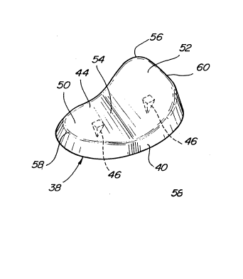
Figure 3 is a perspective view of a trapezial implant
according to the subject invention;
Figure 4 is a plan view of the implant as in Figure 3;
Figure 5 is a front elevational view of the trapezial
implant as in Figure 4;
Figure 6 is a plan view of a first alternative
embodiment of the trapezial implant;
Figure 7 is an end view of the trapezial implant as in
Figure 6;
Figure 8 is a perspective view of a second alternative
emhoAiment of the trapezial implant;
WO94/13227 21 47 611 PCT~S93/12086
Figure 9 is a plan view of the trapezial implant as in
Figure 8; and
Figure l0 is a front elevational view of the trapezial
implant as in Figure ~.
DETAILED DESCRIPTION
, ..
Referring to the Figures,A wherein like numerals
indicate like and correspondi~g parts throughout the
æeveral views, a human hand skeleton is generally shown at
20 in Figures l and 2. The hand 20 includes first 22,
second 24, third 26, fourth 28, and fifth 30 metacarpals.
The first metacarpal 22 is located medial relative to the
fifth metacarpal 30. The hand 20 has a carpus comprising
eight bones, including the trapezium 32.
Some physical conditions, e.g., arthritis, cancer,
trauma, etc., npcessitate arthoplastic surgery to
prosthetically improve the articular surfaces between the
first metacarpal 22 and the trapezium 32. The prior art
has taught both total replacement as well as resurfacing of
the trapezium 32. In many instances, resurfacing is
preferred and more conservative of bone than total excision
of the trapezium 32.
The trapezium 32 is prepared for the hemiarthroplasty
as follows. The carpometacarpal joint of the thumb is
~Y~oeeA through a transverse or curved longitudinal
incision. The superficial sensor branch of the radial
nerve is identified and protected. The abductor pollices
longus tendon is detached at its insertion and the extensor
pollices brevis tendon is retracted. The joint capsule is
opened and preserved. The marginal osteophytes at the base
of the metacarpal 22 and trapezium 32 are removed with a
rongeur. Distal traction of the thumb allows the joint
surfaces to be well-visualized. With either an osteotome
or power sagittal saw, the saddle 34 of the trapezium 32 is
converted to a flat surface 36, and the medial osteophyte
_,~094/13227 214 7 611 PCT~S93/12086
is removed. Excision of this medial spur permits further
debridement of the capsule and reduction of the subluxed
metacarpal. It is imperative that all bony obstruction to
metacarpal reduction be removed. Figure l illustrates the
distal articular portion or saddle 34 resected from the
trapezium 32, such as by conventional procedures as
described above. This saddle 34 is excised to form a
stable, transverse interface 36 to eliminate shear
stresses.
In place of the excised saddle 34, a trapezial
implant, generally indicated at 38 in Figures 2-5, is
attached to the trapezial bone 32, as ehown in Figure 2.
The implant 38 includes a base member 40 preferably
fabricated from an inelastic and abrasion resistant
material, such as commercially pure titanium, other medical
grade metals or alloys, or alumina, zirconia or silica
ceramice. The base member 40 includes a generally planar
proximal attachment surface 42 structured to engage in
surface-to ~r face contact with the interface 36 of the
prepared trapezium 32. The base member 40 further includes
a distal articular surface 44 constructed and adapted to
mimic the natural geometry of the saddle 34.
In addition to the materials listed above, the implant
38 can be fabricated from zirconium with a coating of
zirconium oxide for improved abrasion resistance. The
implant 38 can also be formed of zirconium alloys including
titanium, niobium, hafnium and other metals known to form
stable alloys with zirconium.
An anchor means extends from the proximal attachment
surface 42 of the base member 40 for anchoring the base
member 40 to the trapezium 32. As illustrated in Figures
3-5, the anchor means preferably includes at least a pair
of spaced posts 46 exten~ing perpendicularly from the
proximal attachment surface 42. Each of the posts 46 taper
proximally to a pointed tip 48. As shown in Figures 3 and
WO94/13227 21~7 611 PCT~S93/12086
4, the posts 46 are preferably pentahedral, or pyramidal,
but also may be conical, tetrahedral, or other simple
pointed shape, and have a length substantially less than
prior art cylindrical stems. Such short posts 46 allow
improved conservation of the trapezium 32, as well as
requiring less joint distraction during surgical
implantation. ~`~
During implantation, the pointed posts are self-
aligning and readily received into the softer cancellous
interior of the resected trapezium 32. Thus, it is
unnecessary to prepare receiving pilot holes in the
trapezium 32 for the posts 46. For added stability, the
proximal attachment surface 42 can be coated with
hydroxylapatite or other osteoconductive material prior to
implantation. To further aid fixation, the proximate
attachment surface 42 can be roughened or made porous to
promote bone ingrowth. However, since the joint is under
compressive forces at all times, the posts 46 should be
adequate to achieve fixation.
The distal articular surface 44 of the base member 40
includes a convex medial portion 50, a convex lateral
portion 52, and a concave central portion 54 between the
medial 50 and lateral 52 portions. This complex curvature
of the distal articular surface 44 provides stable support
to the adjacent first metacarpal 22 throughout the entire
arc of thumb circumduction. Further, this unique and
advantageous geometry requires little or no alteration of
the normal carpometacarpal joint capsular support.
As best shown in Figure 4, the subject implant 38
includes a perimeter comprising a convex curvature adjacent
each of the medial 50 and lateral 52 portions and at least
one concave curvature adjacent the central portion 54. In
this preferred configuration, the distal articular surface
44 is generally anatomically representative of a natural
human trapezium, i.e., one without the degenerative effects
21476il
_ tVO 94/13227 ~ PCT~S93/12086
of disease or trauma. The perimeter, therefore, comprises
a somewhat oval shape having a pronounced jutting end
adjacent the lateral portion 52. Therefore, the lateral
portion 52 has a great~r breadth than the medial portion
50.
The medial portion 50 includes a medial crest 58.
Likewise, the lateral portion 52 includes a lateral crest
60 which is of greater length, or elevation, than the
medial crest 58. Each of the medial 58 and lateral 60
crests define and establish the convex curvatures of the
medial 50 and lateral 52 portions, respectively.
Therefore, when placed in operation, the proximal articular
surface of the first metacarpal 22 seats within the concave
central portion 54 and, during circumduction, reacts along
the distal articular surface 44 between the medial crest 58
and the lateral crest 60.
The natural, anatomically correct, curvature of the
distal articular surface 44 enhances articulation of the
first metacarpal 22 and thereby provides substantially
enhanced comfort, as well as reduced likelihood of
abrasion. It will be appreciated that because the base
member 40 illustrated in Figures 2-5 is substantially
anatomically representative of a natural trapezium, right
and left base members 40 are required due to the differing,
mirror-imaged, bones of the trapezium in the natural human
hand.
According to a first alternative embodiment of the
subject invention as shown in Figures 6 and 7, the base
member 40' can be constructed in a symmetrical fashion,
thereby obviating the need for left and right designations.
However, symmetry is achieved at the expense of
performance, since the anatomically correct implant 38
shown in Figures 2-5 has been found through testing to
provide superior results. For convenience, single prime
designations are used to represent corresponding elements
WO94/13227~ 21 ~ 7 6 11 PCT~S93/12086
to those described above. The distal articular surface 44'
of the implant 38' shown in Figures 6 and 7 includes a
perimeter having a generally waisted, or hourglass, oval
shApe. Thus, as shown in the plan view of Figure 6, the
perimeter takes on a somewhat peanut shell shape. Like the
previous shape, the medial portion 50' and lateral portion
52' are each convex and are spaced on opposite sides of a
concave central portion 54'. Because of the symmetrical
nature of the implant 38' of Figures 6 and 7, the elevation
of the medial 58' and lateral 60' crests must be
equivalent.
A second alternative embodiment of the subject implant
38'' is shown in Figures 8-10. Double prime designations
are used to indicate like or corresponding parts with those
~ above for the sake of convenience. The second
alternative implant 38'' has a perimeter shape comprising
a continuously convex curvature. As best shown by the plan
view of Figure 9, the perimeter has a generally ovoid, or
elliptical, shape. The distal articular surface 44''
provides increased surface area in a concave central
portion 54'' compared to either of the previous
embodiments. And, like the first alternative embodiment of
Figures 6 and 7, the implant 38'' is symmetrical so that
left and right designations are not required.
The subject trapezial implant 38, 38', 38'' shown in
the Figures overcomes the disadvantages of the prior art by
means of an anatomically-correct distal articular surface
44 providing stable support to the adjacent first
metacarpal 22 throughout the entire arc of thumb
circumduction. As a result, fewer wear particles are
generated using implants of the invention than obtained
with prior art prostheses. This invention permits material
selection for the implant 38 to include hard, abrasion-
resistant metals, alloys and ceramics rather than softer
elastomers. Further, the anchor means is specifically
structured to reduce joint distraction during implantation,
214761~
~094/13227 PCT~S93/12086
further allowing improved conservation of the bone of the
trapezium 32. Therefore, the subject implant 38 is easier
to install than prior art implants.
Although description of the preferred emhoA;ment has
for~ upon the location of the implant 38 upon the distal
trapezium 32, it will be readily appreciated by those
skilled in the art that equally successful results can be
obt~i~eA when the implant 38, or one of the alternative
implants 38', 38'', is located in other small joints of the
body, e.g., the finger or toe joints.
The invention has been described in an illustrative
manner, and it is to be understood that the terminology
which has been used is intended to be in the nature of
words of description rather than of limitation.
Obviously, many modifications and variations of the
present invention are possible in light of the above
teachings. It is, therefore, to be understood that within
the scope of the appended claims the invention may be
practiced otherwise than as specifically described.
