Note: Descriptions are shown in the official language in which they were submitted.
21~82~5
APPARATUS AND METHOD FOR
VOLUMETRIC CAPILLARY CYTOMETRY
Cross-Reference to Related Applications
This is a continuation-in-part of U.S. Ser. No.
08/018,762 filed on February 17, 1993. The present
application is also related to U.S. patent application
entitled "Method and Apparatus for Cell Counting and Cell
Classification", invented by Ning L. Sitzo and Louis J.
Dietz, filed on the same day as the present application
and owned by the same Assignee. The related application
is incorporated by reference as if fully set forth
herein.
This invention relates to automated cytometry
instruments and procedures and to the identification and
enumeration of cellular components of biological fluids.
Rapid identification and enumeration of the various
components of biological fluids is an important research
and diagnostic aim. ~i n; ~q 1 processing and handling of
samples would contribute to the widespread use of such
techniques.
In the case of enumeration of leukocyte subclasses
of human blood, the need for improved techniques is
especially keen. For example, the usefulness of moni-
toring CD4+ lymphocyte levels in noting the progression
from HIV positive status to AIDS has underscored the need
for a fast, inexpensive, and reliable method to analyze
patient blood samples.
Landay et al., "Application of flow cytometry to the
study of HIV infection," AIDS 4:479-497 (1990) describes
the utility of a technique in understq~n~;ng the biology
of HIV infection. Multiple-color flow cytometric
analysis can be applied to the study of HIV disease by
using various monoclonal antibodies to perform phenotypic
analysis of blood samples. This technique is also useful
2148205
-- 2 --
in other immune system determinAtions, as in evaluating
the status of organ transplant or leukemia patients.
Flow cytometry is a well-known technique wherein
cells may be characterized and separated based on
fluorescent emission. A labeled, mono-dispersed cell
suspension travels through a tube in a fine fluid stream
and is presented to an excitation beam. The emitted
fluorescence of each cell is measured by appropriate
detectors and the cells may be split into droplets and
sorted according to given parameters by electrical and
mechanical means.
Flow cytometry may be used to identify and enumerate
specific subclasses of blood cells. For example, in U.S.
Pat. No. 4,284,412 Hansen et al., lymphocytes which have
been reacted with fluorescently-labeled monoclonal
antibodies are separated from red blood cells and
presented one by one to a fixed detector in a flow
cytometry system. Each cell is characterized by analysis
of forward light scatter, right angle scatter, and
fluorescence. This method requires complex sample
preparation and instrumentation. While flow cytometry
has improved assay reliability and reproducibility in
this application, it generally cannot directly provide
absolute cell counts for lymphocyte subsets. Independent
white blood counts and differential white counts are
required to calculate absolute cell counts per unit
volume. In the usual flow cytometry practice, in order
to distinguish lymphocytes from monocytes and
granulocytes, a lymphocyte gate based on forward and side
light scatter patterns must be established for each
sample.
Flow cytometry is not routinely used for identifying
and enumerating lymphocyte subclasses in the presence of
red blood cells, although U.S. Patent No. 4,727,020
2~482~S
-- 3 --
Recktenwald provides a contrary example. Removal of the
red blood cells, by density-gradient separation or
lysing, increases the time, cost and number of blood-
handling steps per assay. Additional blood-h~n~l;ng
steps increase the potential for exposure to blood-borne
infectious agents. As stated above, the resultant data
produced by the flow cytometry method is inadequate for
some purposes. In order to calculate absolute cell count
per unit volume, flow cytometric data must generally be
combined with additional data obtained from other
methods. Also, because flow cytometers conventionally
utilize a fluid stream passing through a small nozzle,
they may generate aerosols which pose an additional
source of biohazardous materials for laboratory
personnel.
An alternative is to fix sample position relative to
the excitation beam. For example, in U.S. Pat. No.
4,758,727 and its divisional, U.S. Pat. No. 4,877,966,
Tomei et al., a method and apparatus for measurement of
low-level laser-induced fluorescence is described. In
this invention, a coherent laser beam is passed through a
three-dimensional scanner and focused onto a static
target. The target is an object such as a monolayer cell
culture or tissue section. A beam spot, having a size as
small as one micron, is passed back and forth across the
target by a scanner whose path and movement rate are
computer-controlled. Fluorescent light is gathered by a
biased-cut fiberoptic base plate and relayed to a
detector positioned on the opposite side of the target
from the beam.
U.S. Pat. No. 5,037,207, also granted to Tomei et
al., discloses a laser imaging system with ~h~nced
spatial resolution and light gathering efficiency which
allows for digital imaging of a target of varying size,
dependent upon the data retrieval and storage limitations
21~s20s
- 4 -
of the supporting computer system. The system utilizes a
novel optical fiber detector assembly and a rapid scan
for collection of all light from every laser spot to
create a quantitative digital reproduction of the image
on the surface of a target.
U.S. Pat. Nos. 5,072,382, Kamentsky, and 5,107,422,
Kamentsky et al., disclose an apparatus and method for
sc~nn;ng a cell population with a beam to generate
multiparameter optical data based on each cell's specific
location. The scan is made of a surface on which cells
have been deposited. A background level is estimated for
the neighborhood surrounding each cell based on digital
data and corrections are made or the background level.
In "Acousto-Optic Laser-Scanning Cytometer,"
Cytometry 9:1Ql-110 (1988) Burger and Gershman and U.S.
Pat. No. 4,665,553 Gershman et al., a laser-scanning
cytometer is disclosed. An optical scan is made of a
lysed and washed sample in a cuvette by a Bragg cell-
controlled scanner. The cuvette is translated in a
stepwise fashion in one direction relative to the
scanner. The scanner operates in a direction
perpendicular to the direction of cuvette translation and
the scan occurs along the ~ide of the cuvette. Once a
cell is located, a beam optimization algorithm operates
to steady the beam on the cell and measurements of
forward light scatter, orthogonal light scatter, and
fluorescence are made. Then the process is repeated.
In U.S. Pat. No. 5,117,466, Buican et al. describe a
fluorescence analysis system in which data from a flow
cytometer establish identification criteria used by a
confocal laser microscope to virtually sort the cellular
components of a sample. Birefringent optics and Fourier-
Transform technology are used to visually select and
~ 21~82~
-- 5
display cells or subcellular structures having the
desired spectral properties.
In "Fluorescence Analysis of Picoliter Samples,"
Analytical Biochemistry 102:90-96 (1980) Mroz and Lechene
teach a method of handling picoliter-volume samples to
gather fluorescence intensity data. Samples are taken up
via syringe in a single siliconized capillary tube with
oil between the samples. Measurements are made of an
optical fluorescence chamber defined by a pinhole
diaphragm, a microscope ob~ective, and the diameter of
the capillary tube.
U.S. patents granted to Mathies et al. are also
relevant to the field of the present invention. In U.S.
Pat. No. 4,979,824, a high sensitivity detection appara-
tus is described. This apparatus is based on a flowcytometry system and utilizes a spatial filter to define
a small probe volume that allows for detection of
individual fluorescent particles and molecules. Laser
power and exposure time of the sample are chosen for the
best signal-to-noise ratio. Real-time detection of
photon bursts from fluorescent particles is used to
distinguish the number, location or concentration of the
particles from background energy.
In U.S. Pat. No. 5,091,652 Mathies et al., a laser-
excited fluorescent scanner is revealed for S~nn; ngseparated samples using a confocal microscope. The
sample is preferably separated by and detected from an
electrophoresed slab gel, but may also be on a membrane,
filter paper, petri dish, or glass substrate. The confo-
cal microscope forms an illumination volume in the geland the beam is oriented so that background scattering is
minimized by the polarization characteristics of the
scattered light.
2148205
-- 6 --
U.S. Pat. No. 5,274,240 also granted to Mathies et
al. and a continuation-in-part of the above patent,
teaches a laser-excited capillary array scanner. This
invention is primarily intended for fluorescence
detection from an array of capillary tubes cont~in;ng
samples that have been separated by capillary
electrophoresis. The fluorescence detection assembly
employs a confocal system to detect fluorescence from the
interior volumes of each capillary tube.
The current cytometry art generally requires time-
consuming and potentially hazardous sample-handling and
component separation steps. It fails to allow for rapid
volumetric identification and enumeration of sub-
populations of a cell suspension that are present within
a mixed population. The techniques of the prior art
often require trained personnel.
It is therefore an object of the present invention
to provide a quick, simple to use, less expensive, safer,
automated apparatus and method for directly obtAin;ng
counts of specific cellular subsets in biological fluids
in a volumetric manner and which require small volumes of
sample and reagent.
The above object has been achieved with an apparatus
and method for identifying and enumerating the cellular
components of a biological fluid in a volumetric manner
based on the formation of fluorescent complexes and the
optical scanning of a capillary tube cont~ining the
sample in a static and minimally processed form. The
fluorescence is detected from throughout a non-flowing
cell suspension and enumeration may be done in a precise
volume for the purpose of obt~ining absolute cell counts.
"Absolute", as defined herein, means the absolute number
of cells per volume as represented by the volume scanned.
As defined herein, "cell" means a whole cell or a part of
21~8205
-- 7 --
a cell. The complexes are the result of a reaction
between fluorescently-labeled binding agents and
corresponding binding sites present in the cellular
components of the fluid. An excitation laser beam is
directed by an optical scanner to a columnar region of
the capillary tube, the columnar region generally defined
by the interior depth ~;me~ion of the capillary tube and
the beam spot of the laser. A spatial filter of
sufficient pin-hole aperture is chosen to selectively
detect the fluorescence emitted throughout the columnar
region and is disposed between the capillary tube and a
detection means. Because no separation of bound and
unbound fluorescently-labeled binding agent is necessary
in the sample, both are viewed as fluorescence by the
detection means. However, areas of heightened
fluorescence intensity occur where the labeled binding
agents congregate, namely on the binding sites present in
the cellular components of the sample. The detection
means, therefore, records a signal of heightened
fluorescence intensity above a given threshold of
background fluorescence as corresponding to a single
cell.
In the preferred embodiment, a laser creates an
excitation beam of a wavelength of 600 to 1000 nanometers
and is focused onto a capillary tube of rectangular
cross-section from a position directly above the
capillary tube. The spot size of the laser beam at the
point of its intersection with the capillary tube is 5 to
15 microns in diameter, depending upon the expected cell
size, and the illuminated depth dimension of the
capillary tube is 25 to 225 microns. As described later,
there is a relationship between the spot size and the
capillary tube depth dimension. In the present inven-
tion, an excitation beam is scanned in two directions to
impinge upon the outer wall of a transparent capillary
tube that is in a fixed position. The first scan
21~8205
-- 8 --
direction follows a path transverse to the longitll~;nAl
axis of the capillary tube, i.e. the wide portion of the
rectangular cross-section of the capillary tube, and
begins and ends at points that are beyond the lateral
boundaries of the capillary tube. The second scan
direction follows a path along the longit~ n~l axis of
the capillary tube. The scan of a known volume of the
capillary tube, achieved by measuring the beginning and
ending points in the second scan direction, or by
beginning and ending the scan at defined points, can be
used to calculate the presence of a particular
subpopulation of cellular components per unit volume,
since the cross-sectional area of the capillary tube is
known.
The apparatus of the present invention is especially
well-suited to the detection of subclasses of blood
cells. In a typical assay, a sample of whole
uncoagulated blood is obtained and incubated with an
excess amount of fluorescently-labeled antibodies that
are directed toward various cell surface markers present
on blood cell subclasses. The fluorophores are chosen so
that they will activate in the wavelength range of the
excitation beam. This wavelength range has also been
specifically selected to ~;n;~;ze interference due to
autofluorescence from blood components not of interest.
The sample cont~;n;ng fluorescently-labeled antibody in
both complexed and free form is generally diluted and
then inserted into the capillary tube. The tube is then
optically scanned at wavelengths necessary to excite the
fluorophores. Based on fluorescent emission from
specific fluorophores used to label specific antibodies,
the number of cells of a certain type per unit volume can
be quickly determined, as can ratios of cell types
present in the blood or other biological fluid sample.
Z14~ZO~
The instrument and technique of the present
invention quickly detect cellular components of biologi-
cal fluids in a precise volume and require m; n;m~l proc-
essing of the sample. The present invention substantial-
ly cuts down on assay times and costs and requiresminimal hAn~l;ng of samples, an especially important
precaution during the examination of blood samples.
Because no special instruments are necessary for
processing the samples and the number of requisite
reagents is kept to a m;n;mllm~ the pre~ent invention is
well-adapted for use in a clinical setting.
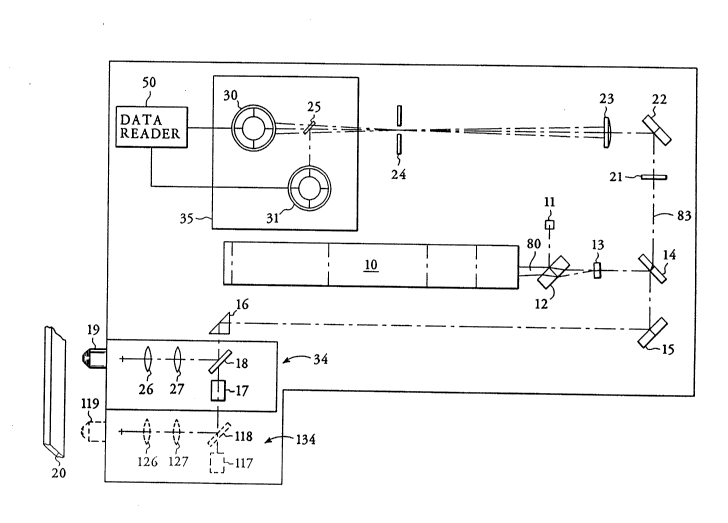
Fig. 1 is a plan view of an apparatus according to
the present invention.
Fig. 2 is a side view of a sample-filled capillary
tube according to the present invention, illustrating an
illuminated columnar region, and both excitation and
emission beams.
Fig. 3 is a perspective view of a sample-filled
capillary tube showing overlapping beam spots and an
illuminated columnar region according to the present
invention.
Fig. 4 is a top view of a sample-filled capillary
tube, showing overlapping beam spots according to the
present invention.
Fig. 5 is a schematic representation of the optical
scAnn;ng path according to a preferred embodiment of the
present invention.
Fig. 6 is a schematic representation of a labeled
cell suspension and the corresponding detector signal.
With reference to Fig. 1, laser 10 generates an
excitation beam 80 that passes first through glass plate
12 which optically co~nn;cates with power monitor 11,
then through laser line filter 13 and through spectral
dispersion means 14 which acts as a mirror for the
selected laser beam wavelength. The spectral dispersion
~ 214~2~5
-- 10 --
device may be, for example, a dichroic beam splitter, a
prism, or a grating. The excitation beam is then
directed to mirror 15 and through right angle prism 16 to
scan assembly 34. In Fig. 1, scan assembly 34 comprises
a galvanometer 17 with attached galvo mirror 18, lenses
26 and 27, and lens 19. Alternatively, the scan assembly
may comprise a multifaceted polygonal mirror. The
excitation beam 80 of the present invention impinges upon
galvo mirror 18 which continually changes position
because it is in cn~llnication with galvanometer 17
thereby causing a change of position of the excitation
beam. Within scan assembly 34, the excitation beam
travels from the galvo mirror 18 through lens 27 then
through lens 26. From lens 26, the excitation beam is
directed through lens 19 so that a focal spot of the beam
may impinge upon the outer wall of a transparent
capillary tube 20.
The excitation beam impinging upon the outer wall
traverses the wall and illuminates a columnar region of
the sample causing fluorescent emission from the sample.
Light collection occurs in an epi-illumination manner.
The emitted fluorescence is collected by lens 19 and
directed back, as retrobeam 83, through scan assembly 34.
Lens 19, seen in Fig. 2, has a central portion for
passage of incident beam 80 and uniform depth of focus of
incident beam 80 through capillary tube 20. Because
fluorescent emission is over a very wide angle,
represented by rays 32a and 32b, fluorescent collection
occurs over a wider portion of objective 19. Returning
to Fig. 1, the retrobeam 83 travels from scan assembly
34 to right angle prism 16 to mirror 15 and spectral
dispersion device 14. Due to its fluorescence emission
wavelength, retrobeam 83 is transmitted through spectral
dispersion device 14 and through bandpass filter 21 to
mirror 22 where it is directed through collimating lens
23. The retrobeam is then selectively passed through
-
2148205
-- 11 --
spatial filter 24 and into the detection means 35. The
spatial filter 24 has a predetermined pinhole aperture of
a diameter that permits passage of only that fluorescence
emission from a region defined by the illuminated segment
within the capillary tube.
Detection means 35 comprises a detection channel
such as detector 30 which reads the fluorescent signal of
the retrobeam 83 and is in communication with data reader
50 which converts it from analog to digital form. The
detector is a light measuring device such as a photo-
multiplier tube or photodiode. The signal is recorded by
data reader 50 as a unit of fluorescence intensity. The
detection means 35 may contain any number of detection
channels. For instance, a spectral dispersion device 25
is positioned between spatial filter 24 and detectors 30
and 31 in Fig. 1 to separate the wavelengths of the
fluorescent emission of the sample and to selectively
direct light of one wavelength to one detector and light
of a second wavelength to a second detector. In this
manner, multiple spectral dispersion devices and multiple
detectors may be incorporated into the detection means
for detection of fluore~cence at different wavelengths
from multiple fluorophores. In a similar manner,
multiple lasers may be utilized for excitation of the
sample at different wavelengths.
A critical feature of the present invention is
illustrated in Fig. 2. Spatial filter 24 is selected
with a pinhole aperture that collects light over a large
numerical aperture, but confines the depth of detection
to the interior depth dimension of the capillary tube.
The spot size of excitation laser beam 80 on the outside
wall of capillary tube 20 is of a generally constant
diameter, and has been chosen to provide uniform
illumination along the depth ~; men sion of the capillary
tube. Thus, the present invention relies upon a
21~8205
- 12 -
dependent relation of the spot size of the excitation
beam, the depth ~;mRn~ion of the capillary tube, and the
pinhole aperture of the spatial filter.
The capillary tube 20 is a transparent sample holder
of known dimensions. The capillary tube preferably has a
rectangular cross-section with a shorter dimension
defining an interior depth of 25 to 225 microns and a
longer dimension defining a width of 1 millimeter. The
length of the capillary tube is not as critical, but the
beginning and ending points of the scan in a direction
along the length of the capillary tube define the precise
volume of the segment scanned. In the present invention,
a capillary tube length of 40 millimeters has generally
been used. The capillary tube is fixedly positioned
directly below the excitation beam so that the scan of
the capillary tube occurs in a top-down manner. The
intersection of the excitation beam and the capillary
tube is generally defined by columnar region 51, as shown
in Figs. 2 and 3. The top dimension of the columnar
region is circular beam spot 33 of 5 to 15 microns. The
size of the beam spot is chosen so that the entire depth
i m~n ~ion of the capillary tube is illuminated.
After a columnar region of the capillary tube is
illuminated and the fluorescence emitted from its
contents is detected and recorded, the optical Sc~nn; ng
means i~ moved to a new position to illuminate a new
columnar region. The movement is of an amount that is
only a fraction of the beam spot size, so that each
illuminated columnar region 51 partially overlaps another
such region 44, as in Fig. 3. The optical scanning means
continues in this manner of illuminating and
fluorescently exciting a region from which fluorescent
emission is detected and recorded, then is moved slightly
to illuminate a new columnar region and to repeat the
process. In the preferred embodiment, the optical
~ 21~820~
- 13 -
sr~nning means follows a scan path in one direction
indicated by arrow 52 that is transverse to the
longitll~inAl axis of the capillary tube, i.e. along its
width, and in the other direction along the length of the
capillary tube, indicated by arrow 53, to form a two-
dimensional array of beam spots. In Fig. 1, the dashed
lines 134 indicate a change of position of scan assembly
34, so that dashed galvanometer 117 and dashed galvo
mirror 118 represent galvanometer 17 and galvo mirror 18
in altered positions. In the same manner, dashed lenses
127, 126 and 119 represent lenses 27, 26, and 19 in
altered positions. As shown in Figs. 3 and 4, the
transverse scan begins and ends at points 54 beyond the
lateral boundaries of the capillary tube. This overscan
is effective in identifying edge anomalies of the
capillary tube.
Fig. 5 shows a schematic representation of scan path
48 from above capillary tube 20, according to the
preferred embodiment of the present invention. The
excitation beam spots are moved along the scan path in a
transverse direction, then snapped back to follow a
closely-spaced parallel path also in the transverse
direction. The process is repeated continually so that
the scan also covers a segment along the longitll~inAl
axis of the capillary tube. In this manner, fluorescence
emission occurs and is detected from any chosen length of
the capillary tube.
The method disclosed in the present invention allows
for analysis of a sample of biological fluid with a
mi nimllm of preparation. According to the present
invention, a biological fluid is incubated with an excess
amount of a binding agent that contains a fluorophore of
known optical characteristics. The fluorescently-labeled
binding agent is selected to react with binding sites
present within the sample. For example, a
~ 2148205
- 14 -
fluorescently-labeled antibody directed to an antigen
present on some cellular component of the biological
fluid may be added to the sample. The labeled binding
agents and the binding sites form fluorescent complexes
that will emit a signal when used with the apparatus of
the present invention.
After the sample is incubated with the labeled
binding agent, it is diluted, if necessary, and then
placed directly into capillary tube 20. No lysing of
components of the biological fluid nor separation of
bound and unbound binding agent is required at any point
in the practice of the method of the present invention.
An optical scan is made of the sample in a volumetric
manner and fluorescence emission is sequentially recorded
from each illuminated columnar region.
The enumeration may occur in an absolute volume,
depending on the desired application, by noting the
beginning and ending points of the lengthwise scan of the
capillary tube and measuring the distance scanned or by
scanning between specific identification marks on the
capillary tube. This quantitation of all of the
fluorescent targets in a fixed, precise volume is a
powerful method of quickly obtA;ning detailed population
data. This volume is fixed by using a uniform cross-
sectional area capillary tube or by independentlymeasuring the volume of the capillary tube between
specific identification marks. Alternatively, a ratio
can be obtained without counting a precise volume, but
rather by comparing relative counts of different
components of the sample.
Data reader 50, in Fig. 1, records events, i.e. an
increase above the background level of fluorescence
exceeding some threshold value, as shown in Fig. 6. The
events correspond to the occurrences of cells of a
21~20~
.
- 15 -
particular type in the sample. Fluorescence emission
occurs from both the binding agent-binding site complexes
45 and from the free binding agent 40, but a more intense
signal 85 relative to background level 80 comes from
areas where the binding agent is clustered, i.e. cells
exhibiting binding sites to which the binding agent is
directed. Therefore, a signal of heightened fluorescence
85 corresponds to a cell, linked by dashed lines in Fig.
6, and is recorded as such.
The method of the present invention does not require
removal of unreacted fluorescently-labeled binding agent.
Dilution of the sample before optical scAnn;ng serves to
improve signal to noise ratios so that fluorescent
imaging according to the present invention occurs in a
quick manner with mi nimAl processing steps. Dilution
also serves to minimize the occurrence of cell overlap in
the sample. In the practice of this invention, optimal
results are obtained when the cells of the sample are on
the order of 10 microns in size and the cell density is
less than 5000 per microliter.
An example of this method's utility is illustrated
by an assay for the dete~minAtion of leukocyte subclasses
present in a blood sample. In a typical assay, a sample
of whole uncoagulated blood is incubated with
fluorescently-labeled antibodies that are directed to
specific cell surface markers. For example, anti-CD4 and
anti-CD8 labeled with fluorophores having different
optical characteristics may be incubated with the whole
blood sample. Within the sample, the leukocytes that
bear either CD4 or CD8 or both cell surface markers will
react with the labeled antibodies to form fluorescent
complexes. After a sufficient reaction time, the whole
blood sample is diluted and inserted into a capillary
tube. The capillary tube is then optically scanned
according to the present invention. The wavelength range
21~205
- 16 -
of the optical scan is selected 80 as to activate the
fluorophores being used and to m;n;m; ze interference due
to autofluorescence from blood components not of
interest. Fluorescent emission corresponding to the
fluorophores that were used to label the anti-CD4 and
anti-CD8 are detected and recorded. The presence of
leukocytes bearing either or both of the cell surface
markers to which these antibodies are directed is then
enumerated. Reæults may be presented as an absolute cell
count per unit volume, by counting the number of cells of
a certain subclass present within a given volume, the
volume being determined by the length of the scan and the
cross-sectional area of the capillary tube. Results may
also be presented as ratios, e.g. CD4/CD8 leukocyte
ratios, by counting the number of cells bearing each of
these cell surface markers and comparing the two. The
usefulness of this last illustration is readily evident,
as this ratio is important in determining the progression
of AIDS.
When an assay is performed to determine leukocyte
subclasses in whole uncoagulated blood using the
technique of the present invention, a two or three minute
wait between placement of the reacted sample into the
capillary tube and the optical scan allows for the
natural density of the numerous red blood cells present
in the sample to cause settling of the red blood cells to
the bottom of the capillary tube and the subsequent
displacement of the white blood cells. This natural
buoyancy effect causes a resultant location of the white
blood cells near the upper portion of the capillary tube
and assists in fluorescence detection because of the top-
down scan geometry of the present invention.
As in the above example, fluorophores with different
optical characteristics can be combined with binding
agents directed to different binding sites, so that the
214820~
- 17 -
presence of multiple reaction moieties in the sample can
be detected. From the precise known volume of the
capillary tube that has been æcanned, a quick reading
will identify the number of cells of a particular sub-
class per unit volume that are present in the sample.
The optical system is simply set to excite each
fluorophore at its crucial wavelength and a detection
channel is created to correspond to the emission
wavelength of each fluorophore.
The apparatus and method of the present invention
are suited to many applications, including those
requiring absolute counts within a known volume. For
example, cell kinetics studies, cell toxicity studies
using intercalating dyes, and in situ hybridization may
be adapted for analysis according to the present
invention. In addition, the volumetric method of the
present invention allows for the avoidance of artifacts
that may be present when immunological and other
biochemical responses are studied in cells located on a
surface rather than in a cell suspension. Although the
cells are detected from within a capillary tube, the
present invention presents the sample in a manner that
allows for flow cytometric-type analysis on relatively
stationary localized cells. Therefore, the cells may be
detected in a location-specific manner or be identified
for subsequent visual ~;n~tion.