Note: Descriptions are shown in the official language in which they were submitted.
WO 94/10343 , PCl`JlJ~i93/10331
~ 21~3~
'
~IET~IODS OF DETECTING ~IC~O~ET;P,STASIS OF PROSTATE C~NCE~
FIEI~D OF T}~E INVENI~ION
This invention is directed to methods of detecting
- prostate cancer.
BACRGRO~ND OF T~E INYENTION
Prostate cancer metastasis will claim the lives of
over 30,000 Americans this year. Boring et al., Cancer
Statistics 1991, 19. The mode of dissemination however,
remains very poorly understood. An almost dogmatic view of
10 metastasis holds that prostate cancer cells first spread
through the prostatic capsule then into th~ lymphatics, and
eventually hematogenously travel to b~ne. Byar et al., Cancer
1~72, 30, 5; Winter, C.C., Surg. ~ynecol . Obstet . 1957 , 105,
136; Kilaris et al., Am. J~ Roentgenol. 1974, 121, 832;
15 McLaughlin et al., J. Urol. 1976, 115, 89; Jacobs, S.C.,
Urology 1983, 21t 337; Batson, O.V., Ann. Surg. 19~0, 112, 138;
Saitoh et al., Cancer 1984, 54, 3078-3084: Whitmore, W.F., Jr.,
Cancer 1973, 32, 1104. However, this model has been based on
histopathologic studies which have significant limitations, and
in actuali~y the sequence of metasta_ic events remain unknown.
Solid tumor animal experiments suggest that only 0.01% of
circulating cancer cells eventually create a single metastatic
deposit. Fidler et al., Science 1982, 217, 998-1001: Liotta et
~- al., Cancer Res. 197~, 34, 997; Schirrmacher, B., Adv. Cancer
25 Res. 1985, 43, 1-32. Ostensibly, a single bone metastasis from
human prostatic adenocarcinoma (PAC) colld be generated by
~ i
.~
L
. , . ~, .. . , ., ., .. .,.. , i ~ . ... .. ~ , . . .
WO94J10343 PCT/US93/10331
~r' .
- 2 -
10,000 circulating cancer cells (2 cell~/l ml blood). In the
past, detection of such a low concentration of cells has been
difficult or impossible. Recently, however, Wu et al. used
keratin-l9 (K-l9) mRNA PC~ to detect breast cancer
5 micrometastasis in patient lymph nodes and bone marrow. Wu et
al., Lab. Inv. 1990, 62, lo9A. Miyomura et al., also reported
the detection of minimal residual acute lymphoblastic leukemia
by PC~ in patients harboring the Philadelphia chromosome.
Miyomura et al., Blood 1592, 79, 1366-1370.
A method of detecting the micrometastasis of prostate
cancer would be greatly desirable.
S~M~RY O~ T~IE INVENTION
In accordance with the present invention, methods of
detecting prostate cancer micrometastasis in a patient are
15 provided comprising the steps of obtaining a sample ofRNA from
a patientls blood and amplifying said RNA with polymerase chain
reaction. The polymerase chain reaction is performed using a
pair of primers which are complementary to separate regions of
the prostate specific antigen gene. These primers may have the
sequences GAGGTCCACACACTGAAGTT (5EQ ID NO: 1) and
CCTCCTGAAGAATCGATTCCT (SEQ ID NO: 2). ~hereafter, the presence
or absence of amplified RNA is detected wherein the presence of
amplified RNA indicates micrometastasis of prostate cancer.
BRIEF DESC~IPTION OF T~E FIG~RES
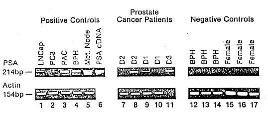
Figure 1 shows an agarose gel in which micrometastasis
is indicated by the presence of a 214 base pair tbp) band.
DETAILED ~ESCRIPTIONiOF T~E INVENTION
In accordance with methods of the present invention,
methods of detecting micrometastasis of prostate cancer in a
30 patient is pro~ided comprising the step of obtaining a sample
of RNA from the patient's blood. Preferably the RNA is
obtained from a blood sample such as a peripheral venous blood
sample. A whole blood gradient may be performed to isolate
nucleated cells and total RNA is extracted such as by the
~V094/10343 21~1~3 ~ PCI`/~SY3/10331 1-
,. ,
- 3 -
RNazole B method (Tel-Test Inc., Friendswood, Texas) or by
modification of methods known in the art such as described in
Sambrook et al., Molecular Cloning: A Laboratory Manual (Cold
Spring Harbor Laboratory, Cold Spring Harbor, NY, 1989).
Thereafter, a polymerase chain reaction may be
performed on the total extracted RNA. Preferably a reverse
transcriptase PCX amplification procedure may be performed in
order to quantify the amount of mRNA amplified. Polymerase
chain reaction methodologies are well known in the art. Innis
et al., PCR Protocols, Academic Press, Inc., San ~iego CA,
1990. Polymerase chain reaction primers may be designed to be
complementary to separate regions of the prostate specific
- antigen (PSA) gene. Henttu et al., Biochem. Blophys. Res.
Comm. 1989, 160, 903-910. By separate regions is meant that a
first primer is complementary to a 3' region of the PSA gene
and a second primer is complementary to a 5' region of the PSA
gene. Preferably, the primers are complementary to distinct,
- separate regions and are not complementary to each other.
PSA is an important marker produced exclusively by
20 prostatic epithelial cells and almost always expressed by
prostate cancer. Stamey et al., J. Urol. 1989, 141, 1076-1083.
Thus, PSA2 (5-GAGGTCCACACACTGAAGTT, S~Q ID N0: 1) and PSA3 (5-
CCTCCTGAAGAATCGATTCCT, SEQ ID N0: 2) oligonucleotide primers
were designed to have high specificity to the PSA gene. A Gene
25 Bank version-70 (Mountain View, CA) search confirmed the
specificity of these primers to PSA and not to the human
glandular kallikrein (HMGR) gene which has high homology to the
PSA gene. Henttu et al, Biochem. Biophys. Res. Comm. 1989,
160, 903-910. PSA2 and PSA3 bind sequences that span intron
30 III of the PSA gene such that PCR amplification yields a 360 bp
DNA and a 214 bp RNA product, thereby eliminating the
possibility of false positives from DNA contamination.
Oligonucleotide primers may be prepared by methods known in the
art such as by standard phosphoramidite chemistry. (See 3
35 Sambrook et al., supra). Following amplification, the presence
or absence of mRNA amplification product may be detected.
Preferably, the PCR product may be run on an agarose gel and
! .
. :. -' , . ' : ,
WO94/10~3 ~3:~ PCT/US93/1033l ~
.
-- 4 --
visualized using a stain such as ethidium bromide. (See
Sambrook et al., supra).
The following examples are illustrative but are not
meant to be limiting of the invention.
EXAMPLES
Example 1 Patient Specimens
Selection of cases was based on the following
criteria. Prostate cancer patients were chosen for analysis if
they had: (1) clinically and/or surgically staged DO-D2 disease
(D0 = elevated tumor markers with no demonstrable metastasis,
Dl = pelvic lymph node in~olvement, D2 = disseminated disease
~ usually to bone) without having received prior hormonal therapy
and who had an elevated serum PSA, or (2) stage D3 disease (D2
disease that is refractory hormonal therapy) with an elevated
PSA Negative control patients consisting of female volunteers,
and patients with benign prostatic hypertrophy (BPH) proven by
biopsy or men who were on a BPH study protocol. Patients who
had surgical manipulation of the prostate during the previous
year were excluded from the study. Positive controls included
a lymph node from a patient with known metastatic PAC tissue
from pathologically pro~en BPH and cD~A PSA plasmid. Henttu et
al, Biochem. Biophys. Res. Comm. 1~89, 160, 90~-910. The
protocol was IRB approved and written consent was obtained.
LNCAP and PC3 human cell lines were obtained from The American
25 Type Culture Collection, (Rockville, MD).
Example 2 Blood Preparation for ~NA Extraction
Approximately six ml of venous blood were obtained
with a standard venipuhcture techniquè using heparinized tubes.
Whole blood was mixed with an equal volume of phosphate
30 ~uffered saline (PBS) which was then layered over eight ml of
Ficoll (Pharmacia Uppsala, Sweden) in a 15 ml polystyrene tube.
The gradient was centrifuged at 200 g for 30 minutes at 5-C.
The lymphocyte and granulocyte layer (approximately 5 ml) was
carefully aspirated and re-diluted up to 50 ml with PBS in a 50
35 ml tube which was then centrifuged at 1800 g for 20 minutes a
W094/10~3 PCT/US93/1033l
21~3~
.. ,. ~
- 5 -
5 C. Supernatant was discarded, and the pellet containing
nucleated cells was used for RNA extraction using the RNazole
B method, as describ~d by the company (Tel-Test Inc.,
Friendswood, Texas~.
5 Example 3 Oligonucleotide pri~ers and probes
PSA2 (5-GAGGTCCACACACTGAAGTT, SEQ ID NO: 1) and PSA3
(5-CCTCCTGAAGAATCGATTCCT, SEQ ID NO: 2) oligonuoleotide primers
were custom designed with high specificity to the PSA gene; a
Gene Bank version-70 (Mountain View, CA) search confirmed the
10 specificity of these primers to PSA and not to the human
glandular kallikrein (HMGK) gene which is 75-85% homology to
~ the PSA gene. Henttu et al, Biochem. Biophys. Res. Comm.
1989, 160 f 903-910. All primers were synthesized and gel
purified by the City of Hope DNA Synthesis La~oratory (Duarte,
15 California). PSA2 and PSA3 bind sequences that span intron III
such that PCR amplification yielded a 360 bp DNA and a 214 bp
RNA product. Previously published actin PCR primer sequences
were used to rule out degraded RNA, and amplification with
actin oligonucleotide primers Al and A2 yislded a 154 bp RNA
20 and a 250 bp DNA product. Ben-Ezra et al., J. ~istochem
Cytochem. 1991, 39, 351-354.
E~ample 4 Polymerase Chai~ Reaction
The reverse transcriptase reaction and PCR
amplification were performed sequentially without interruption
25 in a Per~in Elmer 9600 PCR machine (Emeryville, CA~. 400 ng of
total ~NA in 20 ~l DEPC (Diethyl-pyrocarbonate) treated water
were placed in a 65-C water bath for fi~e minutes then quickly
chilled on ice immediately prior to the addition of PCR
reagents. The 50 ~l total PCR volume consisted of 2 . 5 units
30 Taq polymerase (Perkin Elmer, Emeryville, CA), 2 units AMV c
reverse transcriptase (Boehringer Mannheim, Indianapolis, IN), k
200 ~M each of dCTP, dATP, dGTP, and dTTP (Perkin Elmer, ti
Emeryville, CA), 18 pM each primer, lO mM Tris-HCL, 50 mM KCl,
2 mM MgCl2 (Per~in Elmer, Emeryville, CA) . PCR conditions were
35 as follows: cycle 1 was 42-C for 15 minutes, then 97~C for 15
~ 3 a-
WO94/10343 PCTJVS93/10331
- 6 -
seconds (one cycle); cycle 2 was 95 C for one minute, then 60~C
for one minute and 72~C fQr 30 seconds (15 cycles); cycle 3 was
95-C for one minute, then 60eC for one minute, and 72 degrees
for one minute ~10 cycles); cycle 4 was 95 C for one minute,
5 then 60 for one minute and 72-C for two minutes (8 cycles);
cycle 5 was 72-C for 15 minutes ~one cycle): and the final
cycle was a 4-C hold until sample was taken out of the machine.
The 50 ~1 PC~ products were concentrated down to 10 ~l with
~acuum centrifugation and the entire sample was then run on a
10 thin three perc~nt Tris-borate-EDTA (TBE) garose gel
containing ethidium bromide. All specimens were analyzed at
least twice to confirm a positive or negative outco~e.
- The potential r sk of false positives from cross
contamination was avoided by performing RT PCR in a si.ngle tube
15 without interruption and using filtered pipet tips. Sensitivity
was enhanced by using high amounts of Taq polymerase,
progressively increasing extension times, and analyzing the
entire 5Q ~l PCR product on thin ethidium bromide agarose gels.
These measures ensured a high fidelity assay while maintaining
¦ 20 technical simplicity.
i Prostate human tissue specimens, tissue culture cell
. lines and a PSA cDNA plasmid, cloned and descri~ed by Henttu
and Vihko; Henttu et al., Bioc~em. Biophys. Res. Comm. 1989 ,
~ 160, 903-9l0, were used as positive controls, and they
¦ 25 demonstrated the 214 bp bands as shown in fig.1 top panel. A
¦ pelvic lymph node with metastatic PAC, a primary prostate
¦ cancer, and a BPH specimen all produced strong PSA PCR signals.
The LNCAP and PC-3 human prostate cell line~ produced weaker
signals.
30 EXAMPLE 5 Sequencing
Specificity of these primers to the PSA gene was
confirmed with DNA sequence analysis of the amplified 214 bp
fragment (Figure 1 bottom panel) which in this segment had very
little homology to the HMGX gene. The 214 bp product was
35 purified with a Qiagen PCR Product Purification kit (Qiagen,
Chatsworth, CA) as described by the manufacturer. One microgram
.s
W094/10~3 2 1 ~ ~ 3 ~ ~ PCT/US93/10331
f ,....;
of the PCR product underwent a PCR sequencing reaction by using
the Taq DyeDeoxy Terminator Cycle sequencing kit in a Perkin-
Elmer 9600 PCR Machine, as described by Applied Biosystems
(Applied Biosystems, Foster, CA?. The sequenced product was
S purified using centri-sep columns (Princeton Separations,
Adelphia, New Jersey) as described by the company. This
product was then analyzed with a ABI Model 373A DNA sequencing
system (Applied Biosystems, Foster, CA3 integrated with a
Macintosh IIci computer.
10 Example 6 Detection of Circulating ~ematogenous
Micrometa~tasi~
Twelve prostate cancer patients ~nd 17 control
patients underwent RT PCR analysis on PSA and Actin RNA
extracted from blood, as described in Examples l through 4
(Table l). All cases demonstrated satisfactory RNA quality by
actin PCR (Figure l, bottom row). Of the 12 human prostatic
adenocarcinoma (PAC) patients with metastatic disease, four
cases (33~) had positive PSA signals indicating the presence of
prostatic epithelial cells in the peripheral venous blo~d.
20 These four cases consisted of two sta~e Dl patients, one stage
D2 patient, and one stage D3 patient (N=l) (Figure l, top row).
The 17 negative controls, which consisted of eight volunteer
women and nine men with BPH, all had undetectable PSA mRNA by
RT PCR. These data indicate that RT PCR of the PSA RNA gene
can be used to specifically detect circulating hematogenous
micrometastasis in patients with stage Dl-D3 pathology. These
findings are in agreement with studies by Hamby et al. who
dete~ted circulating PSA positive cells in patients with
metastatic prostate cancer by flow cytology and
immunohistology. Hamby et al., Br. J. Urol. 1992, 69, 392-396.
Micrometastasis was not detected in eight of twelve
prostate cancer patients consisting of two stage D3 patients,
two stage Dl patient~, and four stage DO patients. In order to
enhance the detection of micrometastasis, analysis may focus on
buffy coat cells. Results indicate that the prostate cancer
cells may be more concentrated in the "buffy coat". The PSA
WO~4/10~3 2~3~ PCT~US93/10331 ~:
- 8 -
signal was stronger in the RNA extracted from cells obtained
only from the "buffy coat" (Figure 1, lane 8) compared to those
isolated from the entire Ficoll layer (Figure 1, lane 7) in the
same prostate cancer patient. These findings are in agreement
5 with those of Harty et al. who found that prostatic epithelial
cells migrate into the "buffy coat". Harty et al., J. Surg.
Resr 1979, 26, 411-416.
W094/l0343 _ 9 _ 2 ~ 4 8 3 S Q PCT/USg3/10331 1 ~
SEQUENCE LISTING
(1) GENERAL INFORMATION:
(i) APPLICANT: Croce et al.
(ii) TITLE OF INVENTION: Methods of Detecting
Micrometastasis Of Prostate Cancer
(iii) NUMBER OF SEQUENCES: 2
(iv) CORRESPONDENCE ADDRESS:
(A) ADDRESSEE: Woodcock Washburn Xurtz
Mackiewicz ~ Norris
(B) STREET: One Liberty Place - 46th Floor
(C) CITY: Philadelphia
(D) STATE: PA
(E) COUNTRY: USA
(F) ZIP: 19103
: (v) COMPUTER READABLE FORM:
~ (A) MEDIUM TYPE: DISKETTE, 3.5 INCH, 1.44 Mb STORAGE
: (B) COMPUTER: IBM PS/2
(C) OPERATING SYSTEM: PC-DOS
(D) SOFTWARE: WORDPERFECT 5.1
(vi) CURRENT APPLICATION DATA:
(A) APPLICATION NUMBER: n/a
(B) FILING DATE: Herewith
:~ (C) CLASSIFICATION:
(vii~ PRIOR APPLICATION DATA:
(A~ APPLICATION NUMBER:
(B) FILING DATE:
(viii) ATTORNEY/AGENT INFORMATION:
(A) NAME: Lori Y. Beardell
(B) REGISTRATION NUMBER: 34,293
C) REFERENCE/DOCKET NUMBER: TJU-0722
(ix) TELECOMMUNICATION INFORMATION:
(A) TELEPHONE: (215j 568-3100
(B) TELEFAX: (215) 568-3439
: (2) INFORMATION FOR SEQ ID NO: 1:
` (i) SEQUENCE CHARACTERISTICS:
(A):LENGTH: 20
(B)` TYPE: Nucleic
; (C) STRANDEDNESS: Single
(D) TOPOLOGY: Linear
: (iv) ANTI-SENSE: No
(xi3 SEQUENCE DESCRIPTION: SEQ ID NO: 1:
GAGGTCCACA CACTGAAGTT 20
~ (2) INFORMATION FOR SEQ ID NO: 2:
-~: ` (i) SEQUENCE,CHARACTERI$TICS:
(A) LENGTH: 21
- . (B) TYPE: Nucleic
:~ (C) STRANDEDNESS: Single
(D) TOPOLOGY: Linear
, (iv) ANTI-SENSE: No
~: (xi) SEQUENCE DESCRIPTION: SEQ ID NO: 2:
~: CCTCCTGAAG AATCGATTCC T 21
~ , ,.