Note: Descriptions are shown in the official language in which they were submitted.
WO 94/~0904 2 1 ~ 9 3 1 1 PCI/U~93/10464
APPARATUS AND METHOD FOR
MONITO~ ~N1:)0CARD~ T~L r)URlNG ABLATION
.,~,;, .
~ 5 ;E~ke~l~i~
;4~
~l :
The invention relates to an electrosurgical device, in the form of a catheter,
and ;nstrumentation for use in performing tissuè ablation. ~
~' lO The ablation of organ tissue can be performed during surgical procedures to
treat disease or medical disorders. Ablation of certain cardiac tissue is performed with
increasing frequency to treat certain heart disorders which result in arrhythmia.
The heart is a muscular organ comprislng four separate chambers which
cooperate to purnp blood throughout the body. The heart muscles must contract~ and~
relax in a coordinated sequence in order for blood;to be passed through the circulatory
system in an efficient manner. The heart includes a speclalized system for generating
impulses to cause rhythmical contraction of the heart mu~scle and~or conducting these
impulses rapidly through the;heart. In the proper sequence the atria contract about one
sixth of a second prior to the ventricles. ~This enables extra`filling of the ventricles
bofore they contract to pump blood through the lungs and to other areas of the body.
: :
The basic timing impulse of the heart is g'~enerated in the sinoatrial node
(SA~ node). The SA node has an inherent rhythm which can be modified by the
2~ sympathetic and parasympathetic nervous system. ~ The impulse ~initiated by the SA node~
spreads through the atrium to the ~atrio-ventricular node~(AV node), and then through the
Purkinjefiberstotheendocardialsurfacesofthe~ven~ les.
The rhythmical and conductlon system of the heart; ls susceptible to
disruption by disease. Damage caused to cardiac tlssue~can result in the inability of the
, cardiac conduction pa~thways to properly transmit the electrical impulses generated in the
SA node, leading to aIrhythnlias, or irregular he~beats.; ~Cardiac ~arrhythmias can o~en
~: be detected through electrocardiograms.
Some forms~ of cardiac arrhythmia are able to be~controlled through~
medication. However, other fonns of ~arrhythmia do not respond to medication.
Moreover, medication typ}cally~does not cure the problem, and the dosage ;and~the ~
medication type~ must be; changed periodically to maintain a;continued le~el of control~ of
theproblem.
:` :
;~
W094/I0904 3,~ -2- PCI/tJS93/10464
`~'1
One alternative to medication is the surgical removal of a portion of the
cardiac pathway which is responsible for the arrhythmia. The many dangers associated
with open heart surgery render this a less preferred treatment option. Recently, however,
s it has become possible to intravascularly insert a specialized catheter within the heart? for
positioning adjacent to the conductive tissue responsible for the arrhythrnia. The ?
catheter is adapted to deliver energy (e.g., radio frequency energy) to ablate or destroy
the tissue lesion responsible for an arrhythmia. This has been found to be a relatively
i~, safe and effective techni~ue for eliminating many causes of arrhythmia~ Various
ablation catheters arld techniques for their use are described in U.S. Patent Nos.
4,641,649; 4,785,815; 4,869,248; and 4,896,671.
: :
Cardiac ablation catheters typically have at least one ablation electrode,
;~; positioned at the ~istal end of the catheter, which is adapted to deliver energy to the
`fi 15 tissue lesion. Other electrodes can be proximally positioned on the catheter and used for
; sensing endocardial signals. Ablation may be achieved by the application of electrical
energy, such as radio frequency (RF) or direct current (DC) energy, from a generator
source, through a conductor disposed within the catheter, and to the ablation electrode.
: , :
During the ablation procedure, the ablation electrode is positioned adjacent
to an ablation site, or site of defective tissue. The processes for accurately positioning
the ablation electrode and "mapping" the ablation site~re well known, and generally
,~ involve positioning a multi-electrode "mapping catheter," which may include an ablation
tip, near the lesion, and radiographically visualizing the catheter position while
simultaneously electrically monitoring the heart tissue. Once the~ablation electrode is
accurately positioned, energy, typically in the form o~RF energy, is delivered to the
ablation site by the ablation electrode. ~ ` ~
The goàl of the ablation procedure is to precisely destroy the defective
tissue without damaging any healthy heart tissue. To prevent inadvertent damage of
, healthy tissue, it is desirable to mon~tor both the endocardial signal and the Impedance at
1 the ablation site during the ablation procedure~ Monitoring is normally performed by
i, one or more sensing electrodes proximally positioned on the cathet¢r some distance ,
away from the ablation electrode. Thus, in prior ablation systems, monitoring is not
performed directly at the ablation site. This spatial discrepancy can result in an
imprecisely controlled ablation procedure which may damage some healthy tissue, or fail ~'.
to remove some of the de~ective tissue.
;:i
i~ ` It would thus be advantageous to develop an ablation catheter system,
.~` :
WO ~4/10904 21 qg311 PCr/US93/10464
. suitable for use in cardiac ablation procedures, which measures the local impedance and
the local endocardial signal directly at the ablation site. It would also be advantageous to
develop a system that perforrns these measurements simultaneously with ablation.
~ !
Summary Qf~he Invention
It is thus an object of the invention to provide a catheter suitable for use
with cardiac ablation procedures utiiizing the delivery of RF energy. A further object is
to provide an ablation catheter which provides accurate local monitoring of impedance
o and the endocardial signal at the ablation site, and during an ablation operation. It is also
an object of the invention to provide a method for using such a catheter. Other objects
will be apparent upon reading the disclosure wl~ich~follows. ~
The present invention comprises an ablation catheter having an ablation
lS electrode mounted at a distal end of the catheter and designed to allow positioning of the
ablation electrode adjacent to cardiac tissue. The ablation electrode is coupled to a
remote ablation power source through a low impedance coupling. The ablation electrode
also functions as a sensing electrode, for monitonng the endocardial signal and
preferably also the impedance during the ablation procedure, and is coupled to an
electrode monitor through a lugh impedance coupling. A timing element defines plural
repetitive ab}ation intervals which alternate in non-overlapping fashion with a similar
plurality of quiescent intervals. R~ energy is deliv~ered, to the ablation site during the
ablation intervals, while the local endocardial signal is measured during the quiescent
mtervals.
2s
In one embodiment a first switch element is connected between the low
impedance coupling and the ablation electrode and is controlled by the timing
element to connect the ablation electrode to the power source during ablation intervals,
~ and to disconnect the electrode from the coupling durmg quiescent intervals. A second
i 30 switch element is connected between the electrode monitor and the high impedance
, ~ coupling and is controlled by the timing element to isolate the monitor from the
electrode during ablation intervals and to coup!e them during quiescerit intervals.
In another embodiment of the inventlon~a low-pass filter and an amptifier
; 3s are connected between the second switch element and the elec~ode monitor ~for
improving the quality of the measured endocardial signal. In this embodiment, a third
switch element, connected between the amplifier and the electrode monitor, is sw~tched ``
synchronously with the second switch element. ~ ; ~
WO 94~ 10904 ~ PCI /US93/10464
- 21~9311 4 ~1
; In one preferred embodiment, the E~F power source provides a fifty volt
square wave ablation power signal at R~ frequency. The low impedance coupling
efficiently transmits this RF power to the ablation e}ectrode. The high impedance
coupling is of a type that presents a high impedance at the RF frequency of the power
source, and also allows detection of the low voltage endocardial signal so it can be
measured accurately. ! 7
In the practice of a preferred ablation method, electrodes mounted exterior
l to the chest are actuated to artificial}y pace the heart so that it beats about 120 times per
lo minute during the ablation procedure. The ablation electrode is positioned adiacent to
the ablation site, and delivery of RF energy to the tissue lesion is initiated. The catheter
may have a deflectable catheter tip, with its tip electrode spnng-loaded outwardly to
assure good electrode contact even when the heart moves during the ablation intervals.
lS The invention also contemplates another method when ablation only occurs
while the heart is in a desired part of the cardiac cycle. According to this aspect of the
invention, the ablation power intervals are triggered by timing pulses synchronized with
detection of the R wave. This mode of actuation assures that~the heart is essentially
stationary before delivery of ablation energy, thus minimizing the risk of inadvertently
ablating healthy t;ssue.
In yet a further aspect of a preferred met~od, the impedance at the ablation
site is measured during the ablation inteNals. As is well known, the tissue~impedance
can vary according to several factors such as the fatty tissue distribution in thè patient, or
2s the location of the electrode. However, once ablation commences, changes in impedance
rellect the heating of tissue, denaturing of cellular material, and loss of water from
heated tissue, thus reflecting the degree of ablation of surrounding tissue. Information
Ol1 changes in tissue impedance, taken together with continuous mo~itoring of anarrhythrnic signal, therefore indicates whether the tissue has been~correctly targeted and
sufficiently treated. Thus the impedance along with the measured endocardial signal
provide information for determining when the ablation procedure is complete.
s
In the preferred method~and system ofthe invenhon, ablation~energy is
delivered to the ablation site, during a plur~ality of short, closely-spaced, ablation
3s intervaIs, until the monitored; endocardial signal is free of indications o~ the arrhythmia.
The ablation procedure is normally performed in less than approximately six seconds, -~
and a~ter such treatment, :the absence of the a~rhythmia signal usually means that the
defective tissue has been destroyed. ~ Howevor, improper positioning of the ablation
electrode` may result in ~deiivery of an insufficlent level of pow~r to the tissue, only
WO 94/10904 ~ PCr/US93/10464
~ 5~ 93~;,
stunning, rather than destroying, the defective tissue. Such stunning may cause the
arrhythmia signal to temporarily disappear, so to assure that a lesion has been created in
~,~ the defective tissue, it is preferred to wait about thirty minutes after the disappearance of
. the arrhythtnia signal before withdrawal of the catheter. If the arrhythmia signal does not
.~ 5 return within thirty minutes, there is a high confidence level that the lesion has been
created, and the catheter is then withdrawn. If the rurhy~unia signal does return, the
ablation electrode is FeadJusted and the ablation procedure is repeated.
The endocardial signal is primarily a low frequency signal having most of
its components below several hundred Hz. The highest frequency components of thesignal are in the His-bundie and are about five hundred Hz. These latter signals are the
principal ones detectable in arrhythmia sites. For ~accurate o~servation of the endocardial
signal, applicant samples it at least one thousand times per second, to achieve Nyquist
~ sampling of at least twice the frequency of the highest frequency component.
In one preferred method, the ablation and quiescent intervals alternate five
thousand times per second, and the endocardial signal is satnpled once during each
quiescent interval. The quiescent intervals last for about fifty microseconds, and the
ablation intervals last for about one hundred fifty microseconds. By switching the
. ~o ablation electrode on and off at the preferred sampling frequency of five thousand times
a second, and sampling the endocardial signal and tissue~ impedance beitween active
intervals, significantly faster than the Nyquist rate, applicant~avoids inducing muscle
,j stimulation in the heart Applicant avoids switching~at a lower frequency of about 500
Hz, which would induce muscle contraction in the heart tissue.
Zi~ 2s ~s~
/i
The foregoing features of the invention u ill be understood from the
detailed description of illustrative embodiments to follow, together with the drawings
wherein~
FIGURE 1 is a functional illustration of an ablation catheter apparatus
! according to the invention; ~ -
FIGURE 2~is a timing diagratn representative of the~ switch timing of the
35 invention;
~,~ FIG~JRE 3 is a schomatic representation of the timing circuitry of FIGURE
~$ 1; and
il'. ~: ~ :
~ i
W094/l0904 2149311 6 Pcl/uss3/l~464 !~
FIGURE 4 is a schematic block diagram of another embodirnent of the
invention.
'~1 ' I
S l~)etailed Des~nption of the Invenhon
.
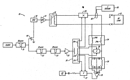
As shown in FIGURE 1, the cardiac ablation system 10 of the present
invention has an ablation electrode 12, typically located at the distal tip of a special
catheter ~not shown) which is coupled to an RF amplifier 14 through a low impedance
lo coupling 16. The low impedance coupling 16 is preferably a transformer having a ratio
i, selected to achieve a specific level of the voltage delivered to the ablatiorl electrode 12.
The RF amplifier 14 is driven by an RF oscillator 18. A first switch 20 is connected
between the low impedance coupling 16 and the ablation electrode 12. ~witch 20 is
opened by application of a signal developed on secondary winding 37a of a transformer
15 36, as discussed further below.
I
The ablation electrode 12 is also coupled, through a high irnpedance
coupling 24, such as a pair of inductors 24a and 24b, to an electrode mon;tor 22. The
hi8h impedance coupling 24 electrically blocks RF energy originating at oscillator 18
from reaching the monitor or its pre-amp. A second switch 26 is provided across the two
leads ofthe high RF impedance coupling 24, which connect via a low-pass filter 28 to an
amplifier 30. A third switch 32 is connected between ~e amplifier 30 and the electrode
monitor 22.
2S During operation of the RF power source, a current sensing loop 62 detects
the instantaneous current, and a processing circuit 65 compares it to the ablation voltagè
and processes the signals to develop an instantaneoùs measure of tissue impedance
across the catheter tip. Impedance sensing may be sccomplished by conventional
circuitry, and details thereof will not be further discussed herein.
FIGIJRE 2! illustrates the timing of the three switches 20, 26 and 32~ In
broad outline, the timing circuitry 34 (as shown in FIGURE 3) produces a set of ontoff
pulses which are coupled, e.g., by a transfonner 36 to the~three switches 20, 26, 32 ; ~ -~
during alternating and non-overlapping ablation intervals and quiescent intervals as~
3s shown in FIGURE 2A. During ablation intervals, first switch 20 is closed to pass the RF
power ~o the ablation electrode~12~ The second switch 26, located beyond à pair of
blocking inductors 24a and 24b, Is also closed to shunt the high impedance coupling 24
;; and thereby isolate the coupling amplifier 30, and elec~ode monitor 22 ~om the high
cu~ent appearing on the ablation electrode.
;. , ~ ; .
~,3 WO 9~/10904 ~ P~r/US93/10464
7 1~93
During the quiescent intervals the first switch 20 and the second switch 26
are opened, thereby removing the low-impedance winding of transfolmer 16 and the low
impedance shunt 26 from the electrode circuit. Switches 20 and 26 are opened after an
s RF die-down interval during a period illustrated by the pulse in FIGURE 2B. The ~hird
switch 32 is also switched, synchronized with the second switch 26, so that it is closed
during an ECG sampling sub-intetval of the quiescent interval. The third switch is
closed, durmg a period illustrated by the pulse in ~IGU~E 2B to pass the filtered
endocardial signal to the electrode monitor 22, only after stray currents in the tissue and
lo circuitry have died down.
~ .
As illustrated in FIGURE 1, a system clock drives the first of three
successive ùnits 51, 52, 53 that determine the quiescent interval, die down interval, and
~ sampling interval. Unit 53 provides a sampling interval defining pulse to a driver
amplifier 55 that is transformer coupled to each switch. In general te~ns, a first timing
pulse, defining a fixed ablation interval shown by way of example to have a fifty
microsecond duration, is applied to the gate of a power field effect transistor 40 to
provide power for one bwrst of RF ablation energy. During the ablation interval,switches 20 and 26 are closed. Thereafter, during he ~uiescent interval, transistor 40
turns off, and after a brief RF die-down interval, a twenty microsecond pulse is applied
to the transformer coupling 36 to simultaneously open switches 20 and 26 and close
switch 32. Alternatively, an additional pulse (FIGURl~,2Cj may be supplied to close
switch 32 during a later part of the twenty microsecond inter/al to pass an amplified
sarnple of the endocardial signal to the monitor 22 after all stray currents in the tissue
2s and circuit~y have died down and arnplifier 30 has stabilized.
This operation is advantageously obtained as illustrated in FIGURE 3,
using several 555 timing chips with suitable resistors and capacitors to successively
define the basic timing interval described above.
FIGURE 4 is another schematic block diagram of the invention. In this
diagram, components are identified with the same refetence characters as used for ;
comparable elements of FIGURE 1. Power FET, and switches 20, 26 and 32 correspond
to switches S 1, S2, S4 and S3, respectively, and the timing interval circuits 51, 52 and 53
3s cor~espond to power one-shots, 61, 62, 63. In this embodiment switch S3 is seen to gate
the input to a second stage ECG amplifier, increasing the overall signal to noise ratio. ~-
As before, one-shot 61 defines a basic quiescent interval, one-shot 62 defines an RF die-
down interval of approximately ten microseconds, and one-shot 63 provides the actual
; switching ¢ontrol for switches S2, S3 and S4. ~ Switch S 1 is controlled directly by one- -~
` WO 94~10904 PCr/US93~10464
2149311 -8-
shot 61. The current sensing and impedance calculating elements are omitted for clar~ty~ I
It will be understood that switch S3 may ~be separately controlled to define a disjoint
'rl ECG sampling interval, following opening of S2 and S4~ i
~`
s In: the presently preferred embodiment, the timing circuitry de~mes an "RF
power on" ablation interval one hundred fifty microseconds in duration, with a ten
microsecond RF die-down and a twenty microsecond endocardial sampling interval
defined in successive sub-mtervals of the fifty microsecond quiescent interval between
ablation intervals. The ablation and quiescent intervals are repeated five thousand times
. 10 per second, so that ampIifier 30, switch 32 (or S3) and monitor 22 form a synchronous
amplifier acting on five thousand samples of the endocardial signal per second~ M~onitor
ii 22 preferably includes a specialized digitai signal: processor, of a type lcnown in the art,
! which incorporates endocardial signal pattern detection and display modules to monitor
^ the cardiac signals of interest and provide a visual and audible~display of the arrhythmia
.~ 15 as the cardiac site is undergoing the ablation treatment~ Thus the cardiac muscle
.' stimulation signals at the site of ablation are detected and displayed contmuously during
the ablation procedure,~ allowing immediate assessment of the accuracy of electrode
,! placement as well as the degree or sufficiency of treatinent~ In this manner a more
¦: refined surgical intervention, with less incidental damage to adjacent tissue is achieved.
. 2Q ~ ~ :
: The invention has been described above in~connection with certain
; illustrated embodiments~ However, varlous additions, sybtractions and modlfications can
be made by those skilled:in the~art without departing from the splrit of the invention, and
. are within the scope of the claims~
2s ~ ~ ~
~; The following claims are intended to cover all generic and specific features
o~the invention, includmg~those objects set forth above~and made apparent from the
J ~ preceding description and:accompanying drawittgs, as well as such modifications:thereto
,; within the scope of the invention, as will occur to those skilled in:the~ art.
ij .~ : :
: ` : :
: ~ ~ :
r ~
1"` ~: ` : : ~