Note: Descriptions are shown in the official language in which they were submitted.
2150~4'~
ATRIAL DEFIBRILLATOR AND METHOD
PROVIDING DUAL RESET OF AN INTERVAL TIMER
BACKGROUND OF THE INVENTION
The present invention generally relates to an atrial
defibrillator for applying cardioverting electrical energy to
the atria of a human heart in need of cardioversion. The
present invention is more particularly directed to a fully
automatic implantable atrial defibrillator which exhibits
improved safety by reducing the potential risk of induced
ventricular fibrillation which may result from the mistimed
delivery of cardioverting electrical energy to the atria of
the heart. More specifically, the atrial defibrillator of the
present invention guards against applying cardioverting
electrical energy to the atria of the heart under conditions
believed to contribute to induced ventricular fibrillation.
Atrial fibrillation is probably the most common cardiac
arrhythmia. Although it is not usually a life-threatening
arrhythmia, it is associated with strokes thought to be caused
by blood clots forming in areas of stagnant blood flow as a
result of prolonged atrial fibrillation. In addition,
patients afflicted with atrial fibrillation generally
experience palpitations of the heart and may even experience
dizziness or even loss of consciousness.
Atrial fibrillation occurs suddenly and many times can
only be corrected by a discharge of electrical energy to the
heart through the skin of the patient by way of an external
~1~00~"~
defibrillator of the type well known in the art. This
treatment is commonly referred to as synchronized
cardioversion and, as its name implies, involves applying
cardioverting or defibrillating electrical energy to the heart
in synchronism with a detected depolarization activation wave
(R wave) of the heart. The treatment is very painful and,
unfortunately, most often only results in temporary relief for
patients, lasting but a few weeks.
Drugs are available for reducing the incidence of atrial
fibrillation. However, these drugs have many side effects and
many patients are resistant to them which greatly reduces
their therapeutic effect.
Implantable atrial defibrillators have been proposed to
provide relief to patients suffering from occurrences of
atrial fibrillation. Unfortunately, to the detriment of such
patients, none of these atrial defibrillators have become a
commercial reality.
Implantable atrial defibrillators proposed in the past
have exhibited a number of disadvantages which probably have
been the cause of these defibrillators failing to become a
commercial reality. Two such proposed defibrillators,
although represented as being implantable, were not fully
automatic, requiring human interaction for cardioverting or
defibrillating the heart. Both of these defibrillators
require the patient to recognize the symptoms of atrial
fibrillation with one defibrillator requiring a visit to a
-2-
CA 02150047 2000-O1-19
physician to activate the defibrillator and the other
der I brI 1 1 ator reCfuiri ng the patleT.''-C t0 aCt? Vate the
defibrillator from external to the patient's skin with a
magnet.
Lmoroved at=ial de_ibrillator5 and lead systems which
exhibit both au~~omati c operati on and i moroved saf ety are f ul l v
described in U.S. Patent No. 5,2~2,~37, issued February 1,
199~-_ in the names oL John M. Adams ar_d C1i trop A. AL ferT'~eSS
:=Or "rmprOVeCI Atr'1.31 Derlbr' 1 LatOr and Met-h00." , and US Patent
No: 5, 433; 729 issued July 18, 1995
in the names of john M. Adams and Clifton A. Alferness, and
Paul R. Kreyenhagen for "Improved Atrial Defibrillator, Lead
Systems, ar_d Method" , which patents are
assigned to the assignee or the present invention.
As disclosed_ in the aforementioned referenced patent and
application, synchronizing the delivery of the defibrillating
or cardioverting electrical activation ( R wave) of the-heart
is important to prevent induced ventricular fibrillation.
Ventricular fibrillation is a fatal arrhythmia which can be
caused by electrical, energy being delivered to the heart at
the wrong time in the cardiac cycle, such as during the T wave
of the cycle. The atrial defibrillators of the aforementioned
referenced applications exhibit improved safety from inducing
ventricular fibrillation by sensing ventricular activations of
-3-
~15U(~4'~
the heart in a manner which avoids detecting noise as
ventricular electrical activations for generating reliable
synchronization signals. Hence, these implantable atrial
defibrillators, by providing such noise immunity in R wave
detection, assure reliable synchronization.
The aforementioned U.S. Patent No. 5,282,837 describes
non-coincident sensing of an electrical activation such as an
R wave at two different areas of the heart to provide a
reliable indication that the sensed electrical activation is
a real or legitimate electrical activation and not noise or
other interference. Non-coincidentally sensed electrical
activations, in accordance with the teachings of U.S. Patent
No. 5,282,837, are considered to be legitimate electrical
activations. Others are considered to be noise or other
interference. The non-coincidentally sensed electrical
activation thus can be relied upon for synchronizing the
delivery of a defibrillating or cardioverting electrical pulse
to the atria.
It has further been observed that during episodes of
atrial fibrillation, the cardiac rate increases to a high rate
and/or becomes extremely variable. At high cardiac rates, the
R wave of each cardiac cycle becomes closely spaced to the T
wave of the immediately preceding cardiac cycle. This may
lead to a condition known in the art as an "R on T" condition,
which is believed to contribute to induced ventricular
-4-
CA 02150047 1999-07-23
fibrillation if the atria are cardioverted in synchronism with
an R wave close to a T wave.
An atrial defibrillator and method which greatly reduces
the risk of inducing ventricular fibrillation during atrial
cardioversion or defibrillation by avoiding applying the
cardioverting electrical energy to the atria at those
instances when increased vulnerability to ventricular
fibrillation may be present is described in the U.S. Patent
No.5,207,219 issued May 4, 1993 to John M. Adams, Clifton A.
Alferness, Kenneth R. Infinger, and Joseph M. Bocek, which
patent is assigned to the assignee of the present invention.
As described in the referenced patent, this is accomplished by
interval timing prior to applying the cardioverting or
defibrillating electrical energy. The time interval between
immediately successive R waves is timed by an interval timer
and the cardioverting or defibrillating electrical energy is
only applied when the interval timer times an interval which
is greater than a preselected minimum interval. This provides
protection from the increased vulnerability to ventricular
fibrillation resulting from a high cardiac rate.
U.S. Patent No. 5,207,219 contemplates, in accordance
with a preferred embodiment, the resetting of the interval
timer responsive to R waves detected in the right ventricle of
the heart. However, while this is generally successful, it
has been learned that R waves detected in the right ventricle
-5-
during atrial fibrillation have highly variable amplitudes.
Hence, as an added measure of safety, it would be desirable to
sense or detect R waves at more than one location of the heart
and reset the interval timer responsive to an R wave detected
at any one of the R wave detection locations. This will
assure reliable timing initiation by the interval timer
notwithstanding the variability of the amplitudes of
depolarization activation waves sensed at any one location of
the heart. Hence, while an R wave may be missed due to an
extremely low amplitude at one location of the heart, it will
still be detected at another location for resetting the
interval timer.
The atrial defibrillator and method of the present
invention greatly reduces the risk of inducing ventricular
fibrillation during atrial cardioversion or defibrillation by
avoiding applying cardioverting electrical energy to the atria
at those instances when increased vulnerability to ventricular
fibrillation may be present. As will be seen hereinafter,
this is accomplished by interval timing prior to applying the
cardioverting or defibrillating electrical energy. The time
interval between immediately successive R waves is timed and
the cardioverting or defibrillating electrical energy is
applied only when a timed interval is greater than a
preselected minimum interval. Timing is reset in response to
sensing a depolarization activation wave in one of a first
-6-
~1~~D~'~
area and a second area of the heart to assure that all R waves
are used for resetting the interval timing.
SOMMARY OF THE INVENTION
The present invention therefore provides an implantable
atrial defibrillator for applying cardioverting electrical
energy to the atria of a human heart. The atrial
defibrillator includes first detecting means for sensing a
depolarization activation wave at a first location of the
heart and generating a first initiation signal. The atrial
defibrillator further includes second detecting means for
sensing the depolarization activation wave at a second
location of the heart and generating a second initiation
signal. The atrial defibrillator still further includes
timing means for timing the time between immediately
successive depolarization activation waves of the heart in
response to at least one of the first and the second
initiation signals for commencing the timing. The atrial
defibrillator still further includes cardioverting means for
applying the cardioverting electrical energy to the atria of
the heart when the atria of the heart are in need of
cardioversion and when the timing means times a time greater
than a predetermined time interval.
The present invention further provides a method of
applying cardioverting electrical energy to the atria of a
human heart in need of cardioversion. The method includes the
~l.~a~4'~
steps of detecting a depolarization activation wave at a first
location of the heart and generating a first initiation signal
responsive thereto. The method further includes the steps of
detecting the depolarization activation wave of the heart at
a second location of the heart and generating a second
initiation signal responsive thereto. The method includes the
further steps of timing the time between successive
depolarization activation waves of the heart and commencing
the timing in response to at least one of the first and the
second initiation signals. The invention still further
includes the step of applying the cardioverting electrical
energy to the atria of the heart when the atria of the heart
are in need of cardioversion and when the time between
immediately successive depolarization activation waves is
greater than a predetermined time interval.
BRIEF DESCRIPTION OF THE DRAWINGS
The features of the present invention which are believed
to be novel are set forth with particularity in the appended
claims. The invention, together with further objects and
advantages thereof, may best be understood by making reference
to the following description taken in conjunction with the
accompanying drawings, in the several figures of which like
reference numerals identify identical elements, and wherein:
Figure 1 is a schematic block diagram of a fully
implantable atrial defibrillator embodying the present
_g_
~1~~~~~
invention in accordance with the preferred embodiment thereof
and shown in association with a human heart in need of atrial
fibrillation monitoring and potential cardioversion of the
atria;
Figure 2 is a series of wave forms representative of
electrical activity of a human heart detected by the atrial
defibrillator of Figure 1;
Figure 3 is a block diagram of the synchronization test
functional stage implemented by the microprocessor of Figure
1;
Figure 4 is a flow diagram illustrating the manner in
which the atrial defibrillator of Figure 1 may be implemented
in accordance with the present invention for reliably
detecting depolarization activation waves of the heart and
applying cardioverting electrical energy to the heart; and
Figure 5 is a flow diagram illustrating the manner in
which the atrial defibrillator of Figure 1 may be implemented
in accordance with the present invention for performing
morphological consistency analysis on detected depolarization
activation waves in conjunction with the flow diagram of
Figure 4.
DETAILED DESCRIPTION OF THE PREFERRED EMBODIMENT
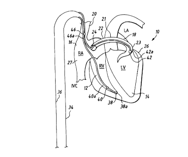
Referring now to Figure 1, it illustrates a fully
implantable atrial defibrillator 30 embodying the present
invention shown in association with a schematically
-9-
illustrated human heart 10 in need of atrial fibrillation
monitoring and potential cardioversion of the atria. The
portions of the heart illustrated in Figure 1 are the right
ventricle 12, the left ventricle 14, the right atrium 16, the
left atrium 18, the superior vena cava 20, the coronary sinus
channel 21 which, as used herein, denotes the coronary sinus
22 and the great cardiac vein 23, the coronary sinus ostium or
opening 24, the left ventricular free wall 26 and the inferior
vena cava 27. In addition, as used herein, the term
"depolarization activation waves" denotes R waves of the heart
cardiac cycle which induce depolarizations of the ventricles
12 and 14.
The atrial defibrillator 30 generally includes an
enclosure 32 for hermetically sealing the internal circuit
elements of the atrial defibrillator 30, an endocardial first
lead 34 and an intravascular second lead 36. The second lead
36 may alternatively comprise two leads. A single lead is
illustrated in Figure 1 so as to not unduly complicate the
figure. The enclosure 32 and the first and second leads 34
and 36 are arranged to be implanted beneath the skin of a
patient so as to render the atrial defibrillator 30 fully
implantable.
The endocardial first lead 34 preferably comprises an
endocardial bipolar lead having electrodes 38 and 40 arranged
for establishing electrical contact with the right ventricle
12 of the heart 10. The electrodes 38 and 40 permit bipolar
-10-
sensing of depolarization activation waves in the right
ventricle between a first pair of locations 38a and 42a within
the right ventricle 12. As illustrated, the lead 34 is fed
through the superior vena cava 20, into the right atrium 16,
and then into the right ventricle 12. As will be appreciated
by those having ordinary skill in the art, a second path for
lead 34 could alternatively be through the inferior vena cava
28, into the right atrium 16, and then into the right
ventricle 12.
The second lead 36 generally includes a first or distal
electrode 42 and a second electrode 46. As illustrated, the
second lead 36 is flexible and arranged to be passed down the
superior vena cava 20, into the right atrium 16, into the
coronary sinus ostium 24, and advanced into the coronary sinus
channel 21 of the heart near the left side thereof. The first
or distal electrode 42 is preferably within the coronary sinus
22 or the great vein 23 of the heart adjacent to the left
ventricle 14 at a location 42a. The electrode 42 is
preferably elongated such that the electrode 42 is within the
coronary sinus 22 and/or the great cardiac vein 23 adjacent
the left ventricle 14 and beneath the left atrium 18. The
second electrode 46 is preferably located at a location 46a
within either the right atrium 16 or the superior vena cava 20
and preferably within the right atrium 16.
As indicated above, the second lead 36 may comprise two
leads. In this preferred embodiment, one of the two leads may
-il-
2~~~~~7
include the first electrode 42 and the other of the two leads
may include the second electrode 46. As noted above, the
first electrode 42 and the second electrode 46 are illustrated
in Figure 1 as combined on a single lead, the second lead 36,
so as not to unduly complicate the figure.
The first electrode 42 together with the second electrode
46 of the second lead 36 provide for the delivery of
defibrillating or cardioverting electrical energy to the
atria. Because the first electrode 42 is located beneath the
left atrium 18 near the left ventricle 14 and the second
electrode 46 is within either the right atrium 16 or the
superior vena cava 20 and above the coronary sinus ostium 24,
the electrical energy applied between these electrodes will be
substantially confined to the atria 16 and 18 of the heart 10.
As a result, the electrical energy applied to the right
ventricle 12 and left ventricle 14 when the atria are
cardioverted or defibrillated will be minimized.
Within the enclosure 32, the atrial defibrillator 30
includes a first or right ventricular (RV) channel 60, a
second or right ventricular-coronary sinus (RVCS) channel 62
and a third or atrial channel 64. The RV channel 60 includes
a first sense amplifier 50, a first filter 52, a second filter
54 and an R wave detector 56. The RVCS channel 62 includes a
second sense amplifier 70, a third filter 72, a fourth filter
74 and a second R wave detector 76. The atrial channel 64
includes a third sense amplifier 80, a fifth filter 82, a
-12-
~1~~~47
sixth filter 84 and an atrial activity detector 86. Within
the enclosure 32, the atrial defibrillator 30 also includes a
microprocessor 88 and a memory 90.
The first sense amplifier 50 includes a first input 92
which is coupled to electrode 38 of the first lead 34 and a
second input 94 which is coupled to electrode 40 of the first
lead 34. The first sense amplifier 50 amplifies the sensed
electrical activity of the heart. The first filter 52
conditions the sensed electrical activity of the heart and
provides at an output 96 an amplified input signal
representative of the electrical activity of the heart such as
depolarization activation waves. sensed by the bipolar
electrodes 38 and 40 in the right ventricle 12 of the heart
10. The first sense amplifier 50 may include one or more gain
stages, and the order of the first sense amplifier 50 and the
first filter 52 may be reversed. That is, the first filter 52
may be coupled between the electrodes 38 and the first sense
amplifier 50. The second filter 54 further conditions the
sensed electrical activity of the heart.
The first R wave detector 56 has an input 98 coupled to
the second filter 54. The first R wave detector 56 produces
an electrical output or an initiation signal corresponding to
the depolarization activation wave sensed by the first sense
amplifier 50 when the amplified input signal received at the
input 98 of the first R wave detector 56 exceeds a threshold.
-13-
~1~00~'~
As a result, the RV channel 60, including electrodes 38,
40, the sense amplifier 50, the first filter 52, the second
filter 54 and the R wave detector 56, forms a first detecting
means for sensing a depolarization activation wave in general
at a first location of the heart, and more specifically,
between a first pair of locations 38a and 42a of the heart for
generating a first initiation signal. The electrodes 38 and
40 and the first sense amplifier 50 form a first sensing means
for sensing the depolarization activation wave. The first R
wave detector 56 forms a first output means for generating the
first initiation signal in response to the first sensing
means.
The RVCS channel 62 preferably operates in a manner
similar to the operation of the RV channel 60. The second
sense amplifier 70 includes a first input 100 which is coupled
to the electrode 42 of the second lead 36 and a second input
102 which is coupled to electrode 38 of the first lead 34.
The second sense amplifier 70 amplifies the sensed electrical
activity of the heart. The third filter 72 conditions the
sensed electrical activity and provides at an output 104 an
amplified signal representative of the electrical activity of
the heart, such as depolarization activation waves sensed by
electrodes 38 and 42. The second sense amplifier 70 may
include one or more gain stages. The fourth filter 74 further
conditions the amplified electrical activity.
-14-
The second R wave detector 76 includes an input 106 for
receiving the amplified signal. The second R wave detector 76
produces an electrical output or an initiation signal when the
amplified input signal provided at the input 106 exceeds a
threshold.
As a result, the RVCS channel, including the electrode
42, the electrode 38, the second sense amplifier 70, the third
filter 72, the fourth filter 74 and the R wave detector 76,
form a second detecting means for sensing a depolarization
activation wave in general at a second location of the heart,
and more specifically, between a second pair of locations 38a
and 42a of the heart for generating a second initiation
signal. The second sense amplifier 70 and the third filter 72
form a second sensing means for sensing the depolarization
activation wave. The R wave detector 76 forms a second output
means for generating the second initiation signal in response
to the second sensing means.
The third sense amplifier 80 senses electrical activity
in the atria 16 and 18 of the heart 10. To that end, the
third sense amplifier 80 includes a first input 110 which is
coupled to electrode 46 and a second input 112 which is
coupled to electrode 42. The fifth filter 82 and the sixth
filter 84 condition the amplified electrical activity sensed
by the third sense amplifier 80. The atrial activity detector
86 includes an input 114 for receiving the conditioned,
amplified electrical activity and an output 114 for providing
-15-
~1~0~4'~
an indication of electrical activity of the heart sensed by
the third sense amplifier 80. As a result, the atrial channel
64, including the electrodes 42 and 46, the third sense
amplifier 80, the fifth filter 82, the sixth filter 84 and the
atrial activity detector 86, form a third detecting means for
sensing activity of the atria of the heart.
The microprocessor 88 is preferably implemented in a
manner as disclosed in the aforementioned U.S. Patent No.
5, 282, 837 and further as described hereinafter with respect to
the flow diagram of Figures 4 and 5. The implementation of
the microprocessor 88 in accordance with this embodiment of
the present invention results in a plurality of functional
stages. The stages include an interval timer 116, an atrial
arrhythmia detector in the form of an atrial fibrillation
detector 118 and a charge delivery and energy control stage
120. The functional stages implemented by the microprocessor
88 also include a synchronization test 119, a sample rate
timer 123, a synchronization timeout timer 125, a shock delay
timer 127 and an RVCS delay timer 129, which will be discussed
further in conjunction with Figures 4 and 5. The
microprocessor 88 is arranged to operate in conjunction with
the memory 90 which is coupled to the microprocessor 88 by a
multiple bit address and data bus 121.
The atrial defibrillator 30 further includes an analog
multiplexer 122, an analog-to-digital converter 124 and a
direct memory access (DMA) controller 126. The output 96 of
-16-
~1~00~7
the f first f filter 52 , the output 104 of the third f filter 72 and
the output 87 of the fifth filter 82 are coupled to the analog
multiplexes 122. In response to control signals received from
the microprocessor 88 at a control input 128, the analog
multiplexes 122 couples signals received from either the first
filter 52, the third filter 72, the fifth filter 82, or a time
sequential combination of these signals to the output 130 of
the analog multiplexes 122. The output 130 is coupled to the
analog-to-digital converter 124, which converts analog signals
received from the output 130 to digital data. The digital
data are conveyed over a multiple bit data bus 132 to the
direct memory access controller 126. The direct memory access
controller 126 conveys digital data, along with storage
address information, over a multiple bit bus 134 to the memory
90. As a result, data received from either the first filter
52, the third filter 72 or the fifth filter 82 are stored by
the DMA controller 126 in the memory 90, without further
intervention by the microprocessor 88.
For determining if the heart 10 is in need of
cardioversion or defibrillation, and to synchronize delivery
of cardioverting or defibrillating electrical energy with
detection of a ventricular activation, the atrial
defibrillator 30 acquires a multi-channel intracardiac
electrogram (EGM) segment. To acquire a multi-channel EGM
segment, the microprocessor 88 conveys control signals to the
control input 128 of the analog multiplexes 122 to cause the
-17-
analog multiplexes 122 to couple a periodic time sequential
combination of two or more of the filter outputs 96, 104, or
87 to the output 130 of the analog multiplexes 122. The
analog-to-digital converter 124 converts sequential analog
signals from each of the requested filter outputs 96, 104, or
87 to digital data. The DMA controller 126 receives the
digital data in a time-multiplexed format and stores the data
in the memory 90.
In this manner, the atrial defibrillator 30 may store
data corresponding to electrical activity of the heart sensed
by each of the first sense amplifier 50, the second sense
amplifier 70 and the third sense amplifier 80. Following
acquisition of an EGM segment by the atrial defibrillator 30,
the data which form the EGM segment may be analyzed by the
microprocessor 88.
The atrial defibrillator 30 further includes a charger
and storage capacitor circuit 140 of the type well known in
the art which charges a storage capacitor to a predetermined
peak voltage level and a discharge circuit 142 for discharging
the storage capacitor within the circuit 140 for a
predetermined time to provide a controlled discharge output of
electrical energy when required to the atria of the heart 10.
To that end, the discharge circuit 142 is coupled to the first
electrode 42 and the second electrode 46 of the second lead 36
for applying the cardioverting or defibrillating electrical
energy to the atria. The atrial defibrillator 30 further
-18-
~1~00~'~
includes a depletable power source 144, such as a lithium
battery, for providing power to the electrical components of
the atrial defibrillator 30.
When the atrial fibrillation detector 118 determines that
the atria 16 and 18 are in fibrillation and thus in need of
cardioversion, the charge delivery control 120 causes the
charger and storage capacitor circuit 140 to charge the
storage capacitor within the circuit 140. The charge delivery
control 120 causes the discharge circuit 142 to discharge the
capacitor of the circuit 140 for applying cardioverting
electrical energy to the atria 16 and 18 in synchronism with
an R wave detected by first sense amplifier 50 and first R
wave detector 56 and second sense amplifier 70 and second R
wave detector 76.
For entering operating parameters into the microprocessor
88, the atrial defibrillator 30 receives programmable
operating parameters from an external controller 146 which is
external to the skin of the patient. The external controller
146 is arranged to communicate with a receiver/transmitter 148
within enclosure 32 which is coupled to the microprocessor 88
over a bidirectional bus 150. The receiver/transmitter 148
may be of the type well known in the art for conveying various
information which it obtains from the microprocessor 88 to the
external controller 146 or for receiving programming
parameters from the external controller 146 which the
receiver/transmitter 148 then conveys to the microprocessor 88
-19-
21~004'~
for storage in internal memory or in the memory 90 within the
enclosure 32.
The receiver/transmitter 148 includes a transmitting coil
152. Such communication circuits are well known in the art
and may be utilized as noted above for receiving commands from
external to the implantable enclosure 32 and for transmitting
data to the external controller 146 from the implanted
enclosure 32.
Referring now to Figure 2, it shows a series of wave
forms representative of electrical activity of the human heart
10 detected by the atrial defibrillator 30 of Figure 1. The
wave forms of Figure 2 are plotted with voltage on the
vertical axis and time on the horizontal axis.
Figure 2 shows a right ventricular intracardial
electrogram (RV EGM) segment 160 which has been detected by
the first sense amplifier 50 between a first pair of locations
38a and 40a. The RV EGM 160 may be converted to digital data
by the analog-to-digital converter 124 and stored in the
memory 90 by direct memory access controller 126 for analysis
by the microprocessor 88. The RV EGM segment 160 includes a
representation of a detected depolarization activation wave or
R wave 162 which has a peak 164. The RV EGM 160 further has
an initial portion 166 with a negative slope and a final
portion 168 with a positive slope. The RV EGM 160 has a peak
width 165 measured at a predetermined voltage 167.
-20-
21~D04'~
Figure 2 further shows a representation of a detected
threshold event 170, as detected by the first R wave detector
56. The second filter 54 receives the RV EGM 160 from the
first filter 52 and, after further conditioning, provides the
RV EGM to the first R wave detector 56. When the signal level
of the RV EGM exceeds a predetermined threshold, the R wave
detector 56 provides an electrical indication such as RV
threshold event 170 to the microprocessor 88. The detected RV
threshold event 170 may be stored in memory 90 as the time at
which the first R wave detector 56 detected a threshold event
in the right ventricle 12 of the heart 10. The microprocessor
88 uses the detected threshold event 170, for example, to
compare the time at which the threshold event 170 is detected
with the timing of the RV EGM 160, as will be discussed
further in conjunction with the flow diagram of Figures 4 and
5.
Figure 2 further shows a right ventricular-coronary sinus
intracardial electrogram (RVCS EGM) segment 172 which has been
detected by the second sense amplifier 70 between a second
pair of locations 38a and 42a. The RVCS EGM 172 may be
converted to digital data by the analog-to-digital converter
124 and stored in the memory 90 by DMA controller 126 for
analysis by the microprocessor 88. The RVCS EGM segment 172
includes a representation of a detected depolarization
activation wave or R wave 174 which has a peak 176. The RVCS
EGM 172 further has an initial portion 178 with a positive
-21-
21~~04'~
slope and a final portion 180 with a negative slope. The RVCS
EGM 172 has a peak width 177 measured at a predetermined
voltage 179.
Figure 2 still further shows a representation of a
detected threshold event 182, as detected by the second R wave
detector 76. When the signal level of the RVCS EGM received
by the second R wave detector 76 exceeds a predetermined
threshold, the second R wave detector 76 provides an
electrical indication such as RVCS threshold event 182 to the
microprocessor 88. The detected RVCS threshold event 182 may
be stored in memory 90 as the time at which the second R wave
detector 76 detected a threshold event between locations 38a
and 42a.
The operation of the atrial defibrillator 30 will now be
described with reference to Figure 4 and with respect to a
preferred embodiment of the present invention. For purposes
of this discussion, it is assumed that the atrial fibrillation
detector 118 has detected an atrial fibrillation episode and
that the storage capacitor within circuit 140 has been charged
to a predetermined peak voltage.
Figure 3 is a block diagram of the synchronization test
functional stage 119 implemented by the microprocessor 88 of
Figure 1. In accordance with the preferred embodiment of the
present invention, after the atrial fibrillation detector 118
has detected an atrial fibrillation episode and after the
storage capacitor within circuit 140 has been charged to a
-22-
predetermined peak voltage, the synchronization test 119
operates to synchronize the delivery of cardioverting or
defibrillating electrical energy from the storage capacitor in
circuit 140 with an R wave detected by the RV channel 60.
The synchronization test functional stage 119 preferably
includes a number of independent tests or checks to verify the
consistency of R waves detected by RV channel 60 and the RVCS
channel 62. Operation of the synchronization test functional
stage 119 will be discussed in further detail in conjunction
with Figures 4 and 5.
These checks includes a timer range check 184 for
verifying that the interval timed. between successive R waves
by the interval timer 116 is greater than a predetermined
minimum and less than a predetermined maximum. The
synchronization test 119 further includes a two-channel
threshold event check 186 for verifying timing between R waves
or threshold events detected by RV channel 60 and RVCS channel
62. The synchronization test functional stage 119 further
includes a two-channel peak test 188 for verifying the time
relationship between peak values of R waves detected by RV
channel 60 and RVCS channel 62.
The synchronization test functional stage 119 still
further includes an RV peak and threshold event check 190 for
verifying the time relation between the peak value and the
detected threshold event measured by the RV channel 60. The
synchronization test functional stage 119 further includes a
-23-
~1~00~7
two-channel morphology check 192 for verifying the amplitudes
and durations of portions of depolarization activation waves
detected by the RV channel 60 and the RVCS channel 62. The
synchronization test functional stage 119 still further
includes a channel-to-channel overlap test 194 for verifying
the time relationship of predetermined portions of the
depolarization activation wave detected by the RV channel 60
and the RVCS channel 62. The synchronization test functional
stage 119 further includes a two-channel noise and amplitude
check 196 for verifying that the depolarization activation
waves detected by the RV channel 60 and the RVCS channel 62
satisfy predetermined noise and amplitude range criteria.
The checks or tests which are included in the
synchronization functional test stage 119 are preferably
performed by the microprocessor 88 in response to instructions
and data stored in the memory 90. The microprocessor 88 may
perform one or more of the tests or checks illustrated in
Figure 3 and may perform other verification tests not
illustrated there.
The operation of the atrial defibrillator 30 will now be
described with reference to Figure 4 and with respect to a
preferred embodiment of the present invention. For purposes
of this discussion, it is assumed that the atrial fibrillation
detector 118 has detected an atrial fibrillation episode and
that the storage capacitor within circuit 140 has been charged
to a predetermined peak voltage.
-24-
~~~oo~~
Referring now to Figure 4, the microprocessor 88 first,
in step 200, resets and initializes elements used for
synchronizing delivery of cardioverting or defibrillating
electrical energy to a detected R wave. The microprocessor 88
resets the sample rate timer 123 (Figure 1), the
synchronization timeout timer 125, the shock delay timer 127
and the RVCS delay timer 129. Also at step 200, the
microprocessor 88 starts the sample rate timer 123 and
initiates a multi-channel EGM data acquisition from the RV
channel filter output 96 and the RVCS channel filter output
104. The sample rate timer 123, upon expiration, signals that
a single data sample has been acquired by the DMA controller
126 from both the RV channel filter output 96 and the RVCS
channel filter output 104. Upon said expiration, the
microprocessor 88 may then process the digital data sample
acquired from each of these two channels.
Also at step 200, the microprocessor 88 starts the
interval timer 116 in response to a ventricular activation (R
wave) detected by either the RV channel 60 or the RVCS channel
62. An R wave is detected if the RV EGM 160 has exceeded a
predetermined threshold in RV channel 60 and the R wave
detector 56 has provided an RV threshold event or initiation
signal 170 to the microprocessor 88 or if the RVCS EGM 172 has
exceeded a predetermined threshold in the RVCS channel 62 and
the R wave detector 76 has provided an RVCS threshold event or
initiation signal 182 to the microprocessor 88. After this
-25-
21~0~14~
signaling of ventricular activation, step 200 is completed and
step 202 is entered.
At step 202, the sample rate timer 123 is tested. If the
sample rate timer 123 has not expired, the microprocessor
returns. If the sample rate timer 123 has expired, at step
204, the microprocessor resets the sample rate timer 123 and
processes the most recently acquired digital data sample
(stored in memory 90) from both the RV channel 60 and the RVCS
channel 62. These two data samples represent a single data
point on an electrogram in the RV channel 60 and the RVCS
channel 62. That is, electrical activity of the heart 10
sensed by the first sense amplifier 50 and the second sense
amplifier 70 is provided by the analog multiplexer 122 to the
analog-to-digital converter 124 for conversion to digital data
and storage by the DMA controller 126 in the memory 90.
During this acquisition, the microprocessor 88 further
processes this data stored in the memory 90 point by point as
it is being acquired by the DMA controller 126 and as signaled
by the sample rate timer 123.
The sample rate timer 123 may be reset to expire after a
predetermined time interval, for example two milliseconds or
four milliseconds. The sample rate timer 123 thus controls
the rate at which data samples representing electrocardiograms
are acquired from the RV channel 60 and the RVCS channel 62
after the atrial fibrillation detector 118 has detected an
atrial fibrillation episode. While the DMA controller 126 is
-26-
~1~00~'~
acquiring electrograms from the RV channel 60 and the RVCS
channel 62, the microprocessor 88 may analyze previously-
acquired electrocardiogram data, for example, by performing
morphology tests described below in conjunction with Figure 5.
It is desirable to apply cardioverting or defibrillating
electrical energy to the atria of the heart immediately
subsequent to detection of an R wave, to minimize the risk of
induced ventricular fibrillation. However, R waves detected
by the RV channel 60 at locations 38a and 40a (Figure 1) tend
l0 to have widely varying amplitudes, particularly when the heart
is in atrial fibrillation. Therefore, there is a risk that
a low amplitude R wave may not be detected by the RV channel
60, meaning the interval timer 116 may not be reset, and
causing the interval timer 116 to measure a falsely long
interval. To ensure that all R waves are detected and cause
the interval timer 116 to be reset, the atrial defibrillator
30, in accordance with the present invention, resets the
interval timer 116 in response to detection of an R wave by
either the RV channel 60 or the RVCS channel 62.
Since the cardiac activity detected by the RVCS channel
62 tends to be noisy, due to atrial fibrillation and other
noise, and because an R wave detected by the RVCS channel 62
tends to be spread out in time, it is preferable to apply
cardioverting or defibrillating electrical energy only in
response to an R wave detected by the RV channel 60. However,
as can be seen from Figure 2, an RVCS EGM 172 detected by the
-27-
~1~004~
RVCS channel 62 may be spread out in time relative to an RV
EGM 160 detected by the RV channel 60. RVCS threshold event
182 may actually be detected earlier in time than an RV
threshold event 170 is detected for the same depolarization
activation wave. Therefore, in accordance with the present
invention, resetting of the interval timer 116 in response to
an RVCS threshold event 182 is delayed by a predetermined
time, such as 20 milliseconds, which is timed by the RVCS
delay timer 129. Resetting of the interval timer 116 in
response to an RV threshold event 170 is preferably not
delayed. The RVCS delay timer 129, which establishes the
delay time for resetting the interval timer 116 in response to
an RVCS threshold event 182 is preferably programmable using
the external controller 146. The dual reset process,
including delaying the resetting of the interval timer 116 in
response to a detected RVCS threshold event 182 allows
application of cardioverting or defibrillating electrical
energy to be accurately synchronized to an R wave detected in
the RV channel 60.
To begin this dual reset process, the microprocessor 88
at step 206 first determines if an RV threshold event has
occurred. An RV threshold event has occurred if the RV EGM
160 has exceeded a predetermined threshold in the RV channel
60 and the R wave detector 56 has provided an initiation
signal 170 to the microprocessor 88.
-28-
~~~oo~~
If an RV threshold event has been detected, at step 208,
the microprocessor 88 determines if the time interval since
the last RV threshold event or the last RVCS threshold event,
measured by the interval timer 116, is within a predetermined
range. The range is determined by minimum and maximum time
intervals which may be set from external to the implanted
atrial defibrillator 30 by means of the external controller
146 and the transmitter/receiver 148. In accordance with the
preferred embodiment of the present invention, the minimum
time interval may be in the range of 500 milliseconds and the
maximum time interval may be in the range of 2,000
milliseconds. If the time interval measured by the interval
timer 116 is less than the minimum time interval or greater
than the maximum time interval, the microprocessor 88 restarts
the interval timer at step 212. No cardioverting or
defibrillating electrical energy will be applied in response
to the RV threshold event detected at step 206, and the
microprocessor 88 will wait until a subsequent RV threshold
event is detected before applying cardioverting or
defibrillating electrical energy. If the time interval
measured by the interval timer 116 is within the predetermined
range of interval times, the microprocessor 88 at step 210
starts the shock delay timer 127 and at step 212 restarts the
interval timer 116.
If an RV threshold event was not detected by the
microprocessor 88 at step 206, at step 214 the microprocessor
-29-
~1~OQ~:'~
88 determines if the RVCS delay timer 129 has expired. The
RVCS delay timer 129 may have been started in response to a
previously detected RVCS threshold event. If the RVCS delay
timer has expired, at step 216, the microprocessor 88 resets
and stops the RVCS delay timer and at step 212 restarts the
interval timer.
At step 218, the microprocessor 88 determines if an RVCS
threshold event has been detected. An RVCS threshold event is
detected if the RVCS EGM 172 has exceeded a predetermined
threshold in the RVCS channel 62 and the R wave detector 76
has provided an initiation signal 182 to the microprocessor
88. In response to detection of an RVCS threshold event, the
microprocessor 88 starts the RVCS delay timer 129 at step 220.
At step 222, the microprocessor 88 determines if the
shock delay timer 127 has expired. The shock delay timer 127
was started at step 210 in response to detection of an RV
threshold event that satisfied the R-to-R interval criterion.
The shock delay timer 127 ensures that cardioverting or
defibrillating electrical energy will only be applied a
predetermined delay time after detection of an RV threshold
event which satisfies the R-to-R interval criterion. For
example, the shock delay timer 127 may delay application of
cardioverting or defibrillating electrical energy by 30-50
milliseconds following detection of an RV threshold event
which satisfies the R-to-R interval criterion. The
predetermined delay time measured by the shock delay timer 127
-30-
~1500~'~
is preferably programmable using the external controller 146.
If the microprocessor 88 determines at step 222 that the shock
delay timer has expired, at step 224 the microprocessor 88
determines if the RV EGM 160 and the RVCS EGM 172 satisfy a
set of predetermined morphological tests or checks,
illustrated in Figure 5.
Referring to Figure 5, it shows a flow diagram
illustrating the manner in which the atrial defibrillator of
Figure 1 may be implemented in accordance with the present
invention for performing morphological consistency analysis on
detected depolarization activation waves in conjunction with
the flow diagram of Figure 4. In performing the steps
illustrated in Figure 5, the microprocessor 88 analyzes data
stored in the memory 90 corresponding to electrical activity
of the heart 10 detected by the first sense amplifier 50 and
the second sense amplifier 70 (Figure 1). To the degree
possible, this analysis is performed on a point-by-point basis
as the data is being acquired as indicated in step 204. This
real-time analysis minimizes the computational time required
to perform the decisions shown in Figure 5 so that
defibrillating electrical energy can be delivered as quickly
as possible after the expiration of the shock delay timer 127.
To ensure that the atrial defibrillator 30 does not apply
cardioverting or defibrillating electrical energy to the atria
of the heart l0 in response to an erroneously detected R wave
or threshold event, the atrial defibrillator in accordance
-31-
~1~004'~
with the present invention performs morphological and
threshold consistency checks in addition to the R-to-R
interval timing test. The morphology of a detected
depolarization activation wave is the shape, duration and
amplitude characteristics of the depolarization activation
wave. As discussed above in relation to Figure 1, a digital
representation of a detected depolarization activation wave
segment is stored in the memory 90 for analysis by the
microprocessor 88. In addition, morphological analysis and
consistency checks are performed in accordance with the
present~invention using R wave segments and threshold events
detected by both the RV channel 60 and the RVCS channel 62.
Before cardioversion or defibrillation can occur, the R wave
segments and threshold events must meet predetermined criteria
to ensure that a detected R wave is a genuine or legitimate R
wave. Thus, cardioverting or defibrillating electrical energy
is applied to the atria of the heart only when the atria of
the heart are in need of cardioversion and when a first signal
detected by the RV channel 60 satisfies a first criterion and
when a second signal produced by the RVCS channel 62 satisfies
a second criterion.
At step 226, the microprocessor 88 determines if
threshold events detected by the RV channel 60 and the RVCS
channel 62 occurred within a predetermined time with respect
to each other, for example 20 milliseconds. The predetermined
time may be programmable using the external controller 146.
-32-
2150147
If a detected R wave is a genuine R wave, it must be detected
in both the RV channel 60 and the RVCS channel 62 within a
predetermined time. If the detected depolarization activation
wave does not pass these criteria, the microprocessor 88
proceeds to step 242 (Figure 4).
At step 228, the microprocessor 88 determines if the
morphological peaks of the depolarization activation wave
segments detected by the RV channel 60 and the RVCS channel 62
occur within a predetermined time period, such as 20
milliseconds. The predetermined time period is preferably
programmable using the external controller 146. In performing
this test, the microprocessor 88 determines the peak 164 of an
RV EGM 160 and the peak 176 of an RVCS EGM 172 (Figure 2). If
the time relationships between the peaks 164 and 176 do not
satisfy these criteria, the microprocessor 88 proceeds to step
242.
Also at step 228, the microprocessor 88 determines if the
widths of the morphological peaks of the detected
depolarization activation wave segments are within a
predetermined peak width time range. For example, the width
165 of the RV EGM 160 (Figure 2) must be less than a maximum
width, such as 50 ms, and greater than a minimum width, such
as 12 ms. Similarly, the width 177 of the RVCS EGM 172 must
be less than a maximum width and greater than a minimum width.
The respective maxima and minima are preferably independently
definable.
-33-
At step 230, the microprocessor 88 determines if the
morphological peak 164 of the R wave detected by the RV
channel 60 occurs within a predetermined time, such as 20
milliseconds, of the RV threshold event 170 detected by the RV
channel 60. The predetermined time interval is preferably
programmable using the external controller 146. The peak 164
of the RV EGM 160 should occur within a predetermined time of
the RV threshold event 170. If the R wave detected by the RV
channel 60 does not satisfy these criteria, the microprocessor
88 proceeds to step 242.
At step 232, the microprocessor 88 determines if the RV
EGM 160 detected by the RV channel 60 and the RVCS EGM 172
detected by the RVCS channel 62 satisfy predetermined
morphology criteria. For example, the microprocessor 88 may
determine if the amplitudes of the initial portion 166 and the
f final portion 168 of the RV EGM 160 are within a predetermined
range of values. Similarly, the microprocessor 88 may
determine if the amplitudes of the initial portion 178 and the
final portion 180 of the RVCS EGM 172 are within a
predetermined range of values. Also, for example, the
microprocessor 88 may determine if the durations of the
initial portion 166 and the final portion 168 of the RV EGM
160 are within a predetermined range of values, and if the
durations of the initial portion 178 and the final portion 180
of the RVCS EGM 172 are within a predetermined range of
values. These tests ensure that cardioverting or
-34-
21~00~'~
defibrillating electrical energy will be delivered to the
atria of the heart 10 only in response to a genuine R wave,
and that the detected R wave is a genuine R wave and is not
contaminated with noise or other interference. If the
detected RV EGM 160 and RVCS EGM 172 do not satisfy these
criteria in step 232, the microprocessor 88 proceeds to step
242.
At step 234, the microprocessor 88 determines if the
duration of the final portion 180 of a detected RVCS EGM 172
exceeds the duration of the final portion 168 of a detected RV
EGM 160 by a predetermined time, such as four milliseconds.
The predetermined time may be programmable in response to
external controller 146. This test helps to ensure that
detected cardiac activity has a physiological origin rather
than an extraneous origin, such as noise. R waves detected by
the RVCS channel 62, such as RVCS EGM 172, generally exhibit
trailing R wave durations, such as final portion 180, that
exceed trailing R wave durations of R waves detected by the RV
channel 60, such as final portion 168. If the detected RV EGM
160 and the detected RVCS EGM 172 do not satisfy these
criteria, the microprocessor 88 proceeds to step 242.
At step 236, the microprocessor 88 determines if detected
signal quality is sufficient to ensure that a genuine R wave
has been reliably detected. For example, the microprocessor
88, in analyzing RV EGM 160 stored in the memory 90, may
determine all portions of the detected RV EGM 160, such as
-35-
initial portion 166 and final portions 168, which exceed a
predetermined amplitude (the amplitude threshold). If the
number of such portions exceeds a predetermined number (the
count threshold) , then the microprocessor 88 may conclude that
the detected RV EGM is noisy and is of insufficient quality.
A similar test may be performed on the detected RVCS EGM 172.
The amplitude threshold and the count threshold are preferably
programmable, for both the RV channel 60 and the RVCS channel
62, using the external controller 146.
l0 An additional test that may be performed at step 236 is
analysis of the maximum amplitude of the detected RV EGM 160
and the detected RVCS EGM 172. For example, the
microprocessor 88 may determine that the peak value 164 of the
detected RV EGM 160 is within a predetermined range of values
and that the peak value 176 of the detected RVCS EGM 172 is
within a predetermined range of values. This ensures that,
for example, the analog-to-digital converter 124 did not
saturate during the R-to-R interval preceding delivery of
cardioverting or defibrillating electrical energy. If the
detected RV EGM 160 and the detected RVCS EGM 172 do not
satisfy these criteria, the microprocessor 88 proceeds to step
242.
The precise ordering of the tests illustrated in steps
226-236 is not critical. Preferably, the detected cardiac
activity must satisfy all the criteria in these steps or tests
before the discharge circuit 142 applies the cardioverting or
-36-
~1~~0~'~
defibrillating electrical energy to the atria of the heart.
However, one or more of these tests may be eliminated while
remaining within the scope of the present invention. In
addition, to conserve energy stored in the depletable energy
source 144, it may be preferable to perform computationally
simple tests first.
Referring again to Figure 4, at step 244, if the detected
ventricular activation has satisfied each of the predetermined
morphology and consistency criteria (steps 226-236), and if
the time interval between the last two immediately successive
ventricular activations as measured by the interval timer is
greater than the preselected minimum time interval and less
than the preselected maximum time interval, and if the atrial
fibrillation detector 118 has determined that the atria are in
need of defibrillation, the charge delivery control stage 120
of the microprocessor 88 causes the discharge circuit 142 to
immediately discharge the electrical energy stored in the
storage capacitor of the circuit 140 for applying the
cardioverting or defibrillating electrical energy to the atria
16 and 18 of the heart 10. Since the microprocessor 88 is
able to process steps 202-222 very quickly after the
occurrence of the last detected ventricular activation and to
process steps 226-236 very quickly after the expiration of the
shock delay timer 127, the discharge circuit 142 will apply
the cardioverting electrical energy to the atria of the heart
substantially coincident or in synchronism with the last
-37-
detected ventricular activation, as determined by the shock
delay timer 127. Preferably, the discharge circuit 76 will
apply the cardioverting electrical energy to the atria of the
heart within 30-50 milliseconds of the last detected
ventricular activation.
At step 242, if the detected ventricular activity did not
satisfy the predetermined morphology and consistency criteria
of any of the tests of steps 226-236, the atrial defibrillator
30 will not apply cardioverting or defibrillating electrical
energy to the atria of the heart in response to the shock
delay timer expiration caused by the RV threshold event
detected at step 206. Instead, the microprocessor 88 resets
and stops the shock delay timer 127 until a subsequent RV
threshold event is detected which meets the R-to-R interval
criteria.
If, at step 222, the shock delay timer 127 had not
expired or if, at step 224, one or more of the morphological
tests did not pass and the shock delay timer was reset at step
242, the microprocessor proceeds to step 246 to determine if
the synchronization timeout timer has expired. The
synchronization timeout timer 125 is used by the
microprocessor 88 to interrupt the shock synchronization
procedure illustrated in Figures 4 and 5 after a predetermined
time. For example, the synchronization timeout timer 125 may
be reset and started at step 200 when the microprocessor 88
begins searching for a detected ventricular activation
-38-
21a~04'~
suitable for synchronizing delivery of cardioverting or
defibrillating electrical energy. The synchronization timeout
timer 125 may expire after, for example, one minute,
indicating that no suitable synchronization condition can be
determined. To prevent continued operation of the shock
synchronization procedure illustrated in Figure 4, which may
deplete the depletable energy source 144, if the
synchronization timeout timer 125 has expired, the
synchronization procedure terminates at step 248.
If at step 246 the synchronization timeout timer 125 has
not expired, the microprocessor proceeds to step 202 and
awaits the next expiration of the sample rate timer 123, which
signals that the DMA controller 126 has acquired another
digital data sample from each of the RV and RVCS channels 60,
62. The sample rate timer 123 expires at regular intervals,
for example every two milliseconds or every four milliseconds.
After every expiration of the sample rate timer 123, the steps
shown in Figure 4 and Figure 5 are potentially executed as
indicated by the decision steps in these figures.
As a result of the foregoing, the atrial defibrillator of
the present invention precludes the application of
cardioverting or defibrillating electrical energy to the atria
of the heart in the presence of a possible vulnerable
condition resulting from a cardiac rate which is too high or
a cardiac rate which is suspected of being highly variable.
In either case, the atrial defibrillator of the present
-39-
~~~a~4'~
invention greatly reduces the risk of inducing ventricular
fibrillation during the application of cardioverting or
defibrillating electrical energy to the atria of the heart.
In addition, the atrial defibrillator of the present
invention precludes application of cardioverting or
defibrillating electrical energy to the atria of the heart
when detected ventricular activations do not meet predefined
morphological and consistency criteria. The atrial
defibrillator of the present invention thus reduces the risk
of falsely detecting ventricular activations and the risk of
inducing ventricular fibrillation during the application of
cardioverting or defibrillating electrical energy to the atria
of the heart in response to such a falsely detected
ventricular activation.
While a particular embodiment of the present invention
has been shown and described, modifications may be made. For
example, the interval timing and morphological criteria of the
present invention may be utilized to advantage in an external
atrial defibrillator wherein an electrode or electrodes
adhered to the surface of the skin of a patient are employed
along with an R wave detector for detecting ventricular
activations and surface pad electrodes are utilized for
applying the cardioverting electrical energy to the atria of
the heart. Such surface detecting and pad electrodes are well
known in the art. Additionally, the invention may be modified
by deleting any or all tests shown in Figure 5. For example,
-40-
~1~Q0~~
RVCS morphology checks could be eliminated or the RV to RVCS
overlap test could be eliminated. Hence, it is therefore
intended in the appended claims to cover all such changes and
modifications which fall within the true spirit and scope of
the invention.
-41-