Note: Descriptions are shown in the official language in which they were submitted.
~151~8fi
WO 94/15653 - PCT/US94/00409
TGF-BETA FORMULATION FOR INDUCING BONE GROWTH
Backqround of the Invention
Field of the Invention
s This invention relates to the use of transforming growth factor-beta (TGF-,6) to induce
bone growth in vivo and to formulations of TGF-,B and tricalcium phosphate useful for this
purpose .
Description of Related Art
The disorders associated with bone loss present major public health problems for0 Western societies. Osteopolosis alone may affect 20 million Americans in the early years of
the next century. Hence, there is wide interest in identifying factors or potential therapeutic
agents that inhibit bone loss and stimulate the formation of healthy new bone.
Bone is an extremely complex, but highly organized, connective tissue that is
continuously remodeled during the life of an adult bv cellular events that initially break it down
15 (osteoclastic resorption) and then rebuild it (osteoblastic formation). This remodeling process
occurs in discrete packets throughout the skeleton, i.e., in both cortical bone and trabecular
bone. It has recently been repo. l~d that mouse bone marrow cells can be stimulated to
generate osteoclasts in the presence of parathyroid hormone-related protein or vitamin D. See
Akatsu et al., Endocrinoloqv, 125: 20-27 (1989); Takahashi et al., Endocrinoloqv, 123: 2600-
20 2602 (1988) and Takahashi et al., Endocrinoloqv, 123: 1504-1510 (1988).
The currently available therapeutic agents known to stimulate bone formation arefluoride, estrogen, and vitamin D. Fluoride clearly increases trabecular bone mass, but
questions remain about the quality of the new bone formed, the side effects observed in some
patients, whether there are bene~icial effects on vertebral fracture rates, and whether
25 increased fragility of cortical bone with subsequent propensity to hip fracture follows.
Another approach is using agents that promote resorption (parathyroid hormone) and
then interrupt resorption (calcitonin). One proposed, but not validated, such sequential
therapeutic regimen is coherence therapy, where bone metabolic units are activated by oral
phosphate administration and then resorption is inhibited by either diphosphonates or
30 calcitonin.
Within the past few years several factors that stimulate osteoblasts were identified in
bone, including TGF-,B, fibroblast growth factor, platelet-derived growth factor, insulin-like
growth factor 1, and ,6`2 macroglobuiin. Of these, TGF-~ and IGF-I were deemed attractive
candidates for factors linking previous bone resorption with subsequent bone formation.
35 Mundy, The Journal of NIH Research, 1: 65-68 (1989).
Other proteins stored in the bone matrix may also be important for bone formation.
When demineralized bone was Injected into the muscle or subcutaneous tissue of rats, a
cascade of events, including chonarogenesls, ensued. Urist, Science,150: 893 (1965!. This
WO 94/15653 ~ g Ç~ PCT/US94/00409
observed activity was due to bone morphogenetic protein (BMP). Since the 1960s several
inve~igdlu,:. have aLle",pted to identify and characterize this activity. Thus, a protein of 22
Kd, called osteogenin, was identified that possessed the activity. Sampath et al., Proc. Natl.
Acad. Sci. USA, 84: 7109 (1987). Three proteins from demineralized ovine bone matrix were
s identified as having this activity. Wang et al., Proc. Natl. Acad. Sci., 85: 9484 (1988) and
Wozney et al., Science, 242: 1528 (1988). These proteins were named BMP-1, BMP-2A, and
BMP-3, the latter two of which belong to the extended TGF-,B family by limited sequence
homology. These workers modified the assay for bone~ induction to show cartilage formation
but did not show that the proteins ultimately stimulate formation of bone.
The TGF~ group of molecules are each dimers containing two identical polypeptidechains linked by disulfide bonds. The molecular mass of these dimers is about 25 Kd.
Biologically active TGF-~ has been defined as a molecule capable of inducing anchorage
independent growth of target cell lines or rat fibroblasts in in vitro cell culture, when added
together with EGF or TGF-a as a co-factor. TGF-~B is se~,eted by virtually all cell types in an
15 inactive form. This latent form can be activated by proteolytic cleavage of mature TGF-~ from
its precursor (at the Arg-Ala bond in position 278). A non-covalent complex is formed from
the association of the mature TGF-~ with the precursor remainder or with a protein binding to
TGF-,~ or with alpha -macroglobulin. This co",,1. Y is disrupted so as to activate the TGF-,~
either by exposure to l~ansienl acidification or by the action of exogenous proteases such as
20 plasmin or plasminogen activator.
There are at least five forms of TGF-~ currently identified, TGF-,B1, TGF-B2, TGF-,B3,
TGF-,~4, and TGF-~5. Suitable methods are known for purifying this family of TGF-,Bs from
various species such as human, mouse, monkey, pig, bovine, chick, and frog, and from
various body sources such as bone, platelets, or placenta, for producing it in recombinant cell
25 culture, and for determining its activity. See, for example, Derynck et al., Nature, 316: 701-
705 (1985); Eu,opean Pat. Pub. Nos. 200,341 published December 10, 1986, 169,016published January 22, 1986, 268,561 published May 25, 1988, and 267,463 published May
18, 1988; U.S. Pat. No. 4,774,322; Seyedin et al, J. Biol. Chem., 262: 1946-1949 (1987);
Cheifetz et al, Cell, 48: 409-415 l1987): Jakowlew et al., Molecular Endocrin., 2: 747-755
30 (1988); Dijke et al., Proc. Natl. Acad. Sci. (U.S.A.), 85: 4715-4719 (1988); Derynck et al., J.
Biol. Chem., 261: 4377-4379 (1986); Sharples et al., DNA, 6: 239-244 (1987): Derynck et
al., Nucl. Acids. Res., 15: 3188-3189 (1987) Derynck et al., Nucl. Acids. Res., 15: 3187
(1987): Derynck et al., EMB0 J., 7: 3737-3743 (1988)); Seyedin et al., J. Biol. Chem., 261:
5693-5695 (1986); Madisen et al., DNA, 7: 1-8 (1988); and Hanks et al., Proc. Natl. Acad.
35 Sci. (U.S.A.), 85: 79-82 11988).
TGF-~3, TGF-,64, and TGF-~5, which are the most recently discovered forms of TGF-,B,
were ide"li~ied bv screenlng cDNA libraries. None of these three putative protelns has been
` 2~514~:
WO 94/1~;653 PCT/US94/00409
isoiated from natural sources, although Northern blots demonstrate expresslon of the
corresponding mRNAs. TGF-,~4 and TGF-,B5 were cloned from a chicken chondrocyte cDNA
library (Jakowlew et al., Molec. Endocrinol., 2: 1186-1195 119881) and from a frog oocyte
cDNA library, respectively. The frog oocyte cDNA library can be screened using a probe
5 derived from one or more sequences of another type of TGF-~. TGF-,~4 mRNA is detectable in
chick embryo chondrocytes, but is far less abundant than TGF-,6`3 mRNA in developing
embryos or in chick embryo fibroblasts. TGF~ 5 mRNA is expressed in frog embryos beyond
the neurula state and in Xenopus tadpole (XTC) cells.
TGF-~ has been shown to have numerous regulatory actions on a wide variety of both
10 normal and neoplastic cells. TGF-,B is multifunctional, as it can either stimulate or inhibit cell
proliferation, lirre er Lialion, and other critical processes in cell function (M. Sporn, Science,
233:532 11986]). For a general review of TGF-,B and its actions, see Sporn et al., J. Cell Biol.,
105: 1039-1045 ~1987), Sporn and Roberts, Nature, 332: 217-219 ~1988), and Sporn and
Roberts, in Sporn and Roberts, ed., Handbook of Experimental Pharmacoloqv: Peptide Growth
15 Factors and Their Receptors 1, Springer-Verlag, New York, pp. 3-15 11990).
The multifunctional activity of TGF-,~ is modulated by the influence of other growth
factors present together with the TGF-,~. TGF-,B can function as either an inhibitor or an
enhancer of anchorage-independent growth, depending on the particular set of growth factors,
e.g., EGF or TGF-a, operant in the cell togeLI"3r with TGF-~6 ~Roberts et al., Proc. Natl. Acad.
20 Sci. U.S.A., 82:119 [19851). TGF-~ also can act in concert with EGF to cause proliferation
and piling up of normal ~but not rheumatoid) synovial cells IBrinkerhoff et al., Arthritis and
Rheumatism, 26:1370 119831)-
Although TGF-~ has been purified from several tissues and cell types, as indicated
above, it is especially abundant in bones IHauschka et al., J. Biol. Chem., 261: 12665 ~19861)
2s and platelets ~Assoian et al., J. Biol. Chem., 258: 7155 l19831). TGF-,B is postulated to be
one of the local mediators of bone generation and resorption, because of its presence in large
amounts in bone and cartilage, because cells with osteoblast and chondrocyte lineage increase
replication after exposure to TGF-,~, and because TGF-,~ regulates differentiation of skeletal
precursor cells. See Centrella et al., Fed. Proc. J., 2: 3066-3073 ~1988).
30 Immunohistochemical studies have shown that TGF-,6` is involved in the formation of the axial
skeleton of the mouse embryo. TGF-,6 is also present in other embryos in the cytoplasm of
osteoblasts in centers of endochondral ossification and in areas of intramembranous
ossification of flat bones, such as the calvarium. Heine et al., J. Cell. Biol., 105: 2861-2876
(1987). Following n situ hybridization of TGF-~1 probes, localization of TGF-,B in both
~s osteoclasts and osteoblasts has been described in development of human long bones and
calvarial bones. Sandberg et al., Development, 102: 461-470 (1988); Sandberg et al., Devel.
WO 94/15653 215 148 6 PCT~US94/00409
Biok, 130: 324-334 ~1988). TGF-,~is found in adult bone matrix (Seyedin et al., Proc. Natl.
Acad. Sci. USA, 82: 2267-2271 [19851, Seyedin et al., J. Biol. Chem., 261: 5693-5695
119861) and appears at the time of endochondral ossification in an in vivo model of bone
formation (Carrington et al., J. Cell. Biol., 107: 1969-1975 119881). Cultured fetal bovine
s bone osteoblasts as well as rat osteosarcoma cells have high mRNA levels for TGF-,~ and
secrete relatively high concentrations of TGF-,~ (Robey et al., J. Cell. Biol., 105: 457-463
1 1 9871).
In certain in vitro models, TGF-,6 was found to stimulate the synthesis of collagen,
osteopontin, osteonectin, and alkaline phosphatase, and to stimulate replication in osteoblast-
lO like cells. See Centrella et al., J. Biol. Chem., 262: 2869-2874 (1987); Noda et al., J. Biol.
Chem., 263: 13916 (1988); Wrana et al., J. Cell. Biol., 106: 915 (1988); Noda et al., J. Cell.
Phvsiol., 1 33: 426 ( 1 987); Ff~iilshi I Ler et al ., Endocrinoloqy, 1 21: 21 2 l 1 987); Centrella et al .,
Endocrinoloqv, 119: 2306 (1986); Roby et al., J. Cell. Biol., 105: 457 ~1987). In other in
vitro models, TGF-,B was found to inhibit proliferation and exp.~ssion of alkaline phosphatase
15 and oslt:ocal~.in. See, for example, Noda and Rodan, Biochem. BioPhvs. Res. Commun., 140:
56 (1986); Noda, Endocrinoloqv, 124: 612 (1989).
Further, while Centrella et al., supra, showed incleased collagen synthesis after
treatment of ostPoblast~ from rat calvaria with TGF-~, Robey et al., supra, could not show
increased synthesis of collagen in fetal bovine bone osteoblasts, postulating that the increased
20 collagen production is secondary to the effects of TGF-,~ on the p~al;r~ ion of osteoblasts. In
organ culture, TGF-,B was .epo.~t:d to stimulate bone resor~,Lion in neonatal mouse calvarias,
but inhibit re:.o.~,Lion in the fetal rat long bone system. See Tashjian et al., Proc. Natl. Acad.
Sci. USA, 82: 4535 (1981); Pfeilsl-irlt:r et al., J. Clin. Invest., 82: 680 (1988). TGF-B activity
was reported to be increased in cultures of fetal rat calvaria and in calvarial cells incubated
25 with stimulators of bone resorption, such as parathyroid hormone, 1,25-dihydroxyvitamin D,
and IL-1 (Petkovich et al., J. Biol. Chem., 262: 13424-13428 119871, PfeilschirLer and
Mundy, Proc. Natl. Acad. Sci. USA, 84: 2024-2028119871). Furthermore, it was reported
that TGF-,B inhibits the formation of osteoclasts in bone marrow cultures. Chenu et al., Proc.
Natl. Acad. Sci. USA, 85: 5683-5687 (1988). The showing that TGF-~ has effects on both
30 osteoclasts and osteoblasts led Pfeilschifter and Mundy, svpra, to propose that it is involved in
the strict coupling of the processes of bone resorption and bone formation characteristic of the
remodeling process in adult bone. It has also been postulated that the local acidic, proteolytic
environment provided by the osteoclasts results in activation of matrix-associated latent TGF-
,6. Oreffo et al., Calcified Tiss. Internatl., 42: Suppl:A15 11988).
In view of the conflicting results reported for in vitro activities, it is not clear whether
in vitro models can be used to predicl the effects of TGF-~ on bone formatlon and It:sor~,lion
in vivo. See Roberts et al., Proc. Natl. Acad. Sci. USA, 82: 119 (1985).
--4--
WO 94/15653 2 ~ S 1 ~ ~ ~ PCT/US94100409
Additional references reporting that TGF-~ promotes the proliferation of connective and
soft tissue for wound healing applications include U.S. Pat. No. 4,810,691 issued March 7,
1989, U.S. Pat. No. 4,774,228 issued September 27, 1988, Ignotz et ai., J. Biol. Chem., 261:
4337 t1986); Varga et al., Biochem. Biophvs. Res. Comm., 138: 974 (1986); Roberts et al.,
5 Proc. Natl. Acad. Sci. USA, 78: 5339 ~1981); Roberts et al., Fed. Proc., 42: 2621 ~1983);
U.S. Pat. No. 4,774,228 to Seyedin et al. TGF-,B stimulates the proliferation of epithelia
~Matsui et al., Proc, Natl. Acad. Sci. USA, 83: 2438 ~19861; Shipley el: al, Cancer Res., 46:
2068 11986]); induces collagen secretion in human fibroblast cultures (Chua et al., J. Biol.
Chem., 260: 5213-5216 ~1~831); stimulates the release of prostaglandins and mobilization of
lO calcium (Tashjian et al., Proc. Natl. Acad. Sci. USA, 82: 4535 ~1985l); and inhibits endothelial
regeneration (Heimark et al., Science, 233: 1078 ~19861).
In wound chambers implanted subcutaneously, TGF-,l~ increased DNA and collagen
production. Sporn et al., Science, 219: 1329 (1983); Sprugel et al., Am. J. Pathol., 129: 601
(1987). Moreover, TGF-,~ produced collagen fibrosis when injected subcutaneously (Roberts et
15 al., Proc. Natl Acad. Sci. USA, 83: 4167-4171 ~1986l) and pronlol~:d healing of skin incisions
in rats (Mustoe et al., Science, 237: 1333 11987l). Nevertheless, although TGF-,t~ induced
chond,ogenesis in muscle-derived cells in vitro (Seyedin et al., Proc. Natl. Acad. Sci. USA, 82:
2267 [19851; Seyedin et al., _1. Biol. Chem., 261: 5693 ~1986l~, it did not produce cartilage in
vivo even when implanted with collagenous suLsl,dtes, a system used for a long time as a
20 bone induction model in animals (Sampath et al., Proc. Natl. Acad. Sci. USA, 84: 7109
119871; Howes et al., Calcif. Tissue Int., 42: 34119881).
New studies have shown a time-dependent appearance of mRNA for TGF-~1 at a
fracture site in a rat and have localized the peptide immunohistocl,e",is 1G~t in the periosteum
of the healing fracture; the same resea,che,:. reported that injections of TGF-,61 into the
~5 perios~:al area of the femur of young rats have caused significant formation of new Cdl ~ilage.
Bolander et al., New York Academv of Sciences, "T~ansfor",il19 Growth Factor-,~s: Chemistry,
Biology and Therapeutics, Mav 18-20, 1989. It has been found that injections of TGF-~1 into
the parietal bone of young rats stimulated periosteal bone formation, resulting in a thickening
of the calvarium. Noda et al., J. Cell. Biol., 107: 48 (1988).
TGF-,t~ was reported to stimulate local periosteal woven bone formation when injected
daily onto the periostea of parietal bones of neonatal rats. Noda and Camilliere,
Endocrinoloqy, 124: 2991-2994 (1989). The fact that TGF-,B increases bone thickness when
applied adjacent to periosteum in vivo is also reported in Joyce et al., J. Cell Biol., 110: 2195-
2207 (1990); Marcelli et al., J. Bone Min. Res., 5: 1087-1096 (1990); Mackie et al., Bone,
35 1 1: 295-300 (1990).
Certain researchers reported that TGF-,~ does not induce bone formation unless it is
ad",;.,;;.te(~:d concurrently with a cofactor, e.g., an osteoinductive factor purified from bovine
WO 94/1~653 l 48 ~ PCT/IJS94/00409
demineralized bone. Bentz et al.. supra. u s Pat. No. 4,843,063 issued June Z7,1989 to
Seyedin et al., and U.S. Pat. No. 4,774.322 issuea September 27, 1988.
The remodeling of bone with TGF-B is also described by Centrella et al., J. Bone and
Jt. Surq., 73A: 1418-1428 (1991). Multiple appiications of TGF-,61 to rat femur induced a
5 profound stimulatory effect with increased deposition of bone at the site of injection. Joyce et
al., J. Bone Min. Res.. 4: 255-259 11989). Additionally, a single local application of TGF-,61 in
a methylc~ ose gel formulation to sites of cartilage damage accelerated the onset and
increased the incidence of bone formation adjacent to the cartilage. Beck et al., J. Bone and
Mineral Research, 6: 961-968 (1991). A single local application of this same formulation in
lO the rabbit skull defect model increased the amount of bone formation in a dose-dependent
manner when measured 28 days after injury. Beck et al., J. Bone Min. Res., 6: 1257-1265
(1991).
Phosphate biomaterials have been prepared and investigated in a number of forms.The most widely studied are biodegradable beta tricalcium phosphate (TCP) and
15 hydroxyapatite. A detailed desc,i~.Lion of the variety of calcium phosphate compositions
studied can be found in deGroot, Bioceramics of Calcium Phosphate, Boca Raton, Florida, CRC
Press, 1983. TCP is used as an in vivo scaffold for bone repair. Perhaps the most consistent
and desirable property of TCP as well as other calcium phosphate cerallli~.s is biocompatibility.
Also, calcium phosphate cera", c are able to bond directly to bone. Driskell, Proc. Ann. Conf.
20 Biomed. Enq., 15: 199 (1973).
While TCP has low impact resistance, it has applicdlion as a bone graft substitute or
extender to the extent that proper fixation can be included during the TCP resor~.tion and bone
repair processes. it has been de",or,al,nled that TCP in granular form can be used as an
autogenous bone extender in the repair of long-bone discontinuities in rabbits. Lemons et al.,
25 First World Biomat. Cong. (Baden, Austria), 1980, 4.10.3 (Abstract). The surgically created
defects filled with 50:50 TCP:autogenous bone healed in slx weeks as compared with four to
six weeks when autogenous bone alone was used. These results indicate that some
applications of the granular TCP may be possible in humans where a degree of stress-beanng
is a factor. Porous TCP has been applied in block form with some success in mandibular
30 discontinuities in dogs. Tortorelli and Posey, J. Dent. Res., 60: Special Issue A:601 (1981)
~abstract) .
The principal clinical application of TCP has been in dentistry. Powdered TCP has
been used for initiating apical closure in teeth and for treating periapical defects.
Biodegradables may play a role as carriers for bone-inductive agents or bone-cell chemotactic
3s factors. Dipolar microspheres or packets of oa~eop-ogenitor cells donated by an individual
may be incorporated within a polymer or ceramic, and in conjunction with characterized bone
inductive proteins can be expected to enhance bone repair and augmentation at any chosen
skeletal site. Hollinger e~ al., Biodearaaable Bone ~eoair Materials, 207: 290-305 (1986).
WO 94/15653 2151~ 8 ~ PCT/US94/00409
TGF-,6 is typically formulated at an acidic pH at which it is active. Various methods for
its formulation include adding 2-5% methylcellulose to form a gel ~Beck et al., Growth Factors,
3: 267-275 119901 reporting the effects on wound healing of TGF-~ in 3% methylcellulose),
adding collagen to form an ointment or suspension (EP 105,014 published 4 April 1984; EP
s 243,179 published 28 October 1987: EP 213,776 published 11 March 1987), or adding a
cosmetically acce,uLdble vehicle to the TGF-~ for a topical formulation (U.S. Pat. No.
5,037,643 issued 6 August 1991).
Additionally, human topical applications containing growth factors such as TGF-,5 are
described in EP 261,599 published 30 March 1988. A slow-release composition of a10 carbohydrate polymer such as a cellulose and a protein such as a growth factor is disclosed in
EP 193,917 published 10 September 1986, A formulation of a bioactive protein and a
polysaccharide is described in GB Pat. No. 2,160,528 granted 9 March 1988. An intranasally
applicable powdery pharmaceutical composition containing an active polypeptide, a quaternary
ammonium compound, and a lower alkyl ether of cellulose is described in EP 193,372
15 published 3 September 1986. See also U.S. Pat. No. 4,609,640 issued 2 September 1986
disclosing a therapeutic agent and a water-soluble chelating agent selected frompolysaccharides, celluloses, starches, dexl,~ses, polypeptides, and synthetic polymers able to
chelate Ca and Mg; and JP 57/026625 published 12 February 1982 d;sclosi"g a preparation
of a protein snd water-soluble polymer such as soluble ce!~u~ose. In addition, a method for
20 e"l,a~i;.ng enzymes in gel beads for use as a biocatalyst is described in U.S, Pat. No.
3,859,169. Also, a method for preparing polyvinyl alcohol gel intended as a transdermal
vehicle for water-soluble synthetic drugs is disclosed in JP 62/205035 published 9 Sept.
1987.
A purified particulate bone mineral product for use in medicine impregnated with a gel-
2s forming protein or polysaccharide such as gelatin is disclosed that may also carry one or moreabso,i,ed drugs such as transforming bone growth factor. WO 90/01955 published 8 March
1990. Use of TGF-B and a biocompatible controlled release polymer is described by Langer
and Moses, J. Cell. Biochem., 45: 340-345 (1991). An osteoinductive pharmaceutical
formulation comprising an anti-fibrinolytic agent such as epsilon amino acid caproic acid or
30 other Iysine analogue or serine protease inhibitor and a cartilage and/or bone inductive protein
such as bone morphogenetic protein is disclosed in WO 91/19510 published 26 December
1991. The formulation may additionally contain a growth factor such as TGF-,~ and may be
encased in a TCP matrix. Biologically active polypeptides based on TGF-,B sequences
disclosed as useful in the treatment of wounds and bone fractures are described in WO
35 90/14359 published 29 November 1990. In addition, TGF-,6 has been disclosed as a
treatment for gingivitis and periodontal disease in the form of implants, microspheres, an
absorbable putty-like ma~rix, or a Polvmeric materlal having the drug impregnated thereon.
WO 90/04974 published 17 Mav 1990. ComDositions with activin, also optionaliv containing
--7--
WO 94tl5653 4,~,~ PCT/US94/00409
a TGF-,~, a bone morphogenetic protein, or bone marrow, have been formulated with
hydroxyapatite and TCP as a dental and orthoPedic implant and for bone growth induction.
WO 92/14481 published 3 September 1992. Also, TGF-,~ formulated for treatment ofinflammatory disorders is described in EP 269,408 published 1 June 1988. Additionally
5 disclosed are cytokines such as TGF-,~ bound to a solid support, which may include ceramics
and polymeric materials as well as insoluble protein materials such as gelatin, collagen, or
albumin. WO 90109798 published 7 September 1990.
Stable lvophilized formulations of polypeptide growth factors such as TGF-~ containing
polymers to impart viscosity to a reconstituted solution or polysaccharides to stabilize against
0 loss of biological activity are described in EP 308,238 published 22 March 1989 and EP
267,015 published 11 May 1988, respectively. See also EP 335,554 published 4 October
1989 on a cosmetic composition suitable for topical application to mammalian skin or hair that
can contain collagen, a gelatin, and powders such as starch and aluminum silicates. Gels with
polymeric material for providing viscosity that may contain a polypeptide growth factor such
15 as TGF-,~ are desc.,ibed in EP 312,208 published 19 April 1989. Collagen-polymer conjugates
in admixture with particulate matter such as TCP are desc~ibed by WO 90tO5755 published 31
May 1990. A cor,l"~lled drug delivery system for place",enL in a periodontal pocket containing
discrete microparticles co",pri:,i"g the drug ~e.g., TGF-,B) and a polymer is described in EP
451,390 published 16 October 1991. A bioactive compound associated with liposomes that
20 may include TGF-,B is described in EP 393,707 published 24 October 1990 and in Strassman
etal., Clin. EXP. Immunol., 86: 532-536 ~1991).
A sustained-release formulation containing an active inyleclient such as TGF andcollagen and a-least one organic acidic compound is described in EP 326,151 published 2
August 1989. TGF-~ in combination with a proteinaceous matrix that may comprise collagen
25 and/or fibrinogen is described by WO 91/03491 published 21 March 1991. A collagen sponge
useful as an implant for a wound-healing matrix for TGF-,B and FGF is described in U.S. Pat.
No. 4,950,483 issued 21 August 1990. A therapeutic drug that contains a growth factor may
be formulated in the form of powder, granules, etc., for example, with gelatin. JP 1-153647
published 15 June 1989. Cicatrising compositions containing activated TGF-,B may be
30 formulated with polysaccharides and humectants such as glycerol. FR 2,667,789 published
17 April 1992.
It has also been known to mix an active medicament unstable to heat with a
biodegradable protein carrier such as collagen, atelocollagen, or gelatin to form a carrier matrix
having sustained-release properties. The resultant mixture is then dried, and the dried material
35 is formed into an appropriate shape, as described in U.S. Pat. No. 4,774,091.It would be desirable to provide a tormulatlon for TGF-,B with the proper conststency
suitable for molding to fill in bone gaps where needed.
WO 94/1i653 2151 18 ~ PCT/US94/00409
Accordingly, it is an object of the present Invention to provide a suitable formulation of
exogenous TGF-,~ to a local site on an anlmal where skeletal (bonyl tissue is deficient so as to
produce in everv case mature, morphologically normal bone at the site of administration where
it is needed.
It is another object to provide a bone-inducing composition that is clinically relevant for
filling in smaller bone defects than is required for prosthetic devices.
It is further object to provide a TGF-,6 formulation with enhanced consistency for
improved application to the desired bone defect slte.
These and other objects will become apparent to those skilled in the art.
Summarv of the Invention
The above objects are achieved bV providing a bone-inducing formulation col"~ ri:,;ng
an effective amount of TGF-,~ and TCP. The TCP includes TCP ceramics as well as TCP
particles. In a specific aspect, this formulation is a bone-inducing formulation comprising
about 0.5 ,ug to about 5 mg TGF-,l~, more preferably 5 ~9 to about 3 mg TGF-,~, adsorbed onto
about 140 mg to about 50 9 TCP particles, preferably granules.
In a prt:~e"t:d aspect, the formulation also contains an effective amount of a polymer
for enhancing consistency of the formulation. More preferably, the polymer is amylopectin. In
a specific aspect, this bone-inducing formulation co",p~i:,es about 0.5 ~9 to about 5 mg TGF-
,~, about 140 mg to about 50 9 TCP particles, and an amount of amylopectin that ranges from
about 0.1:1 to 1:1 amylopectin:TCP, pre~a(ably about 0.25:1 to 0.5:1 amylopectin:TCP.
In another aspect, the invention provides a method of producing a bone-inducing
formulation of TGF-,6 co",pli:,ing a l-l.b.ing an effective amount of a liquid solution of the TGF-,~
with TCP granules for a sufficient period of time to adsorb the TGF-,6 onto the granules and
25 contacting the resulting mixture with an effective amount of amylopectin.
These aspects of the invention enable preparation of a suitable formulation for the
generation of normal mature bone every tlme only where it is required at a particular site.
Preclinical results with TGF-B applied topically as described below show new bone formation
in various animal models.
Brief Description of the Drawinqs
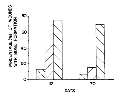
Figure 1 illustrates the percentage of wounds with bone formation when placebo ~left-
most bar), recombinant human TGF-1 (rhTGF-,61 ) at 25 ng/wound (middle bar), or rhTGF-~1 at
100 ng/wound (right-most bar) is applied in the rabbit ear ulcer model at 42 and 70 days after
35 wounding. Maximum bone formatlon was observed at day 42.
Figure 2 illustrates the non-defect end width, an indication of the efficacy in the rabbit
skull defect model, on day 28 post adminlslration of placebo and TCP discs with rhTGF-,~
WO 94/15653 . PCT/US94/00409
2 2~ $1~8~
adsorbed at two different concentrations, where ~ p < 0.05.
Figure 3 illustrates the adsorption ~Inetics of TGF-,~ in the presence of TCP granules
Icircles) and in the absence of TCP granules (squaresl.
Figure 4 discloses a graph of the amount of TGF-,B adsorbed on TCP granules as a5 function of the concentration of TGF-,B in the bathing solution.
Figure 5 illustrates the skull defect area in the rabbit skull defect model on day 28 post
a i"~ L~a~ion of placebo and TCP granules (40-100 mesh) w`lth TGF-~ adsorbed at two
ciir~ :i enL concenl~ dLions, wherein * p < 0.05.
Figure 6 illustrates the skull defect area in the rabbit skull defect model on day 28 post
0 a i",i"i;.l,dLion of placebo and TCP (300 mg)/12% gelatin with TGF-,t~ adsorbed at two ii~elellL
conce"udLions, wherein ~ p < 0.01.
Figure 7 illustrates the skull defect area in the rabbit skull defect model on day 28 post
a i",i"i ,~,dLion of placebo and TCP granules in Iyophilized gelatin with TGF-,~ adsorbed at two
difre,enL concenl,~Lions, wherein ~ p < 0.05.
Figure 8 illustrates total ,eso,~lion surface in the rabbit skull defect model on day 28
post aci",i "~,aLion of a first lot of amylopectln with low endotoxin levels (1), a second lot of
amylopectin with higher endotoxin levels (2), 5 ~m TCP (3), amylope ;Lin + 250 ,um TCP (4),
amylopectin + 10 ,ug TGF-,B (5), amylopectin + 5 ~m TCP + 10 ~9 TGF-,B (6), amylopectin +
250 ,L~m TCP + 10 ~9 TGF-~ (7), and 10 ~9 TGF-,B + 250 ~m TCP + amylopectin (8).Figure 9 illustrates the skull defect area in the rabbit skull defect model on day 28 post
ad", ~ .dlion of formulations 1-8 defined in the legend to Figure 8, wherein ~ p < 0.05.
Figure 10 illustrates release over time of TGF-~ from an amylope..lin/TCP formulation
as analyzed by ELISA, where the open circles are release into normal human serum and the
solid circles are release into PBSI0.5% BSA.
Description of the Preferred Embodiments
A. Definitions:
The polymer for enhancing consistency of the formulation may be any
polysaccharide or insoluble protein material useful for binding the TGF-~ to the TCP to form a
30 smooth, moldable putty or paste. Especiaily pr~rt:"ed are carbohydrates such as agarose,
cross-linked agarose, dextran, cross-linked dextran, inulin, hyaluronic acid, cellulose, cellulose
derivatives such as carboxymethyl cellulose, starch derivatives such as amylopectin, and
insoluble protein materials such as gelatin, including Iyophilized gelatin with glycerol, collagen,
or albumin, or a combination of any of these. The collagen may be chemically conjugated to a
~5 synthetic hydrophiiic polymer and mixed with the TCP as described in W0 90/05755, supra.
The preferred polymer herein is amvioDecnn. most preferably potato amylopectin.
Amylopectin is the branched comDonent of starch: it is formea through chains of D-
-10--
WO 94115653 2 1514 8 ~ PCTIUS94/00409
glucopyranose residues linked together mainlv by (1-->4)-o-D linkages but with 5-6% of (1--
>6)-a-D bonds at the branch points. It is further described in Molecular Bioloay, an
International Series of Monoqrams and Textbooks, The Polysaccharides, Vol. 3, Gerald
Aspinall, ed. (Academic Press, 1985), pp. 216-223.
"Tricalcium phosphate" or "TCP" has a nominal composition of Ca (PO ) and is found
in two different whitlockite crystallographic configurations, a-TCP, and the more stable, ,B-
TCP. It is an extremely biocompatible material used for filling bone and dental defects. It is
described, for example, by Damien and Parsons, J. App. Biomaterials, 2: 187-208 (1991),
Ricci, Biomedical Enqineerinq: Recent DeveloPments~ Saha editor, "Development of a Fast-
Setting Ceramics-Based Grout Material for Filling Bone," p. 475-481 ~1986), Bowers et al., J.
Periodontal, 57: 286-287 (1986). It has also been used with bone morphogenetic protein as a
delivery system. Urist et al., Clin. Ortho~., 187: 277-280 ~1984). TCP is commercially
available from, for example, DePuy, but also may be sy"ll,esi~ed, for example, by the method
described in Biomedical Sciences Instru",en~Lion, Instrument Society of Amerlca, Ed. David
Carlson, Vol. 27, Paper #91-026, Benghuzzi et al., p. 197-203 ~1991). The p,e~"t:d TCP
herein is,~-TCP, and in the examples below, the term "TCP" refers to,~-TCP.
By "bone inducing" is meant pru,,,uLing the formation of morphologically normal,mature bone only at a site where there is a bone deficiency that needs to be replaced. Mature
bone is bone of any type, whether cortical or trabecular, that is ",i.-e~ali~ed as opposed to
20 immature or cartilaginous bone as would be formed in a neonatal model. Morphologically
normal bone is bone that is detected histologically as normal ~i.e., consisting of endochondral
or membranous type lamellar bone and including marrow spaces with osteoblasts and
o:.LeoclasL:j). This is in contrast, for example, to callous formation with a fibrotic matrix as
seen in the first stage of fracture healing. Thus, the bone induction herein is contemplated not
~5 only as acceleralion of bone regeneration, as in a fracture, but also as stimulation of the
formation of bone that is returned to its normal morphological state.
By "skeletal tissue deficiency" is meant a deficiency in bone at any site where it is
desired to restore the bone, no matter how bone deficiency originated, e.g., whether as a
result of surgical intervention, removal of tumor, ulceration, implant, or fracture.
By "bone morphogenetic cofactor" is meant a protein originally found in the bonematrix that induces all of the cascade events involved in the osteoinductlve process in vivo,
including chondrogenesis, vascular invasion, formation of a marrow cavity, and eventually
formation of a bone ossicle. Such factors include the bone morphogenetic proteins as found
in demineralized bone (Urist, Science, 150: 893 119651), osteogenin, a 22 Kd protein with this
activity (Sampath et al., Proc. Natl. Acad. Sci. USA. 84: 7109 ~1987]), a glycoprotein called
osteoinductive factor (U.S. Pat. No. 4,843,063, supra), and BMP-1, BMP-2A, and BMP-3 from
demineralized ovine bone matrix (Wang et al, Proc. Natl. Acad- Sci. USA, 85: 9484119881;
Wozney et al., Science, 242: 1528 ~19881).
WO 94/15653 PCT/US94/Oû409
t 2 ~ 8 ~
The osteoinductive cofactor described in the U.S. patent is isDlated from bone,
preferably a bovine m~taLa,sal bone, wherein the demineralized bone is prepared, non-
collagenous proteins are extracted from the bone, the extract is subjected to gel filtration, the
fraction constituting a low molecular weight (10,000-40,000 daltons~ possessing the greatest
5 chondrogenic activity is s~b,sct3d to ion exchange chromatography, the first fraction CM-1 is
subjected to RP-HPLC, and two peaks of predominantly`28 Kd and 36 Kd
chondrogenic/osteogenic cofactor protein are purified to give single bands on SDS-PAGE.
These cora~,~or:, and the others mentioned above are included in the term "bone
morphogenetic cofactor."
By "osteogenic cell source" is meant a source of viable cells that are capable of
forming bone, as well as viable cells that are precursors to cells capable of forming bone,
including a source of cells capable of recruiting or stimulating cells capable of forming bone.
Suitable such sources include dispersed whole bone marrow cells (obtained by, e.g., aspirâtion
or mechanical agitation), perichondrium, periosteum, or a suitable cell line. For example, the
5 cells may be taken from a site of the animal to be treated adjacent to the deficiency (e.g.,
periosteum stripped from an adjacent site to the defect such as a fracture site or a surgical
excision site~ or from a biopsy site of the animal le.g., one that has been previously accessed,
e.g., the hip), or from bone marrow.
By "animal" is meant any animal having a v~ brale structure, prt:rt:,ably a mammal,
20 and most prefe.ably a human.
By "TGF-~" is meant the family of molecules des.,-il,ed he,~i.-above that have either
the full-length, native amino acid sequence of any of the TGF-,~s from any species, including
the latent forms and associdlt:d or unassociated complex of precursor and mature TGF-,6
I"latent TGF-,6"). Rt:rerence to such TGF-,~ herein will be understood to be a reference to any
25 one of the currently idenliried forms, including TGF-,~1, TGF-~2, TGF-,B3, TGF-,f~4, and TGF-B5,
each of which is represented by certain species indicated in Figure 1 of U.S. Pat. No.
5,158,934 issued October 27, 1992 and latent versions thereof, as well as to TGF-,B species
identified in the future, including polypeptides derived from the sequence of any known TGF-,B
and being at least 75% homologous with the sequence. Members of the TGF-~ family are
30 defined as those which have nine cysteine residues in the mature portion of the molecule,
share at least 65% homology with other known TGF-,B sequences in the mature region, and
compete for the same receptor. In addition, they all appear to be encoded as a larger
precursor that shares a region of high homology near the N-terminus and shows conservation
of three cysteine residues in the portion of the precursor that will later be removed by
35 processing. Moreover, the TGF-,6s appear to have a four- or five-amino-acid processing site.
B. Modes for Carryinq Out the Invention:
The invention Is camed OUI In one aspect by mixing the TGF-,6 with a suitable
--12--
WO 94/15653 21514 8 6 PCT/US94/00409
pharmaceutical carrier, and without the bone morphogenetic cofactor, and ad",inia~ering the
resulting composition locally to a slte on an anlmal where it is desired to induce formation of
normal, adult bone and where a source of osteogenic cells and their precursor cells are present
at the site. If the site does not naturally have a source of osteogenic cells present, the
5 pharmaceutical composition also contains an osteogenic cell source as defined above, in an
amount sufficient to induce bone growth.
Examples of indications where promotion of bone repair at a skeletal site is important
include periodontal disease where root socket heaiing is impaired (tooth socket sites), non-
union fractures, including primary treatment of high risk fractures and adjunctive treatment
10 with bone grafting or bone substitutes for established non-union fractures, large bony defects
caused by trauma or surgery le.g., partial mandibular resection for cancer, large cranial
defects, spinal (vertebral) fusions, correction of severe scolicr s by surgical alignment held in
place with a Harrington bar ~to shorten the six months normally required for a body cast), and
spinal fractures with open reduction (to decrease significantly the period of immobilization)l,
15 and rapid ~ ;on and enhanced fixation of artificial prosll,a-~es and spacer bars, oral
joints, and bone replace",ents.
Examples of the latter include plastic and reconstructive surgery, fixation of pe""dnel,t
dentures into mandible, enhanced fixation of accepted joint prosll,es;s, e.g., hips, knees, and
shoulders ~leading to the acceptance of prostheses that until now have been unacceptable due
20 to rapid loosening and i"~labilil~ such as elbows), and limb salvage procedures, usually
associated with malignancy (the bone shaft may be removed but the articular surfaces are left
in place and connecl~d by a space bar: rapid and enhanced fixation is required for success). If
the site constitutes a periodontal site, i.e., one that involves the teeth, gums, and dental
sockets, the TGF-~ is suitably ad,..ini;,la,~d in conjunction with an exogenously added source
25 of osteogenic cells.
In one preferred embodiment, the TGF-,~ is ad",in; ,l~red by treating a device with the
TGF-,~ composition and implanting the device into the animal at the site of the deficiency, the
composition also containing the osteogenic cell source when the site is deficient in such cells.
The device may consist of any device suitable for implantation, including a molded implant,
30 plug, prosthetic device, capsule, titanium alloy, sponge, or ceramic block. Examples of
suitable delivery vehicles useful as devices are those disrlosed by Nade et al., Clin. OrthoP.
Rel. Res.,181: 255-263 (1982); Uchida et al., J. Biomed. Mat. Res., 21: 1-10 (1987);
Friedenstein et al., Exp. Hematol., 10: 217-227 (1982); Deporter et al., Calcif. Tissue Int., 42:
321-325 (1988); McDavid et al., J. Dent. Res., 58: 478-483 (1979); Ohgushi et al., J.
35 OrthoDaedic Res., 7: 568-578 (1989); Aprahamian et al., J. Biomed. Mat. Res., 21: 965-977
(1986); Emmanual et al., Stain. Tech., 62: 401-409 (1987).
For bone defects involving gaps, such as a dry socket or non-union fracture, a plug
may be used to fill the gap. The plug mav be composed of, for example, hydroxyapatite or
W0 94/156~3 2 i5 ~ ~ PCT/US94/00409
coliagen on which TGF-,B is adsorbed. For larger bone defects resulting from, e.g., trauma or
skeletal reconaL~uction around an ulcer or hip ~rosthesis, the device is prefelablv a made-to-fit
ceramic block. More preferably, the ceramic block comprises 0-100% hydroxyapatite and the
remaining 100-0% TCP, by weight, most preferably 60% hydroxyapatite and 40% TCP.In a specific embodiment for a jaw implant, a calcium carbonate ",c'd-'~'Q material or
InterporeTM molding device is molded to fit the jaw using a 3-dimensional x-ray of the jaw
before surgery, and the molded material is impregnated with TGF-,b'. Then, dispersed bone
marrow from another site of the animal (e.g., frorn:thé hip) is infiltrated into the mold, and the
mold is placed into the jaw for final implantation.
Prefelably, the device is treated with the TGF-B composition (which includes both a
solution and a gel formulation~ for a sufficient period of time to allow adso"~Lion, and to allow
drying in the case of the gel. The concentration of TGF-,~ in the solution or gel and the ~ime of
exposure depend on a number of factors, including the volume of the defect, the potency of
the TGF-~ polypeptide, and the nature of the site to which it is applied, and will be adjusted
15 ar,cord;ngly, As the size of the defect increases, or when the site is other than a bone site,
the concentration of TGF-,~ and the time of presoaking should be increased. The treatment is
for p~eferably at least about 0,5 hour, depending on the factors mentioned above (more
pre~erably at least âbOut 1 hour, and most prefe,dbly 1-2 hours), before implantation, Also
depending on the above considerations, the conce,.l,alion of TGF-,6 in the TGF-,B composition
20 for treating the device is prt:feral,ly at least about 1 ng/ml (more prer.,.ably at least about 1-10
up to 100 ng/ml). The treatment may consist of any mode by which the co",posilion is
applied to the device to deliver effectively the TGF-,6 and the osteogenic cell source. Such
treatment includes, for exa".ple, adsorption, covalent crosslinking, or in.pr~",alion, depending
in part on the nature of the indication.
2s The TGF-,6 compositions to be used in the therapy will be dosed in a fashion consistent
with good medical practice taking into account the nature of the skeletal tissue deficiency to
be treated, the species of the host, the medical condition of the individual patient, the
presence of any other cotreatment drug in the composition, the site of delivery of the agent,
the method of ad~ islralion, the scheduling of ad,..i..;.l.dlion, and other factors known to
30 pra~iLilione,a. Becfluse of differences in host response, significant site-to-site and patient-to-
patient variability exists. For purposes herein, the "therapeutically effective amount" of TGF-
~
is an amount that is effective to induce bone growth, as defined above, at the site of skeletaltissue deficiency.
As a general proposition, the TGF-,B is formulated and delivered to the target site at a
35 dosage capable of eaLablislling at the site a TGF-,~ level greater than about 0.1 ng/ml.
Typically, the TGF-~ concentrations range from about 0.1 ng/ml to 5 mg/ml, preferably from
about 1 to 2000 nglml. These intra-tissue concentrations are maintained preferably by topical
--14--
WO 94/1~653 21 S 14 8 6 P~T/US94100409
~pp ~_Lion and/or sustained reiease.
As noted above, these suggested amounls of TGF-,~ are subject to a great aeal oftherapeutic discretion. The key factor in selecting an appropriate dose and scheduling is the
result obtained. Clinical parameters to determine an endpoint include increases in bone
5 formation and mass and increases in radiographically detectable bone. Such measurements
are well known to those ciinicians and pharmacologists skilled in the art. The TGF-,6
composition is administered locally to the site by any suitable means, including topical and
continuous-release formulation. The active TGF-,~ ingredient is generally combined at ambient
temperature at the appropriate pH, and at the desired degree of purity, with a physiologically
lO acceptable carrier, i.e., a carrier that is non-toxic to the patient at the dosages and
concentrations employed. The camer may take a wide variety of forms depending on the form
of preparation desired for admini~ lion.
To be effective, the TGF-~ is converted by the body to its activated form, i.e., the
mature form is cleaved from its precursor using a suitable enzyme and the resultant complex is
l5 treated with acid or other app~upriale agent to activate the TGF-~. Nevertheless, TGF-,5 is
suitably a.J",in;s~e~ed in an inactive or delayed-release form such as a complex of mature TGF-
with proTGF-,~ not containing mature TGF-,B ti.e., the remaining precursor of TGF-,~), with a
TGF-~ binding protein, or with alpha2-macroglobulin. The latent form is then converted to the
active form either by naturally occurring mechanisms in the local environment or by
20 formulation with TGF-,~ activating agents des.i~iLed above. See, e.g., Gentry et al., Mol. Cell.
BioL, 8: 4162-4168 ~1988); Miyazono et al., J. Biol. Chem., 263: 6407-6415 (1988);
Wakefield et al., J. Biol. Chem., 263: 7646-7654 ~1988); Keski-Oja et al., J. Cell Biochem.
Supr l., 1 l A: 60 (1987); Kryceve-Martinerie et al., Int. J. Cancer, 35:553-558 (1985);
Lawrence et al., Biochem. BioDhvs. Res. Commun., 133: 1026-1034 (1985); Lawrence et al.,
5 J. Cell Phvsiol., 121: 184-188 (1984). Thus, the pH of the TGF-,~ composition may suitably
reflect the conditions necessary for activation.
For the preparation of a liquid composition suitable for impregnation of a device, the
carrier is suitably a buffer, a low molecular weight (less than about 10 residues) polypeptide, a
protein, an amino acid, a carbohydrate including glucose or dextrans, a chelating agent such
30 as EDTA, a cellulose, or other excipient. In addition, the TGF-,B composition is preferably
sterile. Sterility is readily accomplished by sterile filtration through (0.2 micron) membranes.
TGF-~ ordinarily will be stored as an aqueous solution, as it is highly stable to thermal and
oxidative denaturation, although Iyophilized formulations for reconstitution are acceptabie.
Generally, where the bone disorder permits, one should formulate and dose the TGF-,8
35 for site-specific delivery, where the TGF-~ is formulated into a sterile composition suitable for
local application to the desired site.
WO 9411S653 l S i 4 8 6 PCT/IJS94/00409
For local application of the TGF-,6 composition, for example, in the case of a bone
defect that is a crack, e.g., a union fracture, the carrier may be any vehicle effective for this
purpose. For obtaining a gel formulation, the liquid composition is typically mixed with an
effective amount of a water-soluble polysaccharide, polyethylene glycol, or synthetic polymer
s such as polyvinylpyrrolidone to form a gel of the proper viscosity to be applied topically. The
polysaccharide is generaliy present in a gel formulation in the range of 1-90% by weight of the
gel, more preferably 1-20%. Examples of other suitable polysaccharides for this purpose, and
a determination of the solubility of the polysaccharides, are found in EP 267,015, published
May 1 1, 1988.
0 The polysac.. l,a,ide that may be used for the gel includes, for example, cellulose
derivatives such as t,ll,e,ified cellulose derivatives, including alkyl celln~oses, hydroxyalkyl
celluloses, and alkylhydroxyalkyl celluloses, for example, methyl.,el'u~cse, hydroxyethyl
cs'lulose, carboxymethyl cellulose, hydroxypropyl methylcellulose, and hydroxypropyl
cell~ose; starch and fractionated starch; agar; alginic acid and alginates; gum arabic; pullullan;
5 agarose; cc",c.geerian; dt:,~u~ns; dextrins; fructans; inulin; mannans; xylans; ~,~.bina,)s;
cl,ilosal,s; glycogens; glucans; and synthetic biopolymers; as well as gums such as xanthan
gum; guar gum; locust bean gum; gum arabic; tragacanth gum; and karaya gum; and
derivatives and mixtures thereof. The prt:rff"ffd gelling agent herein is one that is inert to
b.olog;cal systems, nontoxic, simple to prepare, and not too runny or viscous, and will not
20 dealabil;~e the TGF-~ held within it.
F~l~rff,~iJly the polysaccharide is an ell,e,iried e e" ~'ose derivative, more preferably one
that is well defined, purified, and listed in USP, e.g., methylceltu'ose and the hydroxyalkyl
cell~'ose derivatives, such as hydroxypropyl cellu'ose, hydroxyethyl cellulose, and
hydroxypropyl methylcellu'ose. Most preferred herein is methylcellulose.
The polyethylene glycol useful for gelling is typically a mixture of low- and high-
molecular-weight polyethylene glycols to obtain the proper viscosity. For exampie, a mixture
of a polyethylene glycol of molecular weight 400-600 with one of molecular weight 1500
would be effective for this purpose when mixed in the proper ratio to obtain a paste.
The term "water soluble" as applied to the polysaccharides and polyethylene glycols is
30 meant to include colloidal solutions and dispersions. In general, the solubility of the cellulose
derivatives is determined bv the degree of substitution of ether groups, and the stabilizing
derivatives useful herein should have a sufficient quantity of such ether groups per
anhydroglucose unit in the cellulose chain to render the derivatives water soluble. A degree of
ether substitution of at least 0.35 ether groups per anhydroglucose unit is generally sufficient.
35 Additionally, the cellulose derivatives may be in the form of alkali metal salts, for example, the
Li, Na, K, or Cs salts.
In a preferred embodiment, the gel contains about 2-5% by weight methylcellulose and
the TGF-~ is present In an amoun~ of about 10-1000 ~9 per ml of gel. More preferably, the
gel consists of about 3% methylcellulose by welght, lactic acid to pH 5.0, and 20-200,ug per
--16--
~0 94/15653 2 I 51 ~ ~ ~ PCT/US94/00409
ml of TGF-,B. This corresponds to a dose of 1 -10 ~9 of TGF-,6 per 50 ~l of gel.For the preparation of a sustained-release formulation, the TGF-~ is suitably
incorporated into a biodegradable matrix or microcapsular particle. A suitable material for this
purpose is a polylactide, although other polymers of poly (o-hydroxycarboxylic acids), such as
5 poly-D-(-)-3-hydroxybutyric acid IEP 133,988), can be used. Additional biodegradable
polymers include poly(lactones), poly(acetals), poly(orthoesters) or poly(orthocarbonates). The
TGF-,6 is also suitably mixed with a biodegradable protein carrier such as collagen,
atelocollagen, for example, one by Koken Co., Ltd., or gelatin to form a carrier matrix having
sustained-release properties; the resultant mixture is then dried, and the dried material is
formed into an appropriate shape, as described in U.S. Pat. No. 4,774,091. Collagen may be
prepared by mincing calf skin and defatting it in chloroform:methanol (1 :1), washing with 4%
EDTA (pH 7.4), and digesting with pepsin solution (pH 2.2; substrate:enzyme ratio, 100:4) for
72 hours at 15C. The collagen solubilized with pepsin is purified by dirrerenlial precipitation
at neutral pH and a salting-out procedure (6% NaCI, pH 3.0, 12 hours) described by Kresina
15 and Miller, Bio.,-l,e",isL,~/, 18: 3089 (1979). The purified collagen is dissolved in 0.01 N HCI (3
mg collagen/ml), sterilized by filtration through a Millipore membrane (pore size 0.45,um), and
freeze-dried. Then it is redissolved in 0.01 N HCI (10 mg collagen/ml) under sterile conditions
and kept in a re~,igelalor until use.
The initial consideration here must be that the carrier itself, or its degradation
20 products, are non-toxic in the target bone site and will not further aggravate the condition.
This can be de~e""i"ed by routine s.;,eeh;,-g in animal models of the target bone disorder or, if
such models are unavailable, in normal animals. For examples of sustained-release
compositions, see U.S. Patent No. 3,773,919; EP 58,481; U.S. Patent No. 3,887,699; EP
158,277; Canadian Patent No. 1,176,565; Sidman et al., BioPolvmers, 22: 547 (1983), and
25 Langer et al., Chem. Tech., 12: 98 (1982).
Controlled delivery of TGF-~ to a site also is suitably accomplished using permeable
hollow cellulose acetate fibers with the TGF-~ that are placed in the site and removed 24
hours later or left for longer periods of time (U.S. Pat. No. 4,175,326). Also, acrylic resin
strips or cast films can be impregnated with TGF-~ and applied to the affected site. In
30 addition, narrow dialysis tubing can be filled with a TGF-,~ solution and placed so as to deliver
TGF-,6 to the appropriate site.
Another preferred method of delivering TGF-,B to the bony site is by way of TCP,including TCP ceramic blocks as described above and TCP particles, which encompass, for
example, granules and powder. While the particles generally can be any size, the preferred
35 particle size of TCP in this invention is > 5,um, more preferably greater than or equal to about
75,~tm. More preferably, the slze of the TCP granules is about 120-420,um, most preferably
about 125-250 ~m, to obtaln a granular DuttY that can be applied to defects that are not so
--17--
WO 94/15653 2 ~ S 1~ ~ ~ PCT/US94/00409
wide as to require implants. The TGF-6 is tvpically adsorbed onto the TCP.
The amount of TCP employed will depend mainly on the type of mammal being treated
and the size of the defect. In humans, the amount of TCP could reach up to about 50 9. The
amount of TGF-B would increase proportionately to TCP. Generally, the amounts range from
about 0.5 ~9 to about 5 mg TGF-B, preferably about 1 ,ug to about 3 mg TGF-B, more
preferabiy about 5 IJ9 to about 1 mg TGF-B, adsorbed onto about 140 mg to about 50 9 TCP
particles, preferably granules. The amount of TGF-B wil! be adjusted downward in accordance
with conventional clinical parameters if there is a biphasic response in which the efficacy of
the TGF-B de~;.eases with increasing TGF-B concentration for the same size defect.
0 Optionally the formulation of TGF-,B and TCP also contains a polymer designed to bind
the components together to improve consistency and ability to mold the resultant putty- or
paste-like material. Examples of such polymers include, but are not limited to, amylopectin,
gelatin, collagen, agarose, dextran, or a mixture of any two or more of these polymers.
Further, the formulation suitably comprises the polymer in conjunction with a co-solvent such
as glycerol, for example, gelatin and glycerol if the formulation is to be Iyophilized before
contact with the TCP and TGF-B mixture.
The polymer is present in the composition in an amount that depends mainly on the
size of the TCP particles being employed, as well as on the type of polymer utilized and the
amount of TGF-B and TCP used.
The TGF-B and TCP may be first mixed before exposure to the polymer, or they may all
be mixed together at the same time, or the TGF-B may be mixed with the polymer and then
with TCP. In a pr~r~..ed mode, the TGF-,~ and TCP are first mixed before the polymer is used
to bind the mixture.
A particularly preferred binding polymer herein is amylopectin, especially in
25 col.,bi"dlion with TCP granules. The method of plepa,d~ion of the amylopectin/TCP
formulation, and possibiy other TCP formulations, can be dependent on the size of the TCP
particles employed. Thus, for example, if the size of the TCP particles is less than about 100
~m, the ingredients may be contacted in any order, including simultaneously mixing the TGF-
~with the amylopectin and TCP or adding the TCP to the amylopectin followed by the TGi--,6.
30 ~iowever, if the size of the TCP granules is greater than about 100,um, the order of mixing
inyl. ' ants may affect the efficacy, at least in one animal model, and thus a preferr.ed method
of producing a bone-inducing formulation of TGF-B for all sizes of TCP granules, and
particularly for larger sizes, comprises admixing an effective amount of a liquid solution of the
TGF-B with the TCP granules for a sufficient period of time to adsorb the TGF-B onto the
granules and contacting the resulting mlxrure with an effective amount of amylopectln.
Conditions that ensure adsorption of the TGF-,~ on the TCP particles are exposing the TCP to
--18--
2151~8~
the TGF-~ at a temperature above about 0C, pr~terably at least abo-lt 5C, mu~e preferably
about 5-40C, still more preferably about 5-30C, and most preferably about roomtemperature. The time of exposure to TGF-,6 is preferably not less than about 5 minutes,
although shorter times may be possible. Then the amylopectin is added and mixed manually
5 with the powder to homogeneity.
A preferred composition comprises about 0.5 ~g to 5 mg TGF-~, about 140 ,u~ to 50 9
TCP particles, pre~erably granules, snd an amount of amylopectin that ranges from about
0.1:1 to about 1:1 (weight/weight) amylopectin:TCP, ~i~terably 0.25:1 to 0.5:1
amylopectin:TCP, depending on the size of the TCP particles. Thus, if the TCP particles are
10 less than 5 ~um, the ratio of amylopectin to TCP is prefersbly about 0.25 to 1, and if the TCP
particles are ~reater than or equal to 75 or 125 ~m, the ratio of smylopectin to TCP is
p~e~elably about 0.5 to 1, and the rstio of TCP:amylopectin:TGF-~B solution is most preferably
1 :0.5:0.5.
The amylopectin may be obtained from any source of starch, such as corn and potato,
15 with potato being preferred. The smylopectin is prefersbly sterilized before use, as by
autoclave or irradiation. To minimize the number of colony forming units ~CFU~ the
amylopectin is suitably dissolved in water to form a solution of about 2-4% and then sterilized
by autoclave ~about 100-120C for no less than about 30 minutes). To remove all the water,
it is also preferably Iyophilized or spray dried.
The composition herein also may suitably contain other peptide growth factors such as
IGF-I, TGF-o, human growth hormone, epidermal growth factor, and PDGF, provided that such
factors do not include the~ bone morphogenetic factors defined above. Such growth factors
are suitsbly present in an amount that is effective for the purpose intended, i.e., to promote
formation of bone.
2s The invention will be more fully understood by ~e~erence to the following examples.
They should not, however, be construed as limitin~ the scope of the invention.
EXAMPLE 1
The TGF-,61 used herein was the recombinant ox~ression product of ~rd.,s~e~;led
human 293 cells as described by EP 200,341, s~lpra, and by Derynck et al., Nature, supra, u
and purified as described in Derynck et al., N~ture, supra. The individusl s~mples of
recombinant human TGF-,B1 (rhTGF-~l ~ were sterilely prepared in methylcellulose containing
20 mM sodium acetate buffer at pH 5.0 and applied as a single topical dose. Selected
concer,l,c~ions of rhTGF-,61 were mixed with methylcell~'ose gel so that the final
3s concentration of methylcellulose was 3%. The vehicle was formulated in a similar manner
without rhTGF-,B1 as a control. The material was stored st 5C until use.
The rst incisionsl model utilized young sdult Si",onsen Albino rats (300-350 9). Full
--19--
WO 94/15653 ~5 ~ PCT/US94/00409
thickness skin incisions were made by cutting through the subdermal panniculus carnosus
musculature following application of Betadinel~ brand antisepLiC and 70% alcohol scrubbing to
disinfect the surgical site. Two pairs of symmetrical transverse incisions ~approximately 2.5
cm) were placed in each animal. A single dose of rhTGF-,61 in methylcellulose was placed into
5 each stainless steel sutured wound bv inserting a 25-gauge needle along the edge of the
wound and below the sutures. The volume of rhTGF-,B1 in 3% methylcellulose placed into
each wound was 0.05 ml. Each rat had two incisions into which rhTGF-,B1 in 3%
methylce - ose was applied. One incision received either vehicle alone (3% methylcellulose~
or no treatment at all. Concentrations of rhTGF-,~1 were 500, 1000, 2000, or 4000 ng/ml.
10 Dose response curves were developed using dose ranges of 5 to 10,000 ng/wound. Animals
were euthanized on day 5, 7, 10, 14, 21, and 28. The entire dorsal skin was excised after
the sutures were removed. Two 8-mm wide strips of skin were collected from each incision
and fixed in 10% neutral buffered formalin for seven days.
New Zealand white male rabbits (2.5-2.8 kg) were purchased from Elkhorn rabbitry.
15 Anesthesia was induced by an intramuscular injection of ketamine hydrochloride/xylazine
hydrochloride mixture. After removal of hair from the ears, the area of the wound was
sterilely prepared using Betadinen brand a.,lisepLic with an alcohol rinse. A circular 6-mm
punch biopsy instrument was used to produce wounds to the depth of the ear cartilage. The
underlying perichondrium was removed with a periosteal elevator and fine scissors. Wounds
20 were treated with 0.025 ml of 3% methylcellulose or 5, 15, 25, 100, 500, or 1000 ng of
rhTGF-,61 in 3% methyl~ ~ ase ~controll. Opsiten surgical dressing was placed over each
wound. An Elizabethian collar was placed around the neck of the rabbits to prevent
mechanical disruption of the wounds by the rabbit.
Studies were also designed to examine short-term and long-term effects of topical
25 rhTGF-,61. Wounds were harvested on days 3, 5, 7, 14, 21, 28, 42, 56, and 70. Wounds
were photographed, cut into hemisections, and fixed in 10% neutral buffered formalin for
histology and morphometric anaiysis. Morphometric analysis included measurements of ~otal
healing wound area, closing wound area, upper wound gap, lower wound gap, area of
collagen, area of granulation tissue, epithelial cell layer length, and bone formation. These
30 measurements were made on a BioQuant IVI ~R & M Biometrics Inc., Nashville, TN) computer
image analysis system.
The rabbit ear ulcers were examined for delayed effects of rhTGF-,~1 on days 21, 28,
42, 56, and 70 following a single application of 25 or 100 ng/wound on the day of wounding.
Bone formation was observed along the wound edges and immediately adjacent to the
35 cartilage. The bone was normal in morphological appearance, consisting of endochondral or
membranous type bone and ossification with marrow spaces. Osteoblasts anri osteoclasts
were present. The percen~age of wounds with bone increased to a maximum of 74% of the
--20--
WO 94/156~3 2 I ~14 8 ~ PCT/US94/00409
treated wounds at day 42 (100 ng/wound) and decreased to 69% by day 70. See Figure 1.
Bone formatlon was observed in less than 12% of placebo-treated wounds.
No bone formation was observed in the ra~ incision model, indicating that bone
formation is induced only at a site that has a source of precursor losteogenic) cells, in this
5 case in the rabbit ear model where the wound was adjacent to perichondrium.
EXAMPLE 2
A rat femur gap model was employed wherein a polyethylene plate 2-mm thick, 8-10mm long, and 4-5 mm wide was pinned to one face of a rat femur with stainless steel pins.
o From the center of the femur a 5-8-mm iong piece of bone was removed. The plate serves to
keep the gap in bone separated. This model is intended to mimic a non-union fracture in a
human .
Set into the gap in the femur is a porous cylindrical 200-to 400-micron ceramic implant
of 60% by weight hydroxyapatite and 40% by weight TCP (Zimmer, Inc.), which is either t1 )
5 implant alone, 12) implant presoaked for 1 hour in a solution of 50 ng/ml rhTGF-,~1 prepared as
desclibed in Example 1 and formulated in Delbecco's medium without serum, (3) implant plus
dispersed whole bone marrow cells obtained from syngeneic rat, and 14) implant plus
dispelsed whole bone marrow cells pretreated with 50 ng/ml of the rhTGF-~1 in Delbecco's
medium described above. A total of 15 rats were used for each of these four groups. One
~o month after implant, the rats were sacrificed and analyzed for histologiG~I changes.
Preliminary results indicate that no bone replacement was observed in the control
without cells or rhTGF-~ nor with rhTGF-,t~ without cells; TGF-~ with cells was found to
accel~,, a~ the rate of bone growth over cells alone. The bone formed with rhTGF-,6 was found
in the interstices of the pores in the ceramic and bridged the gap. The bone formed with the
2s rhTGF-~ was found to be histologically normal.
EXAMPLE 3
A case study was performed using baboons to investigate the effect of TGF-~ on bone
wound healing. The baboon was selected because of the close analogy of its bone kinetics to
30 those of man. A methylcellulose gel of TGF-,B1 was delivered via an analytical bone implant,
and after 22 days the implant was removed from the baboon. Tissue obtained from TGF-,6
implant sites was analyzed using quantitative histomorphometry to determine the mean effect
of TGF-,6 on bone wound healing. Detailed non-quantitative histopathologic evaluation was
also performed.
3s More specifically, four male baboons were implanted with four titanium analytical bone
implants (cages) each, two per tibia in areas of close structural geometry. Holes were drilled
in the tibia to allow implantation. After Implantanon, the baboons were allowed to heal for 41
-
WO 94/156~3 215 l ~ PCT/US94/00409
days. On the 41st dav, all the implant sltes were surgically exposed, tissue was removed, and
the test materials were implanted into the implant cores. Each animal received a normal ~no
treatment~ control, a control with only methylcellulose vehicle, and a low l1 ~rg rhTGF-,B in
methylcellulose) or high (10 ,ug rhTGF-,~ in methylcellulose) dosage of active TGF-,6.
5 Specifically, these formulations each consisted of 1 9 of 3.0% methylcellulose by weight,
lactic acid QS to pH 5.0, and 0, 20, or 200 ~g/ml of rhTGF-,B1 prepared as described in
Example 1. The formulations were poured into size 5 gelatin c~ps~les (Elanco), which were
then placed in the core of the titanium implant and used to deliver 50,ul of each formulation,
with slow dissolution of the capsule. All implant sites within an animal were randomly
lO assigned to one of the four treatments.
Following 22 days of healing, tissue in all implants was retrieved. The tissue samples
were placed in 10% formalin solution, buffered to a pH of 7.0, containing formaldehyde at
3.7% for fixation. Samples were submitted for histopathologic analysis.
The following desc~ ive and quantitative observations were made:
15 1. Bone volume in TGF-,6 sites was lower than control and placebo sites, although not
statistically significant.
2. Osteoblast numbers, volume, and activity were significantly greater in the TGF-~ sites
when compared to either the control or placebo.
3. Osteoclast numbers and activity appeared higher in all four Llealll,enl sites when
20 subjectively compared to control data obtained in previous studies.
4. Residual methylce'lulose was noted and appeared to require phagocytosis before new
trabecular bone could form.
5. TGF-,~ in the presence of methylcellulose matrix was associated with increased numbers of
fibroblast, osteoprogenltor cells, and osteoblasts.
~5 6. No foreign body response or other adverse pathologic reaction to either matrix alone or
matrix and TGF-,~ was observed.
7. Significant pe,iosleal new bone formation was noted over the implants in five TGF-,~ sites
in three animals. Bone formation over the implant to this degree had never been observed in
over 450 titanium implant procedures carried out over the past few years.
30 8. TGF-,6 sites were identified during blinded histologic review in seven out of a total of eight
sites .
9. Methylcellulose sites were identified during blinded histologic review 100% of the time.
Control samples analyzed in this study demonstrated that cancellous tissue formed in
the titanium implant is stratified from inferior to superior aspects of the implant core. The
35 superior portion of the tissue (closest to the cap of the titanium implant) is less mature and
shows greater osteoblastic activity, while tissue near the inferior aspects of the implant and
deep wlthin the medullary compartment Is more mature in morphology and shows a reduced
~1514~
WO 94115653 PCT/IJS94100409
osteoblastic population and activity. In contrast to historical and control samples, the TGF-,B
tissue samples were homogeneous In their high osteoblastic activity throughout the specimen.
Clinical observations of the tissue above and around the supra-periosteal portion of the
titanium implant reveaied pronounced periosteal bone formation. This periosteal bone formed
s large masses over two sites in each of two animals. The masses in these two animals were
highly vascularized, had the clinical appearance of trabecular bone, and varied in size within
one animal. The two masses in each of two animals were approximately 3x2x1.5 cm and
1.5x1x0.5 cm in size. One additional animal demonstrated pronounced periosteal bone
formation over one TGF-,6 site. It is significant that in over 450 titanium implant surgical
lO procedures masses like these have never Tormed over the titanium implants. Histologically,
this periosteal bone formation over five TGF-,6' sites in three baboons was similar to an actively
healing, uncomplicated, fracture callus, i.e., morphologically normal, mature bone formation.
In general, the methylcellulose was well tolerated and no foreign body response was
present in any of the four treatment sites. Additionally, no evidence of cytologic atypia or
15 malignancy was found in either titanium implants or periosteal samples.
EXAMPLE 4
Introduction
The purpose of this study was to evaluate the effects of TGF-~1 in the rabbit skull
20 defect model of bone formation when incorporated into a TCP matrix that was configured as a
thin disc the approximate size of the defect ~12 mm). This was accomplished by measuring
selected bone morphometric parameters from stained histologic sections as well as by
radiographic examination of the excised defect site. Results were compared to defects
ad,l,i,~isl~:red TCP discs without TGF-,~1.
25 Source and Preparation of TGF-~1 and TCP Discs
The rhTGF-,~1 was prepared and purified as described in Example 1. Individual
samples of the active portion of rhTGF-,~1 were prepared under sterile conditions in 20 mM
sodium acetate buffer at pH 5Ø The incorporation of rhTGF-~1 into TCP discs (obtained from
DePuy, Warsaw, Indiana) was done by aseplically incubating TCP in the TGF-,~1 solution for
30 three hours at room temperature. Prior to the incubation, TCP discs were ~erilized by
incubating in 70% ethanol, rinsing thoroughly with sterile normal saline, and drying under UV
- lamp. The average weight of each disc was 153 mg. Two different concentrations of rhTGF-
,B1 were used, 20 and 100,ug/ml. After incubation, each disc was rinsed briefly with sterile
normal saline. The amount of rhTGF-,6`1 adsorbed onto the TCP disc was determined from the
35 changes in the concentration of TGF-~1 incubating solutions bV conventional ELISA methods.
The higher concentration (100,ug/ml) gave the average vaiue of 16 ~g/disc, while 5 ~rg/disc
was the average value from the incubation of the discs with 20 ,ug/ml TGF-,B1.
--23--
WO 94tl5653 PCT/US94/00409
2~ 8~ ~
Animal Suraerv and Treatment
All studies were performed in accordance with the American Association for the
Accledi~Lion of Laboratorv Animal Care (AAALAC~ guidelines. Sixteen male New Zealand
White rabbits (2.8 - 3.2 kg) (Elkhorn Rabbitry, Watsonville, CA) were anesthetized with 0.75
5 ml/kg Hypnorm~ brand anesthesia (Jenssen Pharmaceutica, Beersa, Belgium). The top of the
head and base of the ears were shaved and aseptically prepared for surgery. An elliptical
incision was made over the skull, reflecting the skin flap anteriorly. Similarly, the periosteum
was reflected a,-l~riorl-/ as a flap, exposing the top of the skull. Both skin and periosteal flaps
were covered with sterile, moistened gauze. A 1 2-mm skull defect was selected since, in the
0 absence of treatment, bone does not bridge the gap, but rather a fibrous tissue non-union of
the skull persists. Frame, J. Oral Surq., 38: 176-180 (1980). A sterile trephine attached to
an electric drill was used to produce the defect at the point of intersection between the
sutures of the right and left parietal and frontal bones. The site was liberally irrigated with
physiological saline during the drilling to prevent ove~hed~ing of the bone margins. Care was
15 taken not to puncture or damage the underlying dura. A precut, sterile saline-moistened piece
of GelfilmT~ brand of film (Upjohn, Kalamazoo, Ml) was inserted through the defect overlying
the dura to function as a barrier between the dura and the edges of bone.
Sterile TCP discs or TCP discs with rhTGF-,~1 (5 or 16 ~9) were applied to the defect
filling the defect. The periosteal flap was sutured back in place with 6-0 proline sutures and
20 the skin flap was closed with 4-0 silk. Rabbits were returned to their csges and allowed to
recover. After 28 days rabbits were euthanized with an overdose of barbiturate and the
defect sites were removed with adjace"L normal bone. The defect sites were rinsed in
physiological saline. Sites were fixed in 10% neutral buffered formalin and radiographed using
a FaxitronT~ brand X-ray system and X-omat AR-2 film exposed at 25 KV, 10 s. The fixed
25 tissue samples were then cut in half at the center of the defect parallel to the frontal/parietal
suture. One he""se~;Lion was acid decalcified (Easy-cut'~ reagent, American Histology Reagent
Co., Modesto, CA) and processed by routine histologic methods using hematoxylin and eosin
to stain the 4-~m sections. The other half of the defect was plastic embedded, and
undecalcified sections were processed by routine histologic methods, with the 5-~m sections
30 stained with Goldners' trichrome, von kossa, or toluidine blue.
Goldner's trichrome stained sections were examined using a BioQuant IV"' computer
image analysis system. Selected indices of bone formation and resorption were measured,
including trabecular bone volume (TBV), percentage osteoid surface (%OS), percent~ge
osteoid volume (%OV), mean osteoid width (OW), percentage osteoblasVosteoid (%OblOst~,
35 percentage osteoblast/total surface (%OblTS), total resorption surface (TRS), and number (#)
of osteoclastslsurface length (OclSL). Sections from all animals were analyzed
histomorphornetrically uslng a random stratified sampling scheme that systematicallv
evaluated selected fields from the bony edge of the defect and the entlre area within the
WO 94/lS653 21~1 4 8 6 PCT/US94/00409
defect. Fields were selected using a grid pattern, such that each field within the defect area
had an equal probability of being selected. Approximatelv equal numbers of fields were
evaluated for both the control and treated defects.
The thickness ~width) of bone at the outside edge of the sections (at the edge of the
harvested sample farthest from the detec~ sitel was measured to evaluate the extent of bone
formation at non-defect sites (non-defect end width, NEDW). Defect area that is normally
quantitated radiographically using computer image analysis could not be determined accurately
due to the radiopaque nature of the TCP discs.
Statistical analvsis
Data were analyzed by one factor ANOVA and the Scheffe F-test to dete"",ne
differences between groups. The test of significance was performed at the 95% confidence
interval compared to vehicle control. Each group contained three to four rabbits.
Results
MorPhometric Evaluation. Data from the morphometrlc determinations are presented15 in Table 1. In general, TGF-,61-impregnated TCP discs stimulated a greater degree of bone
formation at the defect site compared to TCP discs without TGF-,B1. Indices of bone
formation, including trabecular bone volume, osteoid width, osteoid volume, and
osteoblastlosteoid were increased in the TGF-~1-treated defects compared to vehicle-treated
defects. In addition, the number of osteoclastslsurface length and total resorption surface
20 were increased in TGF-,~1-treated defects compared to vehicle-treated defects, indicating that
remodeling processes were present. Non-defect end width of bone from defects ad",i"~ d
either 5 or 16 ,ug of TGF-,B1 were greater than placebo-treated defects (see Table 1 and Fig.
2). The only parameters without significance bel.veen groups were osteoid surface and
osteoblastltotal surface.
PCT/US94/00409
WO 94/15653 , 2 lS 1 4 8 ~
.
TABLE 1
Histomorphomeuic Evaluation of Bone Formation
in Skull Defects Applied TGF-,B1 Impregnated onto TCP Discsd
Histomorpho- TGF-,6
metric
ParametersD Vehicle 5 /~a ~!9
l0 TBV (%) 1.64i0.83 21.60+3.43 15 59+2.75C
OW ~mm) 0.003+0.003 0.01 +0.001d 0.009+0.001d
OV (%) 0.76+0.76 9.07+2.17~ 9.19+0.73'
OS (%) 27.68+27.68 46.59i6.42 42.81 +4.30
Ob/TS (%) 36.44 + 31.99 56.86 + 5.04 65.69i1.39
20 Ob/Ost (%) 40.14+40.14 125.43+8.56" 157.99+15.34a
Oc/SL (#) 0+0 0.53iO.09 0.57+0.20
TRS (~) O +0 0.025 + 0.005 0.027 +0.009
NDEW (mm) 1.37+0.11 1 69+0.04d 1 75+0.11d
a Values reported are mean + S.E.M. based. N = 3 for vehicle and 4 each for 5 or 16,ug of
30 TGF~
b TBV is trabecular bone volume; OW is osteoid width, OV is osteoid volume, OS is osteoid
surface, Ob/TS is osteoblast/total surface, Ob/Ost is osteoblastlosteoid, Oc/SL is
osteoclasts/surface length, TRS is total resorption surface, and NDEW is non-defect end
35 width.
'p < 0.01.
p < 0.05.
Due to the radiodense nature of the TCP discs, radiographic defect area was not
determined. While there were apparent differences between placebo-treated and TGF~
treated defects, these differences were not amenable to morphomeTric determinations.
However, the TGF-~1-treated defects appeared slightly more radiopaque, with the defect area
45 and non-defect area blending without sharp border between the edge of the TCP disc and
skull.
Histoloqical evaluation. Histologic evaluation of the defects filled with TCP discs
impregnated with TGF-,B1 indicated an increase in the amount of bone surrounding the TCP
disc. In addition, bone was observed migrating into the surfaces of the disc primarily at the
so margin of the defect, but also on the top and bottom surfaces. The new bone was
chdla~ ed as a mixture of woven (immature) and lamellar (mature) bone by polarized light
- ~0 94/1565~ 2151 ~ 81~ ~T~S94/OU409
mlcroscopic examination. In contrasl, a mlnlmal bonv resPonse was observed histologically in
the TCP discs without TGF~
In summaty, the TCP discs impregnated with TGF-,~1 induced a marked increase In
bone both surrounding the discs as well as migrating into the discs. Bone was chd,~clt:ri~ed
5 histologically as a mixture of immature and mature bone indicating active formation and
resorption processes. Remodeling of bone was subsequently confirmed histomorphometrically
by an increase in both formation and resorption parameters within TGF-~1-treated sites. TCP
discs without TGF-,~1 were minimally inductive at 28 days with only slight amounts of bone
located at the margins of the defect.
These data demonstrate that TCP will function as a carrier for TGF-,B1 and provide a
matrix on which bone can readily form across osseous defects.
EXAMPLE 5
Introduction
The purpose of this study was to evaiuate the effects of TGF-,61 in the rabbit skull
defect model of bone formation when incorporated into 40-100 mesh TCP matrix, wherein the
TCP is supplied as granules. This was accomplished by measuring s~lected bone
morphometric para",dlers from stained histologic sections as well as by radiographic
examination of the excised defect site. Results were compared to defects ad",ini~ered 40-
20 100 mesh TCP without TGF-,B1.
Source and Prd~a,alion of TGF-~ and TCP Matrix
rhTGF-,~1 was prepared as desc,ibed in Example 1. Individual samples of the active
portion of rhTGF-,t~1 were prepared under sterile conditions in 20 mM sodium acetate buffer at
pH 5Ø Two different concentrations of rhTGF-,61 were used, 25 and 100 ~g/ml. Porous TCP
25 granules were used (Peri-OSS~ brand TCP, lot # 7157EL2A2, 40-100 mesh; granules had the
size of 150-420 ~m and were supplied by DePuy and produced from TCP powder by isostatic
pressing and then sintering). The total weight of TCP granules in each dose was 154 mg.
The preparations were obtained by aseptically incubating, at 5C for two hours in a sterile
filter unit, TCP particles in either 20 mM sodium acetate buffer, pH 5, or in the two TGF-,B1
30 solutions of the same buffer.
After incubation, TCP granules were harvested by centrifugation to remove the liquid.
The bathing solutions were then removed from the particles by microcentrifugation through a
filter membrane. Samples that were treated with acetate buffer were labeled as placebo.
Samples treated with 100,ug/ml of TGF-,B1 were labeled as "high" dose and samples treated
35 wlth 25 ~glml of TGF-,B1 were labeled as "low" dose. High aose, as indirectly determined by
ELISA from the difference in the initial and the final bathing concentration, was 13.7+0.2~9
--27--
WO 94/15653 PCT/US94/00409
2~S'~ 4~ --
(+ SD, n=3) and low dose was 2.9+0.1 ~9 (+ SD, n=3). The average weight of TCP
particles in each vial was 154.1 + 3.6 mg ( +SD, n =8).
The amount of rhTGF-,B1 adsorbed onto the TCP granules was determined from the
changes in the conce"L,~ion of TGF-,~1 incubating solutions by conventional ELISA methods.
5 Animal Surqerv and Treatment
The animal surgery and treatment were petformed as described in Example 4. Sterile
TCP or TCP with rhTGF-,~1 (3 or 14 ~9) was applied to the defect filling the defect.
Radiography was performed as described in Example 4, and one hemisection was acid
decalcified and one undecalcified as described in Example 4. Goldner's trichrome stained
o sections were examined using the BioQuant IVI~ computer image analysis system as described
in Example 4. The thickness of bone at the outside edge of the sections was measured to
evaluate bone formation at non-defect sites. In addition, defect area d~ ""ined
radiographically was quantitated using computer image analysis.
SlaLij~Li~al analvsis
Stdli:iLical analysis was done as described in Example 4. Each group contai"ed two to
three rabbits.
Results
AdsorDtion of TGF-~ onto TCP Granules. Figure 3, which shows the adsor~.Lion
kinetics of TGF-,~ on TCP granules, in li~ ~ ~ s that after about 2 hours, the amount adsorl.ed
20 appears to stabilize, with gradual change up to 22 hours. Figure 4 shows the adsorption of
TGF-,6` on TCP granules, wherein the amount of TGF-~ adsG,bed is given as a function of
bathing concellLlaLion of TGF-~. It is seen that the amount of TGF-,~ adsorbed increases
proportionately to the amount of TGF-~ in the bathing solution.
Mor~hometric Determinations. Data from the morphometric dt:Le""il)d~ions are
25 presented in Table 2. In general, TCP with TGF-,l~1 stimulated a greater degree of bone
formation at the defect site co~pared to TCP without TGF-,B1. indices of bone formation that
were increased in defects administered TGF-,61 included osteoid width, % osteoid volume, %
osteoid surface, and % osteoblast/total surface. Trabecular bone volume, an indicator of the
quantity of bone present within the defect, was significant only at p=0.06. Remodeling of
30 bone was present as indicated by an increase in total resorption surface in the TGF-,6'1-treated
defects compared to placebo-treated defects.
--28--
~o 94/15653 2 151 ~ 8 6 pcTluss4lon4os
TABLE 2
Histomorphometric Evaluatlon of Bone Formatlon in Skull Defects
Applied TGF-,B1 Impregnated onto 40-100 Mesh TCP Granulesd
Histomorpho- TGF-B
metric
Parametersb Vehicle 3 ~9 14 llq
0 TBV (%~ 1.09+1.09 20.22+5.40' 26.18+8.46
OW (mm) 0.002 + 0.002 0.009 + 0.001 ' 0.008 + 0 001 C
OV (%) 0.03+0.03 9.67+0.75' 6.50+2.22'
OS (%) 1.39+1.39 33.38+2.66C 34.14+9 78C
Ob/TS (%) 1.67+1.67 33.42+6.71' 38.91 +8.28C
Ob/Ost (%) 40.00+40.00 98.83+12.20 118.34+7.65
Oc/SL (#) 0.33+0.33 0.57+0.07 0.65+0.24
TRS ( % ) 0.006 + 0.006 0.042 + 0.015C 0.041 + 0 004C
' Values tt:pG-I~d are mean + S.E.M. based. N 5 3 for vehicle, 2 and 3 for 3 or 14,ug of
TGF-,~1, respe~;~ively.
30 b Abbreviations are defined in footnote b of Table 1.
c p < 0.05.
Radiographic defect area was d~5L~:-,.-i-,ed using image analysis techniques. The defect
areas were 60.57+9.69, 22.86+7.07, and 8.27+8.27 for placebo, 2, and 14~9 TGF-,61,
respectively (Fig. 5). The dose-responsive decrease in defect area was significant for both
levels of TGF-,~1 (p < 0.05). In general, the placebo-treated defects were radiolucent and the
TGF-,61-treated defects were radiopaque, except for small centrally located regions in the
40 defects treated with 3 ~9 TGF-,~1.
Histoloqical evaluation. TCP granules (40-100 mesh) with TGF-B1 induced a variable
response upon histologic examination. Generally, in defects acJ...;n;~le.ed TCP without TGF-
,B1, the predominant response was a mild chronic inflammation with a mixture of fibrous
connective tissue bridging the defect and surrounding the granules of TCP. Minimal bone
45 growth from the margins of the defect were observed in the control group. Defects
administered TGF-,B1 in TCP induced a much greater bone response with complete bridging in
some cases. However, there was mild fibroplasia located within the central pornon of the
defect and surrounding TCP granules from defects ad...inistered 3,ug of TGF-,B1. Sometimes
--29--
215148~
WO 94/15653 - PCT/US94/00409
the bone formed over or around the granuiar area with the granules surrounded primarily by
fibrous connective tissue. A similar response was observed in defects treated with 14,L~g of
TGF-,B1, with less fibroplasia and more bone formation especially around the granules of TCP.
In summary, radiographs of defect sites after 28 days indicated complete defect
5 closure with 14 ~9 rhTGF-,~1 in 150 mg TCP, having induced a marked increase in bone both
surrounding the dorsal and ventral region of TCP granules as well as migrating into the
granules. The new bone formed within the defects was cha(a.leri~ed histologically as a
mixture of immature and mature bone. This indicates active formation and reso" ~ion
processes that are natural to bone healing. Remodeling of bone was confirmed
o histomorphometrically by an increase in both bone formation and resorption pa,a",~le,~ within
TGF-,~1-treated sites. The results indicate that the defect area is much lower after application
of the TCP granules with 25 ~g/ml ~3,ug/wound site) TGF-~ and is even still lower after
application of the TCP granules with 100 ~g/ml (14 ~g/wound site) TGF-~. TCP granules
without TGF-,~1 were minimally inductive at 28 days with only slight amounts of bone located
5 at the margins of the defect.
These data show that TGF-,~ in association with TCP without other carriers such as
gelatin functions as a potent bone inducing growth factor, providing a matrix on which bone
can readily form across osseous defects.
EXAMPLE 6
Introduction
The purpose of this study was to evaluate the effects of TGF-,t~1 in the rabbit skull
defect model of bone formation when incorporated into TCP granules (150-420~m) with 12%
gelatin that was configured as a disc approximating the size of the defect (12 mm). This was
~5 accomplished by measuring selected bone morphometric parameters from stained histologic
sections as well as by radiographic examination of the excised defect site. Results were
compared to defects administered TCP in gelatin without TGF-,B1.
Source and Preparation of TGF-~ and TCP Granules with Gelatin
rhTGF-,B1 was prepared as described in Example 1. Individual samples of the active
30 portion of rhTGF-,6'1 were prepared under sterile conditions in 20 mM sodium acetate buffer at
pH 5Ø
The solution of rhTGF-,61 in 12% gelatin was prepared by dissolving gelatin (type A,
300 Bloom grams) in 20 mM sodium acetate, pH 5.0 with moderate heat. The gel solution
was sterilized by membrane filtration while it was still very warm. As the solution was cooled
35 to a temperature below 50C, an appropriate aliquot of the sterile rhTGF~ 1 solution was
added and homogeneously mixed. After mixing, 400 ~l of this gelatin-TGF-~1 solution was
--30--
- ~ 94/15653 21 S 14 8 6 PCT/U59J/UI)409
pipetted into 5-ml vials that contained 300 mg of TCP granules (150-420 ~m, DePuy). The
preparation was allowed to congeal. By varving the added volume of TGF-,61 solution, the
final doses were 0, 5, and 21.5 ~9 TGF-,B1 per disc of TCP-gelatin.
- Animal Surqerv and Treatment
The animal surgery and treatment were performed as des~;,ibed in Example 4. Sterile
TCP/gelatin or TCP/gelatin with TGF-,~1 (5 or 21.5,L~g) was applied to the defect filling the
defect. Radiography was performed as described in Example 4, and one hemisection was acid
decalcified and one undecalcified as described in Example 4. Goldner's trichrome stained
sections were examined using the BioQuant IV'~ computer Image analysis system as described
10 in Example 4. In addition, the defect area determined radiographically was quantitated using
computer image analysis.
Statistical analvsis
Statistical analysis was done as described in Example 4. Each group contained 5 to 6
rabbits.
s Results
Morphometric Determinations. Data from the morphometric determinations are
presented in Table 3. In general, TGF-~1 formulated in TCP and 12% gelatin stimulated a
much greater degree of bone formation at the defect site compared to TCP in 12% gelatin
without TGF-,B1. All indices of bone formation were increased in defects administered either 5
20 or 21.5 ~9 TGF-,~1. The number of osteoclasts/surface length and total rcsor~lion surface
were increased in defects treated with 5,ug, but not 21.5,ug, TGF-~1, indicating that
remodeling processes were present at least for the lower dose of the growth factor.
TABLE 3
25 Histomorphometric Evaluation of Bone Formation in Skull Defects
Applied TGF-B1 in 300 mg TCP/12% Gelatin'
Histomorpho- TGF-~
30 metric
Parametersb Vehicle 5 ~9 21.5 Llq
TBV (%) O 14.11 +2.12' 20.20+4.09'
35 OW (mm) O 0.01 +,001C 0.01 +0.001'
OV (%) O 7.13+0.99' 8.34+1.52'
OS (%) O 41,15 + 3,01 C 50.38 + 2.84'
Ob/TS (%) O 43.34+6.28' 57.34+3.86'
Ob/Ost (%) O 104.37 + 14.04~ 114.17 + 5.34c
--31--
W094/15653 2iS14~6 PCT/11594100409
OclSL (#~ 0 0.47iO.11 ~ 0.23 + 0.11
TRS (%) 0 0.029+0.009C 0.012+0.006
a Values reported are mean + S.E.M. N = 5 for vehicle, and 6 each for 5 or 21.5 ~9 of TGF- -
,61 .
b Abbreviations are defined in footnote b of Table 1
'p < 0.01.
p < 0.05.
Radiographic defect area was d~Le,l",ned using image analvsis techniques. The defect
areas were 67.49 + 6.57, 37.89 + 5.14, and 23.24 + 7.99 for placebo, 5, and 21.5 ~9 of
TGF-~1, respeclively (Fig. 6). The dose-responsive decrease in defect area was signiricar,~ for
both levels of TGF-,51 (p < 0.01). F~? ' ~gld~hs were difficult to interpret morphometrically
20 since the granules of TCP were radiopaque. However, the general d!Jpedldllce of the TGF-,~1-
treated defects was denser, especially within the center of the defect.
Histoloqic Evaluation. TCP in 12% gelatin with TGF-,B1 induced a variable response
upon histologic examination. Generally, the predo", ,ant response was a mild chronic
inflammation with a mixture of fibrous connective tissue bridging the defect and surrounding
25 the granules of TCP in the defect ad",i";~l,3,ed TCP in 12% gelatin without TGF-,61. Minimal
bone growth from the margins of the defect were observed in the placebo group. Defects
ad",in;~lered TGF-~1 in TCP and 12% gelatin induced a much greater bone response with
col",c!~,te bridging in most defects ad",i";;.Lert:d 5 ~9 of TGF-~1. However, there was a mild
chronic inflammatory response with fibroplasia located within the central portion of the defect
30 and surrounding TCP granules from defects administered 5 ~9 of TGF-,~1. Four of five defects
ad",i";~Lered 21.5,ug of TGF-,61 were completely bridged with bone. However, mild chronic
inflammation in this group was still evident at each site with variable amounts of fibrous
connective tissue intermingled with granules of TCP.
In summary, the TCP granules in 12% gelatin with TGF-,61 induced a marked increase
35 in bone both surrounding the space occupied by the granules as well as interspersed in the
granules. Bone was characterized histologically as a mixture of immature and mature bone,
indicating active formation and resorption processes. Remodeling of bone was subsequently
confirmed histomorphometrically by an increase in both formation snd resorption parameters
within TGF-~1-treated sites. When compared to the TGF-,B1-impregnated TCP disc studv,
40 however, the values from histomorphometry were lower in the TCP granule/1Z% gelatin
tormulation. indicating that the bone response was not as vigorous. Also, the formulation
melted rapidly and was not easily contormable to the defect.
--32--
WO 94115653 2151~ g 6 PCT/US94/00409
These data demonstrate with the other examples that TCP will function as a carrier for
TGF-,B1 and provide a matrix on which bone can readily form across osseous defects. It is
believed that the mild chronic inflammation would resolve with time as bone replaced the
granules of TCP.
The same experiment is expected to yleld similar results using a gelatin/agarosemixture containing for example about 0.05-1% ~weight/weight) agarose with an exemplary
amount being 0.25% to increase the melting point of the composition
EXAMPLE 7
lO Introduction
The purpose of this study was to evaluate the effects of TGF-~61 in the rabbit skull
defect model of bone formation when incorporated into TCP granules with 2% Iyophilized
gelatin that was configured as a disc of material approximating the size of the defect (12 mm).
This was acco",plished by measuring selected bone morphometric para",eLers from stained
15 histologic sections as well as by radiographic examination of the excised defect site. Results
were compared to defects a.J",i,.i~Le,ed large granules of TCP in Iyop~ ~d gelatin without
TGF-Bl -
Source and Pie,~a,dLion of TGF-~ and TCP Fal ~icles and Gelatin
rhTGF-~B1 was prtua,ed as desc~ibed in Example 1 and formulated in TCP with 2%
20 gelatin as follows. A solution of 2% gelatin (type A 300 Bloom grams) with 2% glycerol was
prepared in 20 mM sodium acetate, pH 5.0 and sterilized by filtration. An aliquot amount of
sterile TGF-,B1 solution (20 or 50 ~9) was added into the gelatin mixture at a temperature of
about 50C and homogeneously mixed at that te",pe(alure to form a gel solution. TCP
particles (500 mg sized at 420-2000 ~m) were weighed into sterile siliconized vials. The gel
25 solution ~0.5 ml) was then added onto the TCP granules sufficiently to cover all the granules.
The preparation was subsequently Iyophilized by conventional Iyophilization technology. The
final doses in these preparations were 20 and 50 ~9.
Animal Surqerv and Treatment
The animal surgery and treatment were performed as described in Example 4. Sterile
30 large granules of TCP in Iyophilized 2% gelatin without TGF-,t~ (placebo) or with TGF-~1 (20 or
50 ~9) were applied to the defect filling the defect. Radiography was performed and one
her"iseL;Lion was acid decalcified and one undecalcified as described in Example 4. Goldner s
trichrome stained sections were examined using the BioQuant IVIY computer image analysis
system as described in Example 4. The thickness of bone at the outside edge of the sections
35 was measured to evaluate bone formation at non-defect sites. In addition defect area
determined radiographically was quantltated using computer image analysis.
Statistical analvsis
--33--
WO 94/15653 2'i~ ~ 4~ 6 PCT/11594/00409 ~
Statistical analysis was done as described in Example 4. Each group contained four to
five rabbits.
Results
Morphometric Determinations. Data from the morphometric determinations are
5 presented in Table 4. In general, the baseline values for the placebo control group were
relatively high, indicating that large granules of TCP in Iyophilized gelatin induced bone
formation to a greater extent than other formulations. However, 20 ug TGF-~1 stimulated
more bone formation at the defect site compared to TCP in gelatin without TGF-~1 as
indicated by an increase in trabecular bone volume and % osteoblast/total surface. In
10 contrast, 50 ug TGF-,61 did not induce an increase in bone formation compared to the TCP
placebo except for % osteoblast total surface. Non defect end width was similar between
groups.
TABLE 4
11iaLumor~ hometric Evaluation of Bone Formation in Skull Defects
Applied TGF-,61 with Large Granules of TCP in Lyophilized Gelatin'
Histomorpho- TGF-~
2 o metric
Pa(al~ lab yehicle 20 va 50 uq
TBV (%) 15.66 + 1.74 28.93 + 2.65C 15.43 + 2.58
25 OW ~mm) 0.011 +0.003 0.009 +0.001 0.009 +0.001
OV (%) 5.04 + 0.58 7.31 + 2.10 8.25 + 1.23
OS ( %) 30.27 + 3.89 41.42 + 6.68 40.68 + 4.25
Ob/TS (%) 35.95 + 2.24 50.47 + 3.98 54.12 + 4.84
Ob/Ost (%) 123.99 + 11.03 129.62 + 17.65 140.54 + 22.04
35 Oc/SL (#) 0.32 + 0.39 0.56 + 0.16 0.33 + O.18
TRS (%) 0.015 +0.003 0.029 +0.009 0.017 +0.007
NDEW (mm~ 1.55+0.11 1.80+0.12 1.90+0.07
' Values reported are mean + S.E.M. based. N = 5 for vehicle, 4 and 5 for 20 and 50,L~g of
TGF-,61, respectivelv-
45 b Abbreviations are defined in footnote b of Table 1.
' p < 0.01 .
p < 0.05.
WO 94/15653 215 14 8 6 PCT/US94/00409
R~di~yldphic defect area was d~ter",ined using image analysis techniques. The defect
areas were 27.22+7.84 4.86i2.25 and 25.01 +7.06 for placebo 20, and 50~9 TGF-~1
respectively (Fig. 7). The decrease in defect area was significanr for 20 ~9 of TGF-61 oniy (p
< 0.01). Radiographs were difficult to interpret morphometrically since the large granules of
5 TCP were radiopaque and uneverily distributed over the defect area. However the general
appearance of the defects ad",i"i~Lered 20 ~9 of TGF-,t~1 was denser and filled the defect.
Histoloaic Evaluation. Histologic examination of bone samples from defects
ad",in;;,l~led TCP in Iyophilized gelatin without TGF-,~1 indicates bone formation at the margin
of the defect bridging approximately 50% of the cross section. Where bone was present
lO there appealed to be trabeculae of bone lined by osteoblasts surrounding the large granules of
TCP. Marrow cavities were present with a typical cellular pattern for bone. In the central
50% of the defect from placebo-treated defects the large granules were surrounded by
fibroblasts and fibrous connective tissue. Defects ~dm ~i;,lered 20 ~19 TGF-~1 in large
granules of TCP and Iyophilized gelatin induced a much greater bone response with complete
15 bridging in all cases. However there were small areas of fibroplasia occasionally located
within the central portion of the defect and surrounding TCP granules. Histologic exa",indlion
indicated that defects ad",;"isL~red 50 /19 TGF-B1 induced a variable response. Two of five
defects ad",in;i,te,ed 50 ~9 TGF-~1 were completely bridged with bone, while 2 of 5 defects
contai"ed predo",;"a"Lly a fibrous response with minimal bone from the margins of the defect.
20 In each case at 50 ~9 TGF-,B1, there was a thick layer of fibrous connective tissue over the
pelioalt:al surface.
In summary the large TCP granules in Iyophilized gelatin with 20 ~9 TGF-~1 induced a
moderate increase in bone both surrounding the space occupied by the granules as well as
int~lspelsed in the granules. Bone was chd,a~ ed histologically as a mixture of immature
25 and mature bone indicating active formation and resorption processes. When compared to
other formulations of TCP without TGF-~1, there was a subalar,lial increase in the baseline
amount of bone in the TCP placebo group. Without being limited to any one theory, this
effect could be attributed to the size of the TCP granules which are known to be conductive
as well as to the Iyophilized gelatin formulation. The low dose of TGF-B1 (20 ~9) induced an
30 increase in bone co",paled to both the TCP placebo and 50 ug TGF-61. While
morphometrically there were fewer parameters that were significantly different from TCP
placebo than in the other TCP studies the low dose of TGF-~1 appeared very comparable to
similar doses of TGF-,~1 in other formulations. In contrast 50,ug TGF-~1 was remarkably
di~er~"t with a much greater degree of fibroplasia and a much more variable amount of bone.
35 This indicates that in this model there Is a blphasic response with TGF-61 similar to other
models of soft tissue wound healing.
WO 94/15653 ~8~ PCT/US94100409
These data further demonstrate that TCP will function as a carrier for TGF-,~ and
provide a matrix on which bone can readily form across osseous defects.
EXAMPLE 8
The purpose of this study was to evaluafe the effects of TGF-,B1 in the rabbit skull
defect model of bone ~ur",alion when incorpo~rated into TCP granules (5,um or 250,um
nominal particle size) with amylopectin that was configured as a malleable putty appro~i",aLi"g
the size of the defect ~12 mm). In addition, the individual components, i.e., TCP ~5 or 250
~m) and two dir~ lots of amylopectin were evaluated to determine which componentlO contributed to the incidence of giant cell rur",aLion observed in this model. Defect sites were
removed 28 days after surgery"adiog,aphed, and processed for histomorphometric
d~5L~r",;"alions. Introduction
In the rabbit skull defect model, there appears to be a foreign body giant cell ,.::,ponse.
The purpose of this study was to evaluate in this model the effects of the individual
15 components of the formulation and combinations of TGF-,~1 and two sizes of TCP granules
Inominal 5 or 250 ~m granules) formulated in two lots of amylope.,li" having di~erenl levels
of endotoxin present. I li .i ~ !e j e examination with measurement of selected bone
morpho",~l,ic pa~ "ete,s from stained histologic sections as well as r ~iOglayhic examination
of the excised defect site were used as criteria for efficacy.
20 Source and PlepalaLion of TGF-B1 and TCPtamvloDectin
Tvpes of Formulations tested. Eight groups of formulations were evaluated for
efficacy in the animal model: two amylopectin controls, two vehicle controls, and the TGF-,~1-
treated groups as des.,,ibed below:
Group 1: Amylopectin with 12 EU/g
25 Group 2: Amylope~;Lin with ~3500 EU/g
Group 3: 5 ~m TCP and amylope-,Lin
Group 4: Amylopectin and 250 ~m TCP
Group 5: Amylopectin and 10,ug TGF-,B1
Group 6: AmylopecLin and 5 ~m TCP and 10 ~9 TGF-,B1
30 Group 7: Amylope~;~i" and 250~m TCP and 10~9 TGF-,~1
Group 8: 1O~LI9 TGF-~1 and 250~m TCP and amylopectin~
Group 8 differs from group 7 only in the order of mixing.
Preparation of Formulations. rhTGF-,61 was prepared as described in Example 1.
Individual samples of the active portion of the TGF-,~1 were prepared under sterile conditions
35 in 20 mM sodium acetate buffer at pH 5Ø Two di~erenl ranges of particle size of TCP were
used to prepare the paste, 5 and 250 Ltm (nominal, range = 5 - 45 ~m and 250-500 ~m,
WO 94/15653 21~14 8 6 PCT/US94/00409
respeclively). Aseptic conditions were malntained throughout the preparation procedure. The
TCP granules were sterilized by 2.5 MRAD gamma irradiation.
Two lots of amylopectin (potato, Sigma Chemical Co.) with different levels of
endotoxin (12 EU/g and >3500 EU/g) were used in Groups 1 and 2, respectively. Only the
s 1 2-EU/g amylopectin was used in Groups 3-8.
The TCP/amylopectin paste for Group 7 was prepared by mixing sterile TCP granules
and sterile amylopectin in the ratio of 4:1 and 2:1 lby weight) for TCP granules with particle
slze of <45 and 250-500,um, respectively. An aliquot of TGF-,61 solution in 20 mM acetate
buffer, pH 5, was added to the solid mixture. The mixing was then performed manually using
a spatula and plate until a uniform mass was obtained. In each preparation, the volume of
TGF-,61 solution was kept constant at the ratio of 1:0.4 (weight of TCP:volume of TGF-~1
solution). The amount of amylopectin/TCP paste administered into each animal was about
500 mg, with the final dose of 10,ug TGF-,61.
For Group 8, the TGF-~ solution was mixed with the TCP sufficiently to become
adsorbed thereon, and then the amylope.,Lin was mixed in to homogeneity.
Animal Surqerv and Treatment
The animal surgery and treatment were performed as described in Example 4 using the
eight groups of formulations defined above. The formulations were malleable, having the
consialenc~r of putty, and were applied to the defect filling the space co",pl~tel~. Radiography
20 was pe,r~,r",ed as described in Example 4, and one hemisection was acid decalcified and one
undecalciried as described in Example 4.
The decalcified and undecalcified stained sections were evaluated for general
characleliali~,s and quality of healing, especially for the presence or absence of a foreign body
giant cell response. In addition, Goldner's L-ichr-r"e stained sections were examined using the
25 BioQuant IV'Y computer image analysis system as described in Example 4. In addition, defect
area dt:Le"",ned radiographically was quantitated using computer image analysis.StaLiaLical analvsis
Statistical analysis was done as described above. Each group contained two to three
rabbits .
~ o Results
- Histoloqic Evaluation. A summary of the histopathologic evaluation is presented in
Table 5. Both lots of amylopectin induced a minimal bone response for foreign body giant cell
response. In contrast, the amylopectin with 5 ~m or 250 ~m TCP granules induced a mixed
response with minimal bone formation and a marked foreign body giant cell response. Defect
- 3s sites adr"ini ,L~red amylopectin with 10 ,ug TGF-~1 but without TCP exhibited extensive new
bone formation with minimal foreign body giant cell response. Ten ~9 TGF-,t~1 admi~ eled to
defects with amylopectln and 5 llm TCP induced a variable response with new bone formation
as well as a moderate giant cell response. When 10 ~9 TGF-,t/1 was mixed with 250 ~m TCP
--37--
WO 94/15653 2 i 5 1~ 8 ~ PCT/USg4/00409
and then mixed with amylopectin the amount of bone formation was increased to a level
similar to the growth factor plus amvlopectln formulation and the degree of giant cell
formation was minimal to moderate. In contrast when the mixing order was reversed such
that the TCP and amylopectin were mixed first the TGF-B1 added less bone and a greater
5 degree of giant cell formation occurred.
TABI E 5
Summary of Histologic Evaluation of
0 Hematoxylin- and Eosin-Stained Sections
Grouo
1 Minimal bone response: connective tissue bridge;
minimal giant cell response.
2 Minimal bone response; connective tissue bridge; minimal giant cell response.
3 Minimal to no bone response; very reactive with numerous giant cells
throughout.
4 Minimal to no bone response; moderate fibrosis; numerous giant cells
surrounding large cavities ~presumably ds- ~ied TCP).
Complete bridging of defect with bone; profound increase in osteoblasts;
minimal signs of chronic inflammation; thick fibrotic capsule overlying bone.
6 75-90% bridging of defect with bone; central area contains moderate fibrosis;
".ode-~le giant cell response with lots of debris (small particles); bone looks
good where it is present; in one sample the bone appears to be primarily
periosteal with gaps at the original cut edges.
7 0-90% bridging of defect with bone; 1/3 - severe giant cell response; small
amount of conne.;li~e tissue; 1/3 - center area moderate giant cell response
with chronic inflammation; 1/3 - new bone looks good funnels down centrally
with mild giant cell response with debris.
8 75-100% bridging of defect with bone; minimal (2/3) to moderate (1/3) giant
cell response; thick fibrous response overlying bone; predo"-i"ant response is
one of large amounts of bone surrounding small cavities (decalcified TCP).
Morphometric Determinations. Data from the morphometric determinations are
45 p,esented in Table 6 and Figure 8. No measurements could be determined in the vehicle
groups that contained either lot of amylopectin. In general TGF-~1 formulated with 250 ~m
TCP then mixed with amylopectin. stimulated a much greater degree of bone formation at the
defect site compared to the other formulations except for the TGF-B1 and amylopectin
combination. Most osteoblastic and osteoclastic indices were increased in the TGF-B1-treated
50 groups co"".~ d to groups without TGF-B1.
--38--
=
~0 94/15653 21514 8 6 PCT/US94/00409
Radiographic defect area was determined using image analysis tecl-r,jq les and is illustrated in
Figure 9. The differences between groups typically depended on the presence or absence of
TGF-,61. The defect area tended to be smaller for the non-TGF-,B1-treated groups that
contained TCP granules that were 250,um. However, radiographs were difficult to i"te,urt:
5 morphometrically due to the radiopa~ity of the TCP granules in all but the first two groups,
i.e., the two lots of amylopectin alone.
TABLE 6
0 Histomorphometric Evaluation of Bone Formation in Skull Defects
l listo",oruhometric' Para"~el~,~
Group Trabec- Osteoid Osteoid Osteoid Osteo- Osteo- Osteo-
ular Width Volume Surface blast/ blast/ clast/
Bone (mm) 1%) (%) Total Osteoid Surface
Vol.1%) Surface (%) length
1%) (O
15 1 0 ~O) O (O) O (O) O (O) O (O) O (O) O (O)
2 0 (0) 0 (O) 0 (O) O (O) 0 (0) 0 ~O) 0 (0)
3 2.68 0.003 1.10 5.41 7.54 46.40 0.10
(2.68) (0.003) (1.10) (5.41) (7.54) (46.40) (0.10)
4 2.27 0 (O) O (O) O (0) 25.01 0 (O) O (0)
(2.27) (25.01)
31.27 0.01 7.63 49.56 51.56 102.51 0.54
(~.28) (0) (1.42) (6.49) (9.26) (5.83) (0.27)
20 6 16.71 0.012 12.06 38.38 52.90 166.75 0.46
(1.99) (0.004) (5.15) (11.39) (6.85) (48.83) (0.11)
7 16.82 0.014 8.21 22.41 28.59 86.9 0.14
(8.54) (0.009) (6.04) (11.51) (14.30) (44.97) (0.07)
8 30.78 0.016 13.20 51.29 45.33 89.55 0.40
(6.26) (0.002) (3.53) (7.02) (4.88) ~6.73) ~0.21)
~ Data are e~.ur~:,sed as mean IS.E. ~/1.).
In summary, results from this study indicate that the amount of endotoxin present in
the amylopectin did not affect the amount of giant cell formation and therefore indicates that
amylopectin should be an adequate carrier for TCP and TGF-,61. In contrast, both the 5,L/m
and 250 ~Jm TCP induced giant cell formation when mixed with the low-endotoxin
amylopectin. It was dete""ined rt:L,oapectively that the 250 ~m TCP contained TCP powdet
- 30 ~particles < 45,um). Since the degree of giant cell formation was less in the 250 ~m TCP
than the 5,vm TCP, without being limited to any one theory, it is believed that the small TCP
granules in the 250 ~m TCP formulation may be contributing to the level of giant cell
formation .
--39--
2~ g~ ~ ~
WO 94/15653 PCT/~S94/00409
The defect area measured from radiographs was similar between groups administered
amylopectin alone or 5 ,um TCP granuies alone. The defect areas for sltes adn,inialt:red
amylopectin and 250 ~m TCP granules (with or without TGF-~1 ) were similar and all were
smaller than sites administered 5 ,um TCP granules alone or amylopectin alone.
Morphometric parameters were similar among the TGF-,B-treated groups.
Histopathologic examination of the eight microscopic slides indicates that the overall response
of the TGF-~1 formulated in 250 ~m TCP was better than that of the TGF-~1 formulated in 5
~m TCP. A moderate to severe giant cell foreign body reaction was observed with the 5 ~m
TCP in amylopectin with or without TGF-~1.
These data indicate that TCP/amylopectin will function as a carrier for TGF-,~ and
provide a matrix on which bone can readily form across osseous defects.
EXAMPLE 9
The purpose of this study was to evaluate the effects of TGF-,B1 in the rabbit long-
5 bone model of bone formation when incorporated into TCP granules (5 ~m or 250 ~m nominal
particle size~ with amylope~;Lin that was configured as a malleable putty the approximate size
of the defect. Defect sites were radiographed and processed for hi;.L~,i"or~.hometric
determinations. Source and PreDaration of TGF-B1 and TCP/amylopectin
rhTGF-~1 was prepared as described in Example 1. Individual samples of the active
20 portion of the rhTGF-,B1 were p,l:pa.ed under sterile conditions in 20 mM sodium acetate
buffer at pH 5Ø A 4% solution of amylopecLin (potato, Sigma Chemical Co.) was prepared
by adding amylope.;lin to water and aler;li~;ng in an autoclave at 100-120C for no less than
30 minutes. The solution was filtered through a 0.22-~m membrane. For removal of all
water, the sterile amyiopectin solution was Iyophilized.
The sterile TGF-,~1 solution was adsori,ed onto the TCP granules (125-250 ~m) byaseptic incubation in a sterile filter unit at 5C for 2 hours as described in Example 5. The
amylope.;Lin was aseptically mixed with the TCP granules upon which the TGF-~1 solution
was adsorbed using plate and spatula to homogeneity.
The proportions of amylopectin, TCP, and volume of water from the TGF-,B1 solution
30 were varied according to the particle size of TCP. In this study, three ranges of particle size
were used, < 5 ~m, > 75 ~m, and > 125 ~m. The proportion (by weight) of
TCP:amylopectin was 1:0.25 when the TCP particles were < 5 ~m. The percentage of water
needed in the mixing was 30% (volume/weight of the total amount of solids). For TCP with
--40--
WO 94/15653 21 51~ ~ 6 PCT/US94/00409
larger particle sizes ( > 75 ~m and > 125 ~m~, the ratio of TCP:amylopectin:TGF-Ig solution
(weight/weight) was 1 :0.5:0.5 .
Release of TGF-,B from TCP/Amviopectin Formulation
Figure 10 shows a graph of the percent of TGF-~ released over time from the
amylopectin/TCP formulation of 250 to 400 ~Im particle size into normal human serum (open
circles) and into phosphate buffered saline (PBS~ containing 0.5% bovine serum albumin (BSA)
(solid circles). The release of the TGF-,B was measured by standard ELISA method s. It can be
seen that the TGF~ is released much more quickly from the TCP/amylopectin in normal human
serum than in the PBS.
Animal Suraerv and Treatment
A model of repair of long bone discontinuities in rabbits was devised based on that
provided in Lemons et al., supra. All studies were performed in accordance with the AAALAC
guidelines. Male New Zealand White rabbits (2.8-3.2 kg) ~Elkhorn Rabbitry, Watsonville, CA)
were anesll,eLi ed with 0.75 ml/kg Hypnorm~ brand ane~ll,esia (Jenssen Pharmaceutica,
Beersa, Belgium). The right forelimb from each rabbit was shaved and asepLi y prepared for
surgery. An incision was made over the anterior-medial aspect of the forearm ~radium/ulna),
re~le~ling the skin laterally. The muscles surrounding the radius were bluntly re~le.;led from
the field of view and about 1.5 cm of the radius was exposed. A 10-mm section of the mid-
shaft radius was removed using an electric drill while liberaliy illigc~Li~g with physiological
20 saline during the drilling to prevent ovt:.l,edLing of the bone margins. Care was taken not to
damage the adjacenL ulna. After the 1 0-mm section of bone was removed, the gap was
packed with sterile gauze to faciliLdte hel"G:,~a:.is. The defect site was subsequently irrigated
to eliminate any small particles of bone.
Three groups were evaluated in the initial preliminary investigations. The vehicle
25 control group was treated with a formulation consisting of sterile TCP t125 ~m particle size)
mixed with amylopectin to homogeneity. The TGF-,B1-treated group was treated with a
mixture of TCP, amylopectin, and 15 ~9 TGF-,~1 formulated as described above. The third
treatment group consisted of a 1 0-mm defect without any treatment. The formulation with
TGF-,~1, amylopectin, and TCP was malleable, having the consistency of putty, and was
30 applied to the defect filling the space completely. The reflected muscles were sutured back in
piace and the skin was closed with 4-0 silk. Rabbits were returned to their cages and allowed
to recover.
Immediately after surgery and weekly thereafter, the surgical site from each rabbit was
,adiog,~phed to monitor healing. After 28 days rabbits were euthanized with an overdose of
35 barbiturate and the radius and ulna were removed and excess soft tissue (i.e., muscle) was
dissected away from the bone and defect site. Sites were fixed in 10% neutral buffered
formalin. The fixed tissue samples were then acid decalcified (using Easy-cutl~ reagent,
--41--
WO 94/1565~ 215 1 ~ 3 $ PCT/US94/00409
American Histology Reagent Co. Modesto CA) and serial sections were processed by routine
histologic methods using hematoxylin and eosin to stain the 4-~lm sections.
The decalcified stained sections were evaiuated for general chard~ iaLiCs and quality
of healing. In addition representative longitudinal sections taken from the center of the defect
5 were examined using a Bio~uant IVI~ computer image analysis system. Selected indices of
bone formation and resorption were measured including TBV %OS %OV OW, %Ob/Ost,
%OblTS TRS and #OclSL. Sections from all animals were analyzed histomorphometrically
using a random stratified sampling scheme that systematically evaluated selected fields from
the bony edge of the defect and the entire area within the defect. Fields were selected using
l0 a grid pattern such that each field within the defect area had an equal probability of being
selected. Approximately equal numbers of fields were evaluated for both the control and
treated defects.
Statistical analvsis
Statistical analysis was done as desclibed above. Each group contained 3 to 4
5 rabbits.
Results
Preliminary results from radiographs indicate that the defect filled more rapidly in
rabbits ad",i"ial~,ed TGF-,B1 formulated with TCP pluâ amylopectin than the untreated control
or the groups with TCP plus amylopecli,) alone. The defects ad",i"i~le,ed TGF-61 tended to
20 be filled with radiodense material by 21 days while defects ad",i";sle,ed TCP plus
amylopectin alone were less dense radiographically at 21 or 28 days. Defects that were
untreated ~I, led minimal filling within the 28-day observation period. Histologic data is
~pected to confirm the radiographic data in that the TGF-~1 ITCP/amylopectin formulation is
expected to increase most if not all histomorphometric parameters exa",il)ed to a greater
25 extent than the other two control formulations.
The result of wetting the TCP with the TGF-B first before adding amylopectin rather
than adding the TGF-~ to the mixture of TCP and amylopectin was a better pharmacological
effect. Without being limited to any one theory, it is believed that the better efficacy of the
preparation wherein TGF-,B is first adsorbed onto TCP is due to the ability of the osteoblasts to
30 form around the TCP particles where the TGF-,B was localized.
These data further indicate that TCP/amylopectin will function as a carrier for TGF-,~
and provide a matrix on which bone can readily form across osseous defects. The
TCP/amylope~ lil1 formulation is preferred in that it does not melt as rapidly as those with
gelatin and could evenly disperse the large TCP granules yet be malleable and formable to
35 regular defects like a putty.
--42--
WO 94/15653 21 5 1 ~ ~ ~ PCT/US94/00409
EXAMPLE 10
The purpose of this study was to formulate the TGF-,~ in collagen and TCP.
Collagen CN IProdex, Inc., Princeton, NJ) was sterilized by ethylene oxide. The matrix
was prepared by mixing an appropriate aliquot of rhTGF-,61 solution prepared as described in
5 Example 4 with TCP ( < 5 ~m) and collagen aseptically using plate and spatula. The
proportion of TCP:collagen:water was 6:1:6 (weight: weight: volume). The volume of water
needed was replaced by the sterile TGF-,B1 solution. The final dose that can be acl~ L~Ied
for the rabbit skull defect model is about 8-10 ,ug of TGF-,~1, dependi~I9 on the defect size.
The amount of TCP powder in each studied anlmal can be about 500-750 mg.
The radiographic data indicated that the formulation of collagen + TCP + 10 ~9 TGF-
,t~ was significantly (p < 0.05) more efficacious than collagen alone in the rabbit skull defect
model described above.
--43--