Note: Descriptions are shown in the official language in which they were submitted.
WO 94/13205 21~ 1~ 9 3 PCTIUS93/11872
Apparatus and method for sterotaxic radiosurgery and radiotherapy.
The present invention relates generally to an apparatus for and method of
carrying out stereotaxic radiosurgery and/or radiotherapy on a particular target5 region within a patient utilizing previously obtained reference data indicating the
position of the target region with respect to its surrounding area which also
contains certain nearby reference points. The present invention relates more
particularly to a number of improvements to the method and apparatus disclosed in
copending United States Patent Application Serial No. 07/600,501 in the name of
10 John R. Adler, filed October 19, 1990, which application is incorporated herein by
reference.
The term stereotaxic radiosurgery refers to a procedure in which a beam of
radiation is used to render a target region, that is, a particular volume of tissue,
specifically tumorous tissue, necrotic, as is well known. Typically, this requires in
1~ the neighborhood of 2000 to 3000 rads of radiation. The term stereotaxic
radiotherapy refers to a procedure in which a beam of radiation is applied to the
target region for therapeutic, non-necrotic purposes. The amount of radiation
typically utilized in this latter case is an order of magnitude less than a necrotic
dose, for example between 200 and 300 rads of radiation. By target region is
20 meant a specific volume of particular configuration which is to be treated with the
required radiation dosage for the intended purpose. The target region may also be
referred to, for example, as a dose contour.
As will become apparent hereinafter, the various features of the present
invention are equally applicable to both stereotaxic radiosurgery and stereotaxic
SUBSTITUTE SHEET (RULE 26)
WO 94/13205 PCT/US93/11872
2151~L9~ -2-
radiotherapy. However, for purposes of ease, the term stereotaxic radiosurgery will
be used herein (both in the specification and appended claims) to refer to both
stereotaxic radiosurgery and stereotaxic radiotherapy. Thus, for example, a
radiosurgical beam recited herein is intended to refer to such a beam and also a5 radiotherapeutic beam.
As indicated above, the copending Adler patent application is incorporated
herein by reference. As will be described in more detail hereinafter, each
stereotaxic radiosurgical apparatus disclosed in the Adler application is designed to
carry out radiosurgery on a particular target region within a patient utilizing
10 previously obtained reference data, for example 3-dimensional mapping data,
indicating the position of the target region with respect to its surrounding area
which also contains certain nearby reference points, for example existing bone
structure or implanted fiducials. In accordance with this procedure, means are
provided for directing a radiosurgical beam of radiation into the target region. In
15 order to ensure that this radiosurgical beam is accurately directed into the target
region, a number of diagnostic beams of radiation, actually target locating beams,
are directed into and through the surrounding area of the target region and the
information derived from these latter beams of radiation is used along with the
previously obtained reference data to accurately aim the radiosurgical beam into the
20 target region. While this overall procedure is quite satisfactory for its intended
purpose, the present invention provides for a number of improvements.
As will be described in more detail hereinafter, an apparatus is disclosed
herein for carrying out stereotaxic radiosurgery (or radiotherapy) on a particular
target region within a patient, especially a target region which is irregular in shape
25 as contrasted with a typical spherically shaped target region. This apparatus,
which is designed in accordance with the present invention, utilizes means for
generating a radiosurgical beam of radiation and beam aiming means. The beam
aiming means includes a robotic arm in a preferred embodiment and serves to
SUBSTIME SHEET (RULE 26)
=~ ~
WO 94/13205 3~ 21 S 1~ 9 3 PCT/US93/11872
support the beam generating means in a way which directs the radiosurgical beam
along a beam path through the target region.
t
In accordance with one feature of the present invention, the radiosurgical
apparatus disclosed herein includes means for moving the beam aiming means
5 along a predetermined, non-circular and non-linear path transverse to the beam path
while, at the same time, the beam path is directed into the target region. In this
way, the radiosurgical beam can be directed through the target region from
particular treatment points along the non-circular and non-linear path so as to
define a specific non-spherical target region. In the actual embodiment disclosed
10 herein, this predetermined, non-circular and non-linear transverse path is a specific
spiral path which has been selected so that the non-spherical target region is in the
shape of a specific ellipsoid. In this way, an irregular shaped tumor can be treated
more effectively than heretofore possible by providing a target region or dose
contour approxi",ali"g but entirely surrounding the irregular shaped tumor. In the
15 past, a tumor of this shape required a series of spherical dose contours which, in
many cases, covered more or less of the tumor than necessary.
In a preferred, actual working embodiment of the apparatus disclosed
herein, its beam aiming means includes a robotic arm which is free to move in atleast three dimensions in order to follow the non-circular, non-linear transverse path
20 recited immediately above. As a result, in accordance with a second feature of the
present invention, in order to protect the patient, the apparatus is provided with
emergency stop means separate from and independent of the beam aiming means
and its robotic arm for automatically stopping all movement of the robotic arm and
automatically turning off the radiosurgical beam if the robotic arm deviates from its
25 intended non-circular, non-linear transverse path.
In addition to the features just discussed, the apparatus disclosed herein
includes a unique procedure for ensuring that the radiosurgical beam is accurately
directed into the target region at substantially any point in time during radiosurgery,
SUBSTtTUTE SHEET (RULE 26)
WO 94/13205 2~S1493 4 PCT/US93/1187
that is, in substantially real time. Like the apparatus disclosed in the Adler
application recited previously, the apparatus disclosed herein is provided with
previously obtained reference data indicating the position of the target region with
respect to its surrounding area which also contains`certain nearby reference points.
5 The disclosed apparatus, like Adler's, also utilizes a plurality of diagnostic or target
locating beams of radiation to obtain substantiàl7y real time location data which is
compared with the previously obtained reference data for determining the location
of the target region in sul)~L~r,lially real time. However, as will be described in
more detail hereinafter, the apparatus disclosed herein carries out this target
10 locating procedure in accordance with a unique, specific temporal sequence that
allows the apparatus to function in a rapid but reliable manner.
The present invention will be described in more detail hereinafter in
conjunction with the drawings, wherein:
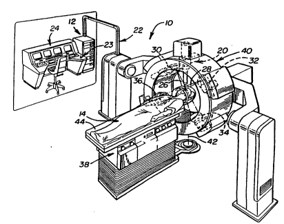
FIGURE 1 illustrates, in isometric view, one embodiment of the apparatus
15 described in the previously recited Adler copending patent ~ppl.~ation;
FIGURE 2 illustrates, schematically, diagnostic X-ray imaging and
accelerator focusing aspects of the Adler arrangement;
FIGURE 3 illustrates, in a view similar to FIGURE 1, an alternative
embodiment of an apparatus described in the Adler ~pp'.cation;
FIGURE 4 illustrates, schematically, a system block diagram in accordance
with both the Adler apparatus and the apparatus designed in accordance with the
present invention;
FIGURE 5 illustrates, in a view similar to FIGURE 3, part of the arrangement
found in FIGURE 3 but modified to incorporate a feature of the present invention,
25 specifically an arrangement for automatically stopping all movement of the robotic
arm forming part of the arrangement shown in FIGURE 3 in the event that the
robotic arm deviates from its movement along a predetermined transverse path;
FIGURE 6 illustrates, schematically, a block diagram corresponding to the
emergency stop feature shown in FIGURE 5;
SUBSTITUTE SHEET (RULE 26)
WO 94/13205 2 f 51 4 9 ~ PCTIUS93/1187~
FIGURES 7 and 8 illustrate, diagrammatically, the way in which the robotic
arm of the arrangement, as illustrated in FIGURE 3, moves along a non-circular,
non-linear path, specifically a spiral path, in order to provide a non-spherical target
region more specifically a target region which is ellipsoid in shape; and
FIGURE 9 illustrates, diagrammatically, the way in which the apparatus are
operated, temporally speaking, in accordance with the present invention.
Turning now to the drawings, wherein like components are desig"aled by
like reference numerals throughout the various figures, attention is first directed to
FIG. 1 which illustrates a stereotaxic radiosurgical apparatus designed in
10 accordance with one embodiment of the present invention and generally indicated
by the reference numeral 10. As indicated previously, the present invention is
directed to a number of improvements in the stereotaxic radiosurgical apparatus
described in the previously recited Adler patent application. Therefore, with
particular regard to apparatus 10, most of the components making up this
15 apparatus are identical to corresponding col"poner,l~ of the apparatus illustrated in
FIG. 1 of the Adler application. In order to more fully appr~cial~ the various
features of the present invention, these corresponding components will be
discussed first.
As illustrated in FIG. 1, overall apparatus 10 includes data storage memory
in, for example, a data processor such as a microprocessor 12 or in an auxiliarydevice such as a disk or a storage unit 13 (FIG. 4). The microprocessor 12 or the
storage unit 13 has stored therein a 3-dimensional mapping of at least a portion of
a living organism, i.e., of a patient 14. If the storage unit 13 is present, the 3-
dimensional mapping data, normally in digital form, will generally be loaded into the
25 microprocessor 12 for comparison purposes. The mapping covers a mapping
region 16 (see FIG. 2) which includes and is larger than a target region 18 within
the patient which is being selectively irradiated. The mapping region 16 of FIG. 2
is essentially the portion of the cranium 1~ of the pabent 14 so that bone structure
is present to serve as an alignment reference. If desired, three or more fiducials 19
SUBSTITUTE SHEET (RULE 26)
WO 9~/13205 2 l S 1 4 9 3 -6- PCT/US93/11872
can be implanted, in which case it is not necessary to utilize bone structure as an
alignment reference. This could be done for treatments of the brain but could beparticularly desirable or necessary in less bony areas of the body.
., q
The 3-dimensional mapping can be obtained by conventional techniques.
5 For example a CAT scan (CT) can be utilized to obtain this image or magnetic
resonance imaging (MRI) can be used to obtain this mapping. As is well known
CT or computerized tomography operates through measurement of the differential
absorption of X-ray beams and treats the resulting data by Fourier transforms. MRI
utilizes the nuclear magnetic resonance property to obtain a 3-dimensional mapping.
10 Apparatus for carrying out both procedures is available commercially. Furthermore,
the data is available in digitized form whereby it can be readily stored in the
memory unit 13 and/or in the microprocessor 12.
A beaming apparatus 20 is provided which, when activated, emits a
collimated surgical ionizing beam of a strength sufficient to cause the target
15 region 18 to become necrotic. One beaming apparatus which can be utilized is in
the nature of a linear accelerator, preferably an X-ray linear accelerator, although
other ionizing radiation sources could be used as can other ionizing radiations.Such X-ray apparatus is available commercially. It has also been described in a
number of texts including "The Physics Of Radiology", 3rd Edition, 5th printing, by
20 A.E. Johns and J.R. Cunningham, 1974, Charles C. Thomas, publisher, Springfield,
Illinois. A radio frequency wave is produced by a power supply, modulator and
power tube and is fed into the accelerator 20 via a wave guide 22. The velocity of
the wave increases as it passes down the tube.
Electrons can be given an energy of, for example, 6 Mev in a 2 meter long
25 tube. The electrons can be impinged upon a target where X-rays are produced in a
beam collimated in a desired direction. Such apparatus is available from variousmanufacturers including, for example, Varian. The preferred apparatus, an X-ray
linear accelerator, preferred because of its relatively small size and relatively light
SUBSTIME SHEET (RULE 26)
W O 94/13205 21~ 1 ~ 9 3 PCTrJS93/11872
weight, is manufactured by Schonberg Radiation Corporation of Santa Clara,
California and is marketed under the trademark MINAC.
r
On operator activation of a switch, for example, a switch 23 on a control
console 24, the beaming apparatus 20 can be activated.
As illustrated in FIGS. 1 and 2, means are provided for passing first and
second diagnostic or target locating beams 26 and 28 through the mapping
region 1 6, the beams being laterally extensive sufficiently to provide projections of
the mapping region. The first and second diagnostic beams 26 and 28 are at a
known non-zero angle relative to one another. In the particular embodiment
illustrated in FIGS 1 and 2 the beams 26 and 28 are orthogonal to one another.
However, any angle can be utilized so long as it is non-zero. Beams 26 and 28 are
generated respectively by respective diagnostic X-ray generating apparatus 30 and
32. Image receivers 34 and 36, r~specli~ely, in the embodiment of Figs. 1 and 2,image amplifiers, receive the beams 26 and 28 and pass the resulting electrical
1 5 signals, with amplification if desired, to the microprocessor 12 where they are
compared with the 3-dimensional mapping.
As is shown in FIG. 4, the image receivers 34 and 36 are connected to the
microprocessor 12. The image receivers 34 and 36 can themselves provide digital
signals or an A/D converter can be present as part of or in association with the20 microprocessor whereby images detected by the image receivers 34 and 36, which
are representative of two di~r~r,l planar regions of the n,apping region 16, can be
compared in digital form with the 3-dimensional mapping (in digital form) of themapping region 1 6. Utilizing conventional geometric calculation techniques the
precise location of the target region 1 8 which is to be irradiated is thereby fully
25 known.
Means are provided for adjusting the relative positions of the beaming
apparatus 20 and the patient 14 as needed in response to data which is
SUBSTITUTE SHEET (RULE 26)
-
WO 94/1320S PCT/US93/11872
215~493 -8-
representative of the real time location of the target region 18 in such a manner
that the collimated beam, when activated, is continuously focused on to the target
region 18. In the particular embodiment illustrated in FIG. 1, the means for
adjusting the relative positions of the beaming apparatus and the patient comprises
5 a gantry 40 to which the beaming apparatus 20, the diagnostic X-ray generators 30
and 32 and the image receivers 34 and 36 are mounted along with conventional
apparatus for lowering and raising the operating table 38 and for rotating it about
an axis 42 and for tilting the top 44 of the operating table 38 about a longitudinally
extending axis, all as illustrated by arrows in FIG. 2. The broad range of
10 adjustment of the relative positions of the gantry 40 and the patient 14 allows the
collimated beam to be continuously focused on the target region while the healthy
tissue through which the collimated beam passes is changed, as by rotating the
beaming apparatus 20 through as much as 360 about the patient. Previous
apparatus was limited to about 180 rotation. Generally, it is preferable to keep the
1 5 patient 14 relatively ~L~lionaly and to move the gantry 40.
The foregoing discussion of apparatus 10 related to the components of that
apparatus in common with the corresponding Adler apparatus (FIG. 1 in the Adler
application). Before discussing Applicant's improvements to apparatus 10, attention
is directed to an alternative stereotaxic radiosurgical apparatus which is illustrated
20 in FIG. 3 and generally designated by the reference numeral 10'. This particular
apparatus corresponds in many respects to the ~ler~ul~ic radiosurgical apparatusillustrated in FIG. 3 in the Adler patent application. As was the case with apparatus
10, the si",ilarilies between apparatus 10' and the Adler apparatus will be
discussed before the various improvements provided by applicant. At the outset,
25 however, it should be noted that one primary difference between apparatus 10' and
apparatus 1 0 illustrated in FIG. 1 is that the former does not utilize a gantry 40 and
it is not necessary to move operating table 138. Rather, as will be seen, a beamgenerating device forming part of apparatus 10 is supported by means of a robotic
arm movable in at least three dimensions.
SUBSTITUTE SHEET (RULE 26
WO 94/13205 21 51 ~ 9 3 PCT/US93/11872
\ g
Turning specifically to FIG. 3, apparatus 10' is shown including a beaming
apparatus or generator 120 which is supported and positioned by a processor
controllable robotic arm mechanism 46 having six axes of motion and six degrees
of freedom (three translation and three rotation), whereby the beaming generator5 can be moved freely about the patient's body, up or down, longitudinally along the
patient's body, or laterally along the patient's body. Such robotic arm mechanisms
are commercially available from, for example, GMF Robotics of Santa Fe Springs,
California, and are sold under the designation S-420F. Other such readily available
robotics arm mechanisms are available from Adept Robotics, San Jose and
10 Cincinnati Milicron. Utilizing such a mechanism, the radiation beam, which iscollimated ionizing radiation beam, can be targeted on the site of treatment, that is,
the target region, from substantially any desired direction. Thus, this embodiment
allows the collimated beam to pass through any particular region of the healthy
tissue for much less time than was the case with the prior art apparatus.
The means for passing first and second diagnostic beams 126 and 128
through the mapping region 18 in the FIG. 3 embodiment is in the nature of a pair
of X-ray generators 130 and 132 which can be permanently mounted, for example,
to the ceiling (not shown). Approprial~ image receivers 134 and 136 serve to
produce electronic images representative of the respective first and second images
20 of the respective first and second projections within the mapping region 16 in the
patient. The electronic images are passed to the microprocessor 12, going through
an A/D converter if the images themselves are not already digital, whereupon
comparison can take place. Signals are then generated by a second processor 12'
(the controller for mechanism 46) which serves as a remote extension to
25 multiprocessor 12 to control the positioning of the overall operation of the robotic
arm mechanism including a mechanism whereby the positioning of the beaming
apparatus 120 is adjusted to assure that the collimated surgical beam which it
produces is focused on the target region 18 that is to be irradiated. In FIG. 3,processor or controller 12' is shown separate and distinct from processor 12
30 since. in an actual embodiment, controller 12' forms part of the overall mechanism
SUBSTITUTE SHEET ~RULE 26)
~0 91/13205 PCTIUS93/11872
2~5 ~ 49 3 -10- ~
46. However, the controller could be incorporated directly into processor 12.
Thus, in the block diagram of FIG. 4, controller 12' could be depicted either as part
of computer or processor 12. or as part of LINAC Manipulator 46 (which is the
robotic mechanism 46 in FIG. 3). .
FIG. 4 illustrates, in system block diagram form, operation of the logic by
which the apparatus of FIG 1 or FIG 3 can be controlled. The 3-dimensional
mapping, which covers a mapping region 16, is stored, for example, on tape in
tape drive 13. Signals from the image receivers 34,134 and 36,136 are passed to
the processor 12. Control signals from the processor 12 are passed back to the
image receivers 34,134 and 36,136 and/or the diagnostic x-ray generating
apparatus 30,130 and 32,132 to activate them at desired time intervals or at
operator command, all as indicated in FIG 4. Signals from the processor 12 are
passed to the robotic arm mechanism 46 (actually its controller 12') or to the
gimbal 40 thus conlr~lli"g its positioning with return signals from the gimbal 40 or
robotic arm mechanism 46 indicative of positioning status being returned to the
processor 12. The beaming apparatus 20,120 is normally activated by the
processor 12 only when it is properly focused on the target region 18 and is
normally otherwise not activated. However, it is possible to leave the beaming
apparatus 20,120 on so long as exposure time of non-target regions in the patient
14 is sufficiently restricted so as to preclude radionecrosis of non-target tissue.
The collimated beam can be retargeted on the target region from any selected
direction thus providing the capability of irradiating from multiple directions.Operator controls are provided by the operator control console 24 which includesan operator display 48. Safety interlocks 50 are also provided for discontinuingoperation of the processor 12 and of the beaming apparatus 20,120 in instances
when such is necessary.
Basically, the image receivers 34,134 and 36,136 provide images which are
separated in time by selected time intervals. These images are compared in the
processor 12 with the CT scan which has generally been loaded into the processor
SUBSTITUTE SHEET (RULE 26)
21S1~93
WO 94tl3205 PCT/US93/11872
12 from the tape drive 13 and the positioning of the gimbal 40 or robotic arm
mechanism 46 is adjusted as necessary to retain focussing of the collimated
beam generated by the beaming apparatus 20120 upon the target region 18 within
the mapping region 16 in the patient. The gimbal 40 or the robotic arm mechanism5 46 can desirably be moved either continuously or in steps while the collimatedbeam is kept focused upon the target region 18 thus nlillillli~illg the extent to
which any healthy tissue in the path of the beam is exposed to ionizing radiation.
In general it should be noted that apparatus and method of the present
invention can be utilized subsL~nlially anywhere on the body. In those regions
10 where there is no bone present to provide necessary markers from which the target
region 18 can be located it may be necessary to insert the three fiducials 19 so as
to provide artificial landmarks. It is also possible to use one or two fiducials if they
are shaped to provide directional indications of their spatial orientation and/or if
enough bone is present to provide one or more partial landmarks. The use of
15 fiducials may even be desirable in locations in the body where sufficient bone is
present since the fiducials may provide a better or more precise system for locating
the target region 18 which is to be irradiated.
Having described apparatus 10 and apparatus 10 to the extent that they
correspond to each stereotaxic radiosurgical apparatus described in the copending
20 Adler patent app.cation attention is now directed to a number of improvements to
these apparatus provided by Applicant in accordance with the present invention.
These improvements include (1) an el"ergency stop ar,~nge",ent which is
applicable to apparatus 10 and which is illustrated in FIGS. 5 and 6 (2) a unique
technique for establishing a non-spherical target region 18 which is also especially
25 applicable to apparatus 10 and (3) a unique temporal procedure for operating the
radiosurgical beam and the diagnostic target locating beams in order to
continuously locate the target region in substantially real time. This latter feature is
applicable to both apparatus 10 and apparatus 10 and the relevant timing diagramis illustrated in FIG. 9.
SUBSTIME SHEET (RULE 26)
WO 94/13205 PCT/US93/11872
2i51~9~ -12-
Turning specifically to FIG. 5, processor controllable robotic arm mechanism
46 forming part of overall apparatus 10' is shown supporting beaming apparatus
120 over patient 136. The rest of the overall apparatus illustrated in FIG. 3 has
been omitted from FIG. 5 for purposes of clarity. On the other hand, apparatus 10'
5 is shown in FIG. 5 including three devices A, B and C which are fixedly mounted in
spaced-apart relationship to one another on tower 200 which, itself, is fixedly
mounted to the ceiling by suitable means, as indicated at 202. At the same time,at least three and preferably more than three devices 1, 2, 3 and so on of a
different type are mounted on the rearward body 204 of generating apparatus 120
10 which, mechanically speaking, forms an extension of robotic arm 206 which, inturn, comprises part of overall robotic arm mechanism 46. Each of the devices A,B, and C serves as a combination l,ansl"ill~r/receiver alternatively transmitting
coded infrared pulse signals and receiving back coded ultrasound pulse signals.
On the otherhand, each of the devices 1, 2, 3 and so on serves as a
1 5 receiver/ll~nsr"ill~r which is specifically designed to receive uniquely coded infrared
pulsed signal from each of the devices A, B and C and l,~n~l"il back a
correspondingly coded pulsed ultrasound signal. These various devices cooperate
with one another in the manner to be described so as to continuously monitor theprecise position and orientation of beam generating apparatus 120, and therefore20 the radiosurgical beam itself in order to shut down the apparatus if the beamdeviates from its i"lended path. To this end, for reasons which will become
apparent, apparatus 10' is provided with eight of these latter receiving/llansn,illil,g
devices, devices 1, 2 and 3 mounted to the front face of body 204, devices 4 and5 mounted on its side face, device 6 on its back face and two additional devices 7
25 and 8 mounted on the opposite side face of body 204, although not shown in FIG.
5.
Devices A, B and C and devices 1-8 form part of an overall emergency stop
arrangement 208 which is illustrated diagrammatically in FIG. 6. As will be
described her~i"a~ltr in conjunction with FIGS. 7 and 8, apparatus 10' is designed
30 in accordance with a second feature of the present invention such that its beam
SUBSTIME SI~EET (RULE 26)
2I51~3O 94/13205 PCT/US93/11872
-13- ~ .
generating apparatus 120, and therefore its beam, is intended to move along a
predetermined, 3-dimensional path transverse to the path of the beam. The
processor controllable robotic arm mechanism 46 and the multiprocessor computer
12 are designed to cooperate with one another in order to accomplish this.
5 However, in the event of a computer error causing the beam generating apparatus
120 to deviate from its path, the patient could be placed in danger without a
backup system for shutting down the apparatus under such circumstances.
Arrangement 208 serves as that backup.
Emergency stop arrangement 208 not only includes the three fixedly
1 0 mounted devices A, B and C and the devices 1-8 mounted for movement with
beam generating apparatus 120, but also multiprocessor computer 12, as shown in
FIG. 6. As the robotic arm 206 is caused to move the beam generating apparatus
120 along its intended path of movement, device A transmits infrared pulsed
signals which are specifically coded to device 1. If device 1 is in the field of view
15 of device A at that point in time, it will receive the signals and in response thereto
transmit back a cor~espor,di"yly coded pulsed ultrasound signal which is received
by device A and processed through computer 12 with readily providable,
conventional means using time of flight information to e~l~blish the position ofdevice 1 with respect to device A at that instant. Thereafter, device A carries out
20 the same procedure in cooperation with device 2 using coded infrared signals
unique to device 2, and then device 3, device 4 and so on. After device A carries
out a full sequence of position monitoring communications with the devices 1-8
(with devices B and C turned off), device A is turned off along with device C, and
device B is caused to carry out the same procedure, and then device C (with
25 devices A and B turned off).
Each of the devices A, B and C can only communicate with those Devices
1-8 that are within its field of view. In the case of overall arrangement 208,
devices 1-8 are positioned such that, at any possible position of robotic arm 206,
at least three of the devices 1-8 will always be in the field of view of the operating
SUBSTITUTE SHEET (RULE 26)
WO ~4/13205 PCTtUS93/11872
2iS~49~ -~4-
devices A, B or C. In that way, when the emergency stop arrangement 208 goes
through one complete monitoring cycle (one full A-sequence, B-sequence and C-
sequence), at least three points on the beam generating apparatus will have beenlocated for each device A, B and C. With a suitable and readily providable
5 algorithm within computer 12, this positional information is utilized to determine
whether beam generating apparatus is, indeed, on its intended path of movement at
that point in time or has deviated from its intended path. If the latter, the computer
is interconnected with both the robotic arm mechanism 46 and the beam generatingapparatus 120 for automatically shutting down the apparatus, that is, for at least
10 automatically stopping movement of the robotic arm and turning off the
radiosurgical beam. This is carried out entirely independent of the servo feedback
relationship between the robotic arm mechanisr" 46 and its controller or processor
12' which is used as a primary means for guiding generating apparatus 120 along
its intended path. In an actual embodiment, arrangement 208 continuously
15 monitors the position of apparatus 120 during its movement by cycling through A,
B and C monitoring sequences three times each second, as apparatus moves at a
rate of 1 cm/sec to 5 cmlsec.
With particular regard to el"eryency stop arrangement 208, it is to be
understood that the present invention is not limited to the particular positional
20 relationship between the fixedly mounted devices A, B and C and the movable
devices 1, 2, 3 and so on or the speed at which the arrangement cycles through
its monitoring sequences. Nor is the present invention limited to the particularnumber of such devices or the particular devices used. Suitable cooperating
devices can be readily provided in view of the teachings herein. In an actual
25 working embodiment, each of the devices A, B and C and each of the devices 1, 2,
3 and so on are purchased from Litek Advanced Systems Ltd. through its
distributors Celesco Transducers of Los Angeles, California under Model No. VS-
1 1 OPRO.
SUBSTIME SI~EET (RULE 26)
WO 94113205 21~1 ~ 9 3 PCT/US93/11872
/S
Having described emergency stop arrangement 208, attention is now
directed to FiGS. 7 and 8 which illustrate the second feature of the present
invention, specifically a particular way in which processor controllable robotic arm
mechanisl" 46 is operated by computer 12 to move beam gener~li"g apparatus
5 120 in a way which results in the creation of a non-spherical target region 18. As
desc,ibed previously, apparatus 10' is designed so that its beam gener~ling device
120 can be moved by the robotic arm n,echan;~,l" 46 along a pr~del~r"lined path
which is determined by the multipr~cessor computer and which is transverse to the
path of the radiosurgical beam while, at me same time, the beam pam is directed
10 into the target region. In radiosurgical apparatus designed heretofore, movement of
its beam genefaling apparatus has been limited to specific pr~del~r"lined arcs on a
sphere i,lt~nded to establish a spherical target region by directing the radiosurgical
beam through the target region as it moved along those paths.
In accor.lance with the present i"l~.r,tion, mulliprocessor computer 12 is
15 provided wim an algorithm which opeldt~s me robotic arm ",echan;~l" 46 in a way
which causes it to move me beam gene,~ti"g apparatus 120 along a
pr~det~r"lined, non-circular and non-linear path transverse to the beam path while,
at the same time, me beam path is directed into the target region. In mis way, the
radiosurgical beam can be directed through the target region at specific ll~all"e"l
20 points along the non-circular and non-linear pam so as to define a non-spherical
target region. In an actual worWng embodiment of me present invention, computer
12 is provided with an algorimm which causes the beam gene,ali"g apparatus to
move through a particular spiral path which lies on the surface of a sphere, as
illustrated in FIG. 7. The spiral path is generally indicated at 210 on the surface of
25 sphere 212 which, in turn, has its center 214 at the origin of an X, Y, Z coordinate
system. Target region 18 which is shown somewhat irregular and somewhat
elongaled in shape, rather man being spherical in shape, surrounds centerpoint
214.
SUBSTITUTE SHEET (RULE 26)
WO 9~/13205 -1 6- PCT/US93/11872
215~493
In actual operation, beam generating device 120 is caused to move along
spiral path 210 while constantly aiming its beam at point 214 within target region
18. The radiosurgical beam is moved i"ler"~ilL~r~Lly starting at, for example, the
treatment point TP1 and thereafter movlng to treatment point TP2, then TP3 and so
5 on through the spiral path to the last treatment point TPN. The beam generating
device directs its radiosurgical beam into the target region only at the varioustreatment points, and it does so in a stationary position. Between treatment points,
the beam generating apparatus remains off. By selecting a specific spiral path 210
which will be described in detail herei"a~Ler, and by selecting specific treatment
10 points on the path, the beam generating apparatus can be made to provide a dose
contour or target region 18' which is ellipsoidal in shape and which just surrounds
the irregular shaped target region 18 as illustrated in FIG. 7. Specifically, as seen
there, point 214 is at the center of symmetry of the ellipsoid which has its major
axis extending along the Z axis and its minor axes exhnding along the X, Y axes.15 This ellipsoidal shaped dose contour is to be contrasted with the prior art which
has heretofore been limited to spherical target regions. In the case of irregular,
elongated region 18, several adjacent spherical target regions would be required in
order to irradiate this entire target region.
The spiral path 210 illustrated in FIG. 7 and episoidal shaped region 18'
20 have been described within an X, Y, Z coordinate system which provides the
necessary positional reference points for a particular algorithm that establishes
spiral path 210 and treatment points TP1, TP2 and so on. In this regard, sphere
210 is duplicat~d in FIG. 8 along with the X, Y, Z axes. In addition, the radius R
represents the radiosurgical beam path between centerpoint 214 and the output
25 point of generaling apparatus 120 (the point at which the radiosurgical beam is first
generated). The angle ~ corresponds to the angle between the Z axis and the
beam path R while the angle ~ is defined between the X axis and projection of
beam path R on the X-Y plane. Based on these relationships, spiral path 210 may
be defined by the following equations:
SUBSTITUTE S~IEET ~Rl)EE 26)
wo 94/13205 21 51 q 9 3 PCT/USg3/11872
-17-
Zj = Rcos
Xj = Rsin ~j Cos
Yj = Rsin ~j Sin ~j
In each of the equations just recited, i corresponds to a particular treatment
5 point on the curve. These treatment points may vary from application to application
in order to customize the ellipsoidal target region. In one particular embodiment of
the present invention, the treatment points are established by the following
equations where N represents the total number of treatment points
f 6 6 N
~, = 2rc ~;Ni
i= 0,1,2...,N
While the foregoing has been a specific description of how apparatus 10'
10 can generate a particular ellipsoidal shaped target region by moving its beamgenerating apparatus along a specific spiral path, it is to be understood that the
present invention is not limited to this particular application. Beam generatingapparatus can be caused to move along any non-circular, non-linear path to
establish a non-spherical target region in view of the teachings herein. For any15 given patient, a treatment planning strategy is established which includes
determining the shape of the treatment volume, that is, the dose contour or target
region and the amount of radiation, that is, the dose distribution, to be delivered
inside that volume. In the case of curve 210, for example, the amount of radiation
SUBSTITUTE SHEET (RULE 26)
WO 9~/13205 -18- PCT/US93/11872
2~s~4~
dose delivered to the ellipsoidal shaped target region can be varied by varying the
radius R or by changing the strength of the radiosurgical beam. In most cases,
minimizing R is preferable so as to minimize the shadow component of the
radiosurgical beam. By changing the treatment points TP1, TP2 and so on, the
5 ellipsoidal shape of the target region can be varied or customized. The present
invention serves as a flexible working tool for establishing the best treatment
planning strategy for each patient.
Having described the way in which apparatus 10' can be operated to
establish a non-spherical target region by appropriately moving the robotic arm
10 along a predetermined non-circular, non-linear path and the way in which the
robotic arm can be automatically stopped by means of an independent emergency
stop arrangement, attention is now directed to a unique way in which apparatus 10
and 10' are operated in order to ensure that their generating apparatus 20, 120 are
constantly aimed toward the target region. As described previously, each
15 apparatus 10, 10' carries out ~ ol~ic radiosurgery on a particular target region
within a patient utilizing previously obtained ,~er~nce data indicating the position of
the target region with respect to its surrounding area which also contains certain
nearby l~e,~nce points. The apparatus also utilizes a pair of diagnostic beams of
radiation or target locating beams, as they will be referred to in this discussion.
20 These beams are passed through the surrounding area containing the target region
and ,~er~nce points and, after passing through the surrounding area, contain data
indicating the positions of the l~r~nce points within the surrounding area. Thisposition data is collected by cooperating deleclors, as described previously, and
delivered to the multiprocessor computer where the latter compares it with
25 previously obtained reference data for del~",liuing the position of the target region
with respect to each of the reference points during each such comparison. The
radiosurgical beam is accurately directed into the target region in substantially real
time based on this information.
SU~STITUTE SHEET (RULE 26)
WO 94/13205 2 1~1 ~ 9 3 PCT/US93/11872
19
In accordance with a further feature of the present invention, the various
steps just described to ensure that the radiosurgical beam is always directed into
the target region are carried out in a specific temporai order, as illustrated in FIG.
9. The TREATMENT period set forth there refers to a period during which the
5 beam generating device 20 or 120 is stationary and its beam is on. The target
locating period refers to the period between treatment periods and includes a first
subperiod in which the target locating devices are on so as to generate locationdata, a second subperiod during which the location data is compared with the
previously obtained reference data, and finally a third subperiod during which the
10 beam generating device positions the beam, if necessary, to ensure that the beam
is directed into the target based on the last comparison. Note specifically that the
first subperiod, the second subperiod and the third sùbperiod making up the overall
target locating period immediately follow one another and that the target locating
period immediately follows a treatment period. During the target locating period,
15 the beam generating device is caused to move along its transverse path of
movement from one treatment point to another. In an actual working embodiment
of the present invention, each treatment period is between about 0.5 and 1 second
in duration and each of the target locating periods is between about 1 and 2
seconds in duration periods. Thus, generating device 20 or 120 is turned on and
20 off every second or two during operation of the overall apparatus.
While the present invention has been described in connection with specific
embodiment thereof, it will be understood that it is capable of further modification,
and this application is intended to cover any variations, uses, or adaptations of the
invention following, in general, the principles of the invention including such
25 departures from the present disclosure has within known or customary practice in
the art to which the invention pertains and as may be applied to the essential
features hereinbefore set forth, and as fall within the scope of the invention and
limits of the appended claims.
SUBSTIME S~IEET (RULE 26)