Language selection

Search

Patent 2152061 Summary

Third-party information liability

Some of the information on this Web page has been provided by external sources. The Government of Canada is not responsible for the accuracy, reliability or currency of the information supplied by external sources. Users wishing to rely upon this information should consult directly with the source of the information. Content provided by external sources is not subject to official languages, privacy and accessibility requirements.

Claims and Abstract availability

Any discrepancies in the text and image of the Claims and Abstract are due to differing posting times. Text of the Claims and Abstract are posted:

  • At the time the application is open to public inspection;
  • At the time of issue of the patent (grant).
(12) Patent: (11) CA 2152061
(54) English Title: SURGICAL DEPTH MEASURING INSTRUMENT AND METHOD
(54) French Title: INSTRUMENT CHIRURGICAL DE MESURE DE LA PROFONDEUR, ET METHODE CONNEXE
Status: Deemed expired
Bibliographic Data
(51) International Patent Classification (IPC):
  • A61B 5/107 (2006.01)
  • A61B 19/00 (2006.01)
  • A61M 25/06 (2006.01)
(72) Inventors :
  • MAKOWER, JOSHUA (United States of America)
  • THATCHER, ROBERT J. (United States of America)
  • VAN TASSEL, ROBERT A. (United States of America)
(73) Owners :
  • SCHNEIDER (USA) INC. (United States of America)
(71) Applicants :
(74) Agent: SMART & BIGGAR
(74) Associate agent:
(45) Issued: 1999-08-03
(86) PCT Filing Date: 1993-12-07
(87) Open to Public Inspection: 1994-08-04
Examination requested: 1995-06-16
Availability of licence: N/A
(25) Language of filing: English

Patent Cooperation Treaty (PCT): Yes
(86) PCT Filing Number: PCT/US1993/011645
(87) International Publication Number: WO1994/016637
(85) National Entry: 1995-06-15

(30) Application Priority Data:
Application No. Country/Territory Date
08/007,696 United States of America 1993-01-22

Abstracts

English Abstract


A device for measuring the depth below skin level of a blood vessel that has
been punctured in the course of a catheterization or
other interventional vascular procedure comprises a tubular member (18) having
a proximal end (20) and a distal end (22) portion that is
preferably tapered to a small outer diameter. The lumen (24, 26) may have a
stepped diameter extending between the proximal and distal
ends (20) 22). A first portion of the lumen (26) is distally located and is of
a diameter generally equal to the diameter of a guidewire with
which the measuring device is used. The second segment of the lumen (24) is of
a substantially larger diameter. A side entry port (32) is
made through the wall of the tubular member (18) at the distal base of the
larger diameter section of the lumen (24). Graduated markings
on the side wall of the tubular member (18) extend from the side entry port
(32) toward the proximal end (20) of the instrument. When
the instrument is fed over a guidewire into a puncture wound formed through
the skin and into an artery, the wall of the artery can be
accurately located rolative to the skin's surface by noting the graduated
markings on the exterior of the tube (18) at the point where blood
flow enters the side entry port (32) and fills die larger diameter lumen (24).
Rather than having two coaxially disposed lumens as above,
the tubular member may have a double lumen (32, 40), one extending the entire
length for accommodating the guidewire and the other
extending between the tube's proximal end and the side entry port (42).


French Abstract

Dispositif destiné à mesurer la profondeur au-dessous du niveau de la peau d'un vaisseau sanguin qui a été ponctionné au cours de la mise en place d'un cathéter ou de toute autre intervention vasculaire. Ledit dispositif comprend un élément tubulaire (18) ayant une extrémité (20) proximale et une partie terminale (22) distale de préférence effilée jusqu'à présenter un petit diamètre externe. Le passage (24, 26) peut avoir un diamètre étagé s'étendant entre les extrémités proximale et distale (20, 22). Une première partie du passage (26) est située de manière distale et présente un diamètre généralement égal au diamètre d'un fil de guidage avec lequel le dispositif de mesure est utilisé. Le second segment du passage (24) présente un diamètre considérablement plus large. Un orifice (32) d'entrée latéral est percé à travers la paroi de l'élément tubulaire (18) au niveau de la base distale de la partie de gros diamètre du passage (24). Un marquage gradué sue la paroi latérale de l'élément tubulaire (18) s'étend depuis l'orifice d'entrée latéral (32) vers l'extrémité (20) proximale de l'instrument. Lorsque l'instrument est introduit grâce au fil de guidage dans un orifice de ponction traversant la peau, puis dans une artère, la paroi de l'artère peut être localisée avec précision par rapport à la surface cutanée en repérant le niveau du marquage gradué sur l'extérieur du tube au moment où le flux sanguin pénètre dans l'orifice d'entrée latéral (32) et remplit le passage (24) de gros diamètre. Plutôt que d'avoir deux passages coaxiaux comme cela est décrit plus haut, l'élément tubulaire peut posséder un double passage (32, 40), l'un s'étendant sur toute la longueur pour accueillir le fil de guidage et l'autre s'étendant contre l'extrémité proximale du tube et l'orifice (42) d'entrée latéral.

Claims

Note: Claims are shown in the official language in which they were submitted.


THE EMBODIMENTS OF THE INVENTION IN WHICH AN EXCLUSIVE
PROPERTY OR PRIVTLEGE IS CLAIMED ARE DEFINED AS FOLLOWS:
1. A device for measuring the depth below skin level of
a blood vessel that has been punctured in the course of an
interventional vascular procedure comprising:
(a) a tubular member having a proximal end, a
longitudinally tapered distal end portion terminating in a
generally pointed tip and a wall defining a first lumen, said
first lumen extending between said proximal end toward but
short of said pointed tip;
(b) a side entry port extending through said wall
in fluid communication with said first lumen; and
(c) graduated markings on said wall of said tubular
member extending from said side entry port toward said
proximal end of said tubular member.
2. A device for measuring the depth below skin level of
a blood vessel that has been punctured in the course of an
interventional vascular procedure and made to receive a
guidewire therethrough comprising:
(a) a tubular member having a proximal end, a
distal end and a wall defining first and second lumens, said
first lumen extending between said proximal end and said
distal end and comprising a first diameter bore generally
equal to the diameter of said guidewire, and second said lumen
extending from said proximal end toward but short of said
distal end;
(b) a side entry port extending through said wall

-10-


in fluid communication with said second lumen; and
(c) graduated markings on said wall of said tubular
member extending from said side entry port toward said
proximal end of said tubular member.
3. The device as in Claim 2 wherein said first and
second lumens extend parallel to one another.
4. The device as in Claim 3 wherein said first lumen is
tangent to said wall.
5. The device as in Claim 2 wherein said first and
second lumens are concentric with one another, said second
lumen having a diameter larger than that of said first lumen.
6. A device for measuring the depth below skin level of
a blood vessel that has been punctured in the course of an
interventional vascular procedure and made to receive a
guidewire therethrough comprising:
(a) a rigid tubular member having a proximal end, a
distal end and a wall defining a lumen that extends between
said proximal end and said distal end, the diameter of the
lumen in a predetermined distal end zone being of a size
generally equal to the diameter of said guidewire and the
diameter of said lumen in a section of said tubular member
proximal to said predetermined distal end zone being larger
than the diameter of said guidewire, such that said guidewire
will substantially totally occlude said lumen in said
predetermined distal end zone and not in said proximal

-11-


section;
(b) a side entry port extending through said wall
proximate the juncture of said predetermined zone with said
proximal section; and
(c) graduated markings on said wall of said tubular
member extending from said port toward said proximal end.
7. A device for measuring the depth below skin level of
a blood vessel that has been punctured in the course of an
interventional vascular procedure and made to receive a
guidewire therethrough comprising:
(a) an elongated, flexible plastic tubular member
having a wall surrounding a first lumen extending the full
length thereof from a proximal end to a distal end of said
tubular member and of a size to receive said guidewire
therethrough with said guidewire substantially occluding said
first lumen;
(b) a second lumen formed in said tubular member
extending parallel to said first lumen from said proximal end
toward but short of said distal end;
(c) a side entry port extending through said wall
and in fluid communication with said second lumen; and
(d) graduated markings formed on said wall
extending from said side entry port toward said proximal end.
8. The device as in Claim 7 wherein said wall is
tapered to a lesser outer diameter in a zone of a
predetermined length proximal of said distal end.

-12-


9. A method for measuring the depth below skin level of
a blood vessel that has been punctured in the course of an
interventional vascular procedure comprising the steps of:
(a) inserting a guidewire through the puncture into
said blood vessel;
(b) passing over said guidewire a tubular member
having a proximal end, a distal end and a wall defining a
first lumen that extends between said proximal end and said
distal end, the diameter of at least a portion of said first
lumen being of a size generally equal to the diameter of said
guidewire, a second lumen extending toward but short of said
distal end, and said tubular member having a side entry port
extending through said wall to intersect said second lumen and
graduated markings on said wall of said tubular member
extending from said port towards said proximal end;
(c) advancing said tubular member until said side
entry port enters through said puncture in said blood vessel
as indicated by the flow of blood through said side entry port
and the second lumen; and
(d) using said graduated markings for noting the
skin-level position relative to said side entry port at the
point where blood flow is noted.
10. A method for measuring the depth below skin level of
a blood vessel that has been punctured in the course of an
interventional vascular procedure comprising the steps of:
(a) inserting a guidewire through the puncture into
said blood vessel;
(b) passing over said guidewire a tubular member

-13-


having a proximal end, a distal end and a wall defining a
first lumen that extends between said proximal end and said
distal end, the diameter of at least a portion of said first
lumen being of a size generally equal to the diameter of said
guidewire, a second lumen extending toward but short of said
distal end, and said tubular member having a side entry port
extending through said wall to intersect said second lumen and
graduated markings on said wall of said tubular member
extending from said port towards said proximal end;
(c) advancing said tubular member until said side
entry port enters through said puncture in said blood vessel
as indicated by the flow of blood through said side entry port
and the second lumen;
(d) slowly withdrawing said tubular member until a
cessation of blood flow in said second lumen is noted; and
(e) using said graduated markings for noting the
skin-level position relative to said side entry port at the
point where cessation of blood flow is noted.

-14-

Description

Note: Descriptions are shown in the official language in which they were submitted.


'"- - 2 -
1261
Background of the Invention
This invention relates generally to a surgical
instrument used during a vascular interventional procedure,
and more particularly to an instrument for accurately
measuring the depth below the skin surface of the blood vessel
through which the vascular catheter is to be passed.
In a typical interventional procedure, such as the
placement of a vascular catheter, the so-called Seldinger
procedure is used to gain access to a blood vessel. More
particularly, a needle trocar is passed through the skin to
puncture an opening through the wall of an artery. Next, a
dilator may be passed over the trocar to slightly enlarge the
diameter of the puncture wound so that it can accommodate a
tubular introduces. Once the introduces is in place, an
elongated, flexible guidewire may be passed through the
introduces into the blood vessel. An interventional device
may then be advanced over the guidewire and through the
vascular system.
Once the procedure in question has been concluded
and the interventional device has been withdrawn, any guide
catheter, the guidewire and the introduces must also be
removed. A difficulty often arises in stemming the flow of
blood through the entry wound. When only manual pressure is
relied upon to stem the blood flow, considerable time is
required on the part of surgical personnel to maintain the
pressure until clotting has taken place. A device developed
by the applicant has
64680-816


WO 94/l6637 PCTIUS93111645
~ri~42pp1
-2-
been especially designed to more rapidly effect
hemostasis by providing a means for injecting a mass of ~
hemostatic material (collagen) as a plug into the
puncture wound. That device is intended to position
the collagen plug beneath the skin and directly in
contact with the exterior wall of the punctured blood
vessel, but without introducing any of the collagen
plug material through that puncture site. To
accomplish this end, it is important that the depth of
the punctured blood vessel beneath the skin surface be
accurately gauged so that the surgeon can accurately
place the barrel of the puncture sealing device (PSD)
into the wound only to that desired depth, thus
assuring that the plug will not be made to enter the
blood vessel where it could act as a thrombogenic site.
Summary of the Invention
It is accordingly a principal object of the
present invention to provide a measuring instrument or
gauge for accurately assessing the depth below the skin
surface of a punctured blood vessel to be sealed.
In accordance with one embodiment of the
invention, the gauge may comprise a plastic tubular
member having a proximal end, a distal end, a lumen
extending therebetween. Its outside diameter may taper
to a somewhat rounded point at the distal end to
facilitate its insertion into a previously formed
puncture. The lumen is preferably of a reduced
diameter proximate the distal end. It is of a size
that receives a guidewire therethrough with only a
slight clearance. A short predetermined distance
proximal of the distal end segment the lumen is of a
larger diameter. The larger lumen segment of the
tubular member is in fluid communication with a side
entry port formed through the wall of the tubular


WO 94116637 PCT/US93I11645
2.~~2~~~.
-3-
member at a location which is a short predetermined
distance proximal of the distal end of the device.
Provided along the outer wall surface of the device are
suitable markings, such as graduations, colored bands,
etc., extending proximally from the side entry port
which can be used to reference the location of the side
entry port relative to the skin surface. In use, the
measuring device is passed over the proximal end of the
guidewire and inserted into the puncture wound formed
to through the skin, into the blood vessel, and slowly
advanced until blood is first seen to flow freely
through the larger lumen segment of the measuring
device and the proximal end. This reflects the fact
that the side entry port has just entered the punctured
blood vessel. The physician may then view the
graduated markings on the exterior barrel of the
instrument and note from the markings relative to the
skin's surface the depth of the side entry port where
blood flow through the lumen is first noted.
Because the device subsequently used to inject the
hemostatic mass has corresponding markings on its
exterior barrel surface, that puncture sealing device
can be inserted to the same depth as earlier noted on
the gauge of the present invention, thus insuring that
its depth will ultimately position the hemostatic mass
against the exterior wall of the blood vessel and not
into it.
In accordance with an alternative embodiment, the
tubular measuring device may have a lumen that does not
extend completely to the distal end but only form the
proximal end to the side entry port located a
predetermined distance proximal of the distal end. The
alternative device is used in much the same manner but
without the use of a guidewire.


WO 94I16637 PCT/LTS93/11645
-4-
Brief Description of the Drawings
The foregoing features, objects and advantages of
the invention will become apparent to those skilled in
the art from the following detailed description of a
preferred embodiment thereof, especially when
considered in conjunction with the accompanying
drawings in which like numerals in the several views
refer to corresponding parts.
Figure 1 is a plan view of the surgical depth
measuring instrument comprising a preferred embodiment
of the present invention;
Figure 2 is an enlarged longitudinal cross-
sectional view of the instrument of Figure 1;
Figure 3 is a side elevation of an alternative
embodiment of the present invention;
Figure 4 is a cross-sectional view taken along the
line 4-4 in Figure 3;
Figure 5 is a cross-sectional view taken along the
line 5-5 in Figure 3;
Figure 6 is a side elevation of another
alternative embodiment;
Figure 7 is a cross-sectional view taken along the
line 7-7 in Figure 6; and
Figure 8 is a cross-sectional view taken along the
line 8-8 in Figure 6.
Detailed Description of the Invention
With reference to Figure 1, there is indicated
generally by numeral 10 a first embodiment of the depth
measuring instrument of the present invention. It is
illustrated in greatly enlarged form as being disposed
within a puncture wound formed through the skin 12 and
the subcutaneous adipose tissue 14, with the distal end
portion of the instrument entering a blood vessel, such
as artery 16. The instrument itself is seen to


WO 94/16637 PCT/US93/11645
....
-5-
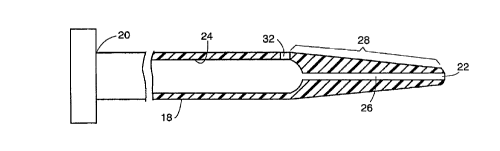
comprise a rigid tubular member 18, preferably formed
from a transparent plastic material and having a
proximal end 20 and a distal end 22. As is apparent in
the cross-sectional view of Figure 2, the device
includes first and second lumens 24 and 26 that
together run the full length of the instrument. The
distal end or tip 22 is slightly rounded to render it
more atraumatic. Also, the distal end portion of the
instrument is tapered over a zone identified by bracket
28 to facilitate its passage through the puncture wound
previously formed through the skin layer 12 and the
wall of the blood vessel 16.
The lumen 26 extending longitudinally through the
zone 28 is of a diameter approximately equal to the
outside diameter of a guidewire 30 with which the
measuring instrument of the present invention may
conveniently be used. The guidewire thus blocks the
flow of blood through the distal end opening. The
lumen 24, however, is of substantially larger diameter
than the diameter of the guidewire 30 so that it will
fit loosely within the lumen 24.
With continued reference to Figure 2, numeral 32
identifies a side entry port which extends through the
wall of the instrument at a predetermined location
proximal of the distal end 22 of the instrument. The
port 22 is drilled or otherwise formed to be proximate
the junction between the larger lumen 24 and the
smaller lumen 26.
Figure 1 shows that the outer surface of the
tubular member is scribed or embossed otherwise
provided with graduated markings with the zero point at
the location of the side port 32. Rather than using
scale-like markings, colored bands may be utilized.
In use, and in accordance with the Seldinger


WO 94I16637 PCT/US93/11645
-6-
technique, after the guidewire 30 is inserted through
a trocar so that its distal end is within the lumen of
the artery 16, the measuring instrument 10 of the
6
present invention may be fed over the guidewire and
advanced through the skin layer 12. Because the O.D.
of the guidewire 30 is approximately equal to the
diameter of the bore or lumen 26, it serves to block
blood flow through that lumen as the distal tip 22 is
made to enter through the puncture wound in the artery
wall 16. However, as the instrument 10 is slowly
advanced further into the wound, a point is reached
where the side entry port 32 passes through the artery
wall. Because the site of the side entry port is at
the base of the larger diameter lumen 24, it will not
be blocked by the presence of the guidewire and blood
will flow through the side entry port to fill the
larger diameter lumen 24. At the location where the
surgeon first notices blood rising up the instrument to
its open proximal end, he may note the graduated
marking on the exterior of the barrel at the level of
the skin surface. This provides an accurate
measurement of the distance below the skin surface of
the opening formed in the artery wall. Once the
measurement has been taken, the instrument 10 may be
removed from the guidewire and the interventional
procedure can be carried out.
At the conclusion of the interventional procedure,
the puncture sealing device described in the
aforereferenced Makower et al. application may be
assembled onto the guidewire and advanced therealong
until the distal tip of that instrument is at a depth
below the skin surface corresponding to the measurement
that had been earlier taken using the instrument of the
present invention. The hemostatic plug can then be


WO 94I16637 PCT/US93111645
ejected from the PSD so as to reside in the puncture
wound at a location abutting the exterior blood vessel
wall 16.
In accordance with a first alternative embodiment
which is particularly illustrated in Figure 3 and the
cross-sectional views of Figures 4 and 5, the measuring
instrument or gauge comprises an elongated flexible
plastic tubular member 30 which may be formed from
silicon rubber or other suitable material and which has
a first lumen 32 extending the entire length thereof
from its proximal end 34 to its distal end 36. The
diameter of this first lumen is sufficiently large so
that it can receive a guidewire 38 of a predetermined
lesser diameter therethrough where that guidewire 38
effective occludes blood flow through the lumen 32.
The flexible tubular member 30 has a second lumen 40
extending from the proximal end 34 thereof to a side
entry port 42 located a predetermined distance proximal
of the distal end 36 of the device. The guidewire
lumen is preferably located so as to be tangent to the
O.D. of the tube 30 as shown to provide as large an
opening for the lumen 40 as can practically realized.
Again, graduated markings 44 are formed on the
exterior wall surface of the tubular member 30 with the
zero point disposed adjacent the side entry port 42 and
extending in the proximal direction therefrom.
As in the case of the first embodiment, after the
guidewire 38 is made to enter the blood vessel, the
measuring instrument shown in Figure 3 may be advanced
over the guidewire with its distal end 36 entering the
blood vessel. As the measuring instrument is further
advanced along the guidewire, the point is reached
where the physician will note the flow of blood out
from the lumen 40, thus indicating that the side entry


WO 94116637 PCTIUS93/11645
~1~2061
_8_
port 42 has entered the blood vessel in question. Now,
by slowly drawing back the measuring instrument of the
present invention in the distal direction until blood
flow from the proximal end of the lumen 40 ceases, an
accurate reading can be taken from the graduated
markings of the depth below skin level of the side
entry port 42 as it leaves the blood vessel. Again,
this information can then be used when subsequently
inserting the puncturing sealing device. In this way,
the surgeon will know how deep to insert that device
before releasing the hemostatic plug so that the
hemostatic plug will reside against the exterior wall
of the blood vessel in question.
Referring Figures 6-8, there is shown a second
alternative embodiment of the invention. Again, the
instrument comprises a tubular body 46 preferably
formed of either a highly flexible or fairly rigid
plastic material and having a proximal end 48 and a
distal end 50. To aid in inserting the instrument of
Figure 6 into a puncture wound, it is preferable that
the distal end portion thereof be tapered to a
generally rounded, atraumatic tip. A lumen 52 extends
from the proximal end 48 only to a side entry port 54
which is located a predetermined distance proximal of
the distal end 50 of the instrument. Etched or
otherwise formed on the exterior surface of the tubular
body 46 are graduated markings 56 which extend in the
_ proximal direction from the side entry port 54. As is
evident from the cross-sectional views of Figures 7 and
8, the lumen 52 need only extend to the side entry port
and does not extend beyond it to the distal end 50.
The instrument illustrated in Figure 6 would find .
application in situations where no guidewire is
involved. The physician merely inserts the instrument


WO 94l16637 PCT/US93I11645
~'15~20 ~
1
_g_
into the already created puncture wound and advances
the instrument until blood entering the side entry port
54 travels up the lumen 52 to exit at the proximal end
48. At the point at which the blood flow is detected,
the physician may note using the scale 45 the distance
below skin level of the side entry port.
This invention has been described herein in
considerable detail in order to comply with the Patent
Statutes and to provide those skilled in the art with
the information needed to apply the novel principles
and to construct and use such specialized components as
are required. However, it is to be understood that the
invention can be carried out by specifically different
equipment and devices, and that various modifications,
both as to the equipment details and operating
procedures, can be accomplished without departing from
the scope of the invention itself.
What is claimed is:

Representative Drawing
A single figure which represents the drawing illustrating the invention.
Administrative Status

For a clearer understanding of the status of the application/patent presented on this page, the site Disclaimer , as well as the definitions for Patent , Administrative Status , Maintenance Fee  and Payment History  should be consulted.

Administrative Status

Title Date
Forecasted Issue Date 1999-08-03
(86) PCT Filing Date 1993-12-07
(87) PCT Publication Date 1994-08-04
(85) National Entry 1995-06-15
Examination Requested 1995-06-16
(45) Issued 1999-08-03
Deemed Expired 2000-12-07

Abandonment History

There is no abandonment history.

Payment History

Fee Type Anniversary Year Due Date Amount Paid Paid Date
Application Fee $0.00 1995-06-15
Maintenance Fee - Application - New Act 2 1995-12-07 $100.00 1995-08-16
Registration of a document - section 124 $0.00 1996-02-08
Maintenance Fee - Application - New Act 3 1996-12-09 $100.00 1996-07-19
Maintenance Fee - Application - New Act 4 1997-12-08 $100.00 1997-08-13
Maintenance Fee - Application - New Act 5 1998-12-07 $150.00 1998-07-29
Final Fee $300.00 1999-04-21
Owners on Record

Note: Records showing the ownership history in alphabetical order.

Current Owners on Record
SCHNEIDER (USA) INC.
Past Owners on Record
MAKOWER, JOSHUA
THATCHER, ROBERT J.
VAN TASSEL, ROBERT A.
Past Owners that do not appear in the "Owners on Record" listing will appear in other documentation within the application.
Documents

To view selected files, please enter reCAPTCHA code :


To view images, click a link in the Document Description column. To download the documents, select one or more checkboxes in the first column and then click the "Download Selected in PDF format (Zip Archive)" or the "Download Selected as Single PDF" button.

List of published and non-published patent-specific documents on the CPD .

If you have any difficulty accessing content, you can call the Client Service Centre at 1-866-997-1936 or send them an e-mail at CIPO Client Service Centre.


Document
Description 
Date
(yyyy-mm-dd) 
Number of pages   Size of Image (KB) 
Representative Drawing 1999-07-26 1 4
Description 1998-10-07 9 381
Cover Page 1995-11-30 1 17
Abstract 1994-08-04 1 58
Description 1994-08-04 9 399
Claims 1994-08-04 5 191
Drawings 1994-08-04 3 43
Claims 1998-10-07 5 184
Representative Drawing 1998-07-14 1 5
Cover Page 1999-07-26 2 89
Correspondence 1999-04-21 1 33
National Entry Request 1995-06-16 11 474
International Preliminary Examination Report 1995-06-16 14 477
Prosecution Correspondence 1995-06-16 14 524
Prosecution Correspondence 1995-08-18 1 26
Examiner Requisition 1998-01-30 1 40
Prosecution Correspondence 1998-03-26 2 38
Examiner Requisition 1998-06-09 3 121
Prosecution Correspondence 1998-07-14 3 102
Fees 1996-07-19 1 76
Fees 1995-08-16 1 84