Note: Descriptions are shown in the official language in which they were submitted.
21521~2
METHODS OF AND APPARATUS FOR
MEASURING UTEP~INE ELECTRICAL AND MECHANICAL ACTIVITY
1. Field of the Invention
The invention relates to methods of and apparatus for measuring
uterine electrical and mechanical activity. More particularly, the invention
relates to a method and instrument useful for determining the contractile
state of the uterus by recording spontaneous and evoked electrical activity
of the muscle cells of the uterus.
2. Backqround of the Invention and Technical Considerations
Preterm labor is one of the major pathological states most frequently
complicating pregnancy. Preterm birth is the major cause of prenatal morbi-
dity and mortality (75%) and long term neurological handicaps. In spite of
the use of different new tocolytics, the incidence of preterm labor and the
incidence of prenatal morbidity and mortality has not changed over the last
1 5 decades.
The diagnosis of labor (term and preterm) is the most significant pro-
blem faced by obstetricians. Preterm labor is the pathological state most
frequently associated with this dilemma. Moreover, term labor often re-
quires adjuvant therapy to halt or augment labor. However, there is no cur-
rently available method to objectively diagnose when the uterus is prepared
to labor either preterm or term. Since there is spontaneous uterine activity
during pregnancy, it is often not possible to distinguish between physiologi-
; 21521~2
.
cal uterine activity or preterm labor. The state of the cervix is commonly
used as a predictor of preterm birth. However, the softening of the cervix,
as well as the appearance of uterine contractions are relatively late in pre-
term labor.
Antiprogestins induce preparatory changes in the uterus in all stages
of pregnancy. This results in the increase in myometrial responsiveness to
oxytocic stimuli such as oxytocin or prostaglandins. The major effect of
antiprogestins on the uterus is the preparation or conditioning of thë myome-
trium to labor and delivery by inducing intercellular coupling which manifests
itself by an increase in propagation due to an increase in gap junctions.
The uterus is quiescent throughout pregnancy to maintain a tranquil
environment for the growing fetus. At the end of pregnancy normally the
.~
uterus begins to contract forcefully in a phasic manner (labor) to expel the
fetus and other products of conceptions. Abnormally the uterus sometimes
either begins to contract and labor prior to term (preterm labor) or fails to
contract at term. Preterm labor occurs in about 10% of all pregnancies
whereas the incidence of insufficient or absence of contractions at term is
also very high (3 to 13%). In most cases the clinician is faced with the
decision to either inhibit labor or stimulate it depending on the circum-
stances. However, the clinician has only subjective methods (state of cervix
or number of contractions but not force of contraction) on which to base a
decision .
The uterus is now known to pass through a series of steps prior to
and during labor to prepare the muscle to contract in a coordinated, synchro-
2~i nous and therefore forceful manner. These steps include the development
of gap junctions (low electrical resistance contacts), receptors and other
events between and on the muscle cells that aliow the uterus to contract as
a syncytium and react to contractile agents. Contractions of the uterus are
dependent upon electrical activity, therefore the presence of gap junctions
is an important component of this process. These steps are known to be
regulated by various physiological signais (hormones) and can be controlled
2 1 ~ 2
pharmacologically. When the muscle cells pass through this state they be-
come electrically and metabolically coupled. This state allows the uterus to
contract forcefully and frequently. Although this process is icnown to occur
during pregnancy, it also appears during the menstrual cycle and may be
present in various pathological conditions of the uterus such as dysmenor-
rhea, endometriosis, habitual abortion, allergic reactions, etc. However, at
present, the obstetrician or gynecologist has no objective method to evalu-
ate this process. The clinical judgement as to treatment would be greatly
enhanced by procedures which could define the state of the patient's
1 0 uterus.
Numerous studies show that gap junctions are present in almost ail
cells and their presence and function has been associated with normal
physiological control. Gap junctions are also known to be altered either
structurally or functionally in pathological states such as cancer, hypoxia,
inflammation, etc. Many studies demonstrate that one can assess gap junc-
tion presence or function by electrical simulation and recording of electrical
events in adjacent cells.
There have been a number of studies with respect to this matter such
as Miller, S.M., et al., "Improved Propagation in Myometrium Associated
with Gap Junctions During Parturition", American Journal of Physio/ogy,
pages 130-141 (1989), incorporated herein by reference, in which gap junc-
tion measurements were made on uterine tissue of pregnant rats. Additional
studies which are reported in the literature include: Garfield et al., Gap
Junctions: Their Presence and Necessity in Myometrium During Parturition",
Science, Vol. 198, pp. 958-960 (December 2, 1977); Miller et al., "Im-
proved Propagation in Myometrium Associated with Gap Junctions During
Parturition", American Physiologica.' Society, pp. C130-C141 (1989);
Garfield et al., "Modulation of Myometrial Gap Junctions: Toxicological
Implications", /n Vitro Toxico/ogy, A Journa/ of Mo/ecu/ar and Ce//ular
Toxico/ogy, Vol. 3, Number 1, pp. 41-59 (1990); Chwalisz et al., "The Pro-
gesterone Antagonist Onapristone Increases the Effectiveness of Oxytocln
~ 2 1 ~ 2
to Produce Delivery without Changing the Myometrial Oxytocin Receptor
Concentrations", Am. J. Obstet. Gyneco/., Vol. 165, No. 6, Part 1, pp.
1760-1770 (December 1991); Garfield, "Structural and Functional Studies
of the Control of Myometrial Contractility and Labor", The Onsef of Labor:
CeJ/ular & Integrafive Mechanisms, pp. 55-79 (1988); Garfield et al.,
"Effects of the Antiprogesterone RU 486 on Preterm Birth in the Ratn,
American Journal of Obstetrics and Gynecology, Vol. 157, No. 5, pp.
1281-~ 285 (Nov.1987); Demianczuk et al., "Myometrial Electrophysiologic
Activity and Gap Junctions in the Pregnant Rabbit, American Journa/ of
Obstetrics and Gynecology, Vol. 149, No. 5, pp. 485-491 (July 1, 1984);
Garfield, "Control of Myometrial Function in Preterm Versus Term Laborn,
Clinical Obstefricsand Gyneco/ogy, Vol. 27, No.3, pp. 572-591 (September
1984); Puri et al., "Changes in Hormone Levels and Gap Junctions in the Rat
Uterus During Pregnancy and Parturition~, Biology of Reproducfion, 27,
967-975 (1982~; Garfield et al., "Endocrine, Structural, and Functional
Changes in the Uterus During Premature Labor", American Journalof Obste-
trics and Gynecolo~y, Vol. 142, No. 1, pp. 21-27 (Jan. 1, 1982); Garfield
et al., "Appearance of Gap Junctions in the Myometrium of Women During
Labor", Ame~ican Journal of Obstetrics and Gynecology, Vol . 140, No. 3,
pp. 25~260 (June 1, 1981); Garfield et al., "Presence of Gap Junctions in
the Myometrium of Women During Various Stages of Menstruation", Ameri-
can Journal of Obstetrics and Gynecology, Vol. 138, No. 5, pp. 569-574
(Nov. 1, 1980); and Garfield et al., "Are Gap Junctions Necessary for Cell-
to-Cell Coupling of Smooth Muscle?: An Update", Can. J. Physiol. Phar-
macol., Vol. 70, pp. 481-490 (1992); each of which is incorporated herein
by reference. While these studies each recognize various aspects of the
phenomenon of interest, they do not suggest just how one would utilize the
phenomenon in practical medical procedure.
The status (function, location, identification, etc.3 of nerves and their
terminals in tissues can be quantified also by selectively stimulating the
nerves with electrical parameters that do not affect surrounding tissues.
~ 21~2~6~
This so-called field stimulation" has been used in many studies to activate
nerves or their varicosities in tissues to assess, localize and identify nerves
in tissues. Exemplary of such studies are the following articles: Garfield
et al., "A Functional and Structural Study of the Innervation of the Human
Uterusn, American Jouma/of Obstetrics and Gynecology, Vol. 160, No. 1,
pp. 218-228, (Jan. 1989); Bulat et al., nStudies of the Innervation of Rabblt
Myometrium and Cervixn, Can. J. Physio/. Pharmaco/., Vol. 67, p p. 837-844
(19~9); and Buchanan et al., ~Innervation and Gap Junction Formation in the
Myometrium of Pregnant Little Brown Bats, Myotis /ucifugusn, Tf~eAnafomi-
ca/Record 221:611-618 (1988), each of which is incorporated herein by
reference.
Prior methods and instruments for evaluating the status of the uterus
have used external monitors which give little information of quantitative
nature necessary to define the processes described above.
Summary of the Invention
An object of the present method and invention is to measure in vivo
the electrical and mechanical activity of tissues, such as for example, but
not limited to, uterine muscle tissue, to produce a more quantitative, com-
prehensive and analytical framework of the tissue by transferring information
from the tissue to a monitor for assessment by an attending physician or
other party interested in monitoring the tissue.
Upon further study of the specification and appended claims, further
objects and advantages of this invention will become apparent to those
skilled in the art.
The present method and apparatus is applicable to a wide range of
obstetrical, gynecological and other conditions. One such application is
defining the state of the uterus during term and preterm labor. Another
application is monitoring the nonpregnant uterus for indication of conditions
such as infertility and uterine pathology in cycling women. The method and
apparatus is also valuable for use in connection with other tissues other than
21~2~
the uterus such as tissues of the bladder, intestine, heart and other muscular
or nonmuscular (brain, liver, pancreas, etc.) tissues for purposes of evaluat-
ing their normal and abnormal behavior. The method and instrument is also
usable for monitoring tissues in animals, as for example in a veterinary clinic
or for live stock.
In accordance with one specific aspect of the invention, a needle in-
cludes stimulating recording electrodes as well as optional miniature piezo-
electric electrodes embedded along an inner surface thereof. In accordance
with one embodiment, the needle is placed in the uterine wall (i.e., myome-
trium) under ultrasound guidance similar to routine procedures during amnio-
tic fluid sampling. The signals detected by the needle are monitored to pro-
vide measurements indicative of spontaneous and electrically evoked activ-
ity. The needle is connected to a multichannel recorder, stimulator and
computer with software for analysis of signals.
The above-described needle may alternatively be hollow for withdraw-
ing material, such as for example amniotic fluid.
Brief Description of the Drawings
Various other objects, features and attendant advantages of the pre-
sent invention wiil be more fully appreciated as the same becomes better
understood when considered in conjunc~ion with the accompanying draw-
ings, in which like reference characters designate the same or similar parts
throughout the several views, and wherein:
Figure 1 is a side view, partially in phantom, showing a needle con-
figured in accordance with the instant invention inserted in the uterine wall
of a pregnant patient, the needle being connected to a recording device and
the position of the needle being monitored by an ultrasonic scanner;
Figure 2 is an enlarged, side elevational view illustrating the needle of
Figure 1 embedded in the muscle tissue of the uterine wall;
Figure 3 is a further enlarged side view showing a portion of the
exterior surface of the needle shown in Figures 1 and 2;
2152~
Figure 4 is a sectional view taken along lines 4-4 of Figure 3 showing
an array of stimulation electrodes;
Figure 5 is a cross-section taken along lines 5-5 of Figure 3 showing
an array of recording electrodes;
Figure 6 is an enlarged sectional view of a portion of Figure 5 showing
how an individual recording electrode is mounted within the needle;
Figure 7 is a front view of a typical recording apparatus to which the
needle is connected and which provides stimulation signals and receives
response signals;
Figure 8 is a schematic view of smooth muscle tissue in proximity
with the needle of Figures 1-7 illustrating the function of the instant
invention;
Figure 9 is a view sirnilar to Figure 8 showing smooth muscle or other
tissue with nerves proximate the needle of the instant invention and further
i~lustrating an additional feature of the invention;
Figure 10 is a diagrammatical view showing spontaneous electrical
activity between cells detected by recording electrodes;
Figure 1 1 is a diagrammatical view showing evoked electrical activity
between cells;
Figure 1 2.is a linear diagram of an inner surface of the needle accord-
ing to the instant invention with the recording electrodes arranged in a
selected array;
Figure 13 is a diagrammatical view comprising cellular electrical
activity in a muscle with muscle contractions; and
Figure 14 is a diagrammatical view comparing cellular electrical activ-
ity and muscle contractions relating the rate of rise amplitude and the rates
of depolariza~ion and relaxation
Detailed Description
Referring now to Figure 1, there is shown schematically a pregnant
patient 10 with a fetus 12 retained with a uterine wall 14 which defines an
------ ----
~ ~ ~ ~ ~ - -
- ~ ~ ~ ~ ~ - ~
~=
~_ ~ E~ E~
the abdominal wall 18 and embedde~d iin the uterlne wali 1 . . h~e~
a bundle of leads 22 which are connected to a recording apparatus 24.
In accordance with the principles of the instant invention, the uterus
of the pregnant patient 10 is monitored by ultrasonic transducers 26 to pro-
vide an image 28 of the uterine wall on an ultrasonic monitor 30 so that the
~ 10 shank 31 of the needle 20 may be accurately guided and properly embedded
in the uterine wall 14.
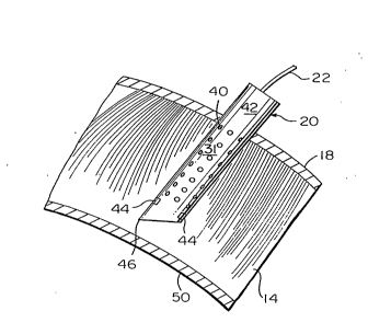
Referring now to Figure 2, a greatly enlarged view of a section of ute-
rine wall 14 is shown with the shank 31 of the needle 20 embedded therein
after having been passed through the abdominal wall 18. The needle 20is
preferably made of stainless steel and has an overall length of about 65 mm.
An array of recording electrodes 40 are disposed on the cylindrical surface
42 of the needle 20 and an array of stimulation electrodes 44 are positioned
proximate the tip 46 of the needle to isolate a portion of the muscle tissue
within the hollow core. The arrays of recording and stimulation electrodes
40 and 44 are completely embedded in the muscle tissue of the uterine wall
~ 14 with the muscle tissue extending into the hollow core of the needle. In
accordance with one embodiment of the invention, the tip 46 of the needle
20 does not penetrate the endometrium 50 which is disposed between the
muscle 14 of the uterine wall and the amniotic fluid 16. In accordance with
2s another embodiment of the invention, the needle 20 is a hollow amniotic
fluid sampling needle which performs fluid sampling alternative to electrical
monitoring .
Referring now to Figure 3, there is shown a side view of the needle
20 where it is seen that the embedded portion of the needle having the array
of recording electrodes 40 and stimulating electrodes 44 has a iength L of
about 2-4 mm and a diameter of about 0.5 to 2 mm. The recording
electrodes 40 are spaced from one another by a distance of about 0.5 mm,
SUBSTITUTE SHEET
~ ' 2~21~2
, while the stimulation electrodes 44 are a height and width of about 0.5 mm.
The tip 46 of the needle 20 is disposed approximately 0.5 mm from the bot-
tom of the array of stimulation electrodes 44 and has a sloping edge 52 ex-
tending from one side of the needle to the tip 46 thereof. The dimensions
defining spaces between various electrodes are suggested dimensions which
may be varied from needle to needle to optimize performance of the needles
~0.
Referring now to Figure 4, where the shank 31 of the needle 20 is
shown in cross-section, the stimulation electrodes 44 are silver plated into
indentations 54 of the wall 56 of the needle. The indentations 54 are first
coated with an insulating material 58 to electrically isolate the stimulation
electrodes 44 from the stainless steel needle shank 31. Each stimulation
electrode 44 has an insulated lead 59 which is led back over the surface 42
of the needle 20 (see Figure 3) into the lead bundle 22 and to the recording
device 24 (see Figure 1). The stimulation electrodes 44 receive either de-
polarized or hyperpolarized current pulses from the recording device 24, with
each pulse having a duration in the range of about 10 to 500 milliseconds,
a frequency in the range of 0.05 to 5 hertz and an amplitude in the range of
about 0.1 to 30 volts.
Referring now to Figures 5 and 6 there is shown the specific structure
of one level of the array of recording electrodes 40 as well as the structure
of a single recording electrode. In the illustrated embodiment, there are four
recording electrodes disposed at 90~ intervals around the wall 56 of the
needle 20. As is seen in the specific embodiment of Figure 6, each record-
ing electrode 40 includes a plate 60 made of silver or another conductive
material which is disposed inside of the needle 20 proximate the inner sur-
face 62 of the wall 56. A layer of electrical insulation 64 is disposed
between the plate 60 and the surface 62 of the wall 56 to electrically isolate
the plate 60 from the stainless steel needle shank 31. An insulated lead 66
extends through a bore 68 through the wall of the needle 56 and a hole 69
in the insulation. As is seen in Figure 1, the lead 66 from each recording
21~21 62
- 10 -
electrode 40 extends back up in the needle and into the lead bundle 22 for
connection to the recording apparatus 24. The sliver plate 60 functions
similar to an antenna and receiver signals generated in the muscle tissue 14
(see Figure 2).
In an alternative apparatus, instead of all the electrodes 40 being re-
cording electrodes, the electrodes may be piezoelectric e~ectrodes 70 which
sense contractual events and transmit these events via insulated leads 71
to the recording apparatus 24. Preferably, the piezoelectric electrodes 70
are disposed between the recording electrodes to provide an alternating
array as is shown in Figure 3.
Referring now to Figure 7, where the recording apparatus 24 is
shown, the recording apparatus includes a stimulator 80 for invoking elec-
trical events in the needle 20. The stimulator 80 is of conventional design
and includes a control for amplitude 82, a control for voltage 84, a control
for duration 86 and a control for frequency 88. The stimulator is connected
via cable bundle 22 to the stimulation electrodes 44 via leads 59. The re-
cording apparatus 24 also includes a monitor 96 with a monitor screen 98
to display readings from the electrical leads 66 and 71 connected to the re-
cording electrodes 40 and piezoelectric electrodes 70, respectively. In a
conventional fashion, the monitor includes controls 100 for selecting various
arrays of electrodes to be detected. For example, the controls may select
the recording electrodes 40 or the piezoelectric electrodes 70 for monitoring.
Finally, recording apparatus 24 also includes controls 102 for amplifying and
filtering the signals relayed over leads 41 and 71 to the monitor 96. A stan-
dard computer 104 is connected to the monitor 24 via cable 106. The com-
puter 104 includes software and a key board for controlling the various
functions of the recording apparatus 24.
Referring now to Figure 8, it is seen that the needle 20 with the
arrays of recording electrodes 40 and piezoelectric electrodes 70 are dis-
posed proximate muscle cells 120 in the smooth muscle tissue of the uterine
wall 14. 8etween each cell 120 and adjacent cells 120, there is schemati-
215~1B~
~ '
cally illustrated what is known a gap junction 122 which is a low resistance
electrical contact that develops prior to and during labor in order to prepare
the smooth muscle tissue 14 for contraction in a coordinated and synchro-
nous manner. Contractions of the uterine wall 14 are dependent upon pro-
pagation of electrical activity between the muscle cells; therefore, the
presence of the gap junctions 122 is an important component of the con-
traction process. Gap junctions are known to be regulated by various
physiological signals produced by hormones and can be controlled pharma-
cologically. When the muscle cells 120 contain open gap junctions, they
become electrically and metabolically coupled which allows the uterus wall
44 to contract forcefully and frequently.
- In accordance with the instant invention, the smooth muscle tissue
14 is stimulated with electrical pulses having parameters that affect only the
cells 120 and not surrounding tissue. Monitoring is initiated by pulsing the
stimulation electrodes 44 (Figures 2-4) with current pulses having a duration
in the range of about 10 to 500 milliseconds at a frequency in the range of
about 0.05 to 5 hertz and at a voltage amplitude in the range of about 0.1
to 30 volts. This stimulation causes spontaneous and electrically evoked
action potentials 126 at the recording electrodes 40 as well mechanical
interactions with the piezoelectric electrodes 70, which signals are trans-
mitted over the leads 66 from the recording electrodes and leads 71 from
the piezoelectric electrodes to the recording apparatus 24. In the recording
apparatus 24 the amplifier 101 modulates the signals using a time constant
of about 1 second at a high frequency filtration band pass in the range of
0.1 to 22 hertz. The thus monitored signals 126 are displayed on the moni-
tor screen 98, then stored PC computer-based hardware and software in the
computer 104 with a sampling rate of about 500 digitized samples per
second .
Referring now to Figure 9, there is schematically shown the needle 12
and recording electrodes 40 in juxtaposition with cells 120 being in a state
where there are gap junctions 122. Muscle tissue, which may be smooth
~1~2162
~
- 12-
, muscle tissue or other muscle tissue, has peripheral nerves 130 therein with
nerve endings 132. It is possible to stimulate nerves 130 and nerve endings
132 by pulsing the stimulation electrodes 44 (Figures 2, 3, 4~ with parame-
ters that do not activate the cells 120. The nerves 130 then act on the cells
120 and the cells generate signals 136 in the cells 120 which are detected
by the electrodes 40. In this way, a physician is able to monitor the effects
of nerve stimulation in isolation on the cells 120.
-Referring now to Figures ~0-15, the recording apparatus 24 stores
signals 126 in the associated computer 104 and extracts the following para-
meters derived from the signals 126:
a. duration of bursts of action potentials 126;
b. propagation velocity of individual action potentials in bursts
following stimulation (measured from change in latency from
successive electrodes 40);
c. patterns of propagation and distance of propagation during
spontaneous and evoked action potentials;
d. entrainment of bursts;
e. velocity and distance of mechanical activity as measured by
the piezoelectric electrodes 70;
f. characteristics of the contractions such as rate of rise and
amplitude as detected by the piezoelectric electrodes; and
g. characteristics of the action potentials such as the rate of rise
of depolarization and plateau, amplitude and the rate of
repolarization .
Considering the aforementioned parameters in more detail, as is seen
in Figure 10, the length of each burst 140 is plotted as a function of time
t to provide the duration of each burst in seconds as detected by the elec-
trodes 40-1 through 40-n. In addition, the computer 104 measures the
action potential frequency in cycles per second by counting the number of
spikes 142 per unit time.
-
~21~2
.
-- 13 --
The propagation obligation velocity of the individual action potentials
126 and evoked potentials 144 is seen from a consideration of Figures 10
and 11, wherein a latency period 150 between selective recording elec-
trodes 40 is shown. In Figure 11, the evoked electrical responses 126 from
the electrodes 40 result from the application of a polarized pulse 152 or a
hyperpolarized pulse 154 applied to the stimulation electrodes 44 (Figures
2, 3 and 4).
Referring now to Figure 12 in combination with Figures 10 and 11,
a computer diagram of an array 156 of the electrodes 40 is shown with the
electrodes arranged in levels and rows above the stimulation electrodes 44.
The computer 104 computes the original and propagation characteristics of
any of the bursts 140 shown in Figure 10. Similarly, from stimulated poten-
tials computer 104 calculates the propagation velocity in distance and dis-
plays this information on the screen 98 of the recording device 24. The en-
trainment of bursts 140 is seen by reference numeral 158 of Figure 10 and
is calculated from the initial latency period 160 between the bursts at each
electrode 40.
Utilizing an approach similar to the approach for monitoring electrical
activity, the computer 104 estimates the velocity and distance of the
mechanical activity detected by the piezoelectrodes 70 (Figure 3) which are
indicative of the contractions of the uterine wall 14. As is seen in Figure
13, the mechanical activity of muscle tissue comprising the uterus wall 14
is identified by a curve 162 which corresponds to the burst 140 indicative
of the underlying electrical activity. Frequency, duration and magnitude of
a contraction of the uterine wall 14 are respectively proportional to the fre-
quency of the bursts 140, the duration of the bursts and the propagation of
the action potential 126 to recruit additional cells 120 (see Figure 8). The
velocity of a contraction is estimated from the latency of contractions at
successive piezoelectrodes 70 with the origin and distance of each contrac-
tual sequence being computed from a computer generated map of the needle
20 such as the map of Figure 12 utilized for detecting action potentials 126.
2~52~2
.
- 14-
Referring now to Figure 14, the characteristics of any contraction
curve 162 or action potentials 126 contained in a burst 140 are isolated by
the program of the computer 104. The rate of rise 164, amplitude 166, rate
of repolarization and relaxation 166 can therefore be estimated.
From the aforedescribed measurements set forth in Figures 10-14,
one can reasonably estimate if tissue, such as the muscle tissue of the ute-
rine wall 14, or other tissue is coupled electrically. In ~ther words, one can
discern if the gap junctions 122 are present, absent or in a closed configura-
tion. The presence of gap junctions 120 is generally indicative of the labor
state for the uterine muscle comprising the uterine wall 14. On the other
hand, the absence of electrical coupling suggests the ambience or closed
state of the junctions and the lack of conditions favorable to labor.
While a preferred embodiment of the invention utilizes a signal needle
20 with both the stimulation electrodes 44 and the recording electrodes 40
thereon, in another embodiment of the invention, the stimulation electrodes
44 and recording electrodes 40 are on sepa-ate needles implanted at diffe-
rent locations. In another embodiment, the electrodes are mounted on the
outside of the needle. In still another embodiment of the invention the
stimulation electrodes 44 are embedded in the tissue being monitored while
the recording electrodes 40 are positioned outside that tissue on the
patient's skin.
While utilization of the apparatus and method has been described
above as especially useful for monitoring the uterine wall 14 during preg-
nancy, gap junctions 122 are present in almost all cells and the presence
and function thereof is associated with normal physiological control. Gap
junctions 122 are also known to be altered either structurally or functionally
in pathological states such as cancer, hypoxia, inflammation and other path-
ological states. Accordingly, it is within the scope of this invention to utilize
the apparatus and methods thereof for medical and biological procedures
other than uterine wall monitoring
-. 21~
- 15 -
From the forego;~g description, one skilled in the art can easily
ascertain the essential characteristics of this invention, and without
departing from the spirit and scope thereof, can make various changes and
modifications of the invention to adapt it to various usages and conditions.