Note: Descriptions are shown in the official language in which they were submitted.
~ WO94/21164 2 f 5 2 2 7 2 PCT~S94/03018
REMOTE SENSING TONOMETRIC CAln~l~ APPARATUS AND M~l~O~
This application is a continuation-in-part of
United States patent application serial no. 035,020, filed
March 22, 1993, which was a continuation-in-part of United
States patent a~pplication, serial no. 014,624, filed
February 8, 1993, which was a continuation-in-part of
copending United States patent application, serial no.
719,097, filed June 20, 1991, which was a continuation-in-
part of copending United States patent application, serialno. 994,721, filed December 22, 1992.
This application hereby expressly incorporates by
reference, the disclosures and drawings of the following
issued U.S. patents: United States Patent Nos. 4,221,567;
4,233,513; 4,273,636; 4,423,739; 4,576,590; 4,480,190;
4,596,931; 4,643,192; 4,671,287 4,859,858; 4,859,859;
4,907,166; 4,914,720; 5,042,522; 5,067,492; 5,095,913;
5,158,083; 5,174,290; and 5,186,172.
R~CK~ROUND AND SUMMARY OF THE lNv~NLlON
This invention relates to medical diagnostic
equipment and methods and is particularly concerned with
hollow viscus tonometry and remote electronic and optical
senslng.
Until the advent of the tonometric method (see
U.S. Patent No. 4,643,192, issued February 17, 1987) few
considered any aspect of acid-base balance when attempting
to monitor or maintain the adequacy of tissue oxygenation.
Yet acid-base balance i5 primarily determined by the
--1--
WO94/21164 2 1 S 2 ~ ~ 2 - PCT~S94/03018
balance between the protons released during the release of
energy by ATP hydrolysis and the resynthesis of ATP by
oxidative phosphorylation. The hydrolysis of ATP generates
150,000 mmols of H+ each day in a resting 70 Kg man. All,
but the 1~ of this fixed acid load excreted by the kidneys
each day, is presumed to be co~su~ed in the resynthesis of
ATP by oxidative phosphorylation. When the delivery of
oxygen fails to satisfy the energy needs of the tissue the
rate of ATP hydrolysis exceeds the rate of synthesis and
the pH falls as the degree of unreversed ATP hydrolysis
ncreases .
Information for determining global tissue
oxygenation has been collected for many years. Eoda, D.,
"`Gastrotonometry' an Aid to the Control of Ventilation
During Artificial Respiration," The Lancet (1959).
However, it is now widely accepted that global measurements
of oxygen delivery, consumption and extraction do not
provide reliable information about the adequacy of local or
even "global" tissue oxygenation in patients. The indirect
measurement of gastric intramucosal pH (pHi) as described
in U.S. Patent Nos. 4,643,192; 5,158,083; 5,186,172
provides clinicians with a mlnlm~lly invasive yet sensitive
means of detecting the development of a tissue acidosis,
and hence inadequacy of tissue oxygenation, in a region of
the body that is one of the first to exhibit an inadequacy
of tissue oxygenation in shock. Use of the measurement has
revealed that some 50~ to 60~ of patients having major
surgery and 80~ of ICU patients develop an intramucosal
~ WO94/21164 215 2 2 7 2 PCT~S94/03018
. . "
acidosis during their illness despite the conventional
appearance of being adequately resuscitated.
The degree and duration of the presence of a
gastric intramucosal acidosis are highly sensitive measures
of the risk of developing ischemic gut mucosal injury and
its putative consequences, namely the translocation of
bacteria and their-.toxins, cytokine release, organ
dysfunctions and failures, and death from the organ
failures. By providing an index of the adequacy of tissue
oxygenation in one of the first parts of the body to
exhibit dysoxia in shock the measurement of gastric
intramucosal pH improves the opportunity to obtain advanced
and accurate warning of impending complications and to
intervene in time to prevent them. More importantly timely
therapeutic measures that restore the intramucosal pH to
normality and "gut-directed" therapies incorporating
measures that reverse an intramucosal acidosis are
associated with an improved outcome. "pH-directed" therapy
has in addition been shown to improve outcome in a
prospective randomized multicenter study of medical and
surgical ICU patients.
The measurements of gastric intramucosal pH have
revealed deficiencies in currently accepted practices. It
has, ~or example, become apparent that empirical increases
in global oxygen delivery may be redundant in some 40~ to
50~ of patients having major cardiovascular surgery who do
not develop a gastric intramucosal acidosis and whose
prognosis is excellent. It is further apparent that the
WO94/21164 215 22~ 2 .~t `l. PCT~S94/03018
vogue of increasing global oxygen delivery to supranormal
levels cannot be relied upon to prevent or to reverse the
presence of an intramucosal acidosis. Of particular
concern is the intramucosal acidosis that may be induced by
measures, notably the transfusion of red blood cells and
dobutamine, that increase global oxygen delivery in
patients who do not have an intramucosal acidosis but whose
global oxygen delivery is considered too low.
THE TONQMETRIC METHO3
The measurement of pH in the most superficial
layer of the mucosa is obtained indirectly by measuring the
partial pressure of carbon dioxide (pCO2; PCO2) in the lumen
of the gut and the bicarbonate concentration in arterial
blood and substituting these two values in the Henderson-
Hasselbalch equation or some modification thereof. See
"Gastric Intramucosal pH as a Therapeutic Index of Tissue
Oxygenation in Critically Ill Patients," Lancet 1992; 339;
195-99, incorporated herein by reference. The indirect
measurement of the pH of the wall of the organ (pH indirect
or intramucosal pH) may be employed because it is believed
or assumed that the pCO2 in the most superficial layers of
the mucosa is in equilibrium with that in the lumenal
contents with which it is in contact. It is further based
upon the assumption that the bicarbonate concentration in
the tissue is the same as that being delivered to it in
arterial blood and that the pKa, 6.l, is the same as that
in plasma.
~ WO94/21164 2 1 5 2 2 7 2 PCT~Sg4/03018
At present, measurements of pCO2 in the lumen of
the stomach are obtained by infusing saline into the
silicone balloon of a gastrointestinal tonometer, allowing
the pCO2 in the saline to equilibrate with that in the lumen
of the gut; recording the equilibration time; aspirating
the saline;-measuring the pCO2 in the saline with a blood
gas analyzerS using a nomogram to derive the steady-state
adjusted pCO2 from the equilibration time and the measured
pCO2; and then derive the intramucosal pH from the steady-
state adjusted pCO2 obtained and the bicarbonateconcentration in a substantially contemporaneous sample of
arterial blood. Again, see U.S. Patent Nos. 4,643,192,
issued February 17, 1987; 5,174,290, issued December 29,
1992; and 5,186,172, issued February 16, 1993; as well as
copending U.S. Applications, Serial Number 719,097, filed
June 20, 1991; Serial Number 994,721, filed December 22,
1992 and Serial Number 014,624, filed February 8, 1993; all
three issued patents being completely and expressly
incorporated herein by reference. The precision of the
measurement of gastric intramucosal pH between healthy
subjects is excellent, the gastric intramucosal pH in a
healthy subject being the same as the pH in his arterial
blood.
The prior art (see U.S. Patent No. 4,643,192) has
recognized that intestinal ischemia, and to a lesser
degree, stress ulceration, are two problems that plague
physicians involved in the management of patients in
intensive care units. Intestinal ischemia, in particular,
WO94/21164 215 22~ 2 PCT~S94/03018
.~ , ,, _
has an insidious onset and may not be detected until days
after the intestine has become completely and irreversibly
compromised. A delay in the diagnosis of intestinal
ischemia may have devastating consequences for a patient.
The availability of means for early diagnosis and
management of patients with these problems would have
immediate applicability in all intensive care units,
especially where the procedure can be conveniently
conducted with reasonable safety and reliability.
It has been established that a fall in the
intramucosal pH may precede the development of intestinal
ischemia and stress ulceration. As discussed in U.S.
Patent No. 4,643,192, which is expressly incorporated
herein by reference, entitled "Hollow Viscus Tonometry" a
fall in intramucosal pH also occurs within minutes of
inducing intestinal ischemia in dogs. The fall in pH in
intestinal mucosa, and hence the likelihood of ischemia or
stress ulceration, can be reliably calculated from a pCO2
(partial pressure of CO2), or other indicia of pH, in
lumenal fluid and the bicarbonate concentration in arterial
blood. The method of calculating the pH in intestinal
mucosal tissue, pursuant to principles set forth in prior
related patents discussed herein, has been validated by
directed measurements under a variety of conditions
simulating clinical problems. A correlation coefficient on
the order of 0.92 to 0.95 has been obtained in each of 16
dogs. The validity of the procedure is inherently
extensible to humans, and indeed may also be useful in
~ WO94/211~ 21~ 2 2 7 2 PCT~S94/03018
assessing the vitality of other hollow organs and tissue.
See R.G. Fiddian~Green et al. "Splanchnic Ischemia and
Multiple Organ Failure".
To measure the pCO2 in the lumen of the gut it has
heretofore been necessary to obtain and remove a sample of
fluid that has been in contact with the wall of the gut for
a certain time period, usually at least half an hour. It
has now been observed that it is somewhat difficult to
manually aspirate the sampling fluid or medium from a
tonometric catheter located in the gut or other internal
focus with any consistency. It is much easier to obtain
such samples from the stomach, but samples obtained from
the stomach frequently contain foreign material that can
damage a gas analyzer.
As taught in prior related patents discussed
herein, the desired sample or samples can be obtained from
the gut using a catheter tube (called a tonometric
catheter) having a walled sampling chamber on the tube with
the sampling chamber being in sample-specific communication
with the hollow interior of the tube. The wall of the
sampling chamber comprises a material which is
substantially impermeable to liquid yet is highly permeable
to gas. One suitable material is polydimethylsiloxane
elastomer.
25 In use the catheter is introduced into a patient
to place the sampling chamber at a desired site within the
gut (or other hollow organ). An aspirating liquid or
medium is employed to fill the interior of the sampling
W094/211~ 15 2 2 7 2 PCTtUS94tO3018
chamber. The sampling chamber is left in place at the
desired sampling site long enough to allow the gases
present to diffuse through the wall of the sampling chamber
into the aspirating liquid. The time should be long enough
for the gases to equilibrate. The liquid impermeable
nature of the sample chamber wall material prevents both
the aspirating liquid from leaking out of the chamber and
also the intrusion of any liquids into the aspirating
liquid. After the appropriate or desired amount of
placement time has elapsed the aspirating liquid is
aspirated along with the gases which have diffused into it.
The sample thus obtained is analyzed for gas content, in
particular for pCO2. In this way the pCO2 within the lumen
of the gut can be reliably measured with the fluid being
free from lumenal debris.
In carrying out the diagnostic method taught in
prior related patents, the pCO2 measurement is utilized in
conjunction with a measurement of the bicarbonate ion
concentration (HCO3-) in an arterial blood sample of the
patient for determining the pH of the tract wall.
Depending upon the particular condition of a
given patient, the catheter may be left in place and
samples may be taken at periodic intervals so that pH
values may be periodically calculated. The procedure has
a high reliability in accurately determining the adequacy
of organ tissue oxygenation, and diagnosing intestinal
ischemia in its incipient stages. Such determination or
detection can be useful in treating the patient so that the
21S2272
WO94/21164 ~CT~S94/03018
potentially devastating consequences resulting from less
timely detection may often be avoided.
While the sampling techniques taught in the prior
related patents discussed herein have provided highly
accurate and reliable results, it has now been observed
that there are instances (in the care of the critically ill
in intensive care units, for example) in which remote
sensing of the organ or organ-wall condition and automatic
determination or calculation of the organ or organ-wall pH
would be advantageous and easier to effectuate. This
method would thus partially or totally eliminate the need
for the somewhat cumbersome manual aspi,ration of the
sampling fluid or medium which fills the sampling chamber.
There is also a need to extend the benefits of tonometric
sampling and sensing to other internal hollow viscus
organs. To this end, there is a need for new and different
tonometric devices specifically adapted to allow sensing
and sampling techniques to be performed with ease in a
clinical environment, and in combination with other
procedures.
The importance and significance of determining
the pH of the wall of a given hollow viscus organ has been
recently dramatically magnified as a result of the recent
recognition that the pH of the wall of a given organ can be
employed to accurately evaluate the vitality and/or
stability of that organ as well as others; this is in
contrast to merely determining whether such an organ is
experiencing an ischemic event. Further, certain organs
21S2272 i
WO94/21164 - PCT~S94/03018
can be selected for monitoring, either alone or in
combination, and evaluation of this organ or these organs
can aid in predicting the overall condition of the patient,
or the onset of a multitude of pathologies, including
predicting or identifying such e~ents as multiple organ
failure. Such a methodology ca~ be employed to greatly
enhance and supplement the monitoring of the critically
ill, for example.
It has also been observed that an unusually large
negative bias is encountered when measuring the pCOz in
saline with certain blood gas analyzers (including those
manufactured by Nova Biomedical, L. Eschweiler and
Mallinckrodt) that have been standardized for blood but not
for saline. The presence or absence of unacceptable bias
may be determined by the use of reference samples of
tonometered saline. The inter-instrumental bias
encountered when measuring arterial blood gases and
especially pCO2 in saline with different blood gas analyzers
requires that each institution derive its own normal values
for meaningful use in clinical practice. It is reported
that the precision of the measurements made within a static
environment may be improved and unacceptable
interinstrumental bias eliminated, in whole or in part, by
using Gelofusine~ (sterile 4~ w/v succinylated gelatine in
saline), a phosphate buffer, bicarbonate-buffered saline,
or mixtures thereof. Unfortunately the diffusional
- characteristics may be altered, in which case the nomograms
provided for the determination of steady-state adjusted pCO2
- 10 -
~ wog4ell64 2152272 ~ PcT/IJsg4l030l8
in saline cannot be used for the determination of
intramucosal pH with these fluids.
The time constant may be reduced to seconds by
using an electrochemical pCO2 sensor directly in the lumen
of the gut and measuring the pCO2 in either liquid or
gaseous luminal contents, as described herein.
Unfortunately, pCO2 sensors are known for their tendency to
drift and cannot be easily recalibrated in vivo.
In one aspect, the present invention provides a
new apparatus and method for remotely sensing organ
condition and conveying a signal, e.g. an electrical
current or optical signal, to an electronic or optical
apparatus located outside the organ under investigation.
In one embodiment, a transducer (or plurality of
transducers) is attached to a tonometric catheter for
introduction into the organ along with the tonometric
catheter. This first sensor generates and conveys a signal
indicative of some desired aspect of organ condition, e.g.,
indicative of the pCO2, pH and/or PO2 level of the organ or
organ-wall. For example, in one preferred embodiment, mean
ambient pCO2, pH and/or PO2 Of lumenal fluid or the like is
measured or monitored via wire or other suitable
electromagnetic energy conveying means to an electronic
circuit which interprets the electromagnetic signal and
produces a report of the organ condition. The electronic
circuit may include an input for receiving a separately
determined signal indicative of the blood pH of the
patient. Using this pCO2, pH and/or PO2 measurement along
WO94/21164 PCT~S94/03018
21S22~2
with blood (preferably arterial) pH data, the electronic
circuit determines the pH of the organ wall under test and
thereby provides information for determining the organ's
current condition or perhaps predicting the organ's future
condition. The electronic circuit may be suitably
constructed from analog components, digital components or
both.
In another embodiment, a pH, pCO2 or PO2 sensitive
colorimetric substance is injected into an area adjacent to
the organ, e.g., into the sampling chamber of the
tonometric catheter, and an optical sensor is employed to
detect color change in order to determine the pH of the
wall of that organ. The optical sensor can either be
disposed in or on the tonometric catheter for introduction
into the area ad~acent the organ or it may be disposed
outside the organ with fiber optic cable optically coupling
the sensor to the tonometric catheter site at which the pH
sensitive substance has been injected.
In another aspect the present invention provides
a variety of new and different tonometric catheter devices
for sensing and/or sampling a fluid or gas property (such
as pH, PO2/ pCO2, and the like) which is indicative of the
condition of an internal organ, in conjunction or
combination with a walled catheter tube adapted for
delivery or draining fluids, such as nasogastric tubes,
urinary catheters, ureteric catheters, intestinal feeding
tubes, wound or abdominal drains (suction or regular) and
- 12 -
_
~ WO94/21164 215 2 2 7 2 PCT~S94/03018
biliary tubes, or other catheters and stents, with or
without remote sensing means for pH, pCO2 and/or PO2.
In still another aspect or embodiment, the device
employs two separate walled catheter tubes, one tonometric
catheter tube for the measurement of a fluid or gas
property, that is in communication with the sampling
chamber; and a ~second walled catheter tube adapted for
delivering or draining fluids.
In yet another aspect or embodiment, the device
employs a walled sampling chamber in communication with a
sensing means, and a second walled catheter tube adapted
for delivering or draining fluids.
Although not originally thought to be feasible or
efficacious, the present invention in yet another
embodiment has also accomplished improved accuracy and
speed by the effective infrared sensor measurement of
liquid or gaseous fluid parameters or compounds of
interest, such as pCO2, anesthetic gases, etc., admixed in
a saseous sampling medium, preferably air. This was
previously not believed to be possible due to the high gas
volumes typically required for accurate infrared
measurements, and because of erroneous measurements
resulting from increased gas densities caused by higher
tonometric sampling medium pressures.
In view of all of the above, it will be
appreciated that tonometric method can now be modified in
a fashion that provides the advantages of reduced
equilibration time (with respect to saline) and without the
WO94/21164 ~ 15 2æ~ ~ PCT~S94/03018
need to recalibrate the sensor in vivo, or remove it for
recalibration. In the improved method, and very generally,
air is employed as the medium, and measurements can be
taken either in discreet samples or continuously. The
sampling medium air is aspirated from the walled sampling
chamber of a tonometric catheter which has been inserted
into the organ of interest (e.g., the gut). The pCO2 of the
aspirated sample is measured by employing a side-stream or
main-stream, drift-free, non-dispersive infrared gas
analyzer. The pCO2 value obtained is then compared with
either (l) the arterial bicarbonate value and/or (2)
another direct or indirect measurement of a "global" or
"systemic" physiologic value (e.g., pH, pCO2 or PO2 of
arterial, venous, umbilical or capillary blood; mixed
venous bicarbonate; arterial oxygen saturation (e.g., as
measured by pulse oximetry); end-tidal pCO2; transcutaneous
(TCpCO2) pCO2) in order to make a determination of the
condition of the organ or if (A) a bicarbonate value must
be obtained and/or (B) what, if any, clinical therapy or
intervention may be necessary or appropriate with respect
to oxygenation of the organ of interest.
In some embodiments, a Raman spectrometer may be
employed, either in line or side stream, in place of the IR
gas analyzer, as it will be appreciated by those skilled in
the art that Raman spectroscopy offers distinct advantages
over the more direct infrared-type measurements in certain
applications.
- 14 -
~ WO94/21164 ~ 2~522 72 ~ PCT~S94/03018
A preferred indirect measurement of a "global" or
"systemic" pCO2 value is an end-tidal CO2 value, or a
transcutaneous CO2 value.
The present invention can successfully use a
gaseous sampli~g medium, such as air, along with known
commercially available non-dispersive infrared
spectrophotometry devices, resulting in high sample and
measurement reliability, faster equilibration, thus
allowing for faster and more frequent intermittent sampling
or even continuous sampling, increased ease of use, and
decreased sources of error, when compared to the prior use
of a liquid sampling medium (such as saline), and a blood
gas analyzer, for example.
Those skilled in the art will readily recognize
the kind of non-dispersive, infrared gas analyzing devices
contemplated by the present invention. Examples of these
devices are those commercially available and marketed by
such companies as Datex, Division of Instrumentarium
Corporation or Novametrix Medical Systems, Inc., for
example. Other examples of such devices and related
equipment are discussed and disclosed in United States
Patent Nos. 4,233,513; 4,423,739; 4,480,190; 4,596,931;
4,859,858; 4,859,859; 4,907,166; 4,914,720; 5,042,522;
5,067,492; 5,095,913, the disclosures and drawings of all
of which are hereby incorporated by reference herein.
Non-dispersive infrared gas analyzers in general
are typically manufactured in either "side-stream" or
"main-stream" configurations. In one, a sample of a volume
WO94/21164 ~: PCT~S94/03018
2~S22rl ~ ~
of gas is taken from a patient's gas flow (such as
respiratory gas flow, a tonometric sampling chamber gas
flow, or both) and conveyed through a sample tube to the
infrared sensor and analyzeri in such a device, the sample
is not typically returned to the patient's gas flow. The
other common type is the so-called in-stream or main-stream
type, which has a sensor apparatus that mounts directly
within the patient's gas flow conduit and senses and takes
measurements as the gas flows past the sensor.
In this regard, a tonometric apparatus according
to the invention can include a temperature measurement
feature, with a built-in thermistor, either in the catheter
device or the sampling chamber itself, or in the system's
processing instrumentation, to measure the sample
temperature as an indication of body core temperature and
for purposes of calibrating or correcting PCO2 (or other
parameters) calculations. Such a feature is especially
desirable in systems using gas samples, due to the
volumetric responses of the gas to changes in temperature.
For further understanding of the invention, its
objects and advantages, reference may be had to the
following specification, the accompanying drawings, and the
information incorporated herein by reference. Also, see
our co-pending and commonly assigned applications serial
no. 719,097, filed June 20, 1991; serial no. 994,721, field
December 22, 1992; and serial no. 014,624, filed February
8, 1993, all of which are completely and expressly
incorporated herein by reference.
~ WO94/21164 21 5 2 2 7 2 PCT~S94/03018
Brief Description of the Drawin~s
Figure 1 ls a view of a first embodiment of the
tonometric catheter;
Figure 2 is a partial view of a tonometric
catheter simi~ar to that of Figure 1, but having optional
sensors mounted-on the inside of the catheter tube;
Figure 3 illustrates the method of use of an
exemplary tonometric catheter in measurement of the pCO2 of
the colon and also of the stomach, the specific embodiment
illustrated for colonic measurement being that of Figure 5
and the specific tonometric catheter for gastric
measurement being that of Figure 4;
Figure 4 is another embodiment of the tonometric
catheter with nasogastric tube;
Figure 4A is a- cross-sectional view of the
tonometric catheter of Figure 4 taken substantially along
the line 4A-4A of Figure 4;
Figure 4B is a cross-sectional view of the
tonometric catheter of Figure 4 taken substantially along
the line 4B-4B of Figure 4;
Figure 5 is yet another embodiment of the
tonometric catheter having multiple sensing/sampling
portions;
Figure 5A is a cross-sectional view of the
tonometric catheter of Figure 5, taken substantially along
the line 5A-5A of Figure 5;
Figure 6 is a detailed view illustrating the
tonometric catheter of Figure 4 in use within the stomach;
- 17 -
WO94/21164 21s22~ 2 PCT~S94/03018 ~
Figure 7 is a detailed view illustrating the
tonometric catheter of Figure 5 in use within the colon;
Figure 8 is a similar view illustrating the
tonometric catheter of Figure 1 in use within the colon;
Figure 9 is an electrical schematic diagram
illustrating one embodiment of .electronic circuit in
accordance with the invention;
Figure 10 is a view of one example of a
tonometric catheter in combination with a urinary catheter;
Figure 11 is a view of another embodiment of a
tonometric catheter in combination with a urinary catheter;
Figure llA is a cross-sectional view of the
tonometric catheter/urinary catheter of Figure 11, taken
substantially along the line llA-llA of Figure 11;
Figure 12 illustrates one preferred example of
the application of a tonometric catheter device, with
remote sensing and recording apparatuses for monitoring and
recording certain critical properties of interest;
Figure 13A is a diagrammatic representation of an
exemplary in-stream, non-dispersive infrared gas analyzer
system usable in the present invention;
Figure 13B is a diagrammatic representation of an
exemplary side-stream, non-dispersive infrared gas analyzer
system in the present invention;
Figure 13C is a diagrammatic representation of an
infrared sensor apparatus usable with the system of either
Figure 13A or Figure 13B;
- 18 -
WO94/21164 ~ 1 S~ 2 7 2 PCT~S94/03018
Flgure 14 is a schematic representation of a
modified Raman system according to the present invention;
Figure 15 is a schematic representation of a
number of alternate variations on the invention;
Figure 16 is a diagrammatic representation of a
manual syringe,~ modified to provide for sample pressure
equalization in the present invention;
DETATT~n DESCRIPTION OF THE PREFERRED EMBODIMENTS
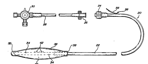
Figure l illustrates a first embodiment of
tonometric catheter 20. The tonometric catheter comprises
a length of suitable tubing 22, one end 32 of which is
closed, and the opposite end of which has a connector such
as a luer-lock 24. Luer-lock 24 is adapted to receive a
complementary fitting 26, which in turn couples through a
second length of tubing 28 to a three-way stopcock 30.
Three-way stopcock 30 may be used to selectively connect
tubing 28 to various sources of irrigation or aspiration.
Other fittings can be used, depending on the particular
application, including those wherein a tonometric catheter
is used in conjunction with an infrared sensing device, a
Raman spectroscopy device, or the like.
Adjacent the closed end 32, tubing 22 is
perforated as at 34. A balloon-like tonometric catheter
membrane 36 is fitted over the closed end so that the
perforations 34 are enclosed, as illustrated. The
tonometric catheter membrane 36 has an internal sleeve
diameter at 38 which forms a tight fit with tubing 22. The
-- 19
WO94/21164 ; r PCT~S94/03018
preferred form of tonometric catheter membrane is
polydimethylsiloxane elastomer. The membrane may be sealed
to the tubing 22 with appropriate adhesive so that the
tonometric catheter membrane is sealed in a closed
relationship to the outer wall of tubing 22, thereby
forming a sampling chamber 40 aa3.acent closed end 32. The
tonometric catheter membrane has a certain elasticity to
allow the membrane to expand when filled with an aspirating
fluid (liquid or gas).
The membrane 36 is preferably constructed such
that at least a portion of it is selectively permeable to
the liquid or gas fluid property of interest. In a
preferred embodiment, it is selectively permeable to carbon
dioxide, and oxygen, so that pCO2 and/or PO2 can be
measured. It is also preferably impermeable to other
materials that would interfere with the desired
measurements, such as proteins and the like. In a highly
preferred embodiment, a gas permeable membrane is employed.
Bonded to either the inner wall (see Figure 2) or
the outer wall of tubing 22 are one or more sensors 42 for
detecting a property indicative of pCO2, PO2, and/or
temperature. Two such sensors are illustrated in Figure l,
bonded to the outside wall of tubing 22 with suitable
adhesive. Figure 2 illustrates the sensor attached to the
inner wall of tubing 22.
In a preferred embodiment, at least a portion of
the tubing, but not necessarily all of it, is made of a CO2
impermeable material, such as those based on polyurethanes,
- 20 -
~ WO94/21164 21522 72 PCT~S94/03018
PVC's, or polyester elastomers derived from the reaction of
dimethylterephtalate l,4-butanediol and ~-hydro-Q-
hydroxypoly (oxytetramethylene). In preferred embodiments,
this material can be PVC or polyurethane.
For purposes of sensing temperature, thermistor
devices are presentiy preferred.
The sampling chamber 40 can be filled with an
aspiration or sampling medium (gaseous or liquid) that is
used to absorb or otherwise provide a means for
incorporating and delivering or measuring the liquid or
gaseous fluids of interest. Such a medium is selected
depending upon many factors, including the properties of
the liquid or gaseous fluids of interest, the type of
sensor 42 employed, and the type of calibration that is
necessary. Such mediums include air, bicarbonate
solutions, bicarbonate-buffered solutions, phosphate-
buffered solutions and saline solution. It might be noted
that gases often behave as fluids and are therefore
frequently considered to be fluids.
As noted above, when the sensor employed does not
require frequent recalibration, the need for the sampling
chamber 40 to be in communication with the proximate end of
the tonometric catheter (that re~ n~ outside the patient)
may be eliminated since no aspiration is needed. However,
in many instances such communication may still be desirable
as aspiration may be required to calibrate the sensor or
sensors, to replace the aspirating or sampling medium with
215 227 2 PCT~S94/03018 ~
a ~resh medium, and to incorporate the gas or gases of
interest.
Another embodiment of the tonometric catheter is
illustrated in Figures 4, 4A and 4B. As illustrated, the
tonometric catheter can be appropr1ately configured to also
serve as a nasogastric tube, either with or without an air
lumen. With reference to Figure 4, the tonometric
catheter 20a comprises a multipassage tubing 62 which
defines three individual passageways or lumens, an optional
air lumen 64, a suction lumen 66 and a tonometric catheter
lumen 68. A tonometric catheter membrane, similar to that
previously described, is attached at an intermediate
location on tubing 62, allowing a portion of the tubing to
extend beyond the end o~ membrane 36 to define the
nasogastric tube 70, or a portion thereof. Tubing 62 is
provided with a plurality of perforations 72 which
communicate between tonometric catheter lumen 68 and the
sampling chamber 40 defined by membrane 36. If desired,
one or more sensors 42 can be included in accordance with
the above teachings, in which case a suitable conductor 56
may be routed through tonometric catheter lumen 68 to exit
at sealed aperture 58.
The nasogastric tube 70 is suitably provided with
a plurality of openings 74 through which the stomach may be
aspirated.
At the opposite end of tubing 62 the tubing
splits to form three separate connections. Optional air
lumen 64 communicates with optional air lumen
~ WO94/21164 215 2 2 7 2 PCT~S94103018
passageway 76, suction lumen connects with suction lumen
passageway 78 and tonometric catheter lumen 68 communicates
with tonometric catheter lumen passageway 80. The
tonometric catheter lumen passageway is fitted with three-
way stopcock 30, similar in function and purpose to thethree-way stopcock 30 described in connection with
Figure l. If desired, a quick connect fitting 82 may be
used to couple the suction lumen passageway 78 with an
aspiration source. As illustrated, the quick connect
fitting preferably has angularly cut ends and a slightly
enlarged midsection, making it easy to insert into the end
of passageway 78 and also into the aspiration hose coupling
(not shown). The enlarged midsection helps form a seal
with the adjoining passageways. Preferably the quick
connect fitting is fabricated of disposable plastic.
Yet another embodiment of the tonometric catheter
is illustrated in Figures 5 and 5A. This embodiment is a
multiple tonometric catheter embodiment employing a
tubing 84 having a plurality of passageways or lumen as
shown in the cross-sectional view of Figure 5A.
Specifically, tubing 84 includes an air lumen 86a which
communicates with the endmost sampling chamber 36a and
three additional tonometric catheter lumens 86b, 86c and
86d, which communicate respectively with sampling chambers
36b, 36c and 36d. As with the other embodiments, each
sampling chamber may be provided with one or more sensors
such as sensors 42. A radiopaque tungsten plug 88 is
positioned within each of the three tonometric catheter
2 15 2 2 7 2 PCT~S94/03018
lumen 86b, 86c and 86d adjacent the distal end of each
sampling chamber, serving to block the remainder of the
tonometric catheter lumen passageway and thereby ensuring
that fluid pressure introduced into each tonometric
catheter lumen will cause t~e associated sampling chamber
to balloon outwardly as requlred during use. Similarly, a
radiopaque tungsten rod 90 is fitted as a plug in the end
of air lumen 86a, serving to terminate the end of the air
lumen passageway. Being radiopaque, the tungsten plugs and
tungsten rod aid in properly positioning the tonometric
catheters by being visible under fluoroscope or x-ray. In
addition, if desired, tubing 84 can be provided with a
radiopaque stripe along all or part of its length.
At the proximal end of tubing 84 the lumen 86a-
86d diverge to define four separate tubes 92a-92d. Each
tube is fitted with a three-way stopcock similar to those
described above. Each sampling connector may optionally be
coded numerically by color, etc. While four approximately
equally spaced sampling chambers have been illustrated in
Figure 5, it will be understood that the invention can be
modified to include a greater or fewer number of sampling
chambers at different spacing as required for a particular
application. It will also be understood that some or all
of the sampling chambers can include one or more sensors
coupled to conductors 56, each preferably routed through
the corresponding lumen passageway.
Referring now to Figure 9, a suitable electronic
monitoring circuit will now be described. In Figure 9, a
- 24 -
~ WO94/21164 21522 72 ~ PCT~S94/03018
pCO2-sensitive CHEMFET semiconductor device 46 has been
shown schematically by the equivalent circuit model
enclosed in dotted lines. The device 46 thus comprises
drain electrode 150, source electrode 152 and reference
electrode 154. The chemically selective system, such as a
membrane system is depicted diagrammatically at 156. The
substrate is grounded as at 158.
Source electrode 152 is coupled to an input lead
of operational amplifier 160 which includes feedback
network diagrammatically depicted at 162. Operational
amplifier 160 senses the drain source current flowing
through device 46 and converts this signal into a voltage
signal which is output on lead 164. The drain source
current changes in accordance with changes in the chemical
system under test. More specifically, as the pCO2 level
changes in the fluid exposed to device 46, the drain source
current changes accordingly. Hence the output voltage
signal on lead 164 is likewise an indication of the pCO2
level of the organ under test. This voltage signal on
lead 164 is coupled to an input of comparator 166 which
also receives a reference voltage Vref, which may be supplied
using a voltage divider network (not shown) or which may
alternatively be provided by a digitally controlled voltage
source 168. The output of comparator 166 is fed to
reference electrode 154 to provide a stable reference bias
voltage. If a digitally controlled voltage source is used,
this reference voltage can be adjusted and calibrated by a
computer circuit yet to be discussed. The voltage ~ignal
- 25 -
WO94/21164 2¦S 22~ 2 PCT~S94/03018 ~
on lead 164 is also fed to an analog to digital
convertor 170, which is in turn coupled to a
microprocessor-based microcomputer 172.
In order to automatically determine the pH of the
wall of the hollow viscus organ under test, a separate gas
analyzer sensor 174 is used to determine the bicarbonate
concentration in the arterial blood of the patient. The
output of sensor 174 is coupled through analog to digital
convertor 176 to microcomputer 172. Microcomputer 172 is
preprogrammed to determine or calculate the pH of the organ
wall using the values provided by analog to digital
convertors 170 and 176. Conversion of pCO2 measurements can
be converted into pH measurements automatically by
microcomputer 172 using various equations and references
disclosed herein or others well-known in the art.
Although many different types of output devices
may be employed, strip chart recorder 178 and CRT
monitor 180 have been illustrated. Strip chart
recorder 178 and monitor 180 are coupled as output devices
to microcomputer 172. Strip chart recorder 178 offers the
advantage of developing an easily readable, permanent
record of the fluctuations in organ wall pH. Monitor 180
offers the advantage of providing digital readout of the pH
value as well as displaying the upper and lower excursions
of pH fluctuation. If desired, microcomputer 172 can be
instructed and/or preprogrammed using keyboard 182 to
compare the instantaneous pH value with doctor-selected
upper and lower alarm limits. If the measured
- 26 -
~ WO94/21164 2 1 5 2 2 7 2 : PCT~S94/03018
instantaneous pH fluctuates outside those limits,
microcomputer 172 can sound an alarm to alert hospital
staff.
While a single semiconductor device 46 has been
illustrated in a~njunction with the electronic circuit of
Figure 9, the circuit may be readily adapted for use with
a plurality of semiconductor devices in order to measure
the pCO2 at different locations substantially
simultaneously. In such an embodiment, the data coming
from each sensor can be fed to a separate I/O port of
microcomputer 172. In the alternative, a single I/O port
can be used with the individual input signals being time
multiplexed.
While some embodiments have been disclosed in
connection with monitoring of the gastrointestinal tract
and the urinary and ureteric tracts it will be appreciated
that its principles are applicable to other hollow internal
organs to monitor tissue or intramucosal pH, pCO2, PO2/
etc., and hence perfusion of those organs. Also while
several detailed constructions for tonometric catheters
have been disclosed, it will be appreciated that other
constructions may be developed which are equally suitable.
The disclosed constructions are presently preferred for the
reason that they are readily fabricated using existing
available materials. Other embodiments may include other,
but equivalent materials for the tonometric catheter
membrane and/or connective tubing. They may also differ in
the specific fabrication details. As an example, the
- 27 -
WO94/21164 2 15 2 ~ 7 2 PCT~S94/030~8 ~
sampling chamber may be eccentric rather than symmetric
about the connective tubing.
As shown, for purposes of illustration, in Figure
lO, the tonometric catheter device according to the present
invention can be employed in com~ination with any number of
different types of urinary ca~heters known to those skilled
in the art. By such an arrangement, the concentrations of
C2/ 2 or other gases of interest, or other parameters, can
be determined and/or monitored, and traditional urinary
catheter operations can be performed, all with a single
combination device.
In Figure lO, the membrane 536 is shown
incorporated into a Foley-type, three-way balloon catheter,
thus making the combination Foley-type urinary and
tonometric catheter a four-way catheter apparatus 520. The
exemplary combination urinary-tonometric catheter includes
a tonometer lumen end 524 in fluid communication with a
sample chamber 540, defined by the membrane 536, in a
manner essentially the same as that described above in
connection with Figure l (with or without a temperature
sensor). The four-way combination catheter apparatus 520
also includes the traditional three-way Foley catheter
components, such as a lumen end 525 in communication with
the Foley balloon 526, for purposes of balloon inflation,
a lumen end 527 for drainage, and a lumen end 528 for
infusing irrigation solutions in order to prevent clot
retention within the bladder, the applications and
functions of all are familiar to those skilled in the art.
- 28 -
~ WO94/21164 2 1 ~ 2 2 7 2 PCT~S94/03018
It should be noted that although the tonometric
catheter arrangement of Figure 1 is shown in Figure 10,
merely for purposes of exemplary illustration, in
conjunction with a three-way Foley-type urinary catheter,
one skilled in ~e art will readily recognize that any of
the tonometric catheter embodiments described and
illustrated herein can be employed in combination with such
a Foley-type urinary catheter, as well as with other
familiar types of urinary catheters, such as a conical tip
urethral catheter having a single eye, a Robinson urethral
catheter, a whistle-lip urethral catheter, a Coudé hollow
olive-tip catheter, Macelot self-retaining four-wing or
two-wing catheter, a Pezzer self-retaining drain, open-end
head (used for cystotomy drainage), or any of a number of
well-known urinary catheter types. See Urol ogy 5th ed .,
W.B. Sanders ed. Vol. 1, p. 512 (1986).
Another embodiment of the tonometric catheter is
illustrated in Figures 11 and llA. As illustrated, the
tonometric catheter is appropriately configured to also
serve as a urinary or ureteric catheter, either with or
without suction, which optionally employs sensors. With
reference to Figures 11 and llA, the tonometric
catheter 220 comprises a multipassage tubing 262 which
defines three individual noncommunicating (between each
other) passageways or lumens, an optional irrigation
lumen 264, a drainage or suction lumen 266 and a tonometric
catheter lumen 268. A tonometric catheter membrane,
similar to that previously described, is attached at a
WO94/21164 2~522~ 2 PCT~S94/03018
distal location on tubing 262, allowing an intermediate
portion of the tubing not extending beyond the end of
membrane 236 to define the uretary or ureteric
catheter 270. Tubing 262 is provided with a plurality of
perforations 272 which communi¢ate between tonometric
catheter lumen 268 and the sampling chamber 240 defined by
membrane 236. If desired, one or more sensors 242 can be
included in accordance with the above teachings, in which
case a suitable conductor 256 may be routed through
tonometric catheter lumen 268 to exit at sealed
aperture 258.
The urinary catheter or ureteric catheter
portion 270 is suitably provided with a plurality of
openings 274 through which the bladder or ureters may be
aspirated or irrigated.
At the opposite end of tubing 262 the tubing
splits to form three separate connections. Irrigation
lumen 264 optionally communicates with irrigation
passageway 276, urinary lumen connects with suction or
drainage lumen passageway 278 and tonometric catheter
lumen 268 communicates with tonometric catheter lumen
passageway 280. The tonometric catheter lumen passageway
is fitted with three-way stopcock 230, similar in function
and purpose to the three-way stopcock 30 described in
connection with Figure l. If desired, a quick connect
fitting 82 as seen in Figure 4 may be used to couple the
suction urinary passageway 278 with an aspiration source.
As illustrated, the quick connect fitting preferably has
- 30 -
~ W094/21164 ~ 215 2 2 7 2 PCT~S94/03018
angularly cut ends and a slightly enlarged midsection,
making it easy to insert into the end of passageway 278 and
also into the aspirat1on hose coupling (not shown). The
enlarged midsection helps form a seal with the adjoining
passageways. Preferably the quick connect fitting is
fabricated of disposable plastic.
Yet another embodiment of the urinary
catheter/tonometric catheter combination illustrated in
Figures ll and llA may employ a multiple tonometric
catheter embodiment employing a tubing having a plurality
of passageways or lumen as shown in the cross-sectional
view of Figure 5A.
In another embodiment of the present invention,
a tonometric catheter may be adopted to deliver a
pharmaceutically-active agent, either for systemic, local
or topical activity, or a combination thereof. For
example, an additional lumen, such as the
irrigation/aspiration lumen 264 shown in Figure ll and llA,
may be used to deliver an active agent. In another
embodiment, a portion of the device may be modified so as
to provide sustained release of the active agent of
interest.
Thus, for example, the problems of nosocomial
infection associated with catheter insertion can be
overcome by incorporating an antimicrobial agent into at
least a portion of the polymeric material used to
manufacture the tonometric catheter, or by coating at least
a portion of the device with a sustained release
- 31 -
WO94/21164 2~s22~ 2 PCT~S94/03018 ~
composition or bacteriostatic coating, or by delivering the
antimicrobial via the tonometric catheter. Such
modifications are well known to those skilled in the art.
See U.S. Patent No. 4,677,14~, incorporated herein by
reference.
Classes of useful agents include bacteriostatic
coatings, antimicrobial agents, nonsteroidal anti-
inflammatory agents, topical anesthetics, topical
vasodilators, metabolic suppressants, and other agents that
could be delivered for absorption at the sites of the
tonometric catheter.
In still other embodiments, conventional gas
analyzers may be employed externally. A device such as
that shown in Figure l (or any of the exemplary catheter
devices described herein) may be used in combination with
a pump or aspiration means (not shown) for continuous or
regular intermittent aspiration of a sample of the
aspirating liquid or medium that is used to fill the
sampling chamber 40. The sample removed by pump or
aspiration means via attachment to the luer-lock 24 can be
optionally designed so that the sample aspirated at each
sampling interval can be brought in contact with an
exterior, separate gas analyzing means or sensor (not
shown) to determine the PO2, PCO2 and/or the like, of the
sample. Such automatic sampling can be conducted employing
a system as shown in Figure 12. In the assembly a sampling
system employs a personal computer to conduct evaluations
- 32 -
~ WO94121164 215 2 2 7 2 - PCT~S94/03018
and analysis of the samples withdrawn from the tonometric
catheter 299.
Pump 203 is loaded with the sampling or
aspirating medium, such as saline or air. Next, valve 201
is activated to withdraw a desired amount of the sampling
fluid. The valve 201 is deactivated and pump 203 is used
to infuse the sampling chamber of the tonometric
catheter 299 using a calibrated amount or, optionally,
until a predetermined pressure is sensed by a pressure
transducer 215. The sampling fluid or medium is allowed to
come to equilibrium with the wall of the organ or area of
interest. Next the "dead space," i.e., the area of the
lumen filled with the sampling fluid that is not in
equilibrium, is removed by activating valve 205, activating
pump 207, activating valve 209 and infusing pump 207; the
waste 219 is discarded. A gaseous sample for analysis can
then be withdrawn by deactivating valve 209, activating
pump 207 to then deliver the gaseous sample to an analyzer
such as an infrared or a Raman gas analyzer (not shown)
that provides data from the sample to the PC 217, and the
evaluation is conducted as described herein.
The sample gas analyzer or a separate gas
analyzer may be optionally employed to determine the
bicarbonate concentration in the arterial blood of the
patient, as described above. Such option is depicted
schematically in Figure 12, wherein a blood gas analyzer or
monitor 250 is provided, with its data output signal being
interfaced with the processing system 217. Such blood gas
WO94121164 2 ~ S 2 ~ ~ 2 . PCT~S94/03018 ~
analyzer continuously monitors the patient's intraarterial
pCO2, pH, PO2, or other parameters of interest by way of a
sensor, such as a fiberoptic sensor placed into the
patient's artery. Examples of commercial available blood
gas analyzers and sensor co~ponents include those marketed
by Puritan-Bennett (PB 3300, see Lundsen, T. et al., ~.
Clin. Monlt. 10:59-66 (1994), herein incorporated by
reference) or by Biomedical Sensors Ltd. (Pfizer)).
These systems (providing continuous arterial pCO2,
pH, and bicarbonate values) can also be interfaced into the
tonometric pCO2systems using infrared or Raman spectroscopy
technology (discussed herein) to provide an actual value of
intramucosal pH, as well as pCO2-gap and pH-gap measurements
each time a tonometer pCO2 or PO2 measurement is taken, thus
providing more timely trend values for these parameters.
This greatly facilitates interpretation of these
measurements, since regional (tonometer pCO2 and
intramucosal pH) and systemic (arterial pCO2 and pH) can be
compared rapidly and directly. It should further be noted
that such an optional blood gas monitoring/analyzing
interface can be advantageously employed whether liquid or
gaseous tonometric sampling is used.
It has also been discovered that the pH of venous
blood provides an excellent measure of the adequacy of
tissue oxygenation of the whole body or organs, including
solid organs, comparable to that achieved in hollow viscus
organs by the method described herein, as well as that
described in the above-mentioned, commonly-assigned
- 34 -
~ WO94/21164 21 5 2 2 7 2 . PCT~S94/03018
applications that relate to the use of a tonometric
catheter to determine the adequacy of tissue oxygenation
via the measurement of the pH of the wall of a hollow,
viscus organ.
In numerous clinical settings it is now common to
monitor the carbon dioxide concentration of the arterial
blood of patients, particularly those who are critically
ill or under anesthesia; this measurement has been
determined to bear a usually predictable relationship to
intramucosal pH. One of the most common non-invasive
techniques for measuring arterial CO2 is doing so indirectly
by measuring the CO2 concentration of the last gas expired
from a patient (so called "end-tidal") during normal
respiration. The arterial CO2 concentration is then
calculated by employing the known correlation between the
end-tidal pCO2 and pCO2 of the arterial blood.
It has been discovered in another aspect of the
present invention that end-tidal CO2 (as well as the
underlying correlation between end-tidal CO2 and the pCO2 of
arterial blood) may also be useful in making clinical
determination of the condition of an organ of interest when
the end-tidal CO2 is compared and contrasted with the pCO2
of air aspirated from a tonometric catheter having a walled
sampling chamber inserted into an organ of interest. These
measurements having the added convenience of both being
measurable by IR or Raman gas analyzers.
However, in order to fully appreciate this, a
detailed understanding of the general tonometric method is
2~S2~ 2 PCT~S94/03018 ~
useful. This background is helpful primarily for the
skilled artisan to fully appreciate the relationship of
moving from the general tonometric method (which employs
PCO2 associated with the wall of the organ of interest and
the bicarbonate concentrations of arterial blood) to even
more indirect but useful measurements.
In accordance with one preferred embodiment of
the present invention, the condition of an organ of
interest is determined in a patient in need of such
determination when the pCO2 associated with the wall of the
organ of interest is sampled and compared to substantially
contemporaneous arterial or venous pCO2 values or, in a
highly preferred embodiment, end-tidal pC2 value(s); the
PCO2 of the wall of the organ may also be compared to:
venous or arterial pCO2 or pH; mixed venous bicarbonate
values; transcutaneous pCO2; arterial oxygenation
(saturation), arterial PO2, umbilical blood gases, capillary
blood gases, and the like.
While not intending to be bound by theory, the
following is offered to put these aspects and embodiments
of the present invention in proper context.
The assumptions upon which the indirect
measurement of intramucosal pH (pHi) are based are valid in
normally perfused tissues. In these circumstances, the
indirect measurement of intramucosal pH is identical to
that measured directly in the submucosal space with a
microprobe.
~ WO94/21164 215 22 7 2 PCT~S94/03018
The indirect measurement of intramucosal pH falls
in parallel with the pH made directly in the submucosal
space when an intramucosal acidosis is induced by
endotoxemia, low-flow or no-flow. In those circumstances
in which the intramucosal acidosis in induced by endotoxin
and flow to the gut is maintained at control levels the
measurements are in close agreement (r=0.945). When
induced by low-flow and especially no-flow the indirect
measurements underestimate the severity of acidosis present
in the submucosal space. The disparity between indirect
and direct measurements observed in low-flow and no-flow
states disappears when blood flow is reestab,lished and the
pH is allowed to return towards normality. Inspection of
the twenty-minute values obtained in Antonsson et al's
study reveals that the degree of dissociation observed
between indirect and direct measurements is a linear
function of the rate of change in intramucosal pH induced.
An additional primary assumption upon which the
validity of the tonometric measurement of the adequacy of
tissue oxygenation is that the bicarbonate concentration in
tissue fluid is the same as that being delivered to it in
arterial blood. It has been postulated that the
dissociation between calculated and measured~pH in low-flow
and especially no-flow states may be due to a dissociation
between arterial and interstitial bicarbonate induced by
the buffering of metabolic acids by tissue bicarbonate.
The hypothesis does not account for the law of
mass action which dictates that the fall in bicarbonate
- 37 -
WO94/211642~S 2 21 2 ` PCT~S94/030~8
concentration induced by the addition of a fixed acid load
to a "closed system" from which CO2 cannot escape, such as
the extracellular fluid compartment, is inhibited by the
accumulation of CO2. The addition of even large amounts of
fixed acid to a "closed system" does not produce a
significant reduction in bicarbonate concentration but does
produce a significant rise in pCO2. A fall in bicarbonate
occurs only when venous blood enters the pulmonary
circulation, an "open system" from which the CO2 added to
the venous blood by the buffering of the fixed acid load in
the dysoxic tissue bed is able to escape. The fall in
arterial bicarbonate thus induced causes, the tissue
bicarbonate to fall by equilibration with the lowered
bicarbonate concentration in arterial blood returning to
the tissue bed. The fall in arterial bicarbonate induced
by the escape of CO2 from the lungs cannot cause a reduction
in tissue bicarbonate concentration in a no-flow state for
it is unable to enter the tissue bed.
The tissue bicarbonate should be the same as that
in arterial bicarbonate perfusing the tissue bed in all
circumstances except perhaps very transiently after a
sudden and large change in arterial bicarbonate induced by
an intravenous bolus of bicarbonate or sudden change in
pulmonary ventilation.
As a precaution, however, it is wise to wait
until the arterial bicarbonate has been stable for some 10
to 15 or better yet 30 minutes before measuring the
- 38 -
~ WO94/21164 21 S 2 2 7 2 PCT~S94/03018
intramucosal pH after an intravenous bolus bicarbonate or
sudden changes in ventilation regimes.
It is therefore suggested that the primary
assumption upon which tonometric measurement of
intramucosal pH ;is based, namely that the tissue
bicarbonate is the same as that in arterial blood, is valid
in many relevant clinical settings, including those in
which the dissociation between measured and calculated
intramucosal pH was greatest. The indirect measurement of
intramucosal pH appears to be an accurate measure of the pH
in interstitial fluid in the most superficial layers of the
intestinal mucosa especially in those circumstances in
which the measurement is of greatest value, namely patients
who appear by all conventional criteria to be adequately
resuscitated. The only circumstance in which the
measurement might be inaccurate for an extended period is
a no-flow state. In this circumstance, the indirect
measurement is so abnormal that the presence of the
intramucosal acidosis should not be missed even if there is
a large discrepancy between actual and assumed
measurements. Transient inaccuracies may be expected
following an intravenous bolus of bicarbonate or sudden
change in pulmonary ventilation.
STOICHIOMETRIC ANALYSIS OF DETERMINANTS OF TISSUE ACIDOSIS
During aerobic metabolism the pH of tissue fluid
is determined by the bicarbonate concentration in tissue
fluid, the CO2 released by oxidative phosphorylation, and
- 39 -
2ls~2~2
WO94121164 PCT~S94/03018
the balance between ATP hydrolysis and resynthesis. In
gastric glands the intracellular pH is the same as the
extracellular pH in acidotic states. The pH of the
extracellular fluid (ECF) is determined by the amount of
metabolic acid present and the ability of the ECF to buffer
the acid. The pCO2 attained following the buffering of a
volatile (H2CO3 from oxidative phosphorylation) or fixed
acid load (protons from ATP hydrolysis) in a closed system,
such as the ECF, may be calculated in the manner described
by Gattinoni and Feriani.
In normoxic tissues 6 mmol of CO2 are produced for
every mmol of glucose consumed in the generation of 38 mmol
ATP. 13.5~ of a volatile carbonic acid load added to ECF
remains after being buffered by proteins and determines the
pCO2 present in the ECF. Assuming that the bicarbonate
concentration in ECF is 25 mEq/1 the metabolism of one mM
glucose gives rise to a pCO2 of 27 mmHg (6x 0.135/0.03). In
normoxic and resting healthy subjects with a tissue
bicarbonate of 25 mEqll the pCO2, determined tonometrically,
is 40 mmHg and the intramucosal pH 7.40. If it is assumed
that the protons released by ATP hydrolysis are exactly
balanced by the protons consumed by ATP resynthesis in
oxidative phosphorylation then the aerobic metabolism of
1.48 mM glucose is required to generate the volatile
carbonic acid necessary to attain the pCO2 of 40 mmHg (27 x
1.48 = 40mmHg) and pH of 7.40 found in normoxic ECF when
the tissue bicarbonate concentration is 25 mEq/1.
- 40 -
~ WO94/21164 215 2 2 ~ 2 PCT~S94/03018
The pCO2 attained from the buffering of the
volatile acids released into normoxic ECF in a tissue bed
should increase as the metabolic rate increases, the
increased demand for oxygen in the absence of replenishment
by flowing blood being met exclusively by an increase in
oxygen extraction ratio. A rise in metabolic rate of the
magnitude seen in an exercising athlete, which may be as
great as 900~, can be expected to cause a rise in
equilibrium pCO2 and hence fall in intramucosal pH in
normoxic tissues. The magnitude of the fall in pH induced
by the rise in pCO2 is offset by the rise in tissue
bicarbonate also induced by the buffering of carbonic acid
(a volatile acid). The rise in metabolic rate observed in
the critically ill is a fraction of that seen in an
exercising athlete. Furthermore the oxygen extraction
ratio is unchanged and more often decreased in septic
patients who exhibit the highest metabolic rate in the
critically ill. In any event, the increased metabolic
demand for oxygen in the critically ill, especially in
those who are septic, is primarily met by an increase in
oxygen delivery, oxygen delivery being "demand-dependent~
in these circumstances. The pCO2 attained by the buffering
of the volatile acid load generated in normoxic ECF should
not, therefore, be significantly influenced by changes in
~ 25 metabolic rate of the order encountered in the critically
ill .
Aerobic glycolysis and associated generation of
CO2 by oxidative phosphorylation decreases in dysoxic states
W094/2ll64 2~522~ 2 ~ PCT~S94/03018 ~
as the availability of oxygen relative to demand decreases.
Thus the fall in tissue pH in severely dysoxic states is
due almost exclusively to the protons released by adenine
nucleotide hydrolysis and thelr interaction with the body
buffers.
If it is assumed~that the intramucosal pCO2 and pH
are solely determined by the amount of volatile and fixed
metabolic acid being buffered in the ECF at the time, the
intramucosal pH can be expected to remain constant as
oxygen delivery is reduced with or without a reduction in
blood flow until the point at which supply-dependency or
dysoxia develops. Below this point the pCO2 in ECF should
rise and the intramucosal pH fall as the contributions by
aerobic metabolism to volatile acid decreases and by
anaerobic metabolism to proton release increases with
further reductions in oxygen delivery.
Intramucosal ~H
The buffering of the protons by tissue
bicarbonate in dysoxic states causes the pCO2 to rise. As
the bicarbonate concentrations in a "closed system", such
as the ECF, is not significantly reduced by the addition of
a fixed acid load, the fall in pH must be inversely related
to the rise in log pCO2 at any given concentration of tissue
bicarbonate. The constant bicarbonate line at 25 mEq/l on
a pH-log pCO2 diagram will show that the pCO2 in normoxic
ECF at a point A to be 40 mmHg and the pH to be 7.40. The
bicarbonate line moves to the right as the equilibrium pCO2
~ WO94121164 2 I 5 2 2 7 2 PCT~S94/03018
rises above 40 mmHg to a point B in dysoxic states and the
tissue pH falls below 7.40. The pH in the dysoxic state
may be determined by extrapolation from the pCO2 intercept
on the constant bic~arbonate line at 25 mEq/l.
The fall in pH induced by dysoxia alone in a
tissue with a known bicarbonate concentration may be
computed from the difference between the pH in the normoxic
and dysoxic states determined from the same constant
bicarbonate line (pH-gap), log of the ratio piCO2/paCO2 (B -
A) or their antilog equivalents (pCO2-gap and H+-gap). It
will be appreciated that pCO2-gap is defined as pCO2-gap =
piCO2-paCO2, and H+-gap = Ha+-Hi+. These determinations of the
magnitude in fall in pH induced by dysoxia are all
dependent upon the assumption that the bicarbonate
concentration in the dysoxic ECF is the same as that
present in normoxic ECF. If it is assumed that the pCO2 in
normoxic ECF is the same as that in arterial blood (paCO2)
and the tissue pCO2 in dysoxic ECF is the same as the
intramucosal pCO2 measured from the lumen of the gut with a
walled sampling chamber tonometer (piCO2) then the actual pH
in dysoxic ECF may be calculated from the following formula
(with pHa = pH of arterial blood):
Intramucosal pH = pHa - (log piCO2 - log paCO2)
= PHa - log piCO2/paCO2
and displayed in a perceptible form, such as human readable
or audible form, or machine readable form. Thus, by
relating the differences between measured values, wherein
the term "difference" does not necessarily mean an
- 43 -
wog4t21164 2~S22~ 2 PCT~S94/03018 ~
arithmetic difference, but refers generally to a comparison
of measurements, for example by employing functions and
formulas, important biological information may be obtained.
CLINICAL IMPLICATIONS
The indirect m~surement of intramucosal pH
provides an accurate diagnostic test for the presence of
macroscopic and clinical evidence of gastric, small
intestinal and large intestinal ischemia in patients. The
sensitivity of the intramucosal pH as a diagnostic test for
gastric ischemia in man is reported to be 95~ and the
specificity lOO~. For severe ischemic colitis after
abdominal aortic surgery the sensitivity is reported to be
lOO~ and the specificity 87~. Of particular relevance to
patients who are critically ill is the inability of those
with an intramucosal acidosis to secrete acid in response
to pentagastrin. Those patients who have a normal gastric
intramucosal pH secrete acid in response to this stimulus.
It has been suggested that the inability to secrete acid in
patients with an intramucosal acidosis may be due to an
energy deficit secondary to a dysoxic state. An energy
deficit is a known cause of stress ulceration in animals
and an impairment of gastric mucosal oxygenation the likely
cause of stress ulceration in patients.
The gastric intramucosal pH, measured following
the administration of an H2-receptor antagonist to avoid
confounding influence of the back diffusion of acid and/or
CO2, is inversely related to the hepatic venous lactate
~ WO94/21164 21 522 72 PCT~S94/03018
concentrations in patients having cardiac surgery (r=-0.71)
and correlates closely with this and other indices of
splanchnic tissue oxygenation (r=0.92). The gastric
intramucosal pH provides, therefore, an index of the
adequacy of splanchnic tissue oxygenation.
The gastri~ intramucosal pH correlates very well
and inversely with systemic blood lactate when it is
abnormally elevated. In many circumstances, however, blood
lactate is normal when the intramucosal pH is low and no
correlation between the variables can be demonstrated.
Indeed a fall in gastric intramucosal pH may precede a rise
in blood lactate in a deteriorating patient by many hours
or even days. Changes in intramucosal pH influence the pH
dependent enzymes regulating carrier mediated afflux of
lactate from muscle and the pH dependent enzyme
phosphofructokinase which regulates the rate of anaerobic
glycolysis. In addition blood lactate is the net effect of
both production by anaerobic glycolysis and consumption by
tissues such as the myocardium. The overall correlation
between the two variables is thus rather poor (r=-0.40) but
nevertheless statistically significant (p=0.026). Thus in
addition to providing indices of gastric mucosal and
splanchnic tissue oxygenation the indirect measurement of
gastric intramucosal pH provides an index of the adequacy
25 of global tissue oxygenation.
The indirect measurement of intramucosal pH
provides a measure of the adequacy of tissue oxygenation in
the most superficial layer of the mucosa, a region of the
- 45 -
WO94/21164 2~$~ PCT~S94/03018
gut rendered relatively hypoxic by the counter current
exchange system within the mucosal vasculature and hence
especially sensitive to alterations in the adequacy of
tissue oxygenation. It also provides a measure of the
adequacy of tissue oxygenation,l~ a region of the body that
is among the first to deve~op an inadequacy of tissue
oxygenation or dysoxia in shock and the last to be restored
to normality with resuscitation. Splanchnic vasculature is
selectively constricted by the endogenous vasoconstrictors
released in shock. For these reasons a fall in
intramucosal pH may occur hours to days in advance of any
other conventional evidence of an inadequacy of tissue
oxygenation, most specifically arterial acidosis, elevation
in blood lactate, hypotension and oliguria.
It is concluded that the indirect measurement of
gastric intramucosal pH provides a sensitive measure of the
adequacy of splanchnic and even global tissue oxygenation
in patients in addition to providing an index of the
adequacy of superficial gastric mucosal oxygenation.
Correlations with acid-base balance and clinical events
The indirect measurement of gastric intramucosal
pH may correlate very closely with the arterial pH (r=
0.67) and other systemic indices of a disturbance in acid-
base balance such as arterial bicarbonate (r=0.50), the
base deficit in extracellular fluid (r=0.60) and base
deficit in blood (r=0.63). This is consistent with the
deduction that gastric intramucosal pH provides an index of
the balance between the protons released by ATP hydrolysis
- 46 -
~ WO94/21164 215 2 2 7 2 PCT~S94/03018
and consumed in the resynthesis of ATP by oxidative
phosphorylation. As with global measurements of blood
lactate changes in systemic acid-base balance provide a
very dampened signal of disturbances in the adequacy of
tissue oxygenation. A fall in intramucosal pH will often
precede a fall in arterial pH by hours or even days.
The predictive value of measurements of gastric
intramucosal pH for outcome are superior to those of the
systemic measures of acid-base balance. Maynard et al, for
example, compared the predictive value of measurement of
gastric intramucosal pH with those of arterial pH and base
excess for death in ICU patients. The likelihood ratio for
intramucosal pH was 2.32, for arterial pH l.52 and base
excess l.47. Logistic regression showed only intramucosal
pH to independently predict outcome. In Boyd et al's
study, the gastric intràmucosal pH was likewise of better
predictive value for outcome than base excess. Clinical
experience has shown that changes in gastric intramucosal
pH correlate far better with the passage of clinical events
than either the arterial pH or base excess. Indeed
abnormalities in these systemic measures of acid-base
imbalance will often occur only as the intramucosal
acidosis is being reversed and the patient's condition is
improving.
Reperfusion after the low-flow and particularly
no-flow states induced in Antonsson et al's validation
study in pigs caused the intramucosal pH to rise and the
arterial bicarbonate to fall. Similarly in patients the
- 47 -
WO94/21164 2~s22~ 2 PCT~S94/03018
reversal of a severe intramucosal acidosis may be
accompanied by a fall in arterial pH and base excess of
abnormally low levels. These observations are consistent
with the consequences described above of reestablishing
perfusion in a dysoxic tissue bed in patients. The pCO2 in
the venous effluent leaving the dysoxic tissue bed is
elevated but the bicarbonate concentration is not
significantly reduced by the buffering of the fixed acid in
the tissue bed. The bicarbonate is only reduced by the loss
of CO2 during the passage of the venous effluent through the
pulmonary circulation (an open system). As dissociation
between the direction of change in the intramucosal and
systemic pH is to be expected after flow is reestablished
through a dysoxic tissue bed.
Intramucosal ~H as a therapeutic tarqet
"Gut-directed" and "intramucosal pH-directed"
therapies may improve outcome. These therapies use a
normal intrmucosal pH or intramucosal pH greater than 7.35
as an additional therapeutic goal in the resuscitation of
patients. This pH was chosen to ensure the pH was
maintained well within the normal limits reported for
normal subjects. The normal limits may, however, differ
from institution to institution with the use of saline and
different blood gas analyzers, a problem solved by the air
sampling medium (IR or Raman pCO2) analysis embodiments of
the present invention. It is furthermore possible that an
end-point other than 7.35 might be more appropriate.
- 48 -
~ WO94/21164 21~ 2;2 ~.~ PCT~S94/03018
Values such as 7.25; 7.30; 7.35; 7.37 etc. may also be
useful.
While it is clearly desirable to maintain a
normoxic state by maintaining the pH-gap at zero, it is not
necessarily desirable to maintain the pHi at normal levels.
There is a considerable body of evidence indicating that
mild degrees of cellular acidosis protect cells in anoxia
and ischemia possibly by limiting the activity of the
autolytic enzymes responsible for cell injury and death.
A cellular acidosis may in addition facilitate carrier-
mediated afflux of lactate from cells and bring the
intracellular pH to an optimal range for anaerobic
glycolysis during anaerobic metabolism. Furthermore the
addition of bicarbonate to the extracellular environment
attenuates the fall in intracellular pH during ATP
depletion and accelerates cell death. The presence of an
actual intramucosal acidosis may, therefore, be desirable
and efforts to correct a metabolic acidosis with
bicarbonate potentially harmful. Indeed the practice of
correcting a metabolic acidosis induced by a cardiac arrest
by the administration of bicarbonate is no longer
recommended.
It is concluded that acid-base balance is
intimately related to the adequacy of tissue oxygenation in
so far as it relates to the balance between the protons
released by ATP hydrolysis and consumed by ATP synthesis
from oxidative phosphorylation. The intramucosal pH is
determined by the pCO2 attained following and buffering of
- 49 -
WO94/21164 2~s22~ 2 PCT~S94/03018 ~
the metabolic acid released into the ECF and the
bicarbonate concentration in ECF at the time--- the "buffer
hypothesis". The intramucosal pH is related to blood flow
only in so far as it relates to the adequacy of tissue
oxygenation. The assumption that tissue bicarbonate is the
same as that in arterial bicarbonate is only valid in the
absence of the generation of an alkaline tide and
associated secretion of acid. The indirect measurement of
gastric intramucosal pH is the sum of the effects of
several determinants of an intramucosal acidosis. It is
relevant to activity of pH-dependent enzymes especially as
they might relate to cellular injury in dysoxic states. By
eliminating the confounding effects of disturbances in
systemic acid-base balance the pH-gap provides a measure of
the acidosis attributable to an imbalance between ATP
hydrolysis and resynthesis, or degree of dysoxia present.
Systemic measures of acid-base balance may be dissociated
from the adequacy of tissue oxygenation upon reperfusion of
a dysoxic tissue bed and correlate poorly with clinical
events relative to the measurement of gastric intramucosal
pH.
In light of all the above, it will be appreciated
that one series of embodiments of the present methods
relate to the use of arterial carbon dioxide concentrations
(measured directly or indirectly, preferably as an end-
tidal carbon dioxide value) as a predictive indicator of
the pH of the most superficial layer of the mucosa of the
- 50 -
~ WO94/21164 21 S 2 2 7 ~ PCT~S94/03018
wall of an internal solid organ, particularly the gut. In
recognizing that
pHi = pHa + log paCO2
piCO2
and that paCO2 is approximately equal to PC02-end tidal/ thus
pHi = pHa + log pCO2endtidal
piCO2
Either or both of these may be employed.
In accordance with the practice of the methods of
the present invention, the pCO2 of the wall of the organ is
determined. This is preferably done by inserting a
15 tonometric catheter with a walled sampling chamber into or
adjacent the organ of interest. The sampling chamber is
filled with a gaseous or liquid sampling medium such as air
or saline. The sampling medium is allowed to come to
equilibrium (equilibrate) with the area so that the pCO2
20 concentration of the sampling medium reflects the pCO2 of
the superficial layer of the mucosa of the organ of
interest. The pCO2 concentration of the sampling medium is
determined, giving piCO2.
In conjunction with the determination of the pCO2
25 of the mucosa, the carbon dioxide concentration in arterial
(paCO2) or venous blood is determined directly or
indirectly. (A highly preferred indirect measure is end-
c tidal pCO2, or pCo2-end tidal) . The two values (e.g., paCO2 and
piCO2) are then subjected to a nomogram, such as those
30 described in equations above, to determine for example, a
pHi value or pH-gap. The time integrated pH-gap can be
- 51 -
WO94/211~ 2~ 2 PCT~S94/03018
u^sed as a parameter for assessing the cumulative effects of
tissue damage over time. The time differentiated PiCO2 can
be used as a parameter to determine the rate and direction
of change in PiCO2 which may be useful in situations when
PiCO2 may change rapidly (e.g, ventilation changes during
ventilator weaving).
In a highly preferred embodiment, the sampling
medium for the walled sampling chamber is air. The air is
aspirated to an IR or Raman spectrometer. In combination,
the measurement of end-tidal pCO2 is employed as a
substitute for the arterial paCO2. The end-tidal
respiratory air is likewise aspirated to an IR or Raman
spectrometer. Both gas analyzing devices are controlled by
a microcomputer, which also affects the selected nomogram
or nomograms which compare the pCO2 of the wall of the organ
(gut) with the end-tidal pCO2 value. The gas analyzing
devices may operate on a single channel, or via multiple
channels.
Additional detection techniques may be performed
on the air aspirated from the patient, either via
respiration or from the tonometric walled sampling chamber.
For example, IR or Raman analyses may be performed to
determine the level of anesthetic gases, such as N2O. The
results of the nomogram are displayed on a monitor (not
shown) in human or machine readable form.
In a highly preferred embodiment, the operation
of one example of an infrared gas analyzer is controlled by
a microcomputer. The microcomputer itself is not, by
- 52 -
~ WO94121164 215 2 2 7 2 PCT~S94/03018
itself, part of the present invention. For this reason and
because one skilled in the relevant arts could routinely
program a general purpose computer to follow the routines
required for this application, the microcomputer will not
be described in detail herein. (See the U.S. patents
incorporated herein by reference.)
Referring now to Figure 13A, a gas analyzer or
detector 320a is shown in accord with the principles of the
present invention. Analyzer 320a is specifically designed
to monitor the concentration of carbon dioxide in the
exhalations of a medical patient--e.g., a patient being
ventilated during a surgical procedure.
The major components of the infrared gas analyzer
320a are a powered unit 322a and a sensor assembly 324a of
a transducer head 326a and an airway adapter 328a. The
transducer head 326a is connected to the unit 322a of the
gas analyzer 320a by a conventional electrical cable 330a.
In the application of the invention depicted in
Figure 13A, the gas analyzer 320a is employed to measure
fluid parameters of interest, similar to the apparatuses
shown and discussed above, except that a gaseous sampling
medium, such as air, is conveyed, either manually or
automatically, as shown above, and analyzed by the infrared
sensor assembly 324a, where the sampling medium is conveyed
to the assembly 324a via one of the above described
tonometric catheter devices. This information can be
effectively employed by medical personnel to monitor the
? 1~Z~2
WO 94/21164 PCT/US94/03018
C~n~;~;~ o$ a patient's internal organ more accurately and
mo're quickly than before.
Figure 13A depicts an in-stream type of infrared
gas analyzer, shown merely for purposes of illustration,
but one skilled in the art wi~ll appreciate that the same
principles apply to the use of a side-stream type IR gas
analyzer, such as that shown in Figure 13B.
Figure 13B depicts a side-stream infrared gas
analyzer, similar to that of Figure 13A, except that the
infrared sensor is located inside the powered unit 322b.
Also, sampling line 331b is used to convey a continuous
gaseous sample from the patient by way of an airway adapter
333b. The gaseous sample is conveyed from the sampling
line 331b through a water trap 335b (in order to remove
condensate) to the sensor located in the powered unit 322b.
Figure 13C schematically depicts an infrared
sensor, which can be used in the infrared sensor assembly
324a of Figure 13A, or in the powered unit 322b of Figure
13B. In Figure 13C, an infrared light source 337c directs
an infrared beam through a gas sample cell 339c (located in
sensor assembly 324a of Figure 13A, or in powered unit 322b
of Figure 13B), which contains the gas sample, to a
detector 341c, which directs its output signal to a signal
processor 343c.
It will be appreciated that Raman spectrometers
(gas analyzers) offer advantages over IR analyzers and may
be employed in the present invention. A Raman spectrometer
is outlined and discussed in Westenskow, D.R., et al.,
~ WO94/21164 21 5 2 2 7 2 PCT~S94/03018
Anesthesiology 70 :350-355 tl989) and Westenskow, D.R. et
al., Biomed. Inst . & Technol . November/December:485-489
(1989), herein incorporated by reference. It will also be
appreciated that a multichannel Raman in combination with
multiple catheters is also contemplated by the present
invention. See Niemczyk, T.M. et al., Laser Focus World
March:85-98 (1993), herein incorporated by reference. The
use of the combination of a tonometric catheter and a Raman
spectrometer allow the measurement of oxygen gas; nitrogen
gas; water; N2O and other anesthetic agents such as
halothane, enflurane, isoflurane and sevoflurane, all of
which exhibit Raman scattering. Raman devices not only
measure pCO2 more accurately, but can measure N2, 2 and H2O
directly. This may reduce the potential error associated
with certain IR techniques, especially where other
substances (N2Oi 2; H2O) may effect the IR pCO2 measurement
due to errors from overlapping wavelengths. The ability to
measure 2 directly with a Raman system instead of employing
two sensors to measure 2 and CO2 as with the IR system is
also important, especially with tonometric samples wherein
the volume of sample may not be sufficient for two
measurements.
By measuring 2 and N2, air leaks may be detected
and detection is highly accurate. For example,
equilibrated tonometric samples could be compared to the
air concentration of 2 and N2. Any samples that "look like
air" to the system would thus be discarded. This may be
especially useful in situations where a pCO2 in the stomach
WO94/21164 PCT~S94/03018 ~
2~ S22~ 2
is, igh (e.g. 80 mmHg) and mixing with air during high
suction from a nasogastric tube may reduce the CO2 level,
but not to zero. In a Raman system, this sample would be
detected as an air leak. However, in an IR system an
inaccurate pCO2 reading may result because the means for
detecting the air leak are- based primarily on the pCO2
reading
Another important advantage of the Raman
spectrometer is the ability to employ a fiberoptic probe
within the sampling chamber 40 of the tonometric catheter.
The fiber optic probe may also be used in combination with
the catheter such that the tip of the fiberoptic probe
resides inside the balloon of the catheter. This approach
allows low or no dead space applications and lends itself
to applications where excessive inflation of the balloon is
not possible or desirable e . g. colon or stomach of a
neonate, within a wound and on surfaces of organs.
The sampling principles used with the Raman
spectrometers are similar to those used with side-stream
monitors, discussed above, in that a sample is aspirated
from the patients respiratory line and analyzed. Thus, to
connect a tonometric catheter of the present invention to
the Raman spectrometer, a pump to infuse and aspirate the
sample may be added. Alternatively, the aspirating pump on
the Raman spectrometer may be modified in a manner to allow
it to infuse the tonometer balloon (intermittently or
continuously), along with its normal function of aspirating
- 56 -
WO94/21164 ~ 5 2 2 7~ PCT~S94/03018
samples for respiratory and anesthetic gas measurements.
This modified system is shown in Figure 14.
As shown in Figure 14, a Raman spectrometer may
contain a gas sample cell 416 between a light source 422
such as a laser, and an output mirror 424. The Raman
scattered light is directed through detection means such as
collection optics, filters, focusing optics and detectors,
known to those skilled in the art, are depicted
collectively at 426 in Figure 14. A microprocessor and
display are generally referred to at 428.
Also shown in Figure 14, in a preferred system of
the present invention employing a Raman spectrometer, a
aspiration and infusion pump 430 is in communication with
a pump switch valve 434 which controls the incoming and
outgoing sample in the sample cell 416. A sample from a
tonometric balloon enters the system as shown at 410 and,
as shown at 412, a respiratory sample may also enter the
system. Both samples then enter a tonometer/respiratory
valve 414 that allows either one of the samples to enter
the sample cell 416 while excluding the other sample. The
sample cell of the Raman spectrometer may be on the order
of 5 microliters, much smaller than the 800 microliter cell
of a typical IR system, and is therefore easily able to
accurately measure even a low volume tonometric sample.
A preferred Raman spectrometer employed in the
present invention is the Rascal~ II, available from Ohmeda
Monitoring Systems, Louisville, CO. The Rascal~ II
incorporates a feature that continuously flushes the sample
- 57 -
WO94/21164 2~7 2 PCT~S94/03018
cell with room air to keep the optics of the sample cell
clean. Because respiratory gases are continuously sampled
at a rate of about 200 ml/min, the typical air flush rate
of about 5 ml/min does not impact the accuracy of the
measurement. In contrast, a~tonometer sample flow may be
slower and the sample volume is less and therefore the air
flush may impact the accuracy of the measurement. Thus, in
a preferred embodiment, this air flush feature may be
modified as shown in Figure 14, to contain an automatic air
intake valve 418 wherein the flow of incoming room air may
be controlled. The automated air intake valve 418 is in
communication with the tonometric/respiratory valve 414
generally through a control interlock (depicted with dashed
line) known to those skilled in the art, wherein the
automated air intake valve 418 will be open when a
respiratory sample is flowing through the
tonometer/respiratory valve 414, and closed when a
tonometric sample is flowing through the valve 414.
It will also be appreciated that with
improvements in solid state technology, a laser system may
be designed to utilize a Raman spectrometer in a main-
stream system. Furthermore, improvements in laser science
will result in smaller size lasers and less noise, cost and
power consumption.
Figure 15 schematically illustrates a biological
filter (biofilter) apparatus 340 being employed in-line,
between an exemplary tonometric catheter apparatus 342 and
the above-discussed exemplary infrared or Raman sensor
- 58 -
~ WO94/21164 215 2 2 7 2 PCT~S94/03018
assembly 324 for filtering out undesirable cont~mln~nts.
The bio-filter 340 can be any of a number of biological
filters known to those skilled in the art and is especially
useful to allow side-stream systems to allow sample return
or in-stream infrared gas analyzer apparatuses to be used
in multi-patient applications. An example of a suitable
biofilter for this purpose is a DualexTM 0.2 micron filter
unit, SLFG 025 XS, manufactured by Millipore Corporation,
Bedford, Massachusetts.
Due to the sensitivity of the current commercial
infrared sensors or detectors to moisture content, and due
to the high moisture content of air sampling-medium based
PCO2 samples coming from both an in v~vo tonometric walled
sampling chamber and end-tidal samples, a moisture filter
or other dehumidifying means is optionally employed. For
example, an air-based pCO2 sample can be passed through
dehumidification tubing 352, such as Nafion~ polymer
tubing, for example. The biofilter 340 and the optional
dehumdification tubing 352 can be used with either the
infrared sensor systems or the Raman sensor systems
described above.
Other methods of eliminating moisture probiems
include employing a heat sink around part or all of the IR
optical path, particularly the lens window where the IR
source passes light. Yet another means includes employing
a water trap or a barrier or filter which is selectively
permeable to water vapor (moisture) and/or the gases of
interest, particularly pCO2.
- 59 -
W094/21164 2~S22~ 2 PCT~S94/03018 ~
It should therefore also be noted that the filter
340 can also optionally include a dehumidifying means,
e.g., a water vapor filter or removal medium, either alone
or in addition to the biological filter, for allowing any
water vapor in the sampling medium or the sampling chamber
to disperse in the environment by delivering the mixture
thereof past a water-vapor-permeable wall or medium.
Figure 15 also schematically illustrates the
addition of a gaseous sampling medium pressure sensor
and/or regulator 350 (optional) for measuring the pressure
of a gas sampling medium, such as air, for example, and/or
for regulating such pressure to be substantially at some
predetermined pressure level, such as atmospheric pressure,
for example, at which the gas analyzer is designed to
operate and give accurate, reliable results.
It will be appreciated that the gaseous sampling
medium pressure sensor and/or regulator 350 is capable of
recording and processing a pressure signal. Until now
there has not been a reliable means for measuring
respiration rate when the patient is breathing on their
own. However, in accordance with the present invention,
small pressure changes induced by the patient's respiration
may be derived from the pressure signal to provide signals
indicating respiration rate (RR). For example, if the
pressure signal resembles a sine curve, wherein the period
of the signal is represented by T, then the respiratory
rate = l/T.
r
- 60 -
21~2272
WO94/21164 PCT~S94/03018
It should also be noted that any of the
embodiments of the sensor assembly 324 can also include
other sensors (other than infrared or Raman~ for measuring
still other parameters. An example would be a paramagnetic
2 sensor or Clark-type polarographic 2 sensor.
It will be appreciated that in a manual system,
wherein a syringe is used to draw the gaseous sample into
the sensor assembly, it has been found to be useful to
provide one or more holes 360 in the syringe body 362 shown
in Figure 16 in order to allow for pressure regulation and
equalization with the atmosphere or some predetermined
pressure level. Alternatively, a pressure-difference
caused by a pump, with or without a pressure regulation or
connection device, as needed, can be employed. In
addition, as is clear from the foregoing discussion, the
system can use a single-tube catheter device or a dual-tube
version, wherein one tube delivers the sampling medium to
the sampling chamber and the other is used to extract it
for measurement.
The gas analyzers described herein, may also be
modified in preferred embodiments to make automated regular
intermittent or continuous measurements of pCO2 by way of a
tonometric catheter. An automated pumping system may be
utilized to withdraw ~intermittently or continuously) the
sample and purge the system. A pressure sensor, such as
that described above, must be available to correct for
measuring chamber pressure and to detect balloon inflation
and deflation. It will be appreciated that the gas
- 61 -
W094/21164 2~S~ 2 PCT~S94/03018
analyzers will be in communication with a computer or other
peripheral equipment, such as a recorder and interfaced by
standard procedures. The analyzers may be programmed, for
example, through the computer to automatically measure and
calculate desired values. For example, in preferred
embodiments, three modes of operation are available and may
be selected from a menu via the computer keypad. The
following is a description of each mode:
MODE 1: Intramucosal pC02 Mode (Defaul t Mode) .
The instrument automatically determines tonometer
intramucosal pCO2 (piCO2) at pre-set intervals (e.g. every
5 min). A digital display and trend of piCO2 may be
displayed.
If arterial pCO2 (paCO2) is entered manually via
the keypad, a pH-gap, defined as pH-gap = (arterial pH -
intramucosal pH), will be calculated. The pH-gap will be
based on the piCO2 at the time paCO2 was measured. A pH-gap
trend may be displayed graphically. The piCO2 trend display
may also display paCO2.
If paCO2 and arterial pH (pHa) are both entered,
the intramucosal pH (pHi) will be calculated. The pHi will
be based on the piCO2 at the time the paCO2 and pHa were
measured. A pHi trend may be displayed graphically.
Respiratory rate is calculated from the measured
pressure in the balloon with an in-line pressure sensor
described above, and may be displayed digitally and as a
trend.
- 62 -
~ WO94/21164 2 1 5 2 ~ ~ ~ PCT~S94/03018
MODE 2: Dual Operation Mode. In this mode, end-
tidal CO2 (EtCO2) is monitored continuously, except when
interrupted during each piCO2 cycle ( e . g. approximately l
min at 5 min intervals). Intramucosal pCO2and EtCO2 may be
displayed as two superimposed trend curves.
If arterial pCO2 (paCO2) is entered manually via
the keypad, a pH-gap, will be calculated. The pH-gap will
be based on the piCO2 at the time paCO2 was measured. A pH-
gap trend may be displayed graphically. The piCO2 trend
display may also display paCO2.
If paCO2 and arterial pH (pHa) are both entered,
the intramucosal pH (pHi) will be calculated. The pHi will
be based on the piCO2 at the time the paCO2 and pHa were
measured. The pHi may be displayed graphically.
MODE 3: End-Tidal CO2 (EtCO2). The system may
also function as a normal EtCO2 monitor.
It will be appreciated that alarm systems
notifying the user of various abnormal conditions may also
be employed in conjunction with the above system. It will
further be appreciated that variables such as body
temperature of the patient, catheter type and elapsed time
since blood gas withdrawal, may also be entered through the
keyboard to allow for greater accuracy in measurements and
thus greater accuracy in calculated values and trends.
Also, an alternative to manually entering the temperature,
it may optionally be measured by measuring the temperature
of the air sample withdrawn from the tonometric catheter or
with a thermistor in the balloon, and displayed.
WO94/21164 1~ 2 2 7 2 ^ PCT~S94/0301B
Accordingly, while several preferred embodiments
of the invention have been disclosed, it will be
appreciated that principles of the invention, as set forth
in the following claims, are applicable to other
embodiments.