Note: Descriptions are shown in the official language in which they were submitted.
2152651
PATENT
243
CORONARY SINUS CHANNEL LEAD AND METIiOD
The present invention generally relates to an intravenous
cardiac lead and method having an improved configuration for
S fixing the lead in a desired position within a vein or an artery
after implantation. The present invention is more particularly
directed to such an intravenous lead for use with an implantable
atrial defibrillator which provides cardioverting electrical
energy to the atria of the heart when the heart is in need of
cardioversion. The intravenous cardiac lead of the present
invention is particularly adapted for implantation in the
coronary sinus or the coronary sinus and the great cardiac vein
of the heart and includes at least one electrode adapted to be
within the coronary sinus or great vein of the heart and a
second electrode adapted to be within the right atrium of the
heart when the lead is fed into the heart to a preferred
position to enable the sensing of atrial activity of the heart
and the delivery of the cardioverting electrical energy to the
atria.
1
s.....
2152654
Atrial fibrillation is probably the most common cardiac
arrhythmia. Although it is not usually a life threatening
arrhythmia, it is associated with strokes thought to be caused
by blood clots forming in areas of stagnant blood flow as a
result of prolonged atrial fibrillation. In addition, patients
afflicted with atrial fibrillation generally experience
palpitations of the heart and may even experience dizziness or
even loss of consciousness.
Atrial fibrillation occurs suddenly and many times can
only be corrected by a discharge of electrical energy to the
heart through the skin of the patient by way of an external
defibrillator of the type well known in the art. This treatment
is commonly referred to as synchronized cardioversion and, as
its name implies, involves applying electrical defibrillating
energy to the heart in synchronism with a detected ventricular
electrical activation (R wave) of the heart. The treatment is
very painful and, unfortunately, most often only results in
temporary relief for patients, lasting but a few weeks.
Drugs are available for reducing the incidence of atrial
fibrillation. However, these drugs have many side'effects and
243app.doc
-2-
CA 02152654 1999-09-20
many patients are resistant to them which greatly reduces their
therapeutic effect.
Implantable atrial defibrillators have been proposed to
provide patients suffering from occurrences of atrial
fibrillation with relief. Unfortunately, to the detriment of
such patients, none of these atrial defibrillators have become a
commercial reality.
Two such proposed defibrillators, although represented as
being implantable, were not fully automatic, requiring human
interaction for cardioverting or defibrillating the heart. Both
of these proposed defibrillators require the patient to
recognize the symptoms of atrial fibrillation with one
defibrillator requiring a visit to a physician to activate the
defibrillator and the other defibrillator requiring the patient
to activate the defibrillator with an external magnet.
An improved implantable atrial defibrillator and lead
system which exhibits automatic operation is fully described in
U.S. Patent No. 5,282,837, issued February 1, 1994, in the names
of John M. Adams and Clifton A. Alferness for ATRIAL
DEFIBRILLATOR AND METHOD, which patent is assigned to the
assignee of the present invention.
243app.doc
-3-
CA 02152654 1999-09-20
The atrial defibrillator disclosed in the
aforementioned referenced patent is truly automatic by including
an atrial fibrillation detector which, responsive to sensed
atrial activity, determines when the atria of the heart are in
S need of cardioversion. When the atrial fibrillation detector
determines that the atria are in fibrillation and thus in need
of cardioversion, the atrial fibrillation detector causes a
cardioverter stage to deliver defibrillating or cardioverting
electrical energy to the atria in timed relation to a detected
ventricular electrical activation (R wave) of the heart. As a
result, the atria are automatically and safely cardioverted.
As also disclosed in the aforementioned cross-referenced
application, the quantity of electrical energy which is required
.~"~:r=> to cardiovert or defibrillate the atria is reduced by an
intravenous lead having an electrode adapted to be within the
right atrium and another electrode adapted to be within the
coronary sinus or the great cardiac vein beneath the left
atrium. The application of the cardioverting electrical energy
across these electrodes not only reduces the energy required to
cardiovert the atria, but also reduces the amount of energy
applied to the ventricles. To place the electrodes in the
243app.doc
-4-
CA 02152654 1999-09-20
positions noted above, the lead is fed down the superior vena
cava, into the right atrium, through the coronary sinus ostium,
and advanced into the coronary sinus and the great cardiac vein.
The lead is also preformed to generally conform to the shape of
the coronary sinus and great vein to assist in holding the lead
in place after implantation.
While the above-mentioned lead is reshaped to conform to the
lead feed patch to assist in holding the lead in place after
implantation, it is desirable to provide the lead with more
positive fixation since the blood flow through the coronary sinus
is in a direction which tends to force the lead in a direction
reverse to the feed path and out of the coronary sinus. Such
positive fixation, however, must permit adequate blood flow
through the coronary sinus and not cause occlusions.
U.S. Patent No. 5 387 233 issued February 7, 1995, in the
names of Clifton A. Alferness and John R. Helland, for
INTRAVENOUS CARDIAC LEAD WITH IMPROVED FIXATION AND METHOD,
assigned to the assignee of the present invention described an
intravenous lead and method of implanting the same which provides
such positive fixation. Fixation of the lead is provided by a
-5-
21~26~4
preformed section of the lead which has a resiliently coiled
configuration. After the lead is implanted within a vein or an
artery, such as the coronary sinus or the coronary sinus and
great cardiac vein, the preformed section is permitted to assume
its coiled. configuration for making substantially continuous
surface contact with inner wall surfaces of the coronary sinus
or great vein in the region of the coiled section. Such surface
contact fixes the lead in place. Thereafter, fibrous tissue
which builds up around the lead assures permanent fixation.
While the lead and method of the copending application
mentioned above provides an elegant solution for fixing an
intravenous lead within an artery or vein, such as the coronary
sinus or the coronary sinus and great cardiac vein, a further
refinement has been realized. This further refinement provides
additional assurance that the lead will remain in a fixed
position after implantation.
As is well known in the art, electrode or lead migration
after implantation can have serious consequences in both being
able to sense heart activity and effectively provide therapy to
the patient. Loss of electrogram signals needed for diagnosis
can occur and energy thresholds for providing needed therapy can
243app.doc
' -6-
2152654
become excessively high. Hence, any improvement towards
electrode and lead fixation is important.
The present invention therefore provides an intravenous
lead for use with a cardiac device and for implantation and
fixation within the coronary sinus or the coronary sinus and
great vein of a human heart. The. lead includes a lead body
adapted to be fed into the coronary sinus and great vein of the
heart, at least one electrode carried by the lead body and
adapted to be coupled to the cardiac device, wherein the lead
body includes a left-handed turned coiled section.
The present invention further provides an intravenous lead
for use with a cardiac device and for implantation and fixation
within the coronary sinus or the coronary sinus and great vein
of a human heart. The lead includes an inner stylet coil, an
outer electrically insulative jacket coaxial with and overlying
said inner stylet coil, and an elongated electrode overlying
said electrically insulative jacket. The inner stylet coil
includes a left-handed turned coiled portion for imparting a
left-handed turned coiled configuration to the lead.
The present invention further provides a method of
implanting an intravenous cardiac lead within the coronary sinus
or the coronary sinus and great vein of the human heart. The
method includes the steps of providing a cardiac lead having a
243app.doc
_7_
z~ ~z~~~
flexible lead body, feeding the lead body to a predetermined
position within the coronary sinus or great vein of the heart,
and imparting a left-handed turned coiled configuration to the
lead body for making substantially continuous surface contact
with inner wall surfaces of the coronary sinus or great vein.
The present invention still further provides an
intravenous lead for use with a cardiac device and for
implantation and fixation within an artery or vein of the heart,
wherein the artery or vein has a direction of curvature. The
lead includes a lead body adapted to be fed into the artery or
vein of the heart, and at least one electrode carried by the
lead body and adapted to be coupled to the implantable cardiac
device. The lead body includes a coiled section wherein the
coiled section is turned in a direction opposite the direction
of curvature of the artery or vein.
The present invention still further provides a method of
implanting an intravenous cardiac lead within an artery or a
vein of the human heart, wherein the artery or vein has a
direction of curvature. The method includes the steps of
providing a cardiac lead having a flexible lead body, feeding
the lead body to a predetermined position within the artery or
vein of the heart, and imparting a coiled configuration to the
lead body for making substantially continuous surface contact
with inner wall surfaces of the artery or vein, the coiled
configuration being turned in a direction opposite the direction
of curvature of the artery or vein.
243app.doc
_8_
2152~6~y
BRIEF DESCRIPTION OF THE DRAWINGS
The features of the present invention which are believed
to be novel are set forth with particularity in the appended
claims. The invention, together with further objects and
advantages thereof, may best be understood by making reference
to the following description taken in conjunction with the
accompanying drawings, in the several figures of which like
reference numerals identify identical elements, and wherein:
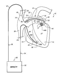
Figure 1 is a schematically illustrated fully implantable
atrial defibrillator shown in use with an intravenous lead
embodying the present invention and in association with a human
heart in need of atrial fibrillation monitoring and potential
cardioversion;
Figure 2 is a perspective exploded view of an intravenous
lead embodying the present invention; and,
Figure 3 is a partial, cross-sectional view, of the lead
of Figures 2 and 3.
Referring now to Figure 1, it illustrates a fully
implantable atrial defibrillator 30 shown in association with a
schematically illustrated human heart 10 in need. of atrial
fibrillation monitoring and potential cardioversion of the
atria. The portions of the heart 10 illustrated in Figure 1 are
the right ventricle 12, the left ventricle 14, the right
243app.doc
_g_
r.
~1~26,~4
atrium 16, the left atrium 18, the superior vena cava 20, the
coronary sinus channel 21 which, as used herein, denotes the
coronary sinus 22 and the great cardiac vein 23, the coronary
sinus ostium or opening 24, and the inferior vena cava 28. In
addition, as used herein, the term "ventricular electrical
activations" denotes R waves of the heart cardiac cycle which
induce depolarizations of the ventricles 12 and 14.
The atrial defibrillator 30 includes circuitry (not shown)
which is contained within an enclosure 32. The enclosure 32
hermetically seals the internal circuit elements of the atrial
defibrillator. The atrial defibrillator is shown in use with an
endocardial first lead 34, and an intravenous second lead 36
embodying the present invention. The enclosure 32 and first and
second leads 34 and 36 are arranged to be implanted beneath the
skin of a patient so as to render the atrial defibrillator 30
fully implantable.
The endocardial first lead 34 preferably comprises an
endocardial bi-polar lead having electrodes 38 and 40 arranged
for establishing electrical contact with the right ventricle 12
of the heart 10. The electrodes 38 and 40 permit bi-polar
sensing of ventricular electrical activations (R waves) in the
right ventricle between a first pair of locations 38a and 40a
within the right ventricle 12. As illustrated, the lead 34 is
fed through the inferior vena cava 28, into the right atrium 16,
and then into the right ventricle 12. As will be appreciated by
those skilled in the art, a second path for lead 34 could
243app.doc
-10-
. , 2152654
~-
alternatively be through the superior vena cava 20, into the
right atrium 16, and then into the right ventricle 12.
The second or intravenous lead 36 embodying the present
invention generally includes a lead body 37 which carries an
elongated distal electrode 44 and an elongated proximal
electrode 46. As illustrated, the lead body 37 is flexible and
includes a preformed section 39 which includes electrode 44 and
which has a resiliently coiled configuration. Because the lead
body 37 is flexible, the preformed section 39 including
electrode 44 may be elongated during implantation to reduce its
effective cross-sectional diameter dimension to permit the
lead 36 to be passed down the superior vena cava 20, into the
right atrium 16, into the coronary sinus ostium 24, and advanced
into the coronary channel 21 of the heart near the left side to
a predetermined position where the electrode 44 is within either
the coronary sinus 22 or the great cardiac vein 23 beneath the
left atrium 18 near the left ventricle 14. The electrodes are
preferably spaced apart relative to one another on lead body 37
so that when electrode 44 is positioned as described above,
electrode 46 is within the right atrium 16 after the preformed
resilient coiled section 39 is permitted to assume its coiled
configuration through the release of the elongation of
section 39. As a result, upon such release, the section 39
makes substantially continuous surface contact with the inner
wall surfaces of the coronary sinus 22 or the great vein, as
illustrated. This surface contact serves to provide positive
243app.doc
-11-
CA 02152654 1999-09-20
fixation of the lead 36 in the position illustrated. The contact
between the coiled section 39 and the inner wall surface of the
coronary sinus 22 or great vein 23 promotes the growth of fibrous
tissue around the lead in the region of section 39 for permanent
fixation of the lead 36.
The distal electrode 44 of lead 36 and the electrode 38 of
the first lead 34 permit bi-polar sensing of ventricular
electrical activations ( R waves ) between a second pair of
locations 38a and 44a of the heart. As will be noted in Figure
1, the spacing between the second pair of locations 38a and 44a
is greater than the spacing between the first pair of locations
38a and 40a. As fully disclosed in United States Patent No. 5,
348, 021 issued September 20, 1994, in the names of John M.
Adams, Clifton A. Alferness and K. Ross Infinger, for IMPROVED
APPARATUS AND METHOD FOR RELIABLY DETECTING A DEPOLARIZATION
ACTIVATION WAVE OF THE HEART AND ATRIAL DEFIBRILLATOR UTILIZING
SAME, which patent is assigned to the assignee of the present
invention, these relative spacings between the first and second
pairs of locations between which ventricular electrical
activations are sensed enable between reliable detection of R
waves.
The electrode 44, together with the proximal electrode 46 of
lead 36, provide for the delivery of defibrillating or
cardioverting electrical energy to the atria. Because the ring
electrode 44 is located beneath the left atrium 18 near the left
ventricle 14 and the proximal electrode 46 is within the right
-12-
2 .~ 5' ~' ~ ~'
atrium 16, the electrical energy applied between these
electrodes will be substantially confined to the atria 16 and 18
of the heart 10. As a result, the electrical energy applied to
the right ventricle 12 and left ventricle 14 will be minimized
when the atria are cardioverted or defibrillated.
To determine when cardioversion or defibrillation of the
atria of the heart 10 is required, the electrodes 44 and 46 also
provide bi-polar sensing of electrical activity in the atria 16
and 18 of the heart 10. A microprocessor (not shown), as
described in the aforementioned U.S. Patent No. 5,282,837,
digitizes the electrical signal provided by the electrodes 44
and 46 and processes the digitized values of the atrial activity
for detecting atrial fibrillation. Such atrial fibrillation
detection may be implemented as described in the aforementioned
U.S. patent.
As will be appreciated by those skilled in the art, the
lead 36 may be implanted as illustrated using the prior art
technique of sliding a guide wire or stylet into a central
passageway of the lead. The guide wire may be preshaped to
assist in guiding the lead 36 along the path previously
described. The stylet not only serves to guide or steer the
lead 36 along the desired path, but in addition, serves to
elongate coiled section 39 to reduce its effective cross-
sectional diameter dimension to permit the lead to be fed into
the heart. Once the lead reaches a predetermined position
within the heart, such as, for example, corresponding to the
243app.doc
-13-
2152654
electrode 44 being located either within the coronary sinus 22
or the great cardiac vein 23, the guide wire is retracted from
the lead.
The retraction of the guide wire from the lead 36 releases
the elongation of the coiled section 39 permitting the coil
section to resiliently assume its coiled configuration as
illustrated. Upon assuming its coiled configuration, the coiled
section 39 will have a cross-sectional outer diameter dimension
corresponding to the inner diameter dimension of the artery or
vein in which it resides and in accordance with this preferred
embodiment, the inner diameter dimension of the coronary
sinus 22 or great cardiac vein 23.
As a result of the foregoing, the coiled section 39 will
make substantially continuous surface contact with the inner
surface of the coronary sinus 22 or great vein 23. This
contact, together with the force exerted by the coiled
section 39 against the inner wall surfaces of the coronary
sinus 22 or great vein 23, provides positive fixation of
lead 36. Also, as a result of the surface contact between
coiled section 39 and the inner surface of the coronary sinus 22
or great vein 23, fibrous tissue will grow around the lead
body 37 in the region of the coiled section 39 to provide
permanent fixation of lead 36.
Even though the coiled section 39 provides positive
fixation of lead 36, it will not adversely effect blood flow
through the coronary sinus 22. Blood within the coronary
243app.doc
-14-
2~ ~2 ~~4
sinus 22 will freely flow through the inner diameter dimension
of the coiled section 39. Also, because of such free flow, the
formation of occlusions through blood clotting will not occur.
As will be further noted in Figure 1, and in accordance
with one aspect of the present invention, the coiled section 39
is a left-handed turned coiled section. The left-handed turned
coiled section for use in the coronary sinus 22 or great vein 23
has unexpectedly been found to have superior fixation qualities
as compared to a right-handed turned coiled section for the same
purpose. This result has actually been observed in practice in
sheep hearts, which have structural characteristics very similar
to the hearts of humans in terms of size and physiology. Over a
dozen leads, each having a right-handed turned coiled section,
have been implanted and nearly one-third of these leads became
dislodged and suffered migration. In contrast, over a dozen
leads, each having a left-handed coiled section, have been
implanted with none of these leads becoming dislodged, and thus
did not evidence migration. Both types of leads were identical
except for the direction in which the coiled sections were
turned. With the leads having the left-handed turned coiled
sections, detected electrogram signals remained of constant
quality, and energy thresholds for cardioverting the atria
remained essentially constant. .
To explain why this unexpected result occurred, it is
postulated that superior fixation is achieved when the coiled
section is turned in a direction which is opposite the direction
243app.doc
-15-
215264
of curvature of the artery or vein, as seen by the lead as it is
fed to its desired position. It is believed that the opposite
turn direction of the coiled section results in a greater
resistance to dislodgement as compared to a turn direction which
is the same as the direction of curvature of the artery or vein.
In the embodiment of Figure 1, it can be seen that the
coronary sinus 22 and great cardiac vein 23 have a direction of
curvature 25 which is to the right, as would be down the lead 36
from a proximal point such as where the lead enters the coronary
sinus ostium 24 to the distal end of the lead which includes
electrode 44. The leads having superior fixation character-
istics were those leads having a left-handed turned coiled
section, as illustrated in Figure 1. Hence, the coiled sections
of those leads were turned in a direction opposite the direction
of curvature of the artery or vein (coronary sinus, great
cardiac vein) in which they were implanted, wherein "direction
of curvature of the artery or vein" is meant to define the
lateral displacement of the lead as seen distally down the lead
from a point proximal to the distal end once the lead has
reached a desired position within the artery or vein. It will
also be noted that the coiled section 44a lies within the
portion of the artery or vein which results in the above-noted
direction of curvature. ..
Referring now to Figure 2, it shows the lead 36 embodying
the present invention in an exploded partial perspective view.
In addition to the structural elements of lead 36 previously
243app.doc
-16-
~~ ~2 ~~4
described, the lead 36 further includes a connector 41 at its
proximal end for coupling the electrodes 44 and 46 to an
implantable cardiac device such as atrial defibrillator 30 of
Figure 1. As is well known in the art, an additional connector
may be included so that each electrode is associated with its
own respective connector.
Preferably, the coiled section is formed to have a free
form cross-sectional outer diameter of, for example, eight (8)
to twelve (12) millimeters. Also, although the coiled
section 39 illustrated in Figure 2 includes two loops, the
coiled section 39 may have any number of loops as appropriate
for a given application.
Figure 3 is a partial cross-sectional view of the lead 36
within the section 39. More specifically, Figure 3 shows in
cross section one coil turn of the coiled configuration of the
lead 36 within the coiled section 39. The lead 36, as
illustrated in Figure 3, includes an inner stylet coil 50, an
outer electrically insulative jacket 52, and the elongated
electrode 44.
The stylet coil 50 is formed by a plurality of closely
spaced small diameter turns of wire. The stylet coil 50 thus
includes a central passageway 54 into which a stylet may be
extended prior to and during the implantation of the-lead 36.
The outer jacket 52 is formed of an electrically
insulative material such as polyurethane or silicone rubber. As
243app.doc
-17-
.-.
2I ~2 ~~4
will be noted in the figure, the insulative jacket 52 is coaxial
with and overlies the inner stylet coil 50.
The electrode 44, like the stylet coil 50, is also formed
from a plurality of closely spaced turns of a conductive wire.
The electrode 44 is preferably preformed with its closely spaced
turns prior to being mounted upon the lead 36.
To impart the coiled configuration to the lead 36 within
the section 39 as illustrated in Figures 1 and 2, either one or
both of the elongated electrode 44 and the inner stylet coil 50
is coiled with a left-handed turn in a portion thereof
corresponding to the section 39 having the coiled configuration.
To that end, the stylet coil 50 may be coiled to form a left-
handed turned helix having comparatively widely spaced turns
before the insulative jacket 52 is slid over the stylet coil 50.
Similarly, the electrode 44 may be coiled to form a left-handed
turned helix having comparatively widely spaced turns prior to
the electrode 44 being slid over the insulative jacket 50. With
either construction, the lead 36 within the section 39 will be
imparted with a coiled configuration for making substantially
continuous surface contact with the inner wall surfaces of the
coronary sinus or the great vein for retaining the lead 36 after
it is implanted.
While particular embodiments of the present invention have
been shown and described, modifications may be made. It is
therefore intended to cover in the appended claims all such
243app.doc
-18-
changes and modifications which fall within the true spirit and
scope of the invention.
243app.doc
-19-