Note: Descriptions are shown in the official language in which they were submitted.
215~136
P-2954 PAI~ENT
OPIICAL BLOOD CULTURE SENSOR
BACKGRO~JND OF T~IE INVE~IION
The present invention relates to a non-invasive method and aL)~a-dlus for det~cting
biological activities in a fluid spe~imPn, such as blood, urine or sputum, where the
specimen and a culture medium are introduced into sealable cont~ine s and are exposed to
conditions enabling a variety of metabolic, physical, and rhPmi~1 changes to take place in
5 the presence of microor~ni~m~ in the sa nple. The biological activity being ~etected by a
variety of chPmi~l sensors that are based on changes in fluorescent~ lifetime and/or
ily.
,
Usually, the presence of microo~ ni~n~ such as bacteria in a patient's body fluid,
20 particularly blood, is d~e....;.-~ using blood culture vials. A small ~lualllily of blood is
injected through a sealing rubber septum into a sterile vial co--~inin~ a culture me~ m-
The vial is incub~tPd at a ~~ ?el~lu~ conducive to b~ctPri~l growth, e.g., 37C, and
monitored for such growth.
2s Common visual inspection involves monitoring the turbidity of the liquid suspension.
2154136
Known instn~m~PntAl methods detect changes in the CO2 content in the head space of the
culture bottles, which is a metabolic by-product of the bacterAl growth. Mo~ ing the
C2 content can be accomplished by con~ ional methods, including radiochP.mi(~Al,
infrared absorption at a C02 spectral line, or plessul~/vacuum measurement. These
s methods, however, require invasive procedures which can result in cross-co.~lA..~inAIi~ n
between vials.
Recently, novel non-invasive methods have been developed which use çhpmic~l
sensors inside a vial. Such sensors often respond to changes in the C2 con~entr~tion by
lo chAn~ing their color or by chAnging their fluoresce.nce hllensily (see, e.g., U.S. Patent No.
4,945,060). The outputs from these sensors are based upon light intensity measurements.
This means that errors may occur, particularly if the light sources used to excite the
sensors, or the photodetectors used to monitor in~e~-.ciliPs, exhibit aging effects over time.
In known automated non-invasive blood culture systems, individual light sources,individual spectral e~cit~tion and emission filters, and individual photodetectors are
arranged adja~,nt to each vial. Such arrangements result in certain station sensitivity
variations from one vial to the next. Due to the fact that most known blood culture sensors
generate only a moderate contrast ratio in the measured photocurrent during bAc~t~PriAl
growth, extensive and time-co~ .in~ calibration procedures and sophi~ti~tP~ dete,cti~n
algo~ ls are required to operate these systems. Moreover, light sources, spectral f~ters,
and photodetectors with extreme narrow specification tolP.r~n~ ~s must be utili7~1.
Even if it would be possible to equalize all vial stations, certain variations in the vial
25 shape would still remain and affect the measured fluorescence photocurrent. In
fluorescence intensity-based sensor arrangements, any drift in the excitation source
21S4136
hllGnsi~y, any change in the photodetector sensi~ivi~y, or any dust coi~lA~..inAIion on an
optical surface will also cause a change in the measured photocurrent. ThelGrolG, the
long-time stability of such an instrument would be poor and frequent re calibration would
be lG~luilGd.
s
The disadvantage of intensity-based sensor arrangements can be overcome by
I~ti1i7.ing fluolGscenl sensors that change their fluolescf.~(ce 1ifP~tim~P, wherein il~lensily
measurement is replaced with time p~r~mp~ter measurement and illle~si~y challges have no
impact on the sensor output signal. Many rhPmi-~A1 sensor mAte.ri~1~ are known that change
10 their fluore~cerlce lifetime with ch~n~ing oxygen con~pnt~tion~ pH, carbon dioxide
con- entration~ or other rhemic~1 parameters (see, e.g., G.B. Patent No. 2,132,348).
A change in sensor fluorescent~e lifetime is commonly monitored by applying a well-
known phase shift method (see, e.g., U.S. Patent No. 5,030,420), wherein the eY~itAtion
5 light is ihll~nsily-mod~ t~d That method results in an intensity-modulated fluorescence
emi~sion that is phase-shifted relative to the ex~itAtion phase. Phase shift angle, 0, being
dependent on the fluorescence 1ifetime, ~, according to the equation:
tan ~ = ~ (l)
where ~ = 27~f, is the circular light modulation frequency.
An inspection of equation (l) reveals that the phase shift method allows for
maximum resolution, dO/d~, under the condition ~ = l. Unfollunal~ly, almost all known
25 pH- or carbon dioxide-sensitive fluorophores have decay times in the range S ns to 500 ps.
In other words, light modulation frequencies, f = l/27~, in the range 32 MHz to 320
21~4136
MHz would be required.
It is possible to accomplish light hllellsily modulation at such high frequencies,
however, this would require acousto-optic or electro-optic modulators which are only
s çffi~ ont in combination with lasers. Moreover, detecting the modulated fluorescence light
would require highly sensitive high-speed photodetectors, such as micro çh~nn~-1-plate
photomultipliers, which are rather expensive. Consequently, all commercial auloLua~ed
blood culture systems are based on intensity monitoring, and none utilize time-resolved
fluolesce"l carbon dioxide sensors.
SUMMARY OF T~IE INVEN'I~ON
The present invention overcomes problems identified in the art by providing a
method and al)pa,~lus for reliably and non-invasively detecting biological activities in
lS blood culture vials using an optical blood culture sensor that is based on time-resolved
fluorescence measurement that avoids the fluorescence intensity 1imit~tions ~ cus~
above.
According to the present invention, a culture medium and blood specimen are
20 introduced into a sealable glass vial having a head space gas nLu~lul~ such that a change in
the gas mixture composition can be monitored by a chemically sensitive composite m~t~ri~1
in the vial. The ch~mi~11y sensitive composite m~teri~1 comprises a nli~lure of two
fluorescent sensor m~teri~1~. The first sensor material exhibits a long fluoresc~nce decay
time and/or a fluorescence intensity that depend on a first ch~mi~1 parameter, such as
2s oxygen concentration. The second sensor material exhibits a fluorescence intensity that
depends on a second chemical parameter, such as pH or carbon dioxide concentration, the
2154136
fluorescence decay time of the second sensor material being at least one order of
m~gnitude shorter than the fluoresc~n~ e decay time of the first sensor m~t~ri~l .
The first and second sensor m~t~ri~1~ are mixed into the same sensor matrix and are
s i11~ 1 with illlensily-modulated excitation light. The modulation frequency is chosen
so that the condition ~1 # l holds for the first sensor material when the fluorescenre
lifçtime has its ."ini.,~u-" value. The fluorescence light emitted by the composite sensor is
monitored using only one photodetector. The fluorescence photocurrent from the
photodetector is split into its AC and DC co~ one lls, that are measured se~a,~lely. A
lO sensor output signal is then gell~,a~d by dividing the measured AC component by the
measured DC component.
The present invention allows both sensor m~tt-ri~1~ to operate in a time-resolved
mode. Therefore, despite the extreme short decay time of the second sensor material, only
15 one relatively low light modulation frequency is r~quired. Therefore, a low-cost light
emi~ting diode (~ED) can be used as the eYcit~tion source. The time-resolved operational
mode cancels out aU drift effects due to light source aging, photodetector sensitivity
changes, dust co"~ ~",h-~lion on optical sl~ ces, and small deformations and/or
disp1~cements of the vials. The present invention thelefo,e results in an extreme long-time
20 stabi1ity for the au~omaled blood culture instrument.
Changes in the sensor output signal can show the same or a different polarity for the
two chemical parameters. If the long decay time of the first sensor material increases, then
the AC/DC ratio decreases. If, e.g., the intensity of the second sensor material increases
2s during bacterial growth, then the AC/DC ratio increases again. Therefore, changes
regarding the two chemical parameters can be identified. In addition, in such a blood
215 4136
culture system, information can be obtained regarding the degree, the speed, and the
relative time delay between oxygen consull~ylion and carbon dioxide production. This
information can then be used for partial i(l~,ntifi~tion of the gluwil~g microorg~ni~m
s According to the present invention, the Uli~lUlte of the ehemi~1 sensor m~teri~1~ is
disposed to the inner wall or to the inner bottom of the glass vial, and is il11lmin~te~1 by an
excit~ti()n light source, preferably a blue LED. The I ~n is connecteA to an electronic
signal source which provides a DC bias and a high-frequency modulation voltage. The
signal source is equipped with a eontrol input to allow for output power control that is
o conllec~ed to a co.ll~uler.
Fluorescence light reem~,r,~ing from the sensor m~teri~l mi~lule is de,teeted by means
of a highly sensitive phûtodetector, such as a photoml-1tirlier. An emission filter is
arranged between the sensor m~te,ri~l and the photodetector to reject back-scattered
ex~,it~tinn light. The signal output of the pholod~eclor is then fed to a first power splitter,
one output of which is conn~ted to the input of a first low-pass filter and then fed to the
cQIll~ulel. The other output of the first power splitter is fed to the input of a first band-
pass filter~ the out~ut of which is conn~cte,d via a first high-frequency voltmeter to the
cQIll~ulcl. The colllpuler is equipped with a data display unit.
Part of the P~it~tion light emitted by the T ~n is coupled to the input of an optical
fiber, and the output of the fiber is arranged in front of a photodiode that acts as a source
monitor. The signal output of the source monitor photodiode is then fed to a second power
splitter, one output of which is connected to the input of a second low-pass filter and then
2s fed to the coll~uLer. The other output of the second power splitter is fed to the input of a
second band-pass filter the output of which is connected via a second high-frequency
2154136
voltmeter to the co~ ul~r.
In operation, the light source illllmin~tes the rhemit~1 sensor m~teri~1~ with intensity-
mod~ te1 e-Ycit~ti~n light. By splitting the fluorescence photocurrent into its high-
s frequency and DC components, by m~Asuring these components se~ ,.te1y, and by
c~1c111~ting the ratio of the two components within the computer, a sensor output signal is
gener~tecl that c~ries information l~gdldillg the response of the two sensor m~teri~1~ to
dirrelcul ~hemi~l pArAmPters.
0 These and other f~lules, objects, benefils and advantages of the present invention
will become more a~ nl upon reading the following detailed description of the ~lcÇ~ d
embo-1im~nt~, along with the appended claims in conjunction with the dl~wings, wherein
reference mlmerAl~ identify cGl~ onding components.
lS DESCRIPIION OF T~IE DRAWINGS
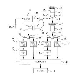
Fig. l shows sch.o.m~ti~11y an optical blood culture sensor arrangement according to
the present invention;
Fig. 2 is a plot showing frequency lifetime product, ~1, versus time for the first
sensor m~t.ori~1 in an aerobic vial;
Fig. 3 is a plot showing fluorescence intensity versus time for the second sensor
material for a strong carbon dioxide producing microorg~ni~m;
2s
Fig. 4 is a plot showing modulation degree, AC/DC, of the combined fluorescence
2154136
emission versus time, based on the individual sensor signals shown in Figs. 2 and 3;
Fig. 5 is a plot showing frequency lifetime product, ~, versus time for the first
sensor m~t~ri~l in an aerobic vial;
s
Fig. 6 is a plot showing fluorescençe inlensily versus time for the second sensor
m~teri~l for a weak carbon dioxide producing microorg~ni~m;
Fig. 7 is a plot showing modulation degree, AC/DC, of the combined fluo~scel~l
o emic.~ion versus time, based on the individual sensor signals shown in Figs. 5 and 6;
Fig. 8 is a plot showing frequency lifetime product, ~, versus time for the first
sensor m~tP.ri~l in an anaerobic vial;
Fig. 9 is a plot showing fluorescence intensity versus time for the second sensor
m~tPri~l for a strong carbon dioxide producing microorg~ni~m;
Fig. 10 is a plot showing modulation degree, AC/DC, of the combined fluorescenceemission versus time, based on the individual sensor signals shown in Figs. 8 and 9;
Fig. 11 is a plot showing modulation degree, AC/DC, of the combined fluorescenceemission versus time, ~ ming an extreme low con~ntr~tion for the second sensor
m~t~.ri~1;
Fig. 12 is a plot showing modulation degree, AC/DC, of the combined fluorescenceemission versus time, ~sllming an optimum concentration for the second sensor material;
2154136
Fig. 13 is a plot showing modulation degree, AC/DC, of the combined fluorescenceemission versus time, ~sllming an extreme high concent~tion for the second sensor
material;
s
Fig. 14 is a plot showing the effect on microorganism growth curves caused by a 20
percent peak-to-peak variation in the second sensor m~teri~l con~pntration;
Fig. 15 is a plot showing relative error of measured modulation degree, AC/DC, in
o percent, ~ ing a 20 pe~ peak-to-peak variation in the second sensor m~tçri~1
concentratic)n;
Fig. 16 is a plot showing modulation degree, AC/DC, of the combined fluorescenceemission versus time, ~s~-ming an extreme low con-~R.ntr~tion for a second sensor m~teri~l
5 with a decreasing fluorescence inlensily in ~s~nse to carbon dioxide production;
Fig. 17 is a plot showing modulation degree, AC/DC, of the combined fluorescenceemission versus time, assuming an o~ um conrertr~tion for a second sensor m~teri~l
with decreasing fluorescence intensity in response to carbon dioxide production;
Fig. 18 is a plot showing modulation degree, AC/DC, of the combined fluorescenreemission versus time, ~s-lming an extreme high concentration for a second sensor m~tPri~l
with a decreasing fluorescence intensity in response to carbon dioxide production;
2s Fig. 19 is a plot showing the effect on microorganism growth curves caused by a 20
present pçak-to-peak variation in the sçcond sensor material concentration, ~sllming a
2154136
second sensor m~t~.ri~l with a decreasing fluorescence hllensily in response to carbon
dioxide production;
Fig. 20 is a plot showing relative error of measured modulation degree, AC/DC, in
s ~ g a 20 ~n~en~ peak-to-peak variation in a second sensor m~teri~l
conc~ntrati-~n, and ~ min~ a second sensor m~t~.ri~l with a decreasing fluorescence
inten.city in response to carbon dioxide production;
Fig. 21 ill~l5~ P~ two growth curves and their first d~ivalives for aerobic sa_ple
0 vials with the curves on the left corresponding to a strong carbon dioxide producer and the
curves on the right to a weak one; and
. * Figs. 21(a) to 21 (d)
Fig. 22 depicts possible parameters of the first derivative plots that could be
~xtr~ctçd as f~lu~s in order to ex~cllte i~ientifi~tic n algo. ;ll~
DETAILED DESCRIPIION
A pl~rc.ncd e~nbodiment of a composite optical blood culture sensor arrangement
embodying the prin-ipl~ and col~ce~ls of the invention is depicted srh~m~ti~ ly in Fig. 1.
20 In this alla"ge~ent, a spe~imPn and culture medium ~ lul~, 4 are introduced into an
optically l~n~ l co,l~hler 1 that is sealed by a cap 2. A ~lule of chtomi~l sensor
m~teri~l~ 3 is disposed to an inner wall 28 or an inner bottom surface 21 of container 1.
The mixture 3 is ill~ in~tPA by an excitation light source 5, preferably a blue T ~n, that is
connected to an electronic signal source 6. Signal source 6 provides a DC bias and a high-
25 frequency modulation voltage to light sour~e 5 over a line 30, and is equipped with apower control input 25 connected by a line 31 to a com~uler 11.
- 10 -
21541~6
Sensor m~tçri~ ul~ 3 comprises a n~lul~ of a first sensor m~tP,ri~l and a secondsensor m~t~,ri~l, wherein the first sensor m~teri~l exhibits a fluore,sc~,nce decay time and/or
a flu~ sc~,nr,e mlensily that depend on a first rh~,mic~l p~r~m~er, such as oxygen
s con~Pntr~tn. The second sensor m~tçri~l, however, exhibits a fluoresc~,nce llellsily that
depends on a second s~h~mi~l parameter, such as carbon dioxide con~e.ntr~tion. The first
sensor m~t~,ri~l can have a fluorescence decay time in the range 0.1 to lOOOllsec and even
in the range 1 to lOOnsec, and the fluorescP,n~e decay time of the second sensor m~t~ri~l is
at least one order of m~gnih~de shorter than the fluolc;scP,nce decay time of the first sensor
o m~t~,ri~l As a result, the invention allows both sensor m~t.-,ri~l~ to operate in a time-
resolved mode and, despite the extreme short decay time of the second sensor m~t~,ri~l,
only one relatively low light modulation frequency is required. Therefore, a low-cost LED
can be used as light source 5. The time-resolved operational mode allows for c~n~ tic~n
of drift effects due to light source aging, photodetector sensitivity changes and small vial
deformations and/or vial displacements.
Fluoresr,e,n-~,e light reemerging from sensor m~tçri~l lllixlu~ 3 is detect~1 by a
photodetector 8, e.g~, a photomultiplier. An emission filter 7 is arranged between ll~ ul~;
3 and photodetector 8 to prevent excitation light from light source 5 re~ching photodetector
8. Photodetector 8 genel~les an output signal on a line 35 that is fed to a first power
spliKer 9. First power spliKer 9 then gen~,.~es two output signals, one of which is
connected by a line 40 to the input of a first low-pass filter 10 that is connP,cted directly to
co"~uler 11. The other output signal of first power splitter 9 is fed by a line 45 to the
input of a first band-pass filter 12 that is connected via a first high-frequency voltmeter 13
2s to computer 11. Computer 11 is connected to a data display unit 14 to display
information.
21~4136
Part of the excitation light emiKed by light source 5 is also coupled into the input of
an optical fiber 15 that is arranged in front of a photodiode 16 that acts as a source
monitor. Source monitor photodiode 16 then gen~ ~es an output signal that is fed to a
s second power splitter 17 via a line 50. Second power splitter 17 then gen~r~tPs two output
signals, one of which is conn~ted by a line 55 to the input of a second low-pass f~ter 18
that is conn~cte~ directly to co~u~ul~,r 11. The other output signal of second power splitter
17 is fed by a line 60 to the input of a second band-pass filter 19 that is conn~ted via a
second high-frequency voltmeter 20 to co"l~u~r 11.
In operation, light source S illl....in~les sensor m~te.ri~l u~L~lulci 3 with ~x~it~tiQn light
that is inlensily-modulated at a circular modulation frequency, ~, having a modulation
degree, m. The intensity modulation of the excitation light can be accomplished using
dirr~ waveforms, but it is advantageous if the light source is periodically mod.ll~te-l
5 In particular, the excit~tion light can be ~imlsoi(1~11y mod~ ted, square-wave mod~ ted or
periodically pulsed. The emitted fluorescence intensity of the first sensor m~teri~1 in
mixture 3 can be described by:
Fl(t)=FOl[l+ ~1 2 ]*sin[~ t- ~ ] (2)
with Fol being the average fluorescen~e intensity, and
~= arctan(~r) (3)
2s being the fluorescence phase shift relative to the excitation modulation phase. The
fluorescence lifetime, ~, may depend on the time, t, according to the expression
2154136
r(t) = k*h(t) (4)
where k is a constant, and h(t) is a time-dependent function that rises from a first, lower
5 level to a second, higher level as a consequence of oxygen consumption during
microor~ni~m growth.
Modulation frequency, ~, is chosen so that the condition ~1 holds for the first
sensor m~teri~l when it has its minin~ value. Fig. 2 depicts the frequency lifetime
lo product, ~, versus time for the first sensor m~t~ l in an aerobic vial.
The average fluorescence intensity, Fol, may also depend on the time, t, according
to an ~ ssion
Fol (t) = k' * h' (t) (4A)
where k' is another constant, and h'(t) is another time-dependent function that also rises
from a first, lower level to a second, higher level as a con~ent e of oxygen con~u~ ion
during microorganism growth.
The fluorescence radiation emitted by the second sensor m~tPri~l in Illi~Ulc; 3 can be
described by the equation
F2 (t) = Fo2 [1 + m* sin(~ t)] (5)
with Fo2 being the average fluorescence intensity of the second sensor material. In
215~136
equation (5) it has been taken into account that the fluorescence decay time of the second
sensor m~tçri~l is extremely short, so that the condition c~ < ~1 holds. Therefore, the
fluorescen~e modulation degree is identi~l to the eY~it~tion modulation degree, m, and the
fluoresce-n~e phase shift is so small that it can be neglected.
The average fluol~scen~e inlensi~y of the second sensor material, Fo2~ may depend
on the time, t, according to the t;A~ ssion
Fo2 (f) = c*g(t) (6)
where c is a constant, and g(t) is a time-dependent function. The function g(t) can rise
from a first, lower level to a second, higher level as a consequence of carbon dioxide
production during microorganism growth. Fig. 3 depicts the average fluorescenre intensity
of the second sensor material versus time. In this case, the microorganism is a strong
15 carbon dioxide producer. The function g(t) can also decrease from a first, higher level to a
second, lower level, as ~ cussecl below.
In an optical blood culture sensor arrangement according to the present invention, the
fluoresc~nre photocurrent, I(t), is given by the expression
I(t) = K(r,d,v)*[F,(t) +F2(f)] (7)
where K(r,d,v) represents a function depending on the photodetector responsivity, r, the
tr~n~mi~.~ion, d, of dilre~ l optical surfaces that show dust cont~min~tion effects, and on
2s the exact vial shape, v.
2I5~136
The overall photocurrent I(t) is split into its AC and DC components, with each
compollent being measured sep~r~te1y, and the AC/DC ratio is calculated within col~uler
11. Based on this, the following sensor output signal, AC/DC, is obtained
01( ) 2 sin[~ t- arctan(~r (t)]+F02(t))msin(~ t)
5 AC Jl + (~T(t))
DC Fol (t)+F~(t)
As can be seen from equation (8), the function K(r,d,v) is c~n~ele~l out. Mol~over,
because the average fluorescence i.~e.-.c;l;~s Fol(t) and Fo2(t) all are proportional to the
excit~ti-~n source Lnensi~y~ aging effects of the ~ ~n are also c~n~l~l out. To avoid errors
l0 in the unlikely event that changes in modulation degree m occur, the sensor arrangement is
provided with means to monitor and control the ~ mod~ tion. This is accomplished by
means of the source monitor photodiode 16 and second power splitter 17.
Fig. 4 shows the modulation degree of the combined fluorescen~e emissions, which5 is calc~ tlY1 within co~?uler ll, and which l~lesenls the sensor output signal. In this
case, oxygen consumption causes a decrease in the output signal. After some time delay,
carbon dioxide pro~uction causes a subsequent increase in the output signal. In Figs. 2 to
4, it has been ~ccllmecl that the microorganism is a strong caIbon dioxide producer. Figs.
S to 7 il111ctr~te expected signals for a weak carbon dioxide producer. Finally, Figs. 8 to
20 10 illustrate the expected signals for an anaelubic vial, where no oxygen changes occu
during microorganism growth.
I
One may ask, how accurately the mixing ratio of the first sensor material and the
second sensor m~t~ri~1 in mixture 3 has to be m~int~ined during production. Fig. ll
25 illustrates a possible worst case scenario with an extreme low concentration of the second
215~136
sensor m~t~ri~l within the IlPil-lure 3. As can be seen, the carbon dioxide-related signal
increase is much weaker than the oxygen-related signal decrease. Fig. 12 shows the sensor
behavior for ~JlilllUIII second sensor m~t~,ri~l con-~,ntr~tion. Here, both the oxygen-related
decrease and the carbon dioxide-related increase are pronounced. Fig. 13 illllst~tes
s another possible worst case scen~rio with an extreme high conr~ntr~tion of the second
sensor m~t~,ri~l within the mixture, in which the oxygen-related decrease and the carbon
dioxide-related increase are weak.
The inflllence of production-related deviations in the sensor mixing ratio from the
lo oylilllu~ mixing ratio is illllstr~ted in Figs. 14 and 15. Fig. 14 shows how growth curves
would be affected by a 20-% peak-to-peak variation in the second sensor m~ter~l
conc~ntr~tion. Fig. 15 shows the relative error in the measured modulation degree in
percent, again for a 20- % peak-to-peak variation in the second sensor m~ter~l
concentr~tion. In practice, the mixing ratio can be controlled easily with an at~ur~cy of
S better than 1%. In other words, the expected impact of production-related variations in the
mixing ratio on the measured growth curves is well below 1%.
The expected.growth curves shown in Figs. 4, 7 and 10-14 are based on the
assumption that the average fluol~scence inlensily of the second sensor m~t~,ri~l, Fo2(t),
20 will increase as a con~uence of carbon dioxide production during microorganism growth.
This results in a dirr~lGIll polarity for sensor output signal changes in response to oxygen
consumption and carbon dioxide production, respectively. While this may be of advantage
sometimes, it could result in a partial signal cancellation, if oxygen consumption and
carbon dioxide production occur siml-lt~neously.
Any signal cancellation can be avoided by selecting a second sensor material that
- 16-
215~136
shows a decrease in intensity in response to carbon dioxide production. In this case, the
expected growth curve shown in Fig. 4 changes to a growth curve as depicted in Fig 17.
The~t;rol~, both oxygen consu~ ion and carbon dioxide production cause a decrease in the
modulation degree, i.e., generate sensor output signal changes that have the same polarity.
s
The plots depicted in Figs. 16, 17, and 18 correspond to the plots shown in Figs.
11, 12 and 13 and illl-str~te the effect of a varying mixing ratio for the two sensor
m~t~ for the case where the second sensor m?(teri~1 exhibits a decreasing fluorescence
illlensily. Figs. 19 and 20 correspond to Figs. 14 and 15, respectively, and illustrate how
o growth curves would be affected by a 20-% peak-to-peak variation in the second sensor
m~t~ri~1 con~Pntr~tion. The conclusions are the same as in the case of a second sensor
m~t~.ri~l showing an increasing fluo,esc~ e ,lllensi~y.
Fig. 21 illnstr~te5 two growth cuNes for aerobic sample vials. The curve on the left
S corresponds to a strong carbon dioxide producer; the curve on the right to a weak one.
Fig. 21 also contains the first derivatives of the growth curves. As can be seen, dirÇ~ltilll
org~ni~m~ may produce dirrel~.ll p,~e,--s that can be uti1ized to ~~ lesu~ emicroorganism i~l~ntifie~ti<)n. Fig. 22 depicts possible parameters of the first derivative
plots that could be extr~cted as features in order to execute i~l~.ntifi~tion algofllllllls.
As described above, the fluoresc~n~ analysis can be ~lrolllled by m~uring the
AC and DC components of the photocurrent, and by calc~ tin~ the AC/DC ratio, which
corresponds to the fluorescence modulation degree. It is also possible to apply other
methods of time-resolved fluorescence analysis.
2s
A further modification of the present invention is possible by measuring the
2ls~l36
fluorescence modulation using a first modulation fr~quency, repeating the same procedure
and applying another modulation frequency, or other modulation frequen~i~.s. By
analyzing the data obtained at dirrelel~l freque.ncies, changes in the first and second
ch-o.min~l paIameters can be isolated, even if these changes would occur ~imll1t~n~ously.
s Sepal~ting the effects of changes in the first and second çh~.min~1 p~r~meter allows for
microorganism itiçntific~tion, because dirrGlGn~ species will generate dirrGlGIl~ time p_U~ s
in regard to the two ch~.min~1 parameters.
In s.. z.y, an optical blood culture sensor accol.ling to the present invention
lO cancels out the effects of variAtions in excitAtion source ihllGnsily, photodetector sensitivity,
optical filter trAncmiC~ion, and vial shape. Moreover, the requirements in ~ in~ a
constant mixing ratio for the two sensors are very modest.
It should be understood that the above-described embodiments are simply illustrative
5 of an a~ ~alus and method embodying the principles and conceGpts of the present
invention. Of course, other suitable variations and mo lifi~tions could also be made to the
app~lus and method described and still remain within the scope of the present invention.
- 18 -