Note: Descriptions are shown in the official language in which they were submitted.
215 416 2 PCT~S94/00~
W094116625
FOR~aRD VIE~IN~ T~GING CA.~V
R~ ~O~ND OF ~R~ TNVRNTTON
l. Field of the ~nvent-;on
The present invention relates generally to a device
and method for ultrasonic intraluminal imaging. More
particularly, an intravascular catheter is provided for imaging
a portion of a blood vessel in a plane ext~n~i~g axially from
the tip of the catheter. The catheter system of the present
invention may also include an additional diagnostic or
interventional work element for use in conjunction with the
imaging element.
2. Des~ription of the Backgro~ Art
Arteriosclerosis, also known as atherosclerosis, is a
common human ailment arising from the deposition of fatty-like
substances, referred to as atheromas or plaque, on the walls of
blood ~ . Such deposits G~ in both peripheral blood
v6~ that feed the limbs of the body and the coronary
ve~^el~ which feed the heart. When the deposits accumulate in
localized regions of a blood v~ , stenoæis, or narrowing of
the v~ Ar chAnn^l~ o~ . Blood flow is lecL~icted and the
person' 5 health is at serious risk.
Numerous approA~h~ for re~tlci nq and removing such
vA~c~llAr depo~its have been ~Lv~ , including balloon
angiopla~ty where a balloon-tipped catheter is used to dilate a
region of atheroma, atheractomy where a blade or cutting bit is
used to sever and remove the atheroma, spark gap reduction in
which an electrical spark burnæ through the plaque and laser
angioplasty where laser energy is used to ablate at least a
portion of the atheroma.
A major difficulty in using such devices is obtAining
images and information on the region of the blood vessel to be
treated. To overcome this difficulty, several te~h~iques have
been ~v~ for intraluminal imaging of v~clllAr vessels.
WO9J/~g~ PCT~S94/00~ ~
Catheters incorporating ultrasonic tr~n~ c~rs for imaging are
disclosed in U.S. Patents Nos. 4,794,931; 5,000,185; 5,049,130;
and 5,024,234. However, these catheters scan in a plane normal
to the catheter axis. While such catheters are very useful for
examining deposits adjacent to their distal tips, they are
generally i~r~pAhle of imaging the vessel downstream of the
catheter.
Such downstream viewing would be u~eful in a variety
of circum~tAn~e~. For example, it would provide a visual
determination of whether there ic a ch~nnpl through which a
guide wire or catheter may be passed. Moreover, downstream
viewing could provide information to help the physician to
determine which type of intravascular device would be most
suitable for reducing the s~enosis. Finally, downstream
viewing can be invaluable as an aid in directing and using
interventional and diagnostic devices and avoiding accidental
penetration of the vessel wall.
U.S. Patent No. 4,S76,177 to Webster describes a
laser catheter having an ultrasonic tr~ cer mounted at a
fixed angle of inclination to the catheter tip. The trAn~
is not movable with respect to the catheter tip however, a~d is
therefore only capable of imaging along a line fixed with
respect to the cathete~ body.
U.S. Patent No. 4,587,972 to Morantte discloses a
catheter appar~tus having an array of transducing elements.
The elements are sequentially excited to obtain an image distal
to the catheter. Such rh~ array devices are very
complicated and therefore costly to fabricate~ Their
resolution and ability to s~eer the beam through a wide range
of angle~ are limited by the number of elements provided.
It would be desirable to provide a catheter apparatus
capable of imaging a blo~d vessel downstream of the catheter
itself. It would be desirable if such a catheter were capable
of sç~nn; n~ a region o~ the blood vessel in a plane located
3S forward of the catheter. Such a catheter should be of
relatively simple design to allow for compact construction and
reliability of use. Additionally, it would be desirable to
combine such a forward viewing catheter with an additional
~ W094/16625 ~ I 5 ~ 1 6 2 PCT~Sg4/00~4
working element to provide the catheter system with a further
diagnostic or interventional capability.
RUMMARy OF ~R~ ~v~.~O~
According to the present invention, a catheter system
and device are provided that are capable of forward or
"downstream" imaging of blood v6s~^l~ and other body lumens and
cavities within a patient. The catheter includeæ a flexible
tllh~l ~r member adapted for insertion into the vessel and an
ultra~onic trAn~Allc~r di~ near the distal end of said
flexible tllh~ r member. A mechAni~m is provided for
oscillating the tr~n~llcer about a transverse axis (i.e., an
axis normal to the longit~lA;n~l axis of the tllhlll~r member.
The ultrasonic transducer is adapted to transmit and receive
ultra onic signals in a direction substantially forward of said
distal extremity and is coupled to external video image
processing circuitry which can thus present the desired forward
view.
In a Cpeci fic embodiment, the ultrasonic tr~n~nrer
i~ coupled to a flexible drive cable rotationally ~ o:cd in a
lumen of the tllhlllAr member. The trAnC~ r i8 me~h~n i cally
coupled to the drive cable by a me~n;cm that C~1~V~ Ls of
rotation of the drive member into pivotal oscillation of the
tr~n~lc~r with respect to the tnhlllAr member. This causes
the transducer to scan a segment of the ves~el in a plane
aligned projecting forward of the catheter.
In another aspect of the invention, an imaging
catheter system is combined with a biopsy device for sampling a
deposit within the vessel. The biopsy device is di~poc~ to
permit simul~Ar?c~ imaging and sampling of the deposit. Other
work elements are possible as well. The~e additional work
elements could include lasers, mech~nical cutters, angioplasty
balloons and the like, all of which are known in the art.
3 ~ ~PT~F D~8CRIPTION OF T~ DRA~ING8
Fig. 1 is a side sectional view of a catheter having
a forward viewing imaging capability according to the present
invention.
4
Fig. 2 is a diagrammatic view of a catheter tip in
accordance with the present invention illustrating the planar
scanning capability.
Fig. 3 is an enlarged side sectional view of a
portion of Fig. 1 showing an assembled mechanism for converting
rotation of a drive shaft into pivotal oscillation of an
ultrasonic transducer.
Fig. 4 - 6 are multi-view orthogonal depictions of
separate parts of the mechanism depicted in Fig. 3.
Fig. 7 is a schematic block diagram of a timing and
control system suitable for use in the present invention.
Figs. 8A - 8C illustrate the parts and assembly of an
inductive coupling device for electrically connecting the
connecting the ultrasonic transducer to the timing and control
system.
Fig. 9 is a side sectional view of a forward viewing
imaging catheter according to the present invention combined
with a biopsy tool for sampling a deposit within a blood
vessel.
DETAILED DESCRIPTION OF THE SPECIFIC EMBODIMENTS
A catheter system according to the present invention
is illustrated in Figure 1. The catheter system comprises a
flexible tubular member 3, an ultrasonic transducer 5 and
drive means 7. Tubular member 3 has a proximal end 9, a distal
end 11 and a central lumen 13 connecting the two. Drive means
7 comprises a drive cable 15 rotatably disposed within central
lumen 13 and a motor coupling 17 at the proximal end of the
drive cable.
Ultrasonic transducer 5 is disposed within a distal
housing 14 at distal end 11 of tubular member 3. The
transducer is adapted for pivotal movement relative to the
tubular member. The distal end of drive cable 15 is connected
to the ultrasonic transducer by a coupling mechanism 19, which
is adapted to convert rotation of the drive cable into pivotal
oscillation of the ultrasonic transducer. Transducer 5 is
shielded by a cover 6. The cover protects the transducer from
interference by tissue within the patient and the patient from
WO94/16625 21 S I 16 2 PCT~S94/00~4
5
internal injury from contact with the rapidly oscillating
trAn~ r. Cover 6 is màde of a acoustically transparent
material to allow the transmission of the acoustic waves sent
and received by tr~n~ cer 5.
5 The pivotal motion of the ultrasonic tr~n~A-~cer may
be conveniently understood with reference to Fig. 2, which
depicts the distal end ll of ~ r member 3, within which
trAnr~llror 5 i8 mounted. TrAn~AllGer 5 pivots within the
tl~h~lAr member about an axis Z and Rweeps back and forth
through an angle ~ lying within a plane X-Y.
Distal housing 14 and coupling me~hA~icm l9 are shown
in greater detail in Fig. 3. The distal housing holds the
coupling mech~ni~m. The coupling mechAni~m comprises three
main parts: a transducer holder 20, which has a receptacle 2l
in which the transducer is held; an actuator 22 for driving the
tr~n~ c~r holder; and a stator 23, about which the trAn~ r
holder pivots.
Figure 4 is a three view orthogonal projection of
distal housing 14. As can be seen therein, the distal housing
has mounting holes 24 and 25 and a ro~YiAl or~ninq 26.
Actuator 22 is depicted in a two view orthogonal
projection in Fig. 5. The actuator has a shaft 28 and an
actuator pin 3l set into concave surface 33. The actuator pin
i8 set at an angle ~ to the center line of the actuator and the
shaft. In a preferred embodiment of the invention, angle ~ is
about 45 de~ e-~5 but the angle may vary without departing from
the principles of the invention.
Figure 6 is a two view projection of tr~n~ cer
holder ~0 and stator 23 disposed therethrough. As mentioned
previously, the tr~nC~llr~r holder has a receptacle 21 for
holding the tr~nr~ er. The tr~n~ er holder also has a slot
36 cu~ into a rounded back surface 38. Slot 36 is adapted for
cooreration with actuator pin 3l of actuator 22 and this will
be described in more detail below.
Referring back to Fig. 3, the integration of the
parts depicted in Figs. 4 - 6 into the catheter system will now
be described. Shaft 28 of actuator 22 is rotatably ~i ~ro~
W094/166~ PCT~S94/00~4 ~
2~$ ~ 6
~hrough COAY;A1 opon;7-g 26 of distal housing 14. The CO~;A1
O7P~n; ~7g acts as a bearing to support the rotating shaft.
The ends of stator 23 are fixed (e.g., by a press
fit) within mounting holes 24 and 2S of the distal housing.
TrAncA1lc~r holder 20 is pivotally disposed about the stator.
A pair of coil~ or windings ~i~ro~ around the stator act as
an inductive coupling 45 for electrical coupling of tr~n~ cer
5 to an associated unL ol means. The construction and
function of inductive coupling 4S is Ai ~Cl~C~ in detail below.
Actuator pin 31 is di~ for ~ooperation with slot
36. The width of slot 36 is slightly greater than the diameter
of actuator pin 31 so that the pin may slide and rotate within
the slot. The pin and slot are thus configured to convert
rotation of actuator 22 into pivotal oscillation of tr~nc~tlcer
lS holder 20 about stator 23.
System control circuitry 50 suitable for controlling
the trAn~ r is illustrated schematically in Fig. 7. The
control circuitry, which can be formed of substantially
conventional equipment, includes a timing and control means 54,
a transmitter 57 and a receiver 58 with a transmit/receive
switch 59, and a display unit 60, typically including a CRT
tube for displaying an image from within the blood vessel.
In operation, timing and CGllLr ol means 54 sends
pulses to transmitter 57. Transmitter 57 generates voltage for
excitation of the tr~ns~ r 5. The tr~n~t~r generates
ultrasonic energy waves which emanate forwardly into the blood
vessel. Portions of the ultrasonic energy waves reflect fro~
tis~ues within the vessel and are reflected back to the
trAnCA1lc~r. The tr~nc~l~cer receives these reflected waves and
cunvelLs them into electrical signals which are sent back to
receiver 58 through cQ~ cting wires 62 and 64. The signals
are amplified and processed by display unit 60, which ~o.l~erLs
the signals into a visual display of the structure of the
ves~el .
3s The tr~nC~lcer is switched between its send and
receive modes by transmit/receive switch 59. Timing and
~o,-LLûl means 54 controls drive motor 67, which may be an open
loop stepping motor or a closed loop servomotor. Motor 67
2154152
W094~16625 PCT~S94/00~4
7
rotates drive cable 15, which, as disr~ ed above, causes the
tr~nCAnc~r to scan back and forth through an arc within the
blood vessel.
The ~c~n~ arc (~ in Fig. 2) will be 90 degrees in
S the emhoA iment depicted where actuator pin 31 is set at a 45
degree angle (~ in Fig. 5) to the axis of actuator shaft 28.
Drive cable 15 is preferably rotated at a constant angular
velocity. Eighteen l~ e~ (1800) rpm is suitable rota~ion
speed for the emhoA;ment depicted. This translates to a
trAn~A~lc~r scan rate of 30 o~cillAtions per recnn~ ~ a rate
sufficient to provide good image detail with an acceptable
image refresh rate. The transducer firing rate is coordinated
with it~ pivotal movement by system controller 50. It will be
understood that the actual tr~n~ncer oscillation rate could be
varied significantly within the scope of this invention.
Electrical signals are carried between the system
controller and the træn~llcer through ~on~llcting wires 62 and
64. The diætal ends of these wires could be attached to the
tr~n~llc~r in a conventional manner, e.g. by soldering. If
this were done, sufficient slack would need to be left in the
wires ~o allow for the pivotal o~cillAtion of the transducer.
Direct ~o~n~ ~ion of wires 62 and 64 to the
trAnC~llcer is problematic howe~L, because of the high speed
cyclic hon~ i ng that directly conn?~ted wires would have to
el,d~Le. As mentioned above, a typical trAnCAll~r oscillation
rate will be about 30 oscillations per -?-Q~ . Under sustAin
use, there is a very significant potential for fatigue failure
either of the wires or the soldered connection between them and
the tr~n~ cor. A failure at either of these points would
disable the ~ystem.
For these reA~on~, it is advantageous to eliminate
the problem of flexing within wires 62 and 64 by using an
indirect connection to electrically couple the wires to the
trAnF~nC~r. An inductive coupling 45 adapted to this purpose
is shown in place in Fig. 3.
The details and construction of inductive coupling 45
are depicted in Figs. 8A - 8C. Figure 8A depicts a stator
assembly in detail. As can be seen therein, a wire channel 80,
WO94/16~ PCT~S94/00~ ~
2 ~ 8
comprising r~nn^l segments 81 and 82, is bored partially
through stator 23 from each end. A w;n~ing groove 85 is cut
into the surface near the middle of the stator and stator holes
87 and 88 are drilled into the stator to co~n~ct wi n~ i ng groove
85 to chann~l segments 81 and 82.
Co..LLoller wire 90 is then fed through one ~h~n~el
segment, turned a number of times around the stator at win~ing
groove 85, and fed out of the other ~h~nnel segment. The turns
of wire 90 within wi~Ainq ~Loo~e 85 form a stator wi~ing 93
around stator 23. The number of turns in wtn~ing 93 may
obviously vary but in one preferred emhoAiment there are
thirteen turns.
A rotator assembly is depicted in detail in Fig. 8B.
A rotator 100 has an inside diameter slightly larger than the
outside diameter of stator 23 so that the rotator may be
disposed to turn about the stator. Along a portion 103 of its
length, rotator 100 has an even larger inside diameter to
accommodate a win~i nq 105 of trAnr~llcor wire 108 and a
retA i n i ~g sleeve 110.
Rotator holes 113 and 114 are bored through the wall
of rotator 100. Tr~n-~llcer wire 108 is fed through one o~ the
holes, wound a number of times about the inside of the rotator
to form a rotator winAi~g lOS, and finally fed back out through
the 5~cQ~A of the holes. Ret~i n i ng sleeve 110 is then fitted
within rotator 100 to hold rotator wi n~ i ng 105 in place.
Rotator wi n~ i ng 105 will typically have the same number of
turns as stator winAing 93; in a preferred embodiment,
thirteen.
As depicted in Fig. 8C, the rotator assembly is
rotatably disposed about the stator a~sembly so that the
windings are aligned with each other to form inductive coupling
45. An electrical current flowing within co~lLLoller wire 90
will pass through stator win~ing 93 ~i~pQ~^~ within rotator
Wi ~ i ng 105 . This will in~llre a corrPcpQ~i ng electrical
current within rotator wi n~ i ~q 105 which will flow through
tr~nC~ r wire 108. The reverse will also be true--a current
flowing through the transducer wire will in~llce a current
within the ~Gl~L~oller wire.
~ W094/166~ 215 4 1 6 2 PCT~S94/00~
Inductive coupling 45 is incorporated into the system
as depicted in Figs. 1 and 3. The two ends of stator 23 are
pre~s fit into mounting holes 24 and 25 of distal housing 14.
The rotator is fixed within trAnA~tlrer holder 20, which pivots
- 5 about the stator. The two ends 91 and 92 of ~ollL~oller wire 90
are rou ed back through tl~hlll;tr m~mber 3, and serve as
onAllcting wires 62 and 64. TrAn~llrer wire 108 is directly
c~r.Acted at each end to tr~n~ c-r 5.
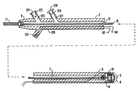
Referring to Fig. 1, ~llh ll ~r member 3 of the catheter
system has a three arm adaptor 120 at its proximal end. A
first arm 122 of three arm adaptor 3 has rQnA~cting wires 62
and 64 routed through it and is adapted for connection with
system ~ oller 50 (Fig. 7).
A rs ~n~ arm 123 of the three arm adaptor has a fill
port 124 and a fill ~-h~nn-l 125 in communication with central
lumen 13 of the t~hl~lAr member. Before imaging, a fluid
suitable for the transmission of ultrasonic signals will be
injected into fill port 124. The fluid will fill the tllhlll ~r
member of the catheter system and flush air bubbles (which
could interfere with imaging) out of the region of the
trAn~A~ er~ along central lumen 13, and through a drain r~nr^
127 and a drain port 128 of a third arm 129 of the three arm
adaptor .
Electrical impulses will then be sent from the
2S o~.,LLoller along ~AnA~r,ting wires 62 and 64 and through stator
winAing 93. These impulses will ;n~llr ~LL_~o~,ling impulses
within rotztor wi nA i ng 105. The ;n~lreA imp~ will be
carried to the tr~n~LAllc-r. The trAn~llc-r will "fire"
repeatedly, ~ASnAi~g ultrasonic wave r~ into the blood
vessel.
The ultrasonic waves will be reflected from
stru~L~e-~ within the blood V~ and returned to the
tr~n~A-~rer. The tr~n~An~-r will receive the reflected waves
and convert them into electrical signals. The electrical
signals will travel back through the inductive coupling and
into ~onA~cting wires 62 and 64, which will then conA~t the
received signals back to the system controller for conversion
by the display into visual images of the blood vessel. During
w094/~6~ 4~2 lo PCT~S94/00~ ~
imaging, the drive motor will continl~Ally rotate the drive
cable causing the tr~nc~llcer to sweep back and forth to scan a
plane within a region of the ~rel lying forward of the
catheter system.
Referring back to Fig. 2, it will be appreciated that
by rotating the tr~n~t~c~r about axis X as it pivots back and
forth within plane X-Y, the trAn~ ce~ may be caused to scan a
series of planes within the blood v~--rl and thereby to image a
three-dimensional region of the blood vessel. In the simplest
case, this may be done by simply rotating the entire catheter
body within the patient's blood vessel. This will cause pivot
axis Z of transducer 5 to rotate about axis X. The surgeon
operating the system can simply form a mental image of a three
dimensional region of the vessel as he rotates the catheter
body through a series of imaging planes.
With further development, mech~n;cal means for
rotating pivot axis Z of the trAnc~llcD~ about axis X could be
devised. This mec~Anical rotation means could even be
synchronized with the equipment for displaying the image, so
that real time three dimensional images could be displayed
directly by the imaging equipment.
A forward viewing imaging catheter system according
to the ~L F ~ent invention may be combined advantageously with
other diagnostic or interventional work elements. Figure. 10
depicts a forward viewing imaging catheter in combination with
a biopsy tool for sampling a deposit within the blood ve~el.
The deposit 129 depicted lies within the imaging plana of the
catheter system. A biopsy tool 132 comprising a tool tip 134
and a tool shaft 135 is di~ within an additional tool
lumen 13~. The system depicted in Fig. 10 may greatly assist a
physician in performing the biopsy ~}o o~ e. The physician
may conveniently view the deposit and the biopsy tip while the
sample is being taken.
Other combinations are possible. For example, a
rotating cutter, a balloon angioplasty device, a laser ablation
device or some other device for treating a stenQsis with the
blood ves~el could conveniently be carried by additional lumen
136. In such a system, the forward viewing capability would
~ wo 94~166~ 2 1 5 ~ 1 6 2 PCT~S94/00464
allow for simultaneous imaging and treatment of the region of
interest within the vessel.
Although exemplary embodiments of the present
invention have been described in some detail herein, the
present examples and embodiments are to be considered as
illustrative and not restrictive. The invention is not to be
limited to the details given, but may be modified freely within
the scope of the Appen~P~ claims, including equivalent
constructions.