Note: Descriptions are shown in the official language in which they were submitted.
WO 94/18893 PCT/US94/0L~62
21~417~
-1 -
5 A LAPAROSCOPIC DISSECTION TENSION RETRACTOR DEVICE AND METHOD
Field of the Invention
This relates to minimally invasive instruments for operali"g through an opening
into a patient's body on tissue therein and more particularly, the ability to stretch and
surgically alter the tissue with a minimally invasive dissector and retractor passing
10 through the opening.
Backqround of the Disclosure
Frequently, during minimally invasive procedures several trocars are placed
through the patient's body for access intc a cavity therein. One trocar could provide
illumination and video, another insufflation and another minimally invasive operative
1 5 instruments.
Surgery through a trocar inserted opening through for example, the tissue of thepatient's abdominal wall has become an important means to Ir,i"i,~ e the extent of
surgical invasion. The lessening of invasion improves the cosmetic result, shortens
recovery and lowers the cost. Minimally invasive intemal surgical procedures and20 equipment are available and in use for a variety of medical operations including gall
- bladder, bowel arld gyllecolog;~al surgery. A proper and simple instrument to retract
and dissect through the opening is needed.
Tension on tissue is the fundan,ental tenet of all surgical technique. When the
dissection field is held under tension, tissue being cut readily spreads away from the
cutting tool. This allows for the safe and rapid ider,l;~ic~ on of underlying structures,
and provides a clear dissection path through which the surgeon can focus in on the
deeper target.
In the minimally invasive setting, with as few as three instrument carrying
openings or portals into the abdomen, the ability to cut tissue under tension is currently
not feasible. The two instruments in use are often taken up holding larger structures
(i.e., the gallbladder, or bowel) out of the way. This compels the surgeon to bite and
tear away the overlying friable fascia using forceps, leaving clumps of tissue and debris
at the sides of the microsurgical site. Often, underlying structures are inadvertently cut
or misidentified due to the inability of the surgeon to create tension at the site.
If the surgeon had a reliable way of plac,ng tension on the tissue at the wound,without giving up other instrumentation or creating another transabdominal puncture,
this could reduce significantly the number of bile duct and vascular injury
WO 94/18893 PCT/US9410L~62
215~1~2 -2-
complications, and increase the ease with which the procedure is accomplished. Bile
duct injuries occur at a rate of 0.2 to 3%, are a serious complication when present, and
sometimes require open surgical revision.
Currently, disposable minimally invasive graspers and dissectors for laparoscopy5 account for millions in sales, with strong growth expected.
There is a wide variety of generic scissor:, and yl~sri.,g forceps, as well as
some slightly more specis~ ed tools intended for yl sF .. ,g specific organs such as the
gall bladder or bowel. Less invasive or minimally invasive surgical procedures are
growing in frequency of use and complexity. Such proceJ-Ires include: laparoscopy,
10 thoracoscopy, endoscopy, etc.
Summarv of the Invention
The dissector and ret,d.,tor may have one or more yl aS~i. 19 or tissue holding
tips at the end of one or more artic~ ted members and a passage through which
another instrument may be passed. The members are designed such that they may
15 be moved away from the axis of their tubular support so that tension may be created
on internal tissue. The passage is designed to allow other instruments (i.e., cutters,
graspers, scissors, or energized devices-laser, ele~tlucautery, scopes, staples and clip
aprl.~r~, etc.) to be passed therethrough so they may be directed towards the tissue
under tension. The members may be separate designed to swing apart, lock together
20 or slide longitudinally with respect to each other, and may be used interchangeably.
The retractor and dissector provides for direct tissue tension and support by the
surgeon while cutting (and that is not always possible with current instrumentation), the
tension members are carried on the tubular support and the instrument for operating
may use the passage therein, reducing the number of openings necessary for
25 operation. Use of the d;sse-,tor and retractor may sl~h '~e the cutting implement with
respect to the tissue under tension in a way not possible with current instrumentation.
The tubular support acts as a safety extension of the opening al!owing instruments to
be exchanged quickly without tne need to move an observation scope during entry.The distal end of the tubular support is located such that rapid exchange of the30 instruments through the tubular support is facilitated. The ~iissector and retractor is
compatible with the techn- jue of not using gas to insufflate the abdomen and may be
used around the scope. It can dissect a path for the scope, and subsequently stabilize
its position in difficult to reach areas, or areas that are moving.
WO 94/18893 PCT/US94/OL~62
~1~417~
The minimally invasive r~.aclor and ~lissector for intemal surgical use on a
patient may have a tubular support for passing into the body. A proximal end on the
tubular support is preferably located outside the patient's body in position to be
~ccessed by the surgeon and may include a gas lock in the nature of a hemostasis5 valve. A distal end on the tubular support is preferably located inside the patient's body
in posilion to provide access for the surgeon. A means movably carried on the tubular
support may have one or more jointed articul~ted members movably posilionable ancap?ble of holding and pulling tissue d;sposed beyond the distal end.
The tubular support includes a passage through which access may be gained
10 suL,st~nlially along an axis thereof during operative procedures on the intemal tissue
of the patient. An instrument capable of moving independent of the means for
cooperative functioning on the tissue most pref~rably simultaneously passes through
the passage from the proximal end to beyond the distal end so the means may position
the tissue relative to the patient and the instrument. The instrument is preferably a
15 surgical tool and the tissue is maneuvered by the means within the body into a position
relative to the surgical tool. The surgical tool may be an ele ~bosurgical device.
The means include at least one member exter,~ y beyond the distal end and
movable relative thereto; the member for holding and pulling tissue within the patient
when the instrument is movable relative to tissue held by the member. The means may
20 altemately include a pair of arms disposed beyond the distal end and movable relative
thereto, the pair of members hold tissue therebetween and the surgical instrument is
movable relative to tissue held between the pair of members. Each member has a
distal tip configured to hold tissue. Each tip may include at least a hook located
II,ereon for access to the tissue or each tip may include a pair of y~aspe~a mounted
25 thereon for gripping or spreading tissue thereadjacent or each tip may include a pair
- of scissors mounted thereon for gripping, spreading or cutting tissue thereadjacent.
The members may be forceps extending beyond the distal end; the forceps can
be operative for opening, closing and holding the tissue. One or more of the members
might include a suction tube for holding and maneuvering tissue beyond the distal end
30 and within the patient.
A control at the proximal end may preferably be provided to manipulate the
means; the control permits holding and pulling tissue disposed beyond the distal end
relative to the axis. The control is capable of moving the instrument independent of the
WO 94/18893 . PCT/US94/OL~62
?.,lSI~ 4
means for oper~li"g on the held and maneuvered tissue within the body beyond thedistal end. The means may be articulated by the control for movement within the body
relative to the axis. The instrument may also be articul~ted by the control for movement
within the body relative to the axis. The control may have a grip for manipulating the
5 means and a handle for operating the instrument.
The tubular support is on one embodiment a pair of U-shaped channels that
cooperate to form an elol)gale tube composed of conjugating parts movable in thedirection of the axis relative to one another.
A method for using a minimally invasive retractor and ~lissector for internal
10 surgery on a patient may have steps. A step may be making an opening for a tubular
support for passing into the body followed by the step of leaving a proximal end on the
tubular support located outside the patient's body in position to be ~ccessed by the
surgeon. Then the next step may be positioning a distal end on the tubular support
inside the palient's body in position to provide access for the surgeon. Another step
15 is moving a means carried on the tubular support and having one or more jointed
articu~otPd members positioned and cap-'le of holding and pulling tissue disposed
beyond the distal end.
The added steps may pre~, ably be holding and pulling tissue disposed beyond
the distal end of the tubular support. The added step of using a passage through the
20 tubular support for access subslarltially along an axis thereof during operative
proceJures on the intemal tissue of the patient may also be followed. A further added
step of moving an instrument in the passage and independent of the means for
cooperative functioning on the tissue simuHaneously may be desirable.
Brief Descri, lion of the Drawinqs
Figure 1 is a perspective view of the minimally invasive retractor ~lissector of the
present invention shown partially cut away to depict the operalion of the members by
the control.
Figure 2 is an enlarged partial perspective view of the members and an
instrument therebetween wherein the tissue when held t~ut can be operated on by the
30 instrument.
Figure 3 is a partial perspective view of a particular tubular support having
cooperative U-shaped conjugating channels that are able to slide axially relative to one
another.
WO 94/188g3 PCT/US94/01362
21$~ 7~
Figure 4 is an enlarged cross sectional view taken along line 4-4 of Figure 3 and
showing the conjugation of the U-shaped chanr,els.
Figure S A through F illustrates in per:,pe.,ti~/e various tip configurations that may
be used with each member as altemates.
Figure 6 is a side cross section showing a way in which the minimally invasive
retractor Jissector may collapse to ft through the tubular support.
Detailed Descii~ulion of the Invention
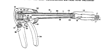
Figure 1 shows a partially cut away perspective view of a minimally invasive
ret.~ctor and ~I;sse~,10r 10 for intemal surgical use on a patient. There is a tubular
10 support 11 for passing into the body such as through a trocar opening or a cannula
placed in the body. In a particular example for laparoscopic use a trocar or cannula
placed through the abdominal wall into an insufflated body cavity would be an
appropriate place for the tubular support 11. The tubular support 11 has a proximal
end 12 located outside the patient's body and posilioned to be ~ccessed by the
15 surgeon; a gas ;ock in the form of a hemostasis valve can be used. A distal end 13 on
the tubular support 11 is positioned inside the body to provide access to the organs
within the insufll~ted cavity. Means 14 are movably carried on the tubular support 11
near the distal end 13 thereof. Specif,c~lly, one or more jointed artic~ ted members
15 are movably positioned and c~pP~le of holding and pulling tissue disposed beyond
20 the distal end 13. The tubular support 11 in Figure 1 includes a passage 16 in the
form of a lumen through which access may be gained su6star,lially along an axis ~A"
thereof during an operative procedure on the intemal tissue of the patient. The
p~s~ge 16 perrnits free axial access to the area between the articu~-ted members 15
for use as an extra way for an additional instrument to enter the body through the
25 p~cs~ge 16 of the tubular support 11.
The additional instrument capable of moving independently of the means 14
provides a cooperative functioning ability to handle the tissue simultanecusly. In
particular, the instrument passes through the passage 16 from the proximal end 12 to
beyond the distal end 13 so that while the means 14 position the tissue relative to the
30 patient and the instrument, the instrument can be independently moved and function.
In one approach, the instrument is a surgical tool and the tissue is maneuvered by the
means 14 within the body into position relative to the surgical tool. A particular surgical
tool could be a scalpel, an electrosurgical device such as a coagulating or cutter, or
WO 94/18893 PCT/US94/0L362
2~
merely video or scope, see Figure 2 for an example of the cooperation of the tool or
instrument and the means 14.
The means 14 include at least one member 15; in the various Figures 1, 2, 3,
and 6, two members 15 are shown but that is not required. If the passage 16 needs
to be larger than it is with two members 15, one member 15 is sufficient, however in
circl"nslances where the maximum flexibility is required, two members 15 may be used
with varying tips 17, as wil! be ex~l~"ed in connectiGn with Figures 5A through F. The
members 15 extend beyond the distal end and are movable relative thereto. In some
of the views, such as Figure 1, 2, 3 and 6, the rnel))bel~ 15 are pivotally mounted o
the distal end 13 of the tubular support 11 and it is also possible that the members 15
could be carried for further axial movement relative to the distal end 13 instead of
pivoting or swinging relative thereto since that might be a more straight forward, simpler
type mechanism wherein the member 15 slides relative to and along an inside wall 18
of the tubular support 11. Figure 6 suggests such an approach. The tip 17 of themember 15 is moveable relative to its member such that when it is positioned within the
cavity and relative to the axis ~A~ of the tubular support 11, tissue may be held.
Consequently, the maneuvering of intemal organs or tissue is easily acco,nr ' shed by
means of having the member 15 carried on the tubular support 11 movable relativethereto.
in Figure 1, there is a control 19 at the proximal end 12 of the tubular support11. The control 19 man;p~ tPS the means 14 and the tips 17. The control 19 permits
holding and pulling tissue disposed beyond the distal end 13 in directions relative to
the axis ~A~ of the tubular support 11. The control 19 permits movjng the instrument
within the passage 16 independent of the means 14 for oper~li"g on the held and
maneuvered tissue within the body beyond the distal end 13 thereof.
Members 15 are pref~,ably in the form of a pair of arms disposed beyond the
distal end 13 and carried for movement relative to the distal ~nd 13 for maximumflexibility in use. Such an arrangement could tend to restrict the window provided by the
passage 16 through the tubular support 11. Shown in the partially cut away portion of
Figure 1 is one form of the control 19 for the members 15 wherein a pair of conjugating
rings 20 carried about the proximal end 12 of the tubular support 11 are used tooperate the tips 17 of the members 15. These rings 20 freely rotate relative to one
another thus allowing the surgeon to rotate the control 19 relative to the tip 17 during
WO 94/18893 PCTtUS94/0L~62
21~172
placement which will make the minimally invasive r~tlactor ~I;sse-,tor 10 easy to use.
An inner ring 21 is connected by a depending cable 22 to the tip 17 so that axial
movement of the cable 22 will close the tip 17 into a clar.l~..,g position. In particular,
if the tip 17 is a forceps 23 as in Figure 5F in a tube 24, then axial movement in the
5 direction of the arrows will draw the forceps 23 into the tube 24 and thus together to
pinch tissue therebetween. The cable 22 extends along the inside wall 18 of the tubular
support 11 from beyond the distal end 13 to the proximal end 12. The cable 22 isguided along the members 15 to the tip 17 to connect to the normally open graspers
25 in Figure 5D or forceps 23 thereat for control thereof.
To operate the cable 22 there is an outer ring 26 circumscribing a depending
wall 27 on the inner ring 21 and conjugating with an inward tumed flange 28 on which
the inner ring 21 sits and within which the depending wall 27 coniug~tes. Lifting the
outer ring 26 will in tum raise the inner ring 21 and axially move the cable 22 attached
to the inside thereof. The outer and inner rings 26 and 21 allow free rotary motion
15 therebetween while control of the closure of the tips 17 rel l I&il 15 unaffected by rotation.
Specifically, to raise the outer ring 26 a bell crank 29 pivotally mounted to the control
19 is used. The bell crank 29 has an inward point 30 that engages the outer ring 26
and a button ~ctu~tor 31 located through the control 19 for easy access by the
surgeon. Push on the button ~chl~tors 31 will lift the cables 22 and the conjugate outer
20 and inner rings 26 and 21 while still allow rotalion of the control 19 relative to the
tubular support 11. Another set of rings 20 are used when there are two sets of tips
17 to be operated. The second set of rings 20 can function the same as the first and
are positioned axially above the first in Figure 1. A se"ated band 32 about the base
of the control 19 may be used to rotate it relative to the positioned tips 17 to enhance
25 use and easing hand position relative to the location of the tips 17.
A dil t:ctiGnal olive 33 is located at the distal end 13 of the tubular support 11 so
that the members 15 mounted thereto are able to swing relative thereto in more than
one plane. In particular, the normal positlon of the members 15 are juxtaposed as
shown in Figure 1 and the members 15 are spring loaded to rest in that position. When
30 it is desired to separate the members 15, then one tip 17 is clamped onto some tissue
as will be explained herein and as shown in Figure 2 so that at least one of themembers 15 is applied. After that the tubular support 11 can be maneuvered to
separate the members 15 so that the free member 15 is pointed to another area of
WO 94118893 PCT/US94/OL~62
-8-
tissue and then clamped thereto. Tension can then be applied to the tissue spanning
between the tips 17 by way of a spreader grip 34 on the control 19. Specifically, the
spreader grip 34 in Figure 1 is connected to rods 35 that pass along the inside wall
from the spreader grip 34 to the members 15 near the directional olive 33. Axial5 movement of the rods 35 pulls on the members 15 to spread them apart at their tips
17. Remembering that the normal positiGn of the members 15 is together the force of
the rods 35 acts to separate then against the spring load. The directional olive 33
allows the members 15 to pivot while maintaining the passage 16 open; that is, the
olive 33 is centrally unimpeded and while able to pivot relative to the distal end 13 does
10 not i"~ ele with the passage 16. The spring loading of the members 15 can be
integral with the members 15 so that they each are made from an elastic material which
is bent to load them into a normally closed position with enough force. Similarly, the
graspers 25 at the tips 17 could be spring loaded to be normally closed and can be
pulled open by the cables 22 although that is not the pr~r..ad approach.
Shown in the cut away in Figure 1 is a latch 36 for each of the A~tu~tor buttons31. There is a tooth edge 37 on the housing that can engage one or more steps oneach s~ct~ tor buttons 31. To release the engage",ent, the ~ tor button is pulled
back against a spring 38 to sephra~e the step from the tooth edge 37. A similar lock
39 is provided for the spreader grip 34 so that the members 15 can be held in position,
i.e. spread apart.
In Figures 3 and 4, an altemate embodiment of the tubular support is shown as
a pair of U-shaped channels 40 that cooperate and form an elongate tube 41
cGn.posed of conjugating parts movable in the direction of the axis ~A~ relative to one
another. Figure 4 specifically shows one way in which the conjugating parts are
associated with one another. It is clear that the relative movement of the U-shaped
channels 40 provides an additional degree of flexibility for the members 15 relative to
one another so that they may be not only be swung apart from one another, but also
moved axially relative to one another. The end cross sectional view of Figure 4 is
enlarged to show the detail of conjugation.
The tubular support 11 can be made out of any material appropriate for the
nature of its use and in particular a medical grade plastics, metals or ceran,ics may be
used, however, the choice of material will undoubtedly be determined by the function
of the particular configuration. While elongate tube 41 can be easily extruded U-shaped
WO 94/18893 PCT/US94/0L362
1 7 ~
channels 40, machining in addition to extrusion, or molding may be needed to obtain
the desired cross-sectional configuration necessary. It is ex~ ected that skilled artisans
will be able to fashion a tubular support 11 from single or multiple pieces in a way
which provides a thin wall and allows a maximum passage 16 theretl,ruugh while
5 providing adequate strength for carrying the members 15 at the distal end 13 thereof.
Flgure 5 illustrates a variety of tips 17; each tip 17 d;sclosed may be used to
hold and/or move tissue or organs within the body. These various tips 17 are located
at the end of the members 15 in position for placement within the body. Specifically,
Figure 5A has a r~t.acti"g hook 42 which can be slid axially in the direction of the
10 arrows to pinch or hold the tissue therewithin. A tube 24 and cooperating hook 42 are
shown for that purpose and the double arrow therein illustrates the directions of axial
movement of the hook 42 relative to the tube 24, although it is prefe"ed that the hook
42 be normally open and merely pulled closed.
Figure SB shows a suction tube which can also be used to hold the tissue.
15 Vacuum applied to the suction tube 24 can be i"le" "itlently activated in order to catch
tissue and hold it. The suction tube 24 should be a semi rigid ",a~erial to afford
maximum control. Figure 5C shows a pair of plates 43 in the nature of a vise which
can be axially moved relative to one another in order to clamp tissue therebetween.
Figure 5D shows a pair of g,aspera 25 in the nature of the jaws of a pliers which may
20 be used to clamp tissue. The y,aspera 25 are normally apart and are pulled together
to hold tissue.
Figure 5E has a pair of tongs 44 which may be used to hold the tissue instead
of Y,i~F..,g it with the teeth o~ the ylaapera 25 shown in Figure 5D. The various
configurations shown are not limiting. Other arrangements may be used as desired for
25 specific needs in connection with the members 15 and medical proce-lures. In
particular, scissGra can be mounted instead of the y,aspera 25 shown in Figure 5D.
Forceps 23 can be mounted so that they are a pair of bent together leaf sprir.gs which
are held within the tube 24; the cable 22 pulls them into the tube 24 or pushes them
axially out of the tube 24 as desired. The bend of the forceps 23 being contained
30 within the tube 24 is attached to the member 15 which is movable relative to the distal
end 13 of the tubular support 11.
In Figure 6 is shown a schematic rendering of how each member 15 could
contain a tip 17 arrangement. The tubular support 11 encases a pair of members 15
WO 94/1~93 PCTrUS94/0~62
2 -1 0-
held therein and when the members 15 are moved axially into and out of the tubular
support 11, they may spread apart or move together laterally, respectively.
Specifically, an instrument within the passage 16 may be moved axially relative
to the tubular support 11 into and out of the body cavity while the means 14 are5 moved, for example, by su:.,y;~lg or rotali"g away from the axis ~A~ such that the
maximum use may be obt~i. ,ed from the entry portal occupied by the tubular support
11 through the patient's body wall. As described, the means 14 may be artic~ ted by
rods 35 connected to the spreader grip 34 and bell cranks 29 to the actuator buttons
31 operate the tips 17 so that the members 15 spread within the body while tissue is
10 held. It is also possible that the rods 35 could be used to slide a pair of distally
supported members 15 axially into and out of the tubular support 11 and rr,ove them
relative to the instrument which can be handled individually and manually as it passes
from the proximal end 12 to the distal end 13 through the p~cs~ge 16. Therefore,while all motion might be axial, it can be independent.
A method for using the minimally invasive retractor and . I;ssector 10 for internal
surgery on a patient includes various steps. Making an opening for the tubular support
11 for passing into the body through, for exam r le, an abdominal wall in a laparoscopic
procedure for removal of gall bladder or a uterus or the like is ordinarily accomF' shed
by a trocar with a flesh cutting end which passes through the body and leaves an20 opening of about 10 mm in diameter. After the opening has been prepared, the tubular
support 11 can be inserted into the opening and positioned within the body so that its
distal end 13is in the cavity and the members 15 associated therewith are in position
for handling tissue therewithin. Another step in the method is leaving the proximal end
12 of the tubular support 11 located outside the pdtient's body and posilioned to be
25 ~ccessed by the surgeon such that control of the means 14 within the body is easily
accomplished. The surgeon may then position the distal end 13 on the tubular support
11 relative to the patient's body and allow access for the surgeon ~y way of the means
14 therein to handle the tissue or organs during a procedure. It is the means 14 which
are moved by the surgeon through the control 19 so that tissue in the body can be held
30 or pulled as desired even though it is disposed beyond the distal end 13 of the tubular
support 11. The tubular support 11 can, to some degree, be moved in and out of the
bociy or relative to the abdominal wall for example such that the distal end 13is
positioned near the tissue or organ of interest, so that the means 14 within the body
WO 94/18893 PCT/US94/0L~62
2 I ~ 1 1 7~
-11-
are able to be used most effectively. The passage 16 through the tubular support 11
can then be used for access subst~ltially aiong its axis ~A~ during the operative
pruceJure on the internai tissue of the patient and in particular a video, surgical device
or other instrument may be passed through the passage 16 into the body cavity. The
5 instrument may thereby may be able to cooperatively function on the tissue
simultaneously but independently of the means 14. The instrument is inserted into a
placed trocar while the distai end 13 thereof is viewed with a camera or scope in
another portai during the entry of the instrument. The surgeon can rotate the tubular
support 11 by its control 19 so that s~tisfs~ctory hand position is achieved. The
10 minimally invasive retractor dissector 10 may then be advanced toward the tissue to be
retracted and/or ~;sse~ted. A member 15 with a tip 17 having a forceps 23 or grasper
25 is clamped on a piece of tissue located slightly lateral with respect to the desired
plane of dissection. The opposite member 15 is positioned by means of the control 19
over toward the other side of the lissection plane and the tip 17 thereon is applied to
15 the tissue thereat. After the tips 17 are in posilion holdins the tissue the members 16
are spread apart by the surgeon with pressure on the grip 34 that cGr,l, ols the member
15 positions. Tension on the .Ji~se~;tion site is thereby obtained and can be gradually
applied as desired and needed by the surgeon.
A second surgical instrument may then be i"se,led through the passage 16 or
20 any other port for ~ I;sse.,1ion of the tissue under tension. As the tissue is rlissected by
the surgeon additionai tension may be applied to pull or spread the fascia away from
the site. As required or desired one or both of the graspers 25 or forceps 23 at the tips
17 of each member 15 can be repositiGned to clamp tissue at a di~erent place forfurther dissection after suitable tension retraction. If required the surgeon can at any
25 time release the cla",i .,9 or tension as easily as the placement was attained. If the
passage 16 is used for the surgicai instrument, then movement of the tubular support
11 will act to guide the surgical instrument. An olive 33 located near the distal end 13
of the tubular support 11 acts to guide the instrument inserted ther~tl,rough during
movement of the tubular support 11 relative to the tensioning members 15.