Note: Descriptions are shown in the official language in which they were submitted.
WO 94/17208 2151265 PCT/US94/00583
METHODS AND DIAGNOSTIC KITS UTILIZING MAMMALIAN
STRESS PROMOTERS TO DETERMINE TOXICITY OF A COMPOUND
TECHNICAL FIELD OF INVENTION
This invention provides methods and
diagnostic kits for identifying and characterizing
toxic compounds. These methods and diagnostic kits
measure transcription or translation levels from genes
linked to native eukaryotic stress promoters,
especially those of mammals. The kits and methods of
this invention utilize at least one stress promoter
from each of the following groups: redox stress, DNA
stress, protein stress and energy/ionic stress. The
invention also provides methods and diagnostic kits for
identifying and characterizing compounds that are toxic
to specific organs, such as skin and the eye, as well
as for each of the individual stresses indicated above.
The methods and diagnostic kits of this invention yield
information concerning the action of a compound on a
subcellular level. This information may be utilized to
design antitoxins to compounds found to be toxic and in
active drug design.
BACKGROUND OF THE INVENTION
At least 55,000 chemicals are presently
produced in the United States. Over 2,000 new
chemicals are introduced into the market each year.
Very few of these chemicals have been comprehensively
tested for acute or chronic toxicity. For example,
less than 1 percent of commercial chemicals have
undergone complete health hazard assessment.
The Environmental Protection Agency ("EPA:")
has the authority to require toxicological testing of a
chemical prior to commercial production, but that
authority is rarely invoked. Less than 10 percent of
new chemicals are subjected to detailed review by the
WO 94/17208 PCT/US94/00583
2.5426 -2-
EPA. In the interest of cost and speedy access to the
market, the EPA often uses the toxicity of previously
tested homologous compounds to gauge the toxicity of a
new chemical.
The potential toxicity of new drugs is
monitored by the Food and Drug Administration ("FDA").
For a New Drug Application (NDA), the FDA typically
requires a large battery of toxicity, carcinogenicity,
mutagenicity and reproduction/fertility tests in at
least two species of live animals. These tests are
required to last up to one year. A two year toxicity
test in rats costs approximately $800,000 [Casarett and
Doull's Toxicology, 4th Edition, M. 0. Amdur et al.,
eds. Pergamon Press, New York, New York, p. 37 (1991)].
Besides cost, animal testing also presents
disadvantages in terms of time, animal suffering and
accuracy. Typical toxicity tests are divided into
three stages: acute, short term and long term. Acute
tests, which determine the LD50 of a compound (the dose
at which 50% of test animals are killed), require some
60-100 animals and a battery of tests for determining
LD50, dose-response curves and for monitoring clinical
end points, other than death. Short term tests usually
involve at least 24 dogs and 90 rats and last from 90
days in rats to 6-,24 months in dogs. Body weight, food
consumption, blood, urine and tissue samples are
frequently measured in the short-term tests. In
addition, dead animals are subjected to post-mortem
examinations. Long term tests are similar to short
term tests, but last 2 years in rats and up to 7 years
in dogs or monkeys.
Animal testing has come under criticism by
animal rights activists and the general public because
of the severe suffering inflicted on the animals.
Moreover, recent evidence calls into question the
accuracy of animal testing. For example, variables,
CA 02154265 2004-06-01
61009-256
-3-
such as animal diet, may impair the predictability of
animal tests in determining carcinogenic properties
[P. H. Abelson, "Diet and Cancer in Humans and
Rodents", Science, 255, p. 141 (1992)]. And prior
determinations on dioxin toxicity, based on guinea pig
testing, are now being reevaluated [B. J. Culliton, "US
Government Orders New Look At Dioxin", Nature, 352,
p. 753_(1991); L. Roberts, "More Pieces in the Dioxin
Puzzle", Research News, October, 1991, p. 377]. It is
therefore apparent that there is an urgent need for a
quick, inexpensive and reliable alternative to toxicity
testing in animals.
several short-term alternative tests are
available. For example, the Ames Assay detects
carcinogens which cause-genetic reversion of mutant
strains of Salmonella tvohimurium. However, the Ames
Assay cannot detect either non-mutagenic carcinogens or
non-carcinogenic toxins. The yeast carcinogen assay
system described in United States patent 4,997,757
overcomes some of the drawbacks of the Ames Assay, but
is still not able to detect non-carcinogenic toxins.
Both of these assays are designed to detect alterations
and mutations at the DNA level only. Therefore, those
prior art tests cannot detect direct damage to proteins
or lipid membranes, nor inhibitors of DNA synthesis.
Moreover, those prior art tests cannot provide
information as to how a mutagen or toxin exerts its
effect.
WO 90/10710 describes the use of a TNF, IL-la
or IL-1fl fused to a reporter gene to detect bacterial
pyrogens. However, the disclosed assay is limited in
that it detects only a particular stress (bacterial
pyrogens) and yields no qualitative information about.
how the pyrogen exerts its toxic effect.
CA 02154265 2004-06-01
61009-256
-3A-
United States Patent 5,589,337 describes an assay which
utilizes a reporter gene fused to bacterial stress promoters
to determine and characterize the toxicity of a compound.
This assay is able to detect damage to proteins or lipid
membranes and inhibition of DNA synthesis. Thus,
WO 94/17208 PCT/US94/00583
q~C~h ~f
~+ - 4 -
this assay provides for the identification of non-
carcinogenic toxins. Unfortunately, the correlation
between bacterial toxicity and toxicity to mammals and
other higher eukaryotes has certain limitations and may
not be an accurate measure of toxicity in higher
animals.
Therefore, there is still a need for an assay
that has the time and cost-saving features of the
bacterial stress assay, but is based on a eukaryotic
cell.
SUMMARY OF THE INVENTION
Applicant has fulfilled this need by
providing an in vitro diagnostic kit and assay method
which identify and characterize the cellular and sub-
cellular effect of a potential toxin on an animal cell.
These kits and methods employ the native stress
promoters of eukaryotic cells, preferably mammalian
cells, and measure the level of transcription or
translation of a gene which is operatively linked
thereto. Depending upon the choice of stress promoters
used, the kits and methods of this invention may be
designed to identify and characterize compounds that
are toxic to the whole animal or to specific organs of
that animal.
In one embodiment, the kits and methods of
this invention characterize the toxicity of a compound
by determining the level of transcription of various
stress genes present in a eukaryotic cell. These kits
and methods employ oligonucleotides that are
complementary or homologous to at least a portion of
various stress gene messenger RNAs to detect
transcription of those genes in the cell. In this
embodiment a single cell is effectively an in vivo
diagnostic reagent for determining what particular
stress a given compound induces.
CA 02154265 2010-01-06
61009-256
-5-
In another embodiment, each of a plurality of similar
eukaryotic cells harbors a different stress promoter operatively
linked to a reporter gene. By exposing each cell separately to
a compound and measuring the expression of the reporter gene
product, the toxicity of that compound may be characterized.
The kits and methods of this invention are
optimally designed to determine the toxicity of a compound
in a matter of days, rather than the months or years
required for animal testing. Furthermore, the kits of this
invention achieve these results for a fraction of the cost
of animal testing and without the objectionable consequences
to live animals. And, the diagnostic kits and methods of
this invention yield direct information about the nature of
a toxin's action on mammalian cells -- something that the
prior art short-term assays fail to do.
In one aspect, there is described a diagnostic kit
for identifying and characterizing a toxic compound
comprising: (a) a eukaryotic cell characterized by: (i) at
least one promoter that responds only to redox stress; (ii) at
least one promoter that responds only to DNA stress; (iii) at
least one promoter that responds only to protein stress; and
(iv) at least one promoter that responds only to energy/ionic
stress, each of said promoters being operatively linked to a
different gene which encodes a different detectable product;
and (b) at least four different oligonucleotide probes, each
of said probes being capable of hybridizing to the mRNA
transcript of a different one of said genes, or to a single
stranded cDNA prepared from said mRNA transcript, wherein said
identification and characterization is achieved by creating a
stress promoter induction profile comprising data identifying
and quantifying the redox, DNA, protein and energy/ionic
stress that the compound causes to the eukaryotic cell.
CA 02154265 2010-01-06
61009-256
-5a-
In another aspect, there is described a diagnostic kit
for identifying and characterizing a toxic compound comprising
at least: (a) a first eukaryotic cell that harbors at least one
promoter or response element that responds only to redox stress;
(b) a second eukaryotic cell that harbors at least one promoter
or response element that responds only to DNA stress; (c) a
third eukaryotic cell that harbors at least one promoter or
response element that responds only to protein stress; and (d) a
fourth eukaryotic cell that harbors at least one promoter or
response element that responds only to energy/ionic stress;
wherein, each of said promoters or response elements is
operatively linked to a heterologous gene encoding a detectable
product, wherein said identification and characterization of
said toxic compound is achieved by creating a stress promoter
induction profile comprising data identifying and quantifying
the redox, DNA, protein and energy/ionic stress that the
compound causes to the eukaryotic cell.
In another aspect, there is described a method for
determining the toxicity of a compound to a eukaryotic cell by
creating a stress promoter induction profile comprising data
identifying and quantifying the redox, DNA, protein and
energy/ionic stress that the compound causes to the cell, the
method comprising the steps of: (a) separately culturing one or
more eukaryotic cells that, in toto, are characterized by:
(i) at least one promoter or response element that responds only
to redox stress; (ii) at least one promoter or response element
that responds only to DNA stress; (iii) at least one promoter or
response element that responds only to protein stress; and (iv)
at least one promoter or response element that responds only to
energy/ionic stress, each of said promoters or response elements
being operatively linked to a gene that encodes a detectable
product; (b) exposing each of said one or more cultures of cells
to said compound; (c) quantifying the detectable product in each
of said cultures; and (d) creating the stress promoter induction
profile from data resulting from step (c) for said compound.
CA 02154265 2010-01-06
61009-256
-5b-
In another aspect, there is described a method of
identifying an antitoxin to a new toxic compound comprising the
steps of: (a) determining the types of stresses caused by said
new toxic compound by the method of the invention;
(b) identifying a known toxic compound that, in the process of
the invention, causes stresses similar to those caused by said
toxic compound; and (c) repeating the method used to determine
the types of stresses caused by said new toxic compound
according to step (a) with the additional step of treating the
eukaryotic cells employed in said method with an antitoxin to
said known toxic compound identified in step (b).
BRIEF DESCRIPTION OF THE DRAWINGS
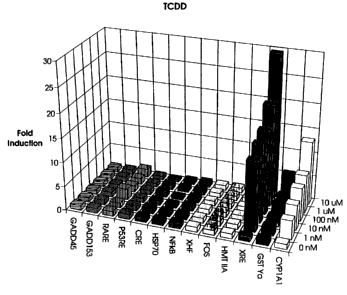
Figure 1 depicts the relative expression of
chloramphenicol acetyl transferase under the control of
different stress promoters in the presence of varying
concentrations of tetrachlorodibenzo-p-dioxin (TCDD).
Figure 2 depicts the relative expression of
chloramphenicol acetyl transferase under the control of
different stress promoters in the presence of varying
concentrations of 3-methyl cholanthrene (3-MC).
Figure 3 depicts the relative expression of
chloramphenicol acetyl transferase under the control of
different stress promoters in the presence of varying
concentrations of benzo[a]pyrene.
Figure 4 depicts the relative expression of
chloramphenicol acetyl transferase under the control of
different stress promoters in the presence of varying
concentrations of cadmium sulfate.
WO 94/17208 PCTIUS94/00583
-6-
Figure 5 depicts the relative expression of
chloramphenicol acetyl transferase under the control of
different stress promoters in the presence of varying
concentrations of dimethyl sulfoxide (DMSO).
Figure 6 depicts the relative expression of
chloramphenicol acetyl transferase under the control of
different stress promoters in the presence of varying
concentrations of ethanol.
Figure 7 depicts the relative expression of
chloramphenicol acetyl transferase under the control of
different stress promoters in the presence of varying
concentrations of methapyrilene hydrochloride.
Figure 8 depicts the relative expression of
chloramphenicol acetyl transferase under the control of
different stress promoters in the presence of varying
concentrations of methyl methanesulfonic acid (MMS).
Figure 9 depicts the relative expression of
chloramphenicol acetyl transferase under the control of
different stress promoters in the presence of varying
concentrations of sodium arsenate.
Figure 10 depicts the relative expression of
chloramphenicol acetyl transferase under the control of
different stress promoters in the presence of varying
concentrations of phorbol 12-acetate-13-myristate
(PMA).
Figure 11 depicts the relative expression of
chloramphenicol acetyl transferase under the control of
different stress promoters in the presence of varying
concentrations of retinoic acid.
DETAILED DESCRIPTION OF THE INVENTION
As used herein, the terms "stress" and
"toxicity" are used interchangeably and refer to the
disturbance of the biochemical and biophysical
homeostasis of the cell.
WO 94/17208 PCT/US94/00583
2154265
-7-
The term "redox stress", as used throughout
this application, refers to conditions which vary from
the normal reduction/oxidation potential ("redox")
state of the cell. Redox stress includes increased
levels of superoxides radicals, increased levels of
peroxides -- both hydrogen peroxide and organic
peroxides --, decreased levels of glutathione and any
other conditions which alter the redox potential of the
cell, such as exposure to strong reducing agents, some
aromatic hydrocarbons, electrophilic compounds,
aldehydes, intracellular thiols, steroids, methyl
cholanthrene, phenobarbital and CC14. The term also
includes any additional conditions which cause
proliferation of peroxisomes.
The term "DNA stress", as used herein, refers
to alterations to deoxyribonucleic acid or to precursor
nucleotides. For example, DNA stress includes, but is
not limited to, DNA strand breaks, DNA strand cross-
linking, exposure to DNA intercalating agents, both
increased and decreased superhelicity, oxidative DNA
damage, DNA alkylation, oxidation of nucleotide
triphosphates and alkylation of nucleotide
triphosphates. The term also includes inhibition of
DNA synthesis and replication and inhibition of mitosis
or meiosis. And the term includes conditions caused by
exposure to growth factors, interferons, tumor
promoters, tumor necrosis factor, phorbol esters,
hydrophobic cytotoxic drugs, inflammatory agents,
mitogens, carcinogens, X-rays, UV radiation and
dimethylnitrosomines.
"Protein stress", as used throughout the
application, refers to alterations to proteins or
= individual amino acids and inhibition of enzyme
functions, as well as perturbations of intracellular
transport of proteins. The term includes, but is not
limited to, denaturation of proteins, misfolding of
WO 94/17208 PCTIUS94/00583
-8-
proteins, chelation of protein cofactors, cross-linking
of proteins, both oxygen-dependent and -independent
oxidation of inter- and intra-chain bonds, such as
disulfide bonds, alkylation of proteins, oxidation of
individual amino acids and protein damage caused by
exposure to heavy metals, such as cadmium and heat.
I use the term "energy/ionic stress" to
encompass conditions which affect ATP levels in the
cell or ionic gradients across a cell membrane.
Examples of energy stress are forced anaerobic
metabolism in the presence of oxygen, perturbations of
electron transport, exposure to uncoupling agents,
membrane depolarization, osmotic shock, exposure to
ions, such as Cat+, exposure to high levels of CAMP and
exposure to ethanol.
The term "cell surface receptor-mediated
stress" refers to those conditions which alter the
transcription level of genes whose expression is
regulated by the interaction of a cell surface receptor
with a ligand. Examples of such stress include
exposure of the skin, eyes or mucous membranes to
irritants, allergens or inflammatory compounds.
The term "stress promoter induction" refers
to conditions which increase the level of expression of
a gene product operably linked to a native stress
promoter or a recombinantly derived stress promoter
which contains a response element. The term "operative
linkage", "operatively linked" or "operably linked"
refers to the positioning of the promoter relative to
the gene such that transcription of the gene is
regulated by the promoter. The term encompasses both
recombinant constructs, as well as the structure of a
naturally occurring promoter and its associated gene.
The term "determining and characterizing the
toxicity of a compound" includes identifying a compound
WO 94/17208
PCTIUS94/00583
-9-
as a toxin and elucidating its mechanism of action
within the cell.
The term "nucleic acid sequences" as used in
this application, includes RNA, single or double-
stranded cDNA or portions thereof, single or double-
stranded genomic DNA or portions thereof, or single or
double stranded synthetic oligonucleotides.
Whereas every gene is controlled by a unique
promoter, genes which respond to identical stresses
contain a common response element within their
promoters. Accordingly, the same response element is
responsible for inducing expression of a family of
genes upon exposure to a certain stress. When isolated
and operably linked to a minimal promoter and a
structural gene, the resulting construct functions like
a stress promoter. This is particularly useful in
dissecting a native stress promoter that responds to
multiple stresses into its component parts.
Individual cells respond to toxic stimuli, in
part, by activating specific genes whose protein
products detoxify the stimuli or repair damage caused
thereby. Eukaryotic cells have large number of genetic
and biochemical responses to damage and stress. At
least 50 different mammalian stress genes have already
been isolated and characterized. These genes are
induced by a variety of chemical and physical stresses
or cellular damage.
Among the chemical stresses which induce one
or more of these identified genes are exposure of the
cell to mercury, heavy metals, nitroxides, aromatic
hydrocarbons, acidity, basicity, alkylating agents,
peroxidizing agents, cross-linking agents, ionophores,
redox active agents, electrophilic compounds,
inflammatory agents, hydrophobic cytotoxic drugs,
ethanol, steroids, uncoupling agents, tumor promoters
and cellular factors, such as tumor necrosis factor,
WO 94/17208 PCT/US94/0058'
-10-
growth factors and interferon. Physical stresses
include exposure to UV radiation, heat or X-rays.
Examples of cellular damage which induce
these identified genes are lipid oxidation, peroxisome
proliferation, DNA strand breaks, DNA alkylation, DNA
cross-linking, DNA oxidation, osmotic imbalance,
protein oxidation, protein misfolding, protein
alkylation, ATP depletion, membrane permeabilization,
glutathione depletion and alterations in signal
transduction. Many more stress genes are believed to
exist. The identification and characterization of
these additional stress genes is highly desirable in
understanding what effects various chemical stresses
have on the cell.
The present invention provides diagnostic
kits and methods for determining and characterizing the
toxicity of a compound in terms of the type of damage
it causes within the cell, i.e., DNA damage, protein
damage, redox damage, energy damage, ionic damage, etc.
According to one embodiment, each diagnostic kit of
this invention comprises a plurality of eukaryotic
cells, each of which harbors at least one promoter or
one promoter element which responds to stress. The
plurality of cells, in toto, must comprise at least one
promoter or promoter element which responds to each of
the aforementioned types of stresses -- redox, DNA,
protein and energy/ionic -- operably linked to a gene
encoding a detectable product.
According to one embodiment, the plurality of
cells in this kit are actually a single cell line,
wherein each cell contains all of the different types
of stress promoters and wherein each of those promoters
is activated upon exposure to the appropriate stress.
In this embodiment, the genes operatively linked to the
stress promoters are most preferably the native stress
genes. In this manner no genetic manipulation need be
WO 94/17208 ' - PCTIUS94/00583
5-
-11-
carried out on the cells prior to running an assay. In
this preferred embodiment, the kits further comprise
oligonucleotides or cDNAs which are complementary to at
least a portion of either the coding or non-coding
strand of the genes under control of the specific
stress promoters. The oligonucleotides are used to
detect and quantify the mRNA transcripts of those genes
or the cDNA complement thereof, either of which may be
the detectable product in this embodiment.
It should be noted that although all
eukaryotic cells contain numerous stress promoters
within their genomes, some of those promoters may or
may not be activatable upon exposure to the proper
stress. This is especially true in higher eukaryotes,
such as mammals. Those cell lines whose stress
promoters do respond to almost all of the appropriate
stresses are preferred in the kits of this invention.
These include primary tissue from mammalian liver,
heart, lung, kidney, brain, or other organ, as well as
mammalian derived cell lines established from these
tissues available from American Type Culture Collection
(ATCC, Rockville, MD). More preferred are HepG2 cells,
HeLa cells and WIL-2 cells. Most preferred are HepG2
cells.
The oligonucleotides employed in the above
diagnostic kits and methods of this invention are
chosen based upon their ability to specifically
hybridize under relatively high stringency conditions
to the either the transcription product of the gene
operatively linked to the various stress promoters or
= its complement (i.e., a single-stranded cDNA reverse
transcribed from that mRNA). The choice of utilizing
= complementary or homologous oligonucleotides depends
upon the method used for detecting the transcription
products. These various methods are described later in
the application.
WO 94/17208 PCT/US94/0058'
-12-
Because the DNA sequence of many mammalian
stress genes are known, hybridizable oligonucleotides
are easily constructed. It should be noted that 1001i
homology or complementarity between the oligonucleotide
and the stress gene mRNA is not required. This is
because the oligonucleotide may be designed based upon
the sequence of a stress gene from a species different
from the source of the cells utilized in the kits and
methods of this invention.
While it is expected that similar stress
genes from different mammalian species will be closely
related, the transcripts from those genes will most
likely not have identical nucleotide sequences.
Accordingly, the oligonucleotides utilized in the kits
and methods of this invention are preferably at least
95% homologous or complementary. Preferably, the
oligonucleotides are between 20 and 500 base pairs
long. Most preferably, the oligonucleotides are
between 50 and 100 base pairs.
More preferably, the oligonucleotides are
synthesized using an oligonucleotide synthesizer,
optionally followed by polymerase chain reaction
("PCR"). In this procedure, an oligonucleotide having
a sequence identical to a portion of either the
template strand or the non-coding strand and within the
coding region of a known, sequenced stress gene is
synthesized. If PCR is to be used to increase the
quantity of oligonucleotide, the oligonucleotide is
synthesized with an additional 6 to 12 nucleotides at
each end. Those extra nucleotides serve as targets for
complementary primers in a PCR reaction. Preferably
the extra nucleotides at each end are complementary to
one another. This allows a ,single primer to prime off
of both the original oligonucleotide and the PCR
product thereof. Most preferably, the extra
WO 94/17208 PCTIUS94/00583
-13-
nucleotides at each end are complementary homohexamers,
i.e., AAAAAA at one end and TTTTTT at the other.
During PCR, one or more labeled nucleotides
are preferably included in the polymerase reaction.
Preferably the label is 32P, biotin or a fluorescent
marker. This results in a labelled product that can be
used directly to detect the level of transcription
product. The advantage of this mixed oligonucleotide
synthesizer/PCR technique is that microgram quantities
of labelled oligonucleotide can be produced in a single
procedure. The resulting oligonucleotides may
optionally be biotinylated following synthesis and
purification.
If the oligonucleotide is used to detect cDNA
reverse transcripts of the transcription product, it is
preferable that they not be labelled. In this
embodiment, it is preferred that the label be
incorporated into the cDNA, rather than the
oligonucleotide.
The design of appropriate oligonucleotide
probes for use in the kits and methods of this
invention is relatively straightforward. Obviously,
they should have high sequence similarity or
complementarity to the stress gene mRNA to which they
are designed to hybridize. The oligonucleotides in any
particular kit should also have approximately the same
melting temperature (Tm) so that a single warming
apparatus (such as a water bath) may be utilized when
carrying out hybridization and subsequent washing
steps. Preferably the oligonucleotides are designed to
have a T. of greater than 70 C in 0.2X SSC. To
determine which portions of the coding regions of the
stress gene to use in designing oligonucleotide probes,
one may utilize a commercially available computer
program, such as OLIGO (National Biosciences, Plymouth,
WO 94/17208 d5, PCT/US94/0058.'
-14-
According to another embodiment, each of the
plurality of eukaryotic cells in the diagnostic kit of
this invention harbors a stress promoter or a stress
response element which is operatively linked to a
heterologous gene encoding a detectable product. In
this embodiment, it is preferable that the same
heterologous gene be linked to the various stress
promoters or response elements in the kit. In this
manner, only a single assay need be performed to detect
induction of any of the stress promoters and stress
response elements. It is also preferable that each
cell within the kit contains only a single stress
promoter or response element/heterologous gene
construct. Thus, the expression of the detectable
product in any given cell in the kit can be
specifically correlated to the induction of a single
.stress promoter or response element.
The diagnostic kits and methods of this
invention employ a plurality of eukaryotic cells,
which, in toto, comprise promoters or response elements
that respond to each of: redox stress, DNA stress,
protein stress and energy stress. The preferred
promoters and response elements of this invention for
use with mammalian cells are listed below in Table 1.
TABLE 1
Preferred Mammalian Stress Promoters
Energy/
Promoter Redox DNA Protein Ionic
CYP1A1 X
GST Ya X X
GADD45 X
GRP78 X X X
JUN X X X
FOS X X X
WO 94/17208 PCTIUS94/00583
-15-
Table 1 (cont'd)
Energy/
Promoter Redox DNA Protein Ionic
XHF X
HSP70 X
MTIIA X X
GADD153 X
ALDH 1 X
HMO X X
CRE X
XRE X
NFkBRE X X
RARE X
ThRE X
PPRE X
THE X
ERE X
p53RE X
Preferably, the promoters or response
elements which respond to redox stress in the methods
and kits of this invention are selected from the
promoters of the CYP1A1, GST Ya, JUN, ALDH1 and HMO
genes and the XRE, NFkBRE, PPRE, RARE, ERE, and ThRE
response elements.
The CYP1A1 gene encodes cytochrome P450 1A1,
an enzyme involved in the metabolism of polycyclic
aromatic hydrocarbons, such as benzo(a)pyrene. The
gene is inducible by aromatic hydrocarbons, plant
flavones and also by tetrachlorodibenzo-p-dioxin
(TCDD), one of the most potent teratogens and tumor
= promoters [L. A. Neuhold et al., Mol. Cell. Biol., 9,
pp. 2378-2386 (1989); Y. Fujii-Kuriyama et al., The
FASEB J., 6, pp. 706-710 (1992); D. W. Nebert et al.,
Env. Health Perspec., pp. 13-25 (1990); R. A. Dixon et
al., Biol. Rev., 61, pp. 239-241 (1986)]. The sequence
of this gene is described in K. Sogawa et al., Proc.
CA 02154265 2004-06-01
61009-256
-16-
Natl. Acad. Sci. USA, pp. 8044-8048 (1986).
The GST Ya gene encodes the glutathione S-
transferase Ya subunit, a unique xenobiotic-responsive
element. The redox stress-sensitive portion of the GST
Ya promoter is strongly induced by electrophilic
herbicides, insecticides and planar aromatic
hydrocarbons such as 0-naphthoflavone and 3-
methylcholanthrene IT. H. Rushmore et al., Proc. Natl.
Acad. Sci. USA, 87, pp. 3826-3830 (1990)]. The
sequence of this gene is described in T. H. Rushmore et
al., supra.
The JUN oncogene codes for c-jun which
participates in the formation of the AP-1 complex -- a
transcriptional activator. Redox stresses which
activate the JUN gene are superoxide radicals and UVA
radiation. The sequence of this gene is described in
R. De Groot et al., EMBO J., 10, pp. 2523-2532 (1991).
The ALDH 2 gene encodes aldehyde
dehydrogenase and is induced by aldehydes and
peroxisome proliferators [D. W. Nebert, Env. Health
Persil., 88, pp. 13-25 (1990)]. The sequence of that
gene is described in L.C. Hsu et al., Proc. Natl. Acad.
Sci. USA, 82, pp. 3771-3775 (1985).
The HMO gene codes for heme oxygenase. The
promoter is induced by the following redox stresses:
oxidative stress, hydrogen peroxides, and-sodium
arsenite [S. T. Keyse and R. M. Tyrell, Proc. Natl.
Acad. Sci. USA, 86, pp. 99-103 (1989)]. The sequence
of this gene is described in that document.
CA 02154265 2004-06-01
61009-256
-17-
The XRE is a redox stress response element.
It responds to xenobiotics, such as aromatic
hydrocarbons (T. H. Rushmore et al., Proc. Natl. Acad.
Sci. USA , 87, pp. 3826-3830 (1990)). The sequence of
this response element is described in that document.
NFkBRE is a redox stress response element
which encodes a transcription factor that is activated
by intracellular thiols [R. Schreck et al., EMBO J.,
10, pp. 2247-2258 (1991); B. Nelson et al., Molec.
Cell. Biol., 8, pp. 3526-3531 (1988)]. It also
responds to DNA stress. The sequence of this response
element is described in K. Leung and G. J. Nabel,
Nature, 333, pp. 776-778 (1988).
PPRE is the peroxisome proliferation response
element. It is a redox stress responsive element that
is induced by peroxisome proliferators [C. Dreyer et
al., Cell, 68, pp. 879-887 (1992)]. The sequence of
this response element is described in that document.
RARE is the retinoic acid response element.
It is a redox stress-sensitive response element that
responds to the steroid hormone retinoic acid and its
analogs [H. de The, et al., Nature, 343, pp. 177-180
(1990)].
ERE is the estrogen response element. It
responds to redox stress that is induced by estrogenic
compounds. The sequence of the ERE is described in V.
Kumar, et al., Cell, 55, pp. 145-156 (1988).
CA 02154265 2004-06-01
61009-256
-18-
ThRE is the thyroid hormone response element.
It responds to redox stress that is induced by thyroid
hormone and its analogs. The sequence of the ThRE is
described in M. Beato, Cell, 56, pp. 335-344 (1989).
Other promoters and response elements which
respond to redox and can be utilized in the kits and
methods of this invention may be selected from those
listed in Table 2, below. In the brief description of
each of these genes and response elements that follows,
the document which discloses the DNA sequence of the
particular gene is indicated in brackets.
UGT encodes a UDP-glucoronosyl transferase
and its redox response is induced by 3-methyl
cholanthrene [T. Iyanagi et al, J. Biol. Chem, 261, pp.
15607-14 (1986)]. CYP11B2 encodes a cytochrome P450
whose redox response is induced by steroids [T.
Kawamoto et al., pros. Natl. Acad. Sci. USA, 89, pp.
1458-62 (1992)). Cu.ZnSOD encodes a superoxide
dismutase that is induced by copper- and zinc-catalyzed
superoxide formation [E. Danciger et al., Proc. Natl.
Acad. Sci. USA, 83, pp. 3619-23 (1986)]. The MnSOD
gene encodes a superoxide dismutase gene which can be
activated by tumor necrosis factor, interleukin-2 and
lipopolysaccharides [M. K. St. Clair and J. C. Holland,
Cancer Res., 51, pp. 939-943 (1991)]. ADPRT encodes a
ribosyl transferase and is induced by oxidative stress.
GP encodes glutathione peroxidase and its redox
response is induced by peroxides [S. Chada, Genomics,
6, pp. 268-71 (1990)]. FAOxase encodes fatty acyl-CoA
oxidase and is induced by peroxisome proliferators [S.
WO 94/17208 PCT/US94/00583
-19-
Miyazawa et al., J. Biol. Chem., 262, pp. 8131-37
(1987)]. PBE encodes a peroxisomal enoyl-CoA
hydratase/3-hydroxyacyl CoA dehydrogenase bifunctional
enzyme and is induced by peroxisome proliferators [J.
K. Reddy et al., Proc. Natl. Acad. Sci. USA, 83, pp.
1747-51 (1986)]. PPAR encodes a peroxisome
proliferator-activated receptor and is induced by
peroxisome proliferators (C. Dreyer et al., Cell, 68,
pp. 879-87 (1992)]. EH encodes an epoxide hydrolase
which responds to redox stress caused by phenobarbital
[R. K. Skoda et al., J. Biol. Chem.., 263, pp. 1549-54
(1988)]. CYP2B2 [J. S. Miles et al., Nucl. Acids Res.,
16, pp. 5783-95 (1988)], CYP2E1 [J. E. Freeman et al.,
Biochem. J., 28, pp. 689-95 (1992)] and CYP3A3 [N. K.
Spurr et al., GenBank Accession number X12387] encode
three different cytochrome P450s. They are responsive
to redox stress caused by phenobarbital (2B2), CC14
(2E1), and aflatoxin, cyclosporin, testosterone and
nifedipine (3A3), respectively. The P450b gene encodes
the cytochrome P450b which is induced by phenobarbital
[C. M. Giachelli, et al., J. Biol. Chem., 264, pp.
7046-7053 (1989). The P450d gene encodes cytochrome
P450d, which is induced by polycyclic aromatic
hydrocarbons, isosafrole, and 3-amino-l-5'H-pyrido[4,3-
b]indole (Trp-P-2) [K. Sogawa et al., J. Biol. Chem.,
260, pp. 5026-5032 (1985). PPa encodes a poly (ADP-
ribose) polymerase and has a redox stress-sensitive
component which responds to lipid peroxidation and
oxidative stress [K. Uchida et al, Biochem. Biophvs.
Res. Comm., 148, pp. 617-22 (1987)]. PKC encodes
protein kinase C and its redox stress-sensitive
component is induced by lipid peroxidation. ALDH1
encodes another aldehyde dehydrogenase that is induced
by aldehydes and peroxisome proliferators [L.C. Hsu et
al., Proc. Natl. Acad. Sci. USA, 82, pp. 3771-75
(1985)]. The NMO1 gene encodes the NAD(P)H menadione
CA 02154265 2004-06-01
61009-256
-20-
oxidoreductase and is induced by various xenobiotics
including planar aromatic compounds, azo dyes and
phenolic antioxidants [L. V. Favreau and C. B. Pickett,
J. Biol. Chem., 266, pp. 4556-4561 (1991)]. The GST2
gene encodes glutathione S-transferase-2 and responds
to similar redox stresses as GST Ya [P. G. Board et
al., Proc. Natl. Acad. Sci. USA, 84, pp. 2377-81
(1987)]. The GAPDH gene encodes glyceraldehyde-3-
phosphate dehydrogenase [L. Ercolani et al., J. BIol.
Chem., 263, pp. 15335-41 (1988)]. The NQO gene encodes
NAD(P)H quinone oxireductase and responds to the same
redox stresses as NMO [A. K. Jaiswal, Biochemistry, 30,
pp. 10647-53 (1991).
The promoters and response elements which
respond to DNA stress that are useful in the methods
and kits of this invention are preferably selected from
the promoters of the GST Ya, GADD45, JUN, FOS, XHF and
GADD153 genes and the THE and p53RE response elements.
The GST Ya gene is described above. Its DNA
stress-sensitive component is induced by alkylated DNA.
The GADD45 gene encodes a growth arrest and
DNA damage responsive protein. The GADD45 gene is
induced by UV irradiation, X-rays, and the DNA damaging
agent, methyl methane sulfonate (MMS). This gene is
described in Q. Than et al., Mol. Cell Biol., 13, pp.
4242-50 (1993).
The JUN gene has a DNA stress-sensitive
component that is induced by UVA radiation, tumor
promoters and growth factors.
The FOS gene encodes the oncogene c-fos. The
DNA stress-sensitive components of its promoter are
induced by tumor promoters and growth factors [E. M.
Haliday, EMBO J., 10, pp. 109-115 (1991)]. The
sequence of this gene is described in F. van Straaten
et al., Proc. Natl. Acad. Sci. USA, 80, pp. 3183-3187
CA 02154265 2004-06-01
61009-256
-21-
(1983).
The XHF gene codes for collagenase and is
activated by mitogenesis, inflammatory agents, W
radiation, and also in response to the tumor promoter,
12-O-tetradecanoyl-phorbol-13-acetate (TPA). The
sequence of this gene is described in P. Angel et al.,
Mol. Cell. Biol., 7, pp. 2256-2266 (1987).
The GADD153 gene is expressed in response to
growth arresting signals and DNA damaging agents [J. D.
Luethy and N. J. Holbrook, Cancer Res., 52, pp. 5-10
(1992)). The sequence of this gene is described in
A.J. Fornace et al., Mol. Cell. Biol., 9, pp. 4196-4203
(1989).
THE is the TPA response element. It responds
to DNA stress induced by phorbol esters. The sequence
of THE is described in P: Angel et al., Cell, 55, pp.
875-85 (1988).
p53RE in the p53 response element. It is
responsive to DNA stress and is induced by X-rays and
MMS. The sequence of the p53RE is described in Q.
Zahn, et al., Mol. Cell. Biol., 13, pp. 4242-4250
(1993).
Other promoters which respond to DNA stress
and are useful in the methods and kits of this
invention are listed in Table 2, below. In the brief
description of each of these gene that follows, the
document which discloses the DNA sequence of the
particular gene is indicated in brackets.
WO 94/17208 PCTIUS94/00583
-22-
The EGR-1 gene encodes an early growth
response factor and is induced by mitogenesis and
phosphatase inhibitors [S. V. Suggs, Nucl. Acids Res.,
18, pp. 4283-89 (1990)]. The GAS 2,3 gene encodes a
gene which responds to growth arrest [C. Schneider et
al., Cell, 54, pp. 787-793 (1988)]. The MGMT encodes
an 0-6-methylguanine methyltransferase and is induced
by alkylated DNA [K. Tano et al., Proc. Natl. Acad.
Sci. USA., 87, pp. 686-90 (1990)]. DNA Pol encodes DNA
polymerase A and is induced by mitogens. TK (which
encodes thymidine kinase) [H. D. Bradshaw Jr. et al.,
Mol. Cell. Biol.], 4, pp. 2316-20 (1984)], DHFR (which
encodes dihydrofolate reductase) [C. Morandi, J. Mol.
Biol., 156, pp. 583-607 (1982)] and PCNA (which encodes
proliferating cell nuclear antigen) [D. Jaskulski et
al., J. Biol. Chem., 263, pp. 10175-79 (1988)] each are
induced by cell proliferation. PGHS encodes
prostaglandin endoperoxidase synthase and is induced by
mitogens [S. A. Kraemer et al., Arch. Biochem.
Biophys., 293, pp. 391-400 (1992)]. LOX encodes a
5/12-lipoxygenase and is activated by exposure to tumor
necrosis factor [P. A. Dixon et al., Proc. Natl. Acad.
Sci. USA, 85, pp. 416-20 (1988)]. ISG15, which encodes
interferon-stimulated gene, is induced by a-interferon
[N. Reich et al., Proc. Natl. Acad. Sci. USA, 84, pp.
6394-98 (1987)]. 2'-5' AS, which encodes 2'-5'
oligoadenylate synthetase, is induced by 9-interferon
[M. Walthelet et al., FEES Lett., 196, pp. 113-20
(1986)]; EH, which is discussed above, contains a DNA
stress-sensitive component which is activated by
carcinogens. CYP2E1 contains a DNA stress-sensitive
element which responds to dimethylnitrosamine. TPO1,
and TP02 encode topoisomerases and are induced by DNA
strand breaks and agents which cause promoter
recombination [P. D'arpa et al., Proc. Natl. Acad. Sci.
USA, 85, pp. 2543-47 (1988); M. Tsai-Pflufelder et al.,
CA 02154265 2004-06-01
61009-256
-23-
Proc. Natl. Acad. Sci. USA, 85, pp. 7177-81 (1988)].
PPa, which was also discussed above, responds to DNA
stress caused by DNA damage. DRA encodes HLA class II
and is induced by interferon gamma [A. J.. Korman et
al., Proc. Natl. Acad. Sci. USA, 79, pp. 6013-17
(1982); D. A. Shackelford et al., Immunol. Rev., 66,
pp. 133- (1982)]. The MnSOD promoter, which is
described above, also contains a DNA stress-responsive
element that is induced by tumor necrosis factor. The
MDR-1 gene encodes a protein which imparts multi-drug
resistance and is mainly induced by hydrophobic
cytotoxic drugs (J. A. Silverman et al., Gene, 106, pp.
229-236 (1991)). The beta-pol gene encodes the DNA
repair enzyme DNA polymerase beta and responds to N-
methyl-N'nitro-N-nitrosoguanidine (MNNG),
mechlorethamine hydrochloride (HN2), and cis-
platinum(II) diamine dichloride (cis-Pt) [S. G. Widen,
et al., J. Biol. Chem., 263, pp. 16992-98 (1988)]. The
stromelysin-1 gene encodes a protein that is induced by
phorbol esters, such as PMA [K. L. Sirum et al.,
Biochemistry, 28, pp. 8691-98 (1989)]. The PCNA gene
encodes proliferating cell nuclear antigen which is
induced by tumor promoters (S. Travali et al., J. Biol
Chem., 264, pp. 7466-72 (1989)].
Promoters which respond to protein stress
useful in the methods and kits of this invention are
preferably selected from GRP78, JUN, FOS, HSP70 and
MTIIA.
The GRP78 gene encodes a 78-kDa protein that
is a major endoplasmic reticulum component. GRP78 is
induced by misfolded proteins and glycosylation blocks
(S. K. Wooden, et al., Mol. Cell. Biol., 11, pp. 5612-
23 (1991). The sequence of this gene is described in
E. Resendez et al., Mol. Cell. Biol., 5, pp. 1212-19
(1985).
CA 02154265 2004-06-01
61009-256
-24-
JUN and FOS, which are described above, both
contain protein stress-responsive elements that are
induced by heat.
The HSP70 gene encodes the heat shock protein
70 and is induced by heat, denatured proteins, amino
acid analogues, heavy metals, anoxia and inhibitors of
energy metabolism [D. D. Mosser et al, Mol. Cell.
Biol., 8, pp. 4736-44 (1988)]. The sequence of this
gene is described in C. Hunt and R. I. Morimoto, Proc.
Natl. Acad. Sci USA, 82, pp. 6455-59 (1985).
MT IIA, which encodes metallothionine IIA, is
induced by heavy metals and glucocorticoids [M. Karin
et al., Nature, 299, pp. 797-802 (1982)]. The sequence
of that gene is described in the above reference.
Other promoters which may be employed in the
kits and methods of this invention to detect protein
stress may be selected from those promoters listed in
Table 2, below, which respond to protein stress. In
the brief description of each of these gene that
follows, the document which discloses the DNA sequence
of the particular gene is indicated in brackets.
MT 1A [R. I. Richards et al, Cell, 37, pp.
263-72 (1984)] and MT III [R. D. Palmitter et al.,
Proc. Natl. Acad. Sci. USA, 89, pp. 6333-37 (1992)]
each encode a metallothionein gene that is induced by
the heavy metal, cadmium. GP contains an element that
responds to the protein damaging heavy metal, selenium.
The preferred promoters and response elements
which respond to energy/ionic stress in the methods and
CA 02154265 2004-06-01
61009-256
-25-
kits of this invention are the promoters of the FOS and
GRP78 genes and the CRE response element.
FOS, which is described above, contains the
cAMP response element ("CRE") (W. J. Roesler et al., J.
Biol. Chem.., 263, pp. 9063-9066 (1988)].
GRP78, which is also described above,
contains an energy/ionic stress responsive element that
responds to calcium ionophores.
CRE is the cAMP response element. It is an
energy/ionic stress-sensitive response element which
responds to increased levels of CAMP [J. Roesler et
al., J. Biol. Chem., 263, pp. 9063-66 (1988)].
Other energy/ionic stress promoters that may
be employed in the kits and methods of this invention,
are listed in Table 2, below.. In the brief description
of each of these gene that follows, the document which
discloses the DNA sequence of the particular gene is
indicated in brackets.
Two cytochrome P450 genes -- CYP11B2 which is
induced by cAMP; and CYP2E1, which is induced by
ethanol -- contain energy/ionic stress responsive
elements. 2'-5' AS contains an element which responds
to energy\ionic stress induced by ethanol. DBH, which
encodes dopamine 9-hydroxylase [B. Grima, Nature, 326,
pp. 707-11 (1987)) and TH, which encodes tyrosine
hydroxylase (A. Lamouroux et al., EMBO J., 6, pp. 3921-
37 (1987)] are both induced by membrane depolarization.
ODC, which encodes ornithine decarboxylase, is induced
by osmotic shock [N. J. Hickok et al., DNA, 6, pp. 179-
87 (1987)]. G6PD encodes glucose-6-phosphate
dehydrogenase and is induced by ATP depletion. PKC
contains an energy/ionic stress-responsive element
which is induced by Na/K ATPase depletion. PVALB
WO 94/17208 PCTIUS94/00583
154'
-26-
encodes parvalbumin and is induced by calcium ions [C.
Lutum et al. GenBank Accession number X63070].
Stromelysin-1 contains an energy/ionic stress-
responsive element which is induced by calcium
ionophores.
TABLE 2
Other Mammalian Stress Promoters
Energy/
Promoter Redox DNA Protein Ionic
UGT X
CYP11 B2 X X
Cu.ZnSOD X
MnSOD X X
NMO1 X
ALDH 2 X
ADPRT X
GP X X
GAS 2.3 X
EGR-1 X
MGMT X
DNA Pol X
beta-pol X
DHFR X
TK X
PCNA X
PGHS X
LOX X
ISG15 X
DRA X
MDR-1 X
2'-5' AS X X
FAOxase X
PBE X
PPAR X
MT 1 A/IIIA X
TH X
DBH X
ODC X
EH X X
CYP2B2 X
WO 94/17208 2 6 PCTIUS94/00583
-27-
Table 2 (cont' d)
Energy/
Promoter Redox DNA Protein Ionic
CYP2E1 X X X
CYP3A3 X
P450b X
P450d X
TPO1 /TP02 X
PPa X X
G6PD X
PKC X X
PVALB X
Stromelysin-1 X X
GST2 X
GAPDH X
NQO X
PCNA X
ARE X
Because response elements can only be
isolated from the promoters which contain them by
recombinant DNA methods, the use of such elements in
the kits and methods of this invention is limited to
embodiments utilizing promoter-heterologous gene
constructs.
In order to operatively link a response
element to a heterologous gene, it must first be
ligated to a minimal promoter. A minimal promoter is
one which constitutively causes a basal expression of a
gene operatively linked thereto. Preferred minimal
promoters are the SV40 minimal promoter, the TK minimal
promoter or the i3-interferon minimal promoter. These
minimal promoters are well known in the art. This
minimal promoter/response element construct is then-
operatively linked to the heterologous gene by well-
known recombinant DNA methods.
Many of the above described promoters, or
functional equivalents thereof, are present in other
WO 94/17208 PCT/US94/00583
-28-
eukaryotes, such as nematodes, yeast, insects,
reptiles, amphibians and plants.
For example, yeast contain a metallothionein
gene, CUP, that responds to protein stress induced by
exposure to heavy metals [T. R. Butt et al., Gene, 27,
pp. 23-33 (1984); T. R. Butt et al., Proc. Natl. Acad.
Sci. USA, 81, pp. 3332-36 (1984)]. Yeast also contain
equivalents of the HSP70 and GRP 78 genes [E. A. Craig,
In Stress Proteins In Biolocty And Medicine, Cold Spring
Harbor Laboratory Press, Cold Spring Harbor, New York,
pp. 301-21 (1990); W. R. Boorstein et al., J. Biol.
Chem., 265, pp. 18912-21 (1990); and M. D. Rose et al.,
Cell, 57, pp. 1211-21 (1990)]. Alcohol dehydrogenases,
a family of yeast genes that are induced by alcohol, an
energy/ionic stress have also been sequenced [T. Young
et al., Basic Life Sci., 19, pp. 335-361 (1982)].
Also, a large number of DNA stress genes have
been identified and sequenced in yeast. These include
MAG, the methyladenine DNA glycosylase, and MGT1, which
respond to DNA alkylation damage [W. Xiao et al., Mol.
Cell. Biol., 13, pp. 7213-21 (1993)]; RAD51, RAD54,
RAD6, RAD23, RAD2, RAD18 and RAD7, all of which respond
to DNA strand breaks [G. Basile et al., Mol. Cell.
Biol., 12, pp. 3235-46 (1992); G. M. Cole et al., Mol.
Cell. Biol., 9, pp. 3314-3326 (1989); K. Madura et al.,
Nucleic Acids Res., 18, pp. 771-78 (1990); Nucleic
Acids Res., 18, pp. 4737-42 (1990); K. Madura et al.,
J. Bacteriol., 166, pp. 914-23 (1990); J. S. Jones et
al., Nucleic Acids Res., 19, pp. 893-98 (1991); J. S.
Jones et al., Nucleic Acids Res., 18, pp. 3281-85
(1990)]; PHR1 which is induced by DNA damaging agents
[J. B. Sebastion et al., Mol. Cell. Biol., 10, pp.
4630-37 (1990)]; RNR2 and RNR3, the yeast
ribonucleotide reductases, which are induced by DNA
damage [S. J. Elledge et al., Mol. Cell. Biol., 9, pp.
5373-86 (1989); S. J. Elledge et al., Gene Dev., 4, pp.
WO 94/17208 PCTIUS94/00583
~G 4255.
-29-
740-51 (1990); Z. Zhou et al., Genetics, 131, pp. 851-
66 (1992)]; CDC9, the yeast DNA ligase [T. A. Peterson
et al., Mol. Cell. Biol., 5, pp. 226-35 (1985)]; UBI4,
another gene that responds to DNA damage [J. M. Treger
et al., Mol. Cell. Biol., 8, pp. 1132-36 (1988)]; and
DDR48, a gene which responds to mutagens [J. M. Treger
et al., Mol. Cell. Biol., 10, pp. 3174-84 (1990)]. In
addition, several other DNA stress genes have also been
identified in yeast [G. W. Robinson et al., Proc. Natl.
Acad. Sci. USA, 83, pp. 1842-46 (1986); S. W. Ruby et
al., Mol. Cell. Biol., 5, pp. 75-84 (1985); E. C.
Friedberg, Microbiol. Rev., 52, pp. 70-102 (1988); T.
McClanahan et al., Mol. Cell. Biol., 4, pp. 2356-2363
(1984)].
The appropriate combination of any or all of
these promoters, as well as other known yeast stress
promoters, may be utilized in the methods and kits of
this invention. It will be understood that if yeast
stress promoters are employed, yeast hosts are
preferred and should be grown under conditions
appropriate for such a host. Such conditions are well
known in the art.
The most preferred kits and methods which
utilize oligonucleotides to detect toxicity comprise
the following stress promoters: ALDH1, CYP1A1, FOS,
GADD153, HMO, HSP70, JUN and MTIIA. The most preferred
kits and methods which utilize reporter gene expression
to detect toxicity comprise the following stress
promoters and response elements: CYP1A1, GST Ya,
GADD45, FOS, XHF, HSP70, MT IIA, GADD153, CRE, XRE,
NFkBRE, RARE and p53RE.
According to another embodiment of this
invention, the diagnostic kits and methods additionally
employ at least one cell surface receptor-mediated
stress promoter. Such kits and methods are
particularly useful for determining and characterizing
CA 02154265 2004-06-01
61009-256
-30-
the toxicity of a compound on external organs, such as
skin, the eye or mucous membranes. The use of cell
surface receptor-mediated stress promoters allows for
the detection of compounds which can cause local
irritation or inflammation of such external organs.
Irritants and inflammatory agents may cause
sub-lethal cell injury that cannot be detected
histologically. Such toxins would not be toxic to an
animal as a whole in a classic sense and thus may
escape detection by methods such as live animal
testing. The use of cell surface receptor-mediated
stress promoters in the kits and methods of this
invention allow for the detection and characterization
of such local irritants or inflammatory agents, as well
as the ability to distinguish between the two on a
subcellular level -- something that whole animal
.testing cannot achieve.
The preferred cell surface receptor-mediated
stress promoters for use in such kits are selected from
the promoters of the IL-1 alpha, G-CSF, GM-CSF, TNF-
alpha, IL-3, IL-6, IL-8, ICAM-1 and stromelysin-1
genes.
The Interleukin (IL)-1 alpha gene encodes a
cytokine that is induced by mitogens,
lipopolysaccharide (LPS), PMA, silica, other cytokines,
and WB irradiation [T. A. Luger et al., J. Invest.
Dermatol., 95, pp. 100S-104S (1990)]. The sequence of
that gene is described in Y. Furutani et al., Nucleic
Acids Res., 14, pp. 3167-79 (1986).
The granulocyte colony stimulating factor (G-
CSF) gene produces a protein that is induced by
endotoxin, interferons, and PMA (T. A. Luger et al., J.
Invest. Dermatol., 95, pp. 100S-104S (1990)]. The
sequence of this gene is described in S. Nagata et al.,
CA 02154265 2004-06-01
61009-256
-31-
EMBO J., 5, pp. 575-581 (1986).
The granulocyte macrophage colony stimulating
factor (GM-CSF) gene encodes a protein that is produced
in response to the same stimuli as G-CSF (T. A. Luger
et al., supra). The sequence of this gene is described
in S. Miyatake et al., EMBO J., 4, pp. 2561-2568
(1985).
The tumor necrosis factor (TNF) alpha gene
encodes a protein that is induced by IL-1 alpha and IFN
gamma [B. J. Nickoloff et al., J. Invest. Dermatol.,
94, pp. 151S-157S (1990)]. The sequence of this gene
is described in D. Semon et al., Nucleic-Acids Res.,
15, pp. 9083-9084 (1987).
The IL-3 gene encodes a product of the same
name and is induced by interferon (IFN) gamma, PMA, and
UVB irradiation [T. A. Luger et al., suvra]. The
sequence of that gene is described in D. R. Cohen et
al., Nucl. Acids Res., 14, pp. 3641-58 (1986).
The IL-6 gene produces a protein that is
expressed in response to other cytokines, bacterial
toxins, viruses, tumor promoters and sodium lauryl
sulphate [T. Hunziker et al., Brit. J. Dermatol., 127,
pp. 254-57 (1992) and T. A. Luger et al., J. Invest.
Dermatol., 95, 100S-104S (1990)]. The sequence of this
gene is described in K. Yasukawa et al., EMBO J., 6,
pp. 2939-45 (1987).
The IL-8 gene is induced by the cytokines IL-
1 alpha, tumor necrosis factor (TNF-alpha), and IFN-
gamma, as well as by LPS, and tumor promoters [I. C.
Oliveira et al., roc. Natl. A cad. i. u SA, 89, pp.
CA 02154265 2004-06-01
61009-256
-32-
9049-53 (1992)]. The sequence of that gene is
described in N. Mukaida et al., J. Immunol., 143, pp.
1366-71 (1989).
The intracellular adhesion molecule (ICAM)-1
gene encodes a protein that is induced by cytokines,
LPS, hydrocortisone, and PMA [S. W. Caughman et al., J.
Invest. Dermatol., 98, pp. 61S-65S (1992)]. The
sequence of this gene is described in B. G. Stade et
al., Immunobiolocrv, 181, pp. 851-56 (1990).
The stromelysin-1 gene contains a cell
surfaced receptor-mediated stress element that is
induced by epidermal growth factor.
Other cell surface receptor-mediated stress
promoters that may be utilized in the kits and methods
of this invention include the promoters of IL-1 beta,
TGF-alpha, IL-10 and M-CSF genes, as well as the
promoters of the genes that encode the cell surface
receptor that regulates the expression of any of the
above genes. In the brief description of each of these
gene that follows, the document which discloses the DNA
sequence of the particular gene is indicated in
brackets.
The IL-1 beta gene is induced by the same
agents as IL-1 alpha (J. J. Huang et al., J. Immunol.,
140, pp. 3838-43 (1988)). The transforming growth
factor (TGF) alpha gene encodes a protein that is
induced by itself as well as by IFN-gamma [F. Iris et
al., Nature Genetics, 3, pp. 137-45 (1993)]. IL-10.is
induced by contact allergens such as
trinitrochlorobenzene (TNCB) and haptens [J. M. Kim et
al., J. Immunol., 148, pp. 3618-23 (1992)]. The other
WO 94/17208 PCTIUS94/00583
-33-
cell surface receptor-mediated stress genes have also
been described in the art.
The diagnostic kits and methods of this
invention rely on the induction of specific stress
promoters or stress response elements and the
transcription and/or translation of a gene operatively
linked thereto.
For embodiments of the invention that employ
a heterologous gene operatively linked to a mammalian
stress promoter or stress responsive element, the
choice of gene is essentially limitless. The only
parameters that are required are (1) that a DNA
sequence encoding the assayable product has been
characterized; and (2) that the product of the gene can
be detected. Sufficient characterization includes
knowledge of the entire coding sequence, availability
of a genomic clone or knowledge of a sufficient number
of restriction sites within the genomic DNA sequence to
allow the gene to be manipulated so as to create an
operative linkage to the stress promoter.
Promoters of most mammalian stress genes are
inducible by more than one type of stress. This is
because such promoters contain within their sequence a
number of stress response elements, each of which is
responsive to a different type of stress. In
embodiments that utilize such multiple stress promoters
it is preferable that another promoter which responds
to only one of the multiple stresses also be employed.
This is true whether native promoter-gene systems or
recombinant promoter-assayable gene fusions are used.
For example, the HMO promoter and the JUN promoter are
induced by both peroxides and by UVA rays. Thus, these
promoters respond to both redox stress and DNA stress.
An NMO1 promoter, which responds solely to oxidative
stress, may be used together with an HMO or JUN
promoter. This combination of promoters allows one to
WO 94/17208 C }~ 2 6 PCT/US94/00583
-34-
determine whether induction of the multiple stress
promoter was due to redox stress or UVA light. In this
manner, the nature of the stress caused by a compound
can be more accurately determined.
According to another embodiment of this
invention, individual response elements of a promoter
may be isolated and then operatively linked to a
mammalian minimal promoter and to a gene which encodes
a detectable product. Thus, expression of the
detectable product in the presence of a compound is
correlated with only one particular type of stress.
In embodiments which employ a gene encoding a
detectable product, the assayable product is preferably
Z-galactosidase (encoded by the lacZ gene),
chloramphenicol acetyl transferase (encoded by the CAT
gene), galactose kinase (encoded by the galK gene), 9-
.glucosidase (encoded by the gus gene), glutathione
transferase, human growth hormone (encoded by the hGH
gene) or firefly luciferase (encoded by the lux gene).
Most preferably, the CAT gene is employed.
The stress promoter-assayable product fusions
harbored by the hosts employed in certain of the
diagnostic kits and methods of this invention may be
made using standard recombinant DNA techniques that are
well known in the art. The choice of techniques
depends upon what is known about the particular stress
promoter to be used in the strain.
If a genomic fragment containing a stress
promoter and its gene have been isolated or cloned into
a vector, the promoter is removed by appropriate
restriction enzyme digests. The promoter fragment is
then isolated and operably linked to a gene encoding an
assayable product in a plasmid. The vector should also
contain a marker, such as Neo, for identifying stable
transfectants. Screening for a functional fusion is
achieved by exposing transfectants to a stress which is
WO 94/17208 PCT/US94/00583
-35-
known to induce the specific stress promoter and
assaying for the detectable gene product.
If the nucleotide sequence of the stress
promoter and its gene is known, polymerase chain
5 reaction technology may be employed to produce
assayable protein fusions. Specifically, one
synthesizes primers which are complementary to the 5'
and 3' ends of the stress promoter portion of the gene,
hybridizes those primers to denatured, total mammalian
DNA under appropriate conditions and performs PCR. In
this manner, clonable quantities of any sequenced
stress promoter may be obtained. Once the stress
promoter DNA has been obtained, it is operatively
linked to a DNA encoding an assayable protein in an
appropriate vector, as described above. Such methods
are well-known in the art.
Constructing operable fusions of stress
promoter response elements to a gene encoding a
detectable product is also carried out by standard
recombinant DNA techniques. Because response elements
are small, DNA encoding them may be produced using an
oligonucleotide synthesizer. Oligonucleotides
corresponding to both strands of the response element
are synthesized, annealed together and cloned into a
plasmid containing a reporter gene under control of a
minimal promoter. Alternatively, the double stranded
oligonucleotides can be allowed to multimerize via self
ligation prior to insertion into a vector. The
multiple copies of the response element allow for
higher expression of the detectable product upon stress
induction.
Embodiments of the present invention that
employ native stress genes as the genes encoding an
assayable product require no genetic manipulation prior
to assaying toxicity.
WO 94/17208 PCT/US94/0058'
21 U ~
-36-
The choice of cell line to use in the kits
and methods of this invention is dependent upon the
assay to be used to determine toxicity. For those
embodiments which utilize stress promoter-assayable
product gene fusions, the cells must be able to produce
the expression product in assayable form. Moreover,
those cells should not constitutively produce the
assayable product from another copy of the gene in
their genome.
For embodiments which utilize the cell's
native stress genes, the choice of cells is based upon
the ability of those genes to be induced by stress.
Preferred cells for embodiments that do not employ cell
surface receptor-mediated stress promoters are HeLa,
HepG2 and WIL-2. For those kits and methods that do
employ cell surface receptor-mediated stress promoters,
the preferred cell line is one derived from the organ
of concern. For example, if the stress kits and
methods are intended to identify compounds which affect
the skin, a skin fibroblast or keratinocyte cell line,
such as SCC12 or its derivatives, such as C6C1, is
preferred. For kits and methods seeking to identify
toxins to the eye, a corneal cell line is most
preferred.
When utilizing stress promoter-assayable
product fusions, it is preferable that each host
employed in the kits and methods of this invention
harbors only one such fusion. In this manner, if a
compound induces expression of the assayable gene
product in any particular host cell, the specific type
of stress caused by the compound can unambiguously be
identified.
It is known that some compounds are not toxic
to mammals in their native form, but become toxic after
being processed by the liver. Therefore, according to
another embodiment of this invention, the compound to
CA 02154265 2004-06-01
61009-256
-37-
be tested in the methods and kits of this invention is
pre-treated with an S9 liver extract. Methods for
preparing an S9 liver extract ("S9") are described by
S. Vennitt et al., In Mutagenicity Testing - A
Practical ADoroach, S. Vennitt et al., eds., IRL Press,
Oxford, England, pp. 52-57 (1984). S9 is
generally a crude homogenate of rat liver
with insoluble particles removed by low
speed centrifugation, but may also be
prepared from human or other mammalian liver.
S9 is incubated with the test compound in a
potassium buffer containing NAD(P)H to
mimic stage I and stage II biotransformation of
compounds normally performed by the mammalian liver
prior to performing the toxicity assay. If, however,
primary mammalian liver cells are utilized in the kits
and methods of this invention, S9 pre-treatment is
unnecessary. The cells will be capable of performing
stage I and II biotransformation of compounds under
assay growth conditions.
Alternatively, the cells utilized in the kits
and methods of this invention are co-cultured with
cells capable of performing stage I and II
biotransformation, preferably, a primary liver cell
line. The biotransformation of the compound being
assayed is, in this instance, performed by those other
cells, rather than enzymatic fractions derived from
liver cells.
Prior to carrying out an assay on a compound
of unknown toxicity using the methods and kits of this
invention, standard curves should be generated
utilizing at least one and preferably at least three
compounds that are known to induce each specific stress
promoter or response element that will be used to
screen the unknown compound.
WO 94/17208 PCT/US94/0058:
-38-
Each known chemical should more preferably be
tested against all of the promoters, not just the
promoter that it is known to induce. And each chemical
should be assayed over a sufficiently wide range of
concentrations to provide a useful standard curve,
preferably 1 picomolar to 1 millimolar as well as at
several time points.
Once the standard curves have been generated,
a computer data base containing those curves is
generated. This database is then used to compare
stress promoter-induction profiles of the compounds to
be tested with those of the known toxins used to
generate the standard curve. Thus, the results for any
untested compound are expressed in terms of relative
toxicity compared to known inducers of stress
promoters.
Each of the characterization and toxicity
determination methods of this invention comprise the
first step of culturing the cells both prior to and
following exposure to a potential toxic compound.
Culture conditions will vary depending upon the cell
type utilized. Most preferably, immortalized human
liver cells (HepG2) are used. Growth of these cells
is performed under standard tissue culture conditions
-- minimal essential medium at 37 C, 516 CO2. The cells
are routinely grown in 165 cm2 flasks until they reach a
density of about 5 x 10⁶ cells/ml.
Following this initial growth, the cells are
subcultured and exposed to the compound to be tested.
A typical assay employs approximately 2.75 x 10⁵
cells/ml. For initial tests on a compound, a series of
10-fold dilutions of the compound should be used.
Another series of dilutions of the compound which have
been pre-incubated with S9 fraction should also be
prepared and added to a second portion of each culture.
A third portion of each culture, which serves as a
WO 94/17208 PCTIUS94/00583
-39-
control, is not exposed to the compound, but otherwise
treated in the same manner as described below.
All of the cultures are then allowed to
incubate at normal growth temperature for a period of
time ranging from 5 minutes to 48 hours. More
preferably, exposure to the toxic or test compound is
for about 2 to 32 hours. Following exposure to the
test compound, the level of assayable product or stress
gene mRNAs are measured.
If the embodiment measuring assayable product
is employed, quantification may be carried out in a
number of ways that are well known in the art. For
example, a colorimetric substrate may be utilized if
the expression product is an enzyme. Appropriate
colorimetric substrates for specific enzymes are well-
known in the art. Alternatively, an assay which
employs specific antibodies, such as an RIA or ELISA,
can be used to detect the expression product.
Depending upon the nature of the assay used,
the buffer conditions of the lysed culture or
supernatant may need to be adjusted. Accordingly,
suitable buffer may be added to the lysed culture or
supernatant so that optimal conditions for the
particular assay are obtained. For example, if the
assayable product is to be detected by an RIA or ELISA
assay, the buffer conditions must be adjusted to a
neutral pH to allow for maximal antibody-antigen
complex formation and to minimize non-specific antibody
binding. Such conditions are well known in the art and
are exemplified by a final buffer condition of 50 mM
phosphate buffer, 150 mM NaCl, pH 7Ø If the
assayable product is an enzyme and detection is to be
achieved by a colorimetric substrate assay, buffer
conditions must be optimized for maximal enzymatic
activity and minimal non-catalytic cleavage of the
CA 02154265 2004-06-01
61009-256
-40-
substrate. These conditions are conventional and vary
depending on the enzyme to be assayed.
In the most preferred embodiment of this
aspect of the invention, the detectable. product is
chloramphenicol acetyl transferase (CAT). Assays for
this enzyme are well-known in the art and are described
in J. Sambrook et al., "Molecular Cloning - A
Laboratory Manual, Second Edition", Cold Spring Harbor
Laboratory Press, pp. 16.60-16.65 (1989). That reference
also describes assay for (3-galactosidase, another assayable
product useful in the methods and kits of this invention
(pp. 16.66-16.67).
In embodiments that utilize. transcription
level to determine stress gene induction, the level of
mRNA transcribed from genes operatively linked-to the
stress promoters utilized in the kits and methods of
this invention must be measured ("stress.gene mRNA").
This requires that total RNA or mRNA be isolated from
exposed cells. This may be achieved by any of the
numerous and well-known methodologies. Commercially
available mRNA or total RNA isolation kits may also be
utilized, such as is available from Strategene [La
Jolla, CA]. Preferably the cells are lysed with
guanidinium isothiocyanate (GTC). The lysate is then
acidified with sodium acetate buffer (pH 5.2) and the
contaminants extracted with phenol. The RNA is then
twice precipitated with ethanol, dried and redissolved
in water.
Once the RNA has been isolated, the level of
stress gene mRNA can be measured in a number of ways,
either directly or indirectly. In the direct method,
oligonucleotides that are complementary to stress gene
mRNA are used. In this method, the mRNA isolated from
the cells is applied to nitrocellulose paper or nylon
membrane filter in a slot blot apparatus. After
WO 94/17208 2j C PCT/US94/00583
-41-
diluting the RNA in the apparatus with appropriate salt
solution (preferably two volume of 20X SSC) and washing
the slots, the nitrocellulose paper or filter is either
baked at 80 C for 2 hours in a vacuum oven or W
crosslinked to fix the RNA. The RNA fixed to the
nitrocellulose is then hybridized to labeled
oligonucleotide probes which are complementary to
stress gene mRNAs under appropriate buffer and
temperature conditions.
An indirect method utilizes oligonucleotides
that are homologous to stress gene mRNAs for detection.
This method measures transcription by using the stress
gene mRNAs as templates for making labelled single
stranded cDNA using reverse transcription. These cDNAs
are then detected and quantitated by hybridizing to
complementary oligonucleotides (or denatured double-
stranded cDNAs) that are bound to a solid support.
Preferably, the solid support is a negatively charged
membrane and the oligonucleotides are modified by the
addition of a positively charged amidite or amino group
on the 3' end prior to binding to the membrane. This
3' modification allows the oligonucleotide to bind to
the membrane only via its 3' end, allowing for more
efficient hybridization than other methods of binding
DNA to a solid support.
In either method, a control representing a
constitutively expressed "housekeeping gene", such as
9-globin, fS-tubulin, S-actin or y-actin, which is not
induced by the specific experimental sample, is also
used. This provides a control for proper growth and
functioning of the cells, as well as the background
standard upon which to calculate the amount of specific
induction. Following hybridization, the amount of
hybridization is quantified. Quantification is
achieved by a method that is consistent with the label
on the oligonucleotide or cDNA. If a radioisotope is
WO 94/17208 cJ PCT/US94/0058'
-42-
used as a label, exposure of the membrane to X-ray film
followed by densitometry tracing or liquid
scintillation counting would be the preferred methods
of quantification. If a fluorescent label is used, a
fluorometer is used for quantification. In this manner
the level of various stress gene inductions can be
measured. If a biotinylated label is used,
quantification is achieved by using streptavidin
conjugated to an enzyme that can yield a measurable
colorimetric product.
It is known that while individual compounds
may not be toxic, combinations of non-toxic compounds
may, in fact be toxic. Therefore, it should be
understood that the kits and methods of this invention
can also be utilized to determine the potential
toxicity of combinations of known and unknown compounds
(eg. drug interactions) in an identical manner to that
described above.
This invention also provides stress-specific
diagnostic kits and methods. For example, the
invention provides redox stress kits and methods; DNA
stress kits and methods; protein stress kits and
methods; energy/ionic stress kits and methods; and
receptor-mediated stress kits and methods. The choice
of promoters to use in these stress-specific kits may
be made from any of the appropriate promoters described
or listed in Tables 1 and 2, above. Preferably these
kits employ at least 3, and more preferably at least 8,
promoters which respond to different subsets of
stresses within the larger group. Most preferably,
these specific kits and methods employ at least 12
promoters in those tables which respond to the
appropriate stress. These kits and methods allow a
more precise and specific analysis of the stresses
caused by a compound.
WO 94/17208 2 1 PCT/US94/00583
-43-
According to another embodiment, the
invention provides a method of identifying an antitoxin
to a compound determined to be toxic by the methods of
this invention. As described above, once a stress
promoter induction profile is generated for an unknown
compound, that profile is compared to profiles of known
compounds in a database. A potential antitoxin to the
unknown compound is a known antidote to a compound
having a similar stress promoter induction profile.
In order to test the efficacy of such an
antitoxin, the stress promoter assay is repeated using
only those hosts containing stress promoters which were
induced by the unknown compound. Each of those hosts
is pre-incubated with varying concentrations of the
proposed antitoxin prior to the addition of an inducing
concentration of unknown compound. If pre-incubation
with the proposed antitoxin decreases or obliterates
the effect of the unknown compound, such an antitoxin
will likely be effective.
Finally, this invention provides a method of
improving active drug design. According to this
embodiment, a new drug is first tested with any of the
above-described kits and methods and its toxicity is
determined. The information provided by such methods
and kits indicates the cellular mechanism of the drug's
toxicity. The portion of the drug that is likely to
cause the particular cellular damage indicated may then
be appropriately modified or eliminated depending upon
the role that portion plays in the drug's
pharmaceutical activity. The resulting modified drug
is then retested with the kits and methods of this
invention to determine if its toxicity has been
sufficiently reduced or eliminated. Drugs improved and
modified by this method are also within the scope of
this invention.
CA 02154265 2004-06-01
.61009-256
-44-
In order that the invention described herein
may be more fully understood, the following examples
are set forth. It should be understood that these
examples are for illustrative purposes only and are not
to be construed as limiting this invention in any
manner.
Certain of the basic molecular biology
techniques described below are not set forth in detail.
Such techniques are well known in the art and are
described in the disclosure of Molecular Cloning - A
Laboratory Manual Second Edition, J. Sambrook et al.,
eds.,' Cold Spring Harbor Laboratory Press, New York
(1989).
EXAMPLE 1
Design And Synthesis of Stress
Gene-Specific Oligonucleotide Probes
The nucleotide sequence of each of the stress
genes described herein is known. Accordingly, design
of specific oligonucleotides is simply a matter of
choosing what portion of the gene to model upon. The
computer program OLIGO allows one to enter the
nucleotide sequence of the gene of interest and analyze
the sequence to determine position, length, and
composition of oligonucleotides which will hybridize to
the sequence of interest at salt concentrations and
temperatures selected by the user. Using this program,
I have designed the following stress gene-specific
complementary oligonucleotides for use in the kits and
methods of this invention:
GADD 153 gene: [SEQ ID NO. 1] 5'-AAAAAAACCCAGTCCAACTA
CAGACATGGCAGCTGAGTCCCTGCCATTCACCTTGGAGACGGTGTTTTTT-3';
XHF1 gene: [SEQ ID NO. 21 5'-AAAAAAGGCCAGTATGCACAGC
TTTCCTCCACTGCTGCTGCTGCTGTTCTGGGGTGTGGTGTCTTI T-3';
WO 94/17208 PCTIUS94/00583
-45-
JUN gene: [SEQ ID NO. 3] 5'-AAAAAACCCCAAGATCCT
GAAACAGAGCATGACCCTGAACCTGGCCGACCCAGTTTTTT-3';
MnSOD gene: [SEQ ID NO. 4] 5'-AAAAAACAACCTGAAC
GTCAACGAGGAGAAGTACCAGGAGGCGTTGGCCAAGGGAGATTTTTT-3';
5 HMO gene [SEQ ID NO. 5] 5'-AAAAAATAGAGCGTCCGCA
ACCCGACAGCATGCCCCAGGATTTGTCAGAGGCCCTTTTTT-3';
GST Ya gene: [SEQ ID NO. 6] 5'-AAAAAAGGAGTTGGGAGCTG
AGTGGAGAAGAAGCCACGACTCTCGCTAGGTCAGTACTCTTTTTT-3';
HSP70 gene: [SEQ ID NO. 7] 5'-AAAAAAGCCGCGGCAGTCG
GCATCGACCTGGGCACCACCTACTCCTGCGTGGGGGTGTTCCAATTTTTT-3';
MDR-1 gene: [SEQ ID NO. 8] 5'-AAAAAAATTACAGCAAGCC
TGGAACCTATAGCCCCTTTAACTTGAGCAGCATCATTTTTTTT-3'
CYP 1Al gene: [SEQ ID NO. 9] 5'-AAAAAACATTCAGGGAAGG
GTTGGGTAGGTAGCGAAGAATAGGGATGAAGTCAGCTTTTTTT-3'
FOS gene: [SEQ ID NO. 10] 5'-AAAAAAATGCTGGAGAAGG
AGTCTGCGGGTGAGTGGTAGTAAGAGAGGCTATCCCCTTTTTT-3'
NMO1 gene: [SEQ ID NO. 11] 5'-AAAAAAGGAATCTCATTTT
CTAGCTTTGATCTGGTTGTCAGTTGGGATGGACTTGCTTTTTT-3'
ALDH2 gene: [SEQ ID NO. 12] 5'-AAAAAACCTCTTGCTTCCC
CGTGTTGATGTAGCCGAGGATCTTCTTAAACTGAGTTTTTTTT-3'
DRA gene: [SEQ ID NO. 131 5'-AAAAAACAGTGGTCAATGTCA
CGTGGCTTCGAAATGGAAAACCTGTCACCACAGGATTTTTT-3'
MGMT gene: [SEQ ID NO. 14] 5'-AAAAAAGGATTGTGAAATG
AAACGCACCACACTGGACAGCCCGTTGGGGAAGCTGGAGCTGTCTTTTTT-3'
2'-5' AS gene: [SEQ ID NO. 15] 5'-AAAAAATTCTTACAATTTT
GGTACCAGTGCTTGACTAGGCGGATGAGGCTCTTGAGTTTTTT-3'
DHFR gene: [SEQ ID NO. 16] 5'-AAAAAAAGTCTTGCATGATCCTT
GTCACAAATAGTTTAAGATGGCCTGGGTGATTCTTTTTT-3'
Cu.ZnSOD gene: [SEQ ID NO. 17] 5'-AAAAAACCAGCACCCCGTCT
CCGCGACTACTTTATAGGCCAGACCTTTTTT-3'
ALDHAI gene: [SEQ ID NO. 18] 5'-AAAAAAAACCGTACTCTCC
CAGTTCTCTTCCATTTCCAGACATCTTGAATCCACCATTTTTT-3'
TK gene: [SEQ ID NO. 19] 5'-AAAAAAGAGTGTCTTTGGC
ATACTTGATCACCAGGCACTTGTACTGAGCAATCTGGTTTTTT-3'
PVALB gene: [SEQ ID NO. 20] 5'-AAAAAAAAACACCTTCTTC
ACATCATCCGCACTCTTTTTCTTCAGGCCGACCATTTTTTTTT-3'
CA 02154265 2004-06-01
61009-256
-46-
TH gene: [SEQ ID NO. 213 5'-AAAAAAGAAGCTCTCAGACAC
GAAGTAGACTGACTGGTACGTCTGGTCTTGGTAGGTTTTTT-3'
EH gene: [SEQ ID NO. 221 5'-AAAAAATCTAGAATATAGG
CAGCCAGACCCACAGGAGAGTCATTCAGAGCAGAGCCTTTTTT-3'
TOP1 gene: [SEQ ID NO. 231 5'-AAAAAAGATAGCGCTCTTCTTC
CCACCATTTCCACTTCTGTTCCTCTTCTTTCTTCTTTTTT-3'
TOP2 gene: (SEQ ID NO. 241 5'-AAAAAAGCCTCTGCCAGTTTTTCTT
CAGTCATCTTCACAACAAATTTCACAGTGGTTTTTTT-3'
MT 1A gene: [SEQ ID NO. 25] 5'-AAAAAATCTCTTCCTTGCAG
GTGGCTCCTGCACCTGCACTGGCTCCTGCAAATGCAAAGAGTTTTTT-3'
The synthesis of the above oligonucleotides
was carried out as follows. The specific
oligonucleotide was synthesized using an automated
oligonucleotide synthesizer [Model 392, Applied
Biosystems: Foster City, CA].
I have also designed the following
oligonucleotides which are homologous to the indicated
stress gene mRNAs, based on the reported nucleotide
sequence of the various genes or cDNAs thereof:
ALDH1 gene: [SEQ ID NO. 26] 5'-AATTGCTATGGCGTGGTAAGTG
CCCAGTGCCCCTTTGGTGGATTCAAGAT-3'
CYP1A1 gene: [SEQ ID NO. 27] 5'-ATCTGAGTTCCTACCTGAACGGT
TTCTCACCCCTGATGGTGCTATCGACA-3'
FOS gene: [SEQ ID NO. 28] 5'- GTACTCCCAGCTGCACTGCT
TACACGTCTTCCTTCGTCTTCACCT-3'
GADD153 gene: [SEQ ID NO. 29] 5'-AGGAGAATGAAAGGAAAGTGGC
ACAGCTAGCTGAAGAGAATGAACGGCTC-3'
GADD45 gene: [SEQ ID NO. 30] 5'-AGTCGCTACATGGATCAATGGGT
TCCAGTGATTAATCTCCCTGAACGGTG-3'
GAPDH gene: (SEQ ID NO. 31] 5'-GTGGTGGACCTGACCTGCCGTCT
AGAAAAACCTGCCAAATATGATGACAT-3'
GST2 gene: [SEQ ID NO. 32] 5'-CAGCCCAAGGAAGCCTCCCATGG
ATGAGAAATCTTTAGAAGAAGCAAGGA-3'
HMO gene: [SEQ ID NO. 33] 5'-CTTACACTCAGCTTTCTGGTGGCG
ACAGTTGCTGTAGGGCTTTA-3'
*Trade-mark
WO 94/17208PCTIUS94/00583
-47-
HSP70 gene: [SEQ ID NO. 34] 5'-AGAAGGACGAGTTTGAGCACAAG
AGGAAGGAGCTGGAGCAGGTGT-3'
JUN gene: [SEQ ID NO. 35] 5'-GCTCAGGGAACAGGTGGCACAGC
TTAAACAGAAAGTCATGAACCACGTTA-3'
MDR1 gene: [SEQ ID NO. 36] 5'-GAAAGGCATCTATTTTTCAATGGT
CAGTGTCCAGGCTGGAACAAAGCGCC-3'
MT1A gene: [SEQ ID NO. 37] 5'-GCACTGGCTCCTGCAAATGCAAA
GAGTGCAAATGCAACTCCTGCAAG-3'
MTIIA gene: [SEQ ID NO. 38] 5'-CCCAGGGCTGCATCTGCAAAG
GGGCGTCGGACAAG-3'
NMO gene: [SEQ ID NO. 39] 5'-ACCACTGTATTTTGCTCCAAGCAGC
CTCTTTGACCTAAACTTCCAGGCAG-3'
PCNA gene: [SEQ ID NO. 40] 5'-ACAAAAGCCACTCCACTCTCTTCA
ACGGTGACACTCAGTATGTCTGCAGA-3'
NQO gene: [SEQ ID NO. 41] 5'-TTGCTCTCGACAGTATCCACAAT
AGCTGACGGCTGGGTGTTTCAGTTTGA-3'
I also designed the following control
oligonucleotides which are homologous to housekeeping
gene transcripts:
ACTG (gamma-actin) gene: [SEQ ID NO. 42] 5'-ACCTTCCAGCA
GATGTGGATTAGCAAGCAGGAGTACGACGAGTCG-3'
BTUB (beta-tubulin) gene: [SEQ ID NO. 43] 5'-TTGAGTGG
ATCCCCAACAATGTGAAAACGGCTGTCTGTGACATCCCACCT-3'.
These oligonucleotides were each modified at
their 3' end by the addition of an amino group so that
they could bind to a negatively charged membrane only
via their 3' end. Such oligonucleotides were
synthesized to order by Operon Technologies, Inc.,
Alameda, CA.
Complementary and homologous oligonucleotide
probes for any of the other stress gene mRNAs that may
be employed in this invention may be similarly designed
using the software described above.
CA 02154265 2004-06-01
.61009-256
-48-
EXAMPLE 2
Toxicity Assay Of An Unknown Compound
Using Radiolabelled Oligonucleotide Probes
I. Direct Quantification of
Stress Gene mRNA by Hybridization
to Oligonucleotide Probes
It is desirable to know if unknown compound
"X" is toxic and, if it is, what type of damage it
causes to mammalian cells.
HepG2 cells are grown in 165 cm2 flasks
containing minimal essential medium (Gibco/BRL,
Gaithersburg, MD) until they reach a density of
approximately 8 x 10⁶ cells/ml. The cells are then
subcultured by diluting them to 5 x 10⁶ cells/ml and
plated at 10 ml/plate. Several plates of each
subculture are exposed to a different concentration of
compound X (1 pM to 1 mM in a series of 10-fold
dilutions). Messenger RNA is isolated from subcultures
after 2, 4, 8, 16 and 32 hours, as described below.
The medium is removed from the cell
monolayers by aspiration and the cells are washed twice
with cold phosphate buffered saline. I then add 2 ml
of cold phosphate buffered saline to the.monolayer and
use a rubber policeman to scrape the lysate into 15 ml
disposable polypropylene tubes. Total RNA was isolated
using the RNAzo1*B reagent (Biotecx Laboratories,
Houston, TX), following manufacturer's directions. The
RNA pellet is dried and redissolved in 10 Al water. A
normal yield of RNA is about 100-200 g/plate.
RNA from each two replicate plates is then
applied to 20 different slots in a slot blot apparatus
as follows. The slot blot apparatus is cleaned prior
to use in 0.1 N NaOH. A piece of nitrocellulose paper
or nylon membrane filter (0.45 m pore size) is briefly
wetted in water and then soaked in 20X SSC for 1 hour
at room temperature. The filter is then placed in the
*Trade-mark
WO 94117208 t5* , PCT/US94100583
-49-
apparatus. The RNA sample from each plate is mixed
with 20 Al of 100% formamide, 7 Al of 37% formaldehyde
and 2 Al of 20X SSC, incubated at 68 C for 15 minutes
and then chilled on ice.
Twenty .g of RNA is applied to each slot in
the apparatus together with two volumes of 20X SSC.
After the solution drains through the filter, the slots
are rinsed twice with 1 ml of 10 X SSC. The filter is
then dried and baked at 80 C for 2 hours in a vacuum
oven. The filter is then cut into strips so that
samples exposed to different concentrations of X for
varying periods of time can be hybridized to individual
stress gene-specific probes. One strip is used for
each separate probe.
Hybridization of the strips to individual
oligonucleotide probes are carried out under well known
conditions for RNA-DNA hybridization. The temperature
and salt concentration for hybridizing various probes
to the RNA will depend upon the nature of the
oligonucleotide. These conditions can be calculated
using well known formulae. Probes to the following
genes are used:
redox stress only: CYP1A1, NMO1, ALDH2;
DNA stress only: XHF, DRA, GADD153 and MDR-1;
protein stress only: HSP70, MT 1A;
redox and DNA stress: GST Ya, HMO and MnSOD;
redox, DNA and protein stress: JUN;
DNA, protein and energy/ionic stress: FOS.
Following hybridization, the strips are
washed, dried and mRNA levels are quantified in one of
two ways. In one method, the strips are exposed to X-
ray film and hybridization quantified by densitometry.
Alternatively the strips are cut into individual slots
and subjected to scintillation counting. Actually,
both methods can be carried out if the former is
performed first.
CA 02154265 2004-06-01
61009-256
-50-
II. Indirect Quantification of
Stress Gene mRNA by Hybridization
of cDNA to Oligonucleotide Probes
A. Cross-Linking of Oligonucleotides
To Negatively Charged Membrane
We separately cross-linked oligonucleotides
homologous to the mRNAs of the following stress and
housekeeping genes to Biodyne*C membranes (Pall
Corporation, East Hills, New York) essentially using
the method described in Y. Zhand et al., Nucleic Acids
Res., 19, pp. 3929-33 (1991):
redox stress only: CYP1A1, ALDH1;
DNA stress only: GADD153;
protein stress only: HSP70;
redox and protein stress: MTIIA;
redox and DNA stress: HMO;
redox, DNA and protein stress: JUN;
DNA, protein and energy/ionic stress: FOS;
control: y-actin.
The Biodyne C membrane was first rinsed
briefly with 0.1 N HC1. We then treated the membrane
for 15 minutes with freshly prepared 20% (w/v) EDC (1-
ethyl-3-(3-dimethylaminopropylcarbodiimide)). The
membrane was then rinsed with H2O and placed on a 96-
well Dot-Blot apparatus. We then applied the amino-
modified oligonucleotides in 0.5M NaHCO3 (pH 8.4) to the
membrane in individual wells using a vacuum for 15 min.
The total volume per well should not to exceed 3 Al.
Each individual oligonucleotides was applied in four
adjacent wells.
After applying the oligonucleotides, we
rinsed the membrane with 1 x TBS/0.1% Tween-20 and then
quenched any remaining active groups on the membrane by
treating with 0.1N NaOH for 10 min. We then rinsed the
membrane with dH2O, air dried, and stored the dry
membrane in a sealed plastic bag.
*Trade-mark
WO 94/17208 PCT/US94/00583
-51-
B. Treatment of Cells
and Isolation of RNA
HepG2 cells are grown in minimal essential
medium in 100 mm cell culture dishes to no greater than
80% confluency. The cultures are then exposed to
various concentrations of compound X and are incubated
at 37'C for the desired length of time. After
aspirating off the media, the cells are washed three
times with 10 ml of a room temperature phosphate
buffered saline solution. 5 ml of the phosphate
buffered saline solution is then added to the cell
culture dish and the cells are scraped from the dish
with a rubber policeman and placed in a 15 ml
centrifuge tube. Cells are the counted with a
hemacytometer and pelleted in a centrifuge. The
phosphate buffered saline is poured off.
If total RNA isolation is desired, we use
RNAzol B (Biotecx Laboratories, Inc., Houston, TX),
following manufacturer's directions. If only mRNA is
desired, the Messenger RNA Isolation Kit (Stratagene
Inc., La Jolla, CA) is used, following the
manufacturer's directions.
3. Reverse Transcription and Optional
PCR of Stress-Specific cDNAs
Total RNA (50 g) or mRNA (2 g) isolated by
the procedures described above is then reverse
transcribed using SUPERSCRIPT II (reverse
transcriptase) (Gibco/BRL, Gaithersburg, MD) and the
following protocol.
The RNA is added to a microcentrifuge tube
with DEPC H2O and oligo-dT primer. The amount of DEPC
H2O added to the reaction mix is determined after-the
volumes of the other reagents are determined. The
oligo-dT primer (0.5 g/ l) is added so that the volume
is 10% of the total end volume. The reaction mixture
WO 94/17208 PCT/US94/0058_'
-52-
is heated to 70'C for 10 min and then quick-chilled on
ice. The following components are added in the order:
5x SUPERSCRIPT first strand buffer (20% of total
volume); 0.1M DTT (10% of total volume); lOmM dNTP mix
(5% of total volume); SUPERSCRIPT II (5% of total
volume). The reaction mixture is centrifuged briefly
and then mixed by repeated pipetting. The mixture is
incubated at 37'C for 1 hour. 2 Al of RNAse A (10
mg/ml) is then added to the reaction mixture and mixed
by pipetting. The reaction mixture is then incubated
at 37'C for another hour. At the end of incubation,
ddH,O is added to a final volume of 450 Al. The cDNA is
precipitated by adding 50 Al of 3M sodium acetate,
followed by 1 ml of 100% ethanol. The mixture is then
centrifuged for 30 minutes in a microcentrifuge at
12,000 x g. The supernatant is removed and the cDNA
resuspended in 100 Al of ddH,O. 2 Al of the resuspended
cDNA is then removed and read in a scintillation
counter. A total of 1,000,000 cpms is required for
hybridization.
The cDNA may be either radiolabelled through
the use of a-32P dCTP (100 MCi) in the reaction or
chemically labelled through the use of digoxigenin-dUTP
using the GENIUS 1 kit (Boehringer Mannheim
Biochemical, Indianapolis, IN).
If an insufficient quantity of stress gene
cDNA is obtained by the above process, PCR may be used
for amplification. Reverse transcription is carried
out as described above, except that no label is
incorporated into the cDNA. Primers based on the
published 5' and 3' coding sequences of the desired
stress gene are used in the PCR reaction.
Amplification of all stress gene cDNAs is carried out
in the same reaction tube.
The total cDNA is diluted 1:10 with dH,O and
10 Al is used for the PCR reaction. The following
WO 94/17208 PCTIUS94/00583
-53-
components are then added to produce the indicated
final concentration: lx TAQ buffer (Boehringer
Mannheim Biochemical); 300 M each dNTP; 25 pmol each
PCR primer; 2.5 units TAQ polymerase (Boehringer
Mannheim Biochemical); dH,O to a total reaction volume
of 50 l. The mixture is mixed by pipetting, followed
by a 2 second centrifugation. Two drops of light
mineral oil are placed on top of the reaction mixture.
PCR is set then carried out for 30 cycles of 1 minute
at 95*C; 2 minutes at Tm + 2'C; 3 minutes at 72'C (10
minutes during last cycle). The PCR products are
labelled with 32P that has been incorporated into nested
primers via a kinase reaction in a one-cycle PCR
reaction.
A nested primer is a primer based on a stress
specific nucleotide sequence located inside of the
sequences upon which the primers for PCR were based.
These primers are synthesized using an oligonucleotide
synthesizer or purchased from a commercial contractor.
The nested primers are labelled by mixing 1.2 l (0.5
g/ l) of nested primer with 1.0 Al of lOx kinase
buffer (0.5M Tris, pH 8.2, 0.1M MgCl2, 50 mM DTT); 5.0
l a-32P ATP; 1.0 Al polynucleotide kinase and 1.8 Al
dH2O. The reaction is mixed by pipetting and incubated
at 37'C for 1 hour. The reaction is terminated by
incubation at 70'C for 10 minutes, followed by a quick
centrifugation and cooling on ice. 90 Al of dH2O are
then added, followed by 100 Al of phenol/chloroform
(1:1). The mixture is mixed well, centrifuged for 10
minutes, and the aqueous phase is then transferred to a
new tube. We then precipitate the labelled primers by
adding 10 Al of 3M sodium acetate, 1 Al *of 10 mg/ml
tRNA, and 400 Al of 100% ethanol and placing on ice for
10 minutes. The mixture is the centrifuged for 30 min
at room temperature. The pellet is washed with 800
ethanol, centrifuged for 5 seconds and dried. The
CA 02154265 2004-06-01
61009-256
-54-
pellet is resuspended with 4 Al dH,O, 0.5 Al 10x TAQ
buffer, and 0.5 l TAQ enzyme.
We then add 5 Al of the labeled nested
primers to the aqueous phase of the above-described PCR
mixture under the. mineral oil and perform an additional
cycle of PCR by heating to 95'C for 2 minutes, cooling
to Tm + 2 *C (of labeled primer) for 2 minutes and
warming to 72'C for 10 minutes.
We then remove 50 l of the aqueous phase to
a new tube, add 50 Al dH,O, 100 l phenol/chloroform,
mix well, and centrifuge for 10 minutes. The aqueous
phase is recovered into a fresh tube. Unincorporated
primers are removed using a spin column.
4. Hybridization
The membrane containing the cross-linked
oligonucleotides is wetted in 15 ml of RAPID HYB buffer
(Amersham, Arlington Heights, IL) by holding one end of
the membrane with a pair of tweezers and slowly
immersing the membrane. The membrane is then
prehybridized by transferring to a Seal-a-Meal bag, the
15 ml of RAPID HYB buffer from the wetting step is
added, and the bag is sealed. It is then immersed in a
68'C shaking water bath for 1 hour. The labelled cDNA
(15,000,000 cpms) is diluted into 1 ml of RAPID HYB and
boiled for 10 minutes followed by quick-chilling on
ice.
After prehybridization, the RAPID HYB is
removed and replaced with 14 ml of fresh RAPID HYB
preheated to 65'C. The labelled cDNA is then added to
the bag and the end is resealed. The bag is then
immersed in a 680C shaking water bath overnight. After
hybridization, the membrane is removed from the bag and
placed into a tray containing low stringency buffer (2x
SSC/0.1% SDS) for 20 minutes at rom temperature. The
membrane is then transferred to high stringency buffer
(0.2x SSC/0.1k SDS; preheated to 45'C), and shaken for
*Trade-mark
WO 94/17208PCT1US94100583
-55-
30 minutes at 45'C. The membrane is then removed from
the high stringency wash, placed on a piece of Whatman
3MM filter and exposed to X-ray film at -70'C overnight
with an intensifying screen. After development, the
autoradiograph is. cut to fit into a 96-well microtiter
plate holder and taped such that the radioactive dots
are aligned with the holes in the holder. The
autoradiograph is then read at 600nm for
quantification.
The results for either of the two above-
described assays are then compared to a database of
standards prepared using the above promoters and known
toxins. By correlating results for X with known
compounds, a toxicity profile can be created. For
example, if X induced the same stress genes as TCDD at
similar concentrations, this would indicated that X is
toxic to whole animals at similar concentrations as
TCDD.
EXAMPLE 3
Construction Of Stress Promoter-CAT Fusions
I prepared an XHF promoter-CAT fusion as
follows. I synthesized two oligonucleotides based upon
the published sequence of the XHF (collagenase)
promoter [P. Angel et al., Mol. Cell Biol., 7, pp.
2256-66 (1987)]. One corresponded to positions -520 to
-501 upstream from the transcription start site primer.
[SEQ ID NO. 44]: 5'-TACCAGGCAGCTTAACAAAG-3'. The other
corresponded to positions +53 to +73 downstream from
the transcriptions start site. [SEQ ID NO. 45]:
5'-ACTGGCCTTTGTCTTCTTTC-3'. The oligonucleotides were
synthesized by Operon Technologies (Alameda, CA). The
oligonucleotides were dissolved in water at a final
concentration of 500 pmoles/ml.
CA 02154265 2004-06-01
.61009-256
-56-
For XHF promoter amplification I mixed 0.1 ug
of Raji (human genomic library) genomic DNA, 20 pmoles
of each of the above two primers, 5 Al of lOX buffer
[500 mM KC1, 100 mM Tris-HC1, pH 8.3, 15 mM MgCl:, 0.1%
gelatin), 5 Al of 2.0 mM of each dNTP and 1 unit of
AMPLITAQ (Taq polymerase) (Perkin-Elmer, Norwalk, CT).
I added water to a total volume of 50 Al and performed
PCR.
The PCR reaction was run at 94 C for 2
minutes, followed by 30 cycles of: 10 seconds at 56 C,
30 seconds at 71 C and 10 seconds at 94 C. The PCR
reaction was completed by incubating the mixture at 56 C
for 1.5 minutes followed by 71 C for 4 minutes. The
reaction product was then electrophoresed on a 1.0%
agarose gel and the amplified sequence excised from the
gel and purified. The isolated fragment was then
kinased and blunt-end ligated into the pBLCAT3 vector
described in B. Luckow et al., Nucl. Acids Res., 15, p.
5490 (1987).
Other stress promoter-CAT fusions may be
similarly prepared using any of the above-cited,
published nucleotide sequences of the various stress
gene promoter regions to design appropriate
oligonucleotide primers for PCR. CAT fusions with the
following stress promoters were prepared for use in the
kits and methods of this invention: XHF, CYP1A1, GST
Ya, MTIIA, FOS, HSP70, GADD45, GADD153 and JUN.
EXAMPLE 4
Construction Of A Response-Element-CAT Fusion
I constructed a xenobiotic response element
XRE-CAT fusion as follows. I first had
oligonucleotides corresponding to both strands of the
XRE synthesized by an independent contractor (Operon
Technologies, Inc., Alameda, CA). The sequence of the
WO 94/17208 PCT/US94/00583
-57-
XRE is described in M. Denison et al., J. Biol. Chem.,
263, pp. 17221-24 (1988). The oligonucleotides were
synthesized with overhanging BamHI compatible ends.
The oligonucleotides were dissolved in water
at a final concentration of 500 pmoles/ml. I then
mixed 50 g of each oligonucleotide together in a
solution containing 500 mM NaCl, 50 mM Tris-HC1, pH
7.8, 1 mM EDTA and boiled for 5 minutes. I then
incubated the solution overnight at 68 C to allow the
strands to anneal to one another. The double stranded
oligos were then electrophoresed on a 12%
polyacrylamide gel and purified by excising the band
and electroeluting the DNA.
The purified response elements were then
kinased and cloned into the BamHI site of pBLCAT2 [M.
Denison et al., J. Biol. Chem., 263, pp. 17221-24
(1988)], just upstream of the tk minimal promoter.
Other response element-CAT fusions may be
similarly prepared using any of the above-cited,
published nucleotide sequences of the various response
elements to design appropriate oligonucleotide primers
for PCR. CAT fusions with the following response
elements were prepared for use in the kits and methods
of this invention: XRE, NFkB, CRE, p53RE and RARE
EXAMPLE 5
Assay Of Toxins Using Stress Promoter-CAT Fusions
Approximately 5 x 10⁴ cells of each of the 14
transformed strains described in Example 3 and 4 above
were separately plated into a row of 12 wells in one of
two 96-well plates. An untransformed human liver cell
line was plated into the wells of the last row of the
second plate to determine cell viability. The cells
were grown in 10% Complete Minimal Essential Media
(Gibco/BRL, Gaithersburg, MD) at 37 C, 5% CO2 until
reaching 90% confluency.
WO 94/17208 PCT/US94/0058'
-58-
We tested five or six different
concentrations of the various chemicals listed in Table
3, below, dissolved in the appropriate solvent in
triplicate.
Table 3
Test Compounds
Compound Solvent Concentration Range
3-MC DMSO 1 nM - 10PM
Sodium arsenate water 1 nM - 10uM
DMSO none .001 - 10% (v/v)
Cadmium sulfate water 100 nM - 10 pM
Benzola)pyrene DMSO 10 nM - 100 pM
Ethanol none .001 - 10% (v/v)
PMA ethanol 3.2 - 200 ng/ml
MMS water 10 ng/ml - 100 jug/ml
Methapyrilene water 10 nM - 100 pM
TCDD DMSO 1 nM - 10 pM
Retinoic acid DMSO 10 nM - 100 pM
The various concentrations were made by performing a
dilution series. For TCDD, we first removed 20 Al of
media from each well in columns 3 through 10 and 10 it
from columns 11 and 12 of the 96 well plates. We
placed 10 Al of a 200 M solution of test chemical into
each well in columns 11 and 12. The liquid in those
wells was mixed well with a multichannel pipetman using
separate pipette tips for each row, and 20 gl was then
transferred from the wells in column 12 to column 10.
The liquid in column 10 was mixed and 20 Al then
transferred to column 8 and the procedure repeated for
the even numbered columns down to column 4. The same
CA 02154265 2004-06-01
61009-256
-59-
procedure was carried out for the odd numbered columns
starting with column 11 and ending with column 3.
Columns 1 and 2 of each row represent untreated
controls. The plates were then incubated at 37 C, 5%
CO2 overnight.
At the end of the incubation the media was
gently aspirated from all of the wells except the cell
viability row on the second plate. The cells in all
but that last row were washed twice with 200 Al of
phosphate buffered saline. After the wash, we added
100 Al of Cell Lysis Buffer (5 mM Mops, 2.5 mM NaCl,
0.38 mM MgC121 0.25% Triton X-100, pH to 6.5 using NaOH)
to each well and incubated 30 minutes at room
temperature. We then combined the lysates in the wells
containing duplicate concentrations of chemical by
transferring the 100 l of lysate in column 12 to
column il, column 10 to column 9, and so on.
We assayed the total protein in each well as
follows. In two fresh 96 well plate we added 190 Al of
1X Protein Assay Reagent (Bio-Rad Laboratories,
Hercules, CA) to each well in column 1 through 7. We
transferred 10 Al of cell lysate from the wells in
column 1 of the toxin test plate to column 2 of the
protein assay plate, from column 3 of the toxin test
plate to column 3 of the protein assay plate, from
column 5 of the toxin test plate to column 4 of the
protein assay plate and so on. The plates were then
incubated for 15 minutes at room temperature and then
the absorbance of each well read at ODm.
The CAT assay was performed using a CAT ELISA
kit (5 Prime-3 Prime, Inc., Boulder, CO) and following
the manufacturer's directions. We used 190 Al of each
cell lysate in the toxin test plates for determining
CAT activity. The CAT assay was allowed to proceed for
three and one-half hours at room temperature. CAT
activity was measured by using a biotinylated anti-CAT
*Trade-mark
WO 94/17208 PCTIUS94/0058?
215~~~~
-60-
antibody, followed by a streptavidin conjugated
alkaline phosphatase, and finally the colorimetric
substrate, p-nitrophenyl-phosphate. Color development
was measured at OD405.
Fold-induction was calculated using the
following formula:
OD4QS (test sample) /OD600 (test sample)
OD405 (control) /OD600 (control)
The results for each of these experiments is
shown graphically in Figures 1 - 11.
EXAMPLE 6
Identification Of Antitoxins
After an unknown compound is found to be a
toxin on the basis of its induction of one or more
mammalian stress promoters, the same process can be
utilized to identify a potential antitoxin.
An unknown compound is demonstrated to induce
the HMO promoter and the GADD153 promoter in any of the
assays described herein. This indicates that the
compound is causing oxidative stress and DNA damage.
One possibility is that the compound is causing
hydrogen peroxide formation in sufficiently high
amounts to result in DNA strand breaks. Ascorbic acid
is known to reduce the number of hydrogen peroxide-
induced DNA strand breaks, and therefore is a potential
antitoxin to this unknown compound.
HepG2 cells are grown as described in Example
2. The cells are then incubated with varying
dilutions of ascorbic acid for 30 minutes. The cells
are then exposed to the concentration of unknown
compound previously determined to be optimum for
inducing the HMO and GADD153 promoters. The assay for
promoter induction (and concomitant stress gene
expression) is then carried out as described in Example
2. If the ascorbic acid-treated cells produce a lower
WO 94/17208 15426 PCT/US94/00583
-61-
level of HMO or GADD153 mRNA transcripts than control
cells, it is considered to be an antitoxin.
While I have hereinbefore presented a number
of embodiments of this invention, it is apparent that
my basic construction can be altered to provide other
embodiments which utilize the diagnostic kits, pro-
cesses and products of this invention. Therefore, it
will be appreciated that the scope of this invention is
to be defined by the claims appended hereto rather than
the specific embodiments which have been presented
hereinbefore by way of example.
WO 94/17208 PCTIUS94/005?"
%~ SkJO -62-
SEQUENCE LISTING
(1) GENERAL INFORMATION:
(i) APPLICANT: XenometirX, Inc. (all states except US)
2860 Wilderness Place
Suite 150
Boulder, Colorado 80301
United States of America
(i) APPLICANT: Farr, Spencer B (US only)
2852 Kalmia Avenue
No. 184
Boulder, Colorado 80301
United States of America
(i) APPLICANT: Todd, Marque D (US only)
8200 North Sheridan Boulevard
No. 904
Westminster, Colorado 80003
United States of America
(ii) TITLE OF INVENTION: METHODS AND DIAGNOSTIC KITS UTILIZING
EUKARYOTIC STRESS PROMOTERS TO DETERMINE TOXICITY OF A
COMPOUND
(iii) NUMBER OF SEQUENCES: 45
(iv) CORRESPONDENCE ADDRESS:
(A) ADDRESSEE: James F. Haley, Jr.
(B) STREET: 1251 Avenue of the Americas
(C) CITY: New York
(D) STATE: New York
(E) COUNTRY: United States of America
(F) ZIP: 10020
(v) COMPUTER READABLE FORM:
(A) MEDIUM TYPE: Floppy disk
(B) COMPUTER: IBM PC compatible
(C) OPERATING SYSTEM: PC-DOS/MS-DOS
(D) SOFTWARE: Patentln Release #1.0, Version #1.25
(vi) CURRENT APPLICATION DATA:
(A) APPLICATION NUMBER:
(B) FILING DATE:
(C) CLASSIFICATION:
(vii) PRIOR APPLICATION DATA:
(A) APPLICATION NUMBER: US 08/008,896
(B) FILING DATE: 21-JAN-1993
(viii) ATTORNEY/AGENT INFORMATION:
(A) NAME: Marks, Andrew S
(B) REGISTRATION NUMBER: 33,259
(C) REFERENCE/DOCKET NUMBER: X-1 CIP
(ix) TELECOMMUNICATION INFORMATION:
(A) TELEPHONE: (212) 596-9000
(B) TELEFAX: (212) 596-9090
(C) TELEX: 14-8367
(2) INFORMATION FOR SEQ ID NO:1:
WO 94/17208 PCT1US94/00583
-63-
(i) SEQUENCE CHARACTERISTICS:
(A) LENGTH: 70 base pairs
(B) TYPE: nucleic acid
(C) STRANDEDNESS: single
(D) TOPOLOGY: linear
(xi) SEQUENCE DESCRIPTION: SEQ ID NO:1:
AAAAAAACCC AGTCCAACTA CAGACATGGC AGCTGAGTCC CTGCCATTCA CCTTGGAGAC 60
GGTGTTTTTT 70
(2) INFORMATION FOR SEQ ID NO:2:
(i) SEQUENCE CHARACTERISTICS:
(A) LENGTH: 69 base pairs
(B) TYPE: nucleic acid
(C) STRANDEDNESS: single
(D) TOPOLOGY: linear
(xi) SEQUENCE DESCRIPTION: SEQ ID NO:2:
AAAAAAGGCC AGTATGCACA GCTTTCCTCC ACTGCTGCTG CTGCTGTTCT GGGGTGTGGT 60
GTCCTTTTT 69
(2) INFORMATION FOR SEQ ID NO:3:
(i) SEQUENCE CHARACTERISTICS:
(A) LENGTH: 59 base pairs
(B) TYPE: nucleic acid
(C) STRANDEDNESS: single
(D) TOPOLOGY: linear
(xi) SEQUENCE DESCRIPTION: SEQ ID NO:3:
AAAAAACCCC AAGATCCTGA AACAGAGCAT GACCCTGAAC CTGGCCGACC CAGTTTTTT 59
(2) INFORMATION FOR SEQ ID NO:4:
(i) SEQUENCE CHARACTERISTICS:
(A) LENGTH: 63 base pairs
(B) TYPE: nucleic acid
(C) STRANDEDNESS: single
(D) TOPOLOGY: linear
(xi) SEQUENCE DESCRIPTION: SEQ ID NO:4:
AAAAAACAAC CTGAACGTCA ACGAGGAGAA GTACCAGGAG GCGTTGGCCA AGGGAGATTT 60
TTT 63
(2) INFORMATION FOR SEQ ID NO:5:
(i) SEQUENCE CHARACTERISTICS:
(A) LENGTH: 60 base pairs
(B) TYPE: nucleic acid
(C) STRANDEDNESS: single
(D) TOPOLOGY: linear
(xi) SEQUENCE DESCRIPTION: SEQ ID NO:5:
AAAAAATAGA GCGTCCGCAA CCCGACAGCA TGCCCCAGGA TTTGTCAGAG GCCCTTTTTT 60
WO 94/17208 PCT/US94/005?"
. ts,05
-64-
(2) INFORMATION FOR SEQ ID NO:6:
(i) SEQUENCE CHARACTERISTICS:
(A) LENGTH: 65 base pairs
(B) TYPE: nucleic acid
(C) STRANDEDNESS: single
(D) TOPOLOGY: linear
(xi) SEQUENCE DESCRIPTION: SEQ ID NO:6:
AAAAAAGGAG TTGGGAGCTG AGTGGAGAAG AAGCCACGAC TCTCGCTAGG TCAGTACTCT 60
TTTTT 65
(2) INFORMATION FOR SEQ ID NO:7:
(i) SEQUENCE CHARACTERISTICS:
(A) LENGTH: 69 base pairs
(B) TYPE: nucleic acid
(C) STRANDEDNESS: single
(D) TOPOLOGY: linear
(xi) SEQUENCE DESCRIPTION: SEQ ID NO:7:
AAAAAAGCAG CGGCAGTCGG CATCGACCTG GGCACCACCT ACTCCTGCGT GGGGGTGTTC 60
CAATTTTTT 69
(2) INFORMATION FOR SEQ ID N0:8:
(i) SEQUENCE CHARACTERISTICS:
(A) LENGTH: 62 base pairs
(B) TYPE: nucleic acid
(C) STRANDEDNESS: single
(D) TOPOLOGY: linear
(xi) SEQUENCE DESCRIPTION: SEQ ID NO:8:
AAAAAAATTA CAGCAAGCCT GGAACCTATA GCCCCTTTAA CTTGAGCAGC ATCATTTTTT 60
TT 62
(2) INFORMATION FOR SEQ ID NO:9:
(i) SEQUENCE CHARACTERISTICS:
(A) LENGTH: 62 base pairs
(B) TYPE: nucleic acid
(C) STRANDEDNESS: single
(D) TOPOLOGY: linear
(xi) SEQUENCE DESCRIPTION: SEQ ID NO:9:
AAAAAACATT CAGGGAAGGG TTGGGTAGGT AGCGAAGAAT AGGGATGAAG TCAGCTTTTT 60
TT 62
(2) INFORMATION FOR SEQ ID NO:10:
(i) SEQUENCE CHARACTERISTICS:
(A) LENGTH: 62 base pairs
WO 94/17208 9 ~lc~ 1 '265 PCTIUS94/00583.
-65-
(B) TYPE: nucleic acid
(C) STRANDEDNESS: single
(D) TOPOLOGY: linear
(xi) SEQUENCE DESCRIPTION: SEQ ID NO:10:
AAAAAAATGC TGGAGAAGGA GTCTGCGGGT GAGTGGTAGT AAGAGAGGCT ATCCCCTTTT 60
TT 62
(2) INFORMATION FOR SEQ ID NO:11:
(i) SEQUENCE CHARACTERISTICS:
(A) LENGTH: 62 base pairs
(B) TYPE: nucleic acid
(C) STRANDEDNESS: single
(D) TOPOLOGY: linear
(xi) SEQUENCE DESCRIPTION: SEQ ID NO:11:
AAAAAAGGAA TCTCATTTTC TAGCTTTGAT CTGGTTGTCA GTTGGGATGG ACTTGCTTTT 60
TT 62
(2) INFORMATION FOR SEQ ID NO:12:
(i) SEQUENCE CHARACTERISTICS:
(A) LENGTH: 62 base pairs
(B) TYPE: nucleic acid
(C) STRANDEDNESS: single
(D) TOPOLOGY: linear
(xi) SEQUENCE DESCRIPTION: SEQ ID NO:12:
AAAAAACCTC TTGCTTCCCC GTGTTGATGT AGCCGAGGAT CTTCTTAAAC TGAGTTTTTT 60
TT 62
(2) INFORMATION FOR SEQ ID NO:13:
(i) SEQUENCE CHARACTERISTICS:
(A) LENGTH: 62 base pairs
(B) TYPE: nucleic acid
(C) STRANDEDNESS: single
(D) TOPOLOGY: linear
(xi) SEQUENCE DESCRIPTION: SEQ ID NO:13:
AAAAAACAGT GGTCAATGTC ACGTGGCTTC GAAATGGAAA ACCTGTCACC ACAGGATTTT 60
TT 62
(2) INFORMATION FOR SEQ ID NO:14:
(i) SEQUENCE CHARACTERISTICS:
(A) LENGTH: 69 base pairs
(B) TYPE: nucleic acid
(C) STRANDEDNESS: single
(D) TOPOLOGY: linear
(xi) SEQUENCE DESCRIPTION: SEQ ID NO:14:
WO 94/17208 PCT/US94/005?
2154265
-66-
AAAAAAGGAT TGTGAAATGA AACGCACCAC ACTGGACAGC CCGTTGGGGA AGCTGGAGCT 60
GTCTTTTTT 69
(2) INFORMATION FOR SEQ ID NO:15:
(i) SEQUENCE CHARACTERISTICS:
(A) LENGTH: 62 base pairs
(B) TYPE: nucleic acid
(C) STRANDEDNESS: single
(D) TOPOLOGY: linear
(xi) SEQUENCE DESCRIPTION: SEQ ID NO:15:
AAAAAATTCT TACAATTTTG GTACCAGTGC TTGACTAGGC GGATGAGGCT CTTGAGTTTT 60
TT 62
(2) INFORMATION FOR SEQ ID NO:16:
(i) SEQUENCE CHARACTERISTICS:
(A) LENGTH: 62 base pairs
(B) TYPE: nucleic acid
(C) STRANDEDNESS: single
(D) TOPOLOGY: linear
(xi) SEQUENCE DESCRIPTION: SEQ ID NO:16:
AAAAAAAGTC TTGCATGATC CTTGTCACAA ATAGTTTAAG ATGGCCTGGG TGATTCTTTT 60
TT 62
(2) INFORMATION FOR SEQ ID NO:17:
(i) SEQUENCE CHARACTERISTICS:
(A) LENGTH: 51 base pairs
(B) TYPE: nucleic acid
(C) STRANDEDNESS: single
(D) TOPOLOGY: linear
(xi) SEQUENCE DESCRIPTION: SEQ ID NO:17:
AAAAAACCAG CACCCCGTCT CCGCGACTAC TTTATAGGCC AGACCTTTTT T 51
(2) INFORMATION FOR SEQ ID NO:18:
(i) SEQUENCE CHARACTERISTICS:
(A) LENGTH: 62 base pairs
(B) TYPE: nucleic acid
(C) STRANDEDNESS: single
(D) TOPOLOGY: linear
(xi) SEQUENCE DESCRIPTION: SEQ ID NO:18:
AAAAAAAACC GTACTCTCCC AGTTCTCTTC CATTTCCAGA CATCTTGAAT CCACCATTTT 60
TT 62
(2) INFORMATION FOR SEQ ID NO:19:
(i) SEQUENCE CHARACTERISTICS:
(A) LENGTH: 62 base pairs
(B) TYPE: nucleic acid
WO 94/17208 PCTIUS94/00583
-67-
(C) STRANDEDNESS: single
(D) TOPOLOGY: linear
(xi) SEQUENCE DESCRIPTION: SEQ ID NO:19:
AAAAAAGAGT GTCTTTGGCA TACTTGATCA CCAGGCACTT GTACTGAGCA ATCTGGTTTT 60
TT 62
(2) INFORMATION FOR SEQ ID NO:20:
(i) SEQUENCE CHARACTERISTICS:
(A) LENGTH: 62 base pairs
(B) TYPE: nucleic acid
(C) STRANDEDNESS: single
(D) TOPOLOGY: linear
(xi) SEQUENCE DESCRIPTION: SEQ ID NO:20:
AAAAAAAAAC ACCTTCTTCA CATCATCCGC ACTCTTTTTC TTCAGGCCGA CCATTTTTTT 60
TT 62
(2) INFORMATION FOR SEQ ID NO:21:
(i) SEQUENCE CHARACTERISTICS:
(A) LENGTH: 62 base pairs
(B) TYPE: nucleic acid
(C) STRANDEDNESS: single
(D) TOPOLOGY: linear
(xi) SEQUENCE DESCRIPTION: SEQ ID NO:21:
AAAAAAGAAG CTCTCAGACA CGAAGTAGAC TGACTGGTAC GTCTGGTCTT GGTAGGTTTT 60
TT 62
(2) INFORMATION FOR SEQ ID NO:22:
(i) SEQUENCE CHARACTERISTICS:
(A) LENGTH: 62 base pairs
(B) TYPE: nucleic acid
(C) STRANDEDNESS: single
(D) TOPOLOGY: linear
(xi) SEQUENCE DESCRIPTION: SEQ ID NO:22:
AAAAAATCTA GAATATAGGC AGCCAGACCC ACAGGAGAGT CATTCAGAGC AGAGCCTTTT 60
TT 62
(2) INFORMATION FOR SEQ ID NO:23:
(i) SEQUENCE CHARACTERISTICS:
(A) LENGTH: 62 base pairs
(B) TYPE: nucleic acid
(C) STRANDEDNESS: single
(D) TOPOLOGY: linear
(xi) SEQUENCE DESCRIPTION: SEQ ID NO:23:
AAAAAAGATA GCGCTCTTCT TCCCACCATT TCCACTTCTG TTCCTCTTCT TTCTTCTTTT 60
TT 62
WO 94/17208 PCT/US94/00582
21542
-68-
(2) INFORMATION FOR SEQ ID NO:24:
(i) SEQUENCE CHARACTERISTICS:
(A) LENGTH: 62 base pairs
(B) TYPE: nucleic acid
(C) STRANDEDNESS: single
(D) TOPOLOGY: linear
(xi) SEQUENCE DESCRIPTION: SEQ ID NO:24:
AAAAAAGCCT CTGCCAGTTT TTCTTCAGTC ATCTTCACAA CAAATTTCAC AGTGGTTTTT 60
TT 62
(2) INFORMATION FOR SEQ ID NO:25:
(i) SEQUENCE CHARACTERISTICS:
(A) LENGTH: 67 base pairs
(B) TYPE: nucleic acid
(C) STRANDEDNESS: single
(D) TOPOLOGY: linear
(xi) SEQUENCE DESCRIPTION: SEQ ID NO:25:
AAAAAATCTC TTCCTTGCAG GTGGCTCCTG CACCTGCACT GGCTCCTGCA AATGCAAAGA 60
GTTTTTT 67
(2) INFORMATION FOR SEQ ID NO:26:
(i) SEQUENCE CHARACTERISTICS:
(A) LENGTH: 50 base pairs
(B) TYPE: nucleic acid
(C) STRANDEDNESS: single
(D) TOPOLOGY: linear
(ii) MOLECULE TYPE: cDNA
(iii) HYPOTHETICAL: NO
(xi) SEQUENCE DESCRIPTION: SEQ ID NO:26:
AATTGCTATG GCGTGGTAAG TGCCCAGTGC CCCTTTGGTG GATTCAAGAT 50
(2) INFORMATION FOR SEQ ID NO:27:
(i) SEQUENCE CHARACTERISTICS:
(A) LENGTH: 50 base pairs
(B) TYPE: nucleic acid
(C) STRANDEDNESS: single
(D) TOPOLOGY: linear
(ii) MOLECULE TYPE: cDNA
(iii) HYPOTHETICAL: NO
(xi) SEQUENCE DESCRIPTION: SEQ ID NO:27:
WO 94/17208 2154265 PCTIUS94/00583
-69-
ATCTGAGTTC CTACCTGAAC GGTTTCTCAC CCCTGATGGT GCTATCGACA 50
(2) INFORMATION FOR SEQ ID NO:28:
(i) SEQUENCE CHARACTERISTICS:
(A) LENGTH: 45 base pairs
(B) TYPE: nucleic acid
(C) STRANDEDNESS: single
(D) TOPOLOGY: linear
(ii) MOLECULE TYPE: cDNA
(iii) HYPOTHETICAL: NO
(xi) SEQUENCE DESCRIPTION: SEQ ID NO:28:
GTACTCCCAG CTGCACTGCT TACACGTCTT CCTTCGTCTT CACCT 45
(2) INFORMATION FOR SEQ ID NO:29:
(i) SEQUENCE CHARACTERISTICS:
(A) LENGTH: 50 base pairs
(B) TYPE: nucleic acid
(C) STRANDEDNESS: single
(D) TOPOLOGY: linear
(ii) MOLECULE TYPE: cDNA
(iii) HYPOTHETICAL: NO
(xi) SEQUENCE DESCRIPTION: SEQ ID NO:29:
AGGAGAATGA AAGGAAAGTG GCACAGCTAG CTGAAGAGAA TGAACGGCTC 50
(2) INFORMATION FOR SEQ ID NO:30:
(i) SEQUENCE CHARACTERISTICS:
(A) LENGTH: 50 base pairs
(B) TYPE: nucleic acid
(C) STRANDEDNESS: single
(D) TOPOLOGY: linear
(ii) MOLECULE TYPE: cDNA
(iii) HYPOTHETICAL: NO
(xi) SEQUENCE DESCRIPTION: SEQ ID NO:30:
AGTCGCTACA TGGATCAATG GGTTCCAGTG ATTAATCTCC CTGAACGGTG 50
(2) INFORMATION FOR SEQ ID NO:31:
(i) SEQUENCE CHARACTERISTICS:
(A) LENGTH: 50 base pairs
(B) TYPE: nucleic acid
(C) STRANDEDNESS: single
(D) TOPOLOGY: linear
WO 94/17208 PCT/US94/0058"
2152
-70-
(ii) MOLECULE TYPE: cDNA
(iii) HYPOTHETICAL: NO
(xi) SEQUENCE DESCRIPTION: SEQ ID NO:3l:
GTGGTGGACC TGACCTGCCG TCTAGAAAAA CCTGCCAAAT ATGATGACAT 50
(2) INFORMATION FOR SEQ ID NO:32:
(i) SEQUENCE CHARACTERISTICS:
(A) LENGTH: 50 base pairs
(B) TYPE: nucleic acid"
(C) STRANDEDNESS: single
(D) TOPOLOGY: linear
(ii) MOLECULE TYPE: cDNA
(iii) HYPOTHETICAL: NO
(xi) SEQUENCE DESCRIPTION: SEQ ID NO:32:
CAGCCCAAGG AAGCCTCCCA TGGATGAGAA ATCTTTAGAA GAAGCAAGGA
(2) INFORMATION FOR SEQ ID NO:33:
(i) SEQUENCE CHARACTERISTICS:
(A) LENGTH: 44 base pairs
(B) TYPE: nucleic acid
(C) STRANDEDNESS: single
(D) TOPOLOGY: linear
(ii) MOLECULE TYPE: cDNA
(iii) HYPOTHETICAL: NO
(xi) SEQUENCE DESCRIPTION: SEQ ID NO:33:
CTTACACTCA GCTTTCTGGT GGCGACAGTT GCTGTAGGGC TTTA 44
(2) INFORMATION FOR SEQ ID NO:34:
(i) SEQUENCE CHARACTERISTICS:
(A) LENGTH: 45 base pairs
(B) TYPE: nucleic acid
(C) STRANDEDNESS: single
(D) TOPOLOGY: linear
(ii) MOLECULE TYPE: cDNA
(iii) HYPOTHETICAL: NO
(xi) SEQUENCE DESCRIPTION: SEQ ID NO:34:
AGAAGGACGA GTTTGAGCAC AAGAGGAAGG AGCTGGAGCA GGTGT 45
WO 94/17208 2 1 5 4 2 b' 5 PCTIUS94/00583
-71-
(2) INFORMATION FOR SEQ ID NO:35:
(i) SEQUENCE CHARACTERISTICS:
(A) LENGTH: 50 base pairs
(B) TYPE: nucleic acid
(C) STRANDEDNESS: single
(D) TOPOLOGY: linear
(ii) MOLECULE TYPE: cDNA
(iii) HYPOTHETICAL: NO
(xi) SEQUENCE DESCRIPTION: SEQ ID NO:35:
GCTCAGGGAA CAGGTGGCAC AGCTTAAACA GAAAGTCATG AACCACGTTA 50
(2) INFORMATION FOR SEQ ID NO:36:
(i) SEQUENCE CHARACTERISTICS:
(A) LENGTH: 50 base pairs
(B) TYPE: nucleic acid
(C) STRANDEDNESS: single
(D) TOPOLOGY: linear
(ii) MOLECULE TYPE: cDNA
(iii) HYPOTHETICAL: NO
(xi) SEQUENCE DESCRIPTION: SEQ ID NO:36:
GAAAGGCATC TATTTTTCAA TGGTCAGTGT CCAGGCTGGA ACAAAGCGCC 50
(2) INFORMATION FOR SEQ ID NO:37:
(i) SEQUENCE CHARACTERISTICS:
(A) LENGTH: 47 base pairs
(B) TYPE: nucleic acid
(C) STRANDEDNESS: single
(D) TOPOLOGY: linear
(ii) MOLECULE TYPE: cDNA
(iii) HYPOTHETICAL: NO
(xi) SEQUENCE DESCRIPTION: SEQ ID NO:37:
GCACTGGCTC CTGCAAATGC AAAGAGTGCA AATGCAACTC CTGCAAG 47
(2) INFORMATION FOR SEQ ID NO:38:
(i) SEQUENCE CHARACTERISTICS:
(A) LENGTH: 35 base pairs
(B) TYPE: nucleic acid
(C) STRANDEDNESS: single
(D) TOPOLOGY: linear
(ii) MOLECULE TYPE: cDNA
WO 94/17208 PCTIUS94/0058"
-72-
(iii) HYPOTHETICAL: NO
(xi) SEQUENCE DESCRIPTION: SEQ ID NO:38:
CCCAGGGCTG CATCTGCAAA GGGGCGTCGG ACAAG 35
(2) INFORMATION FOR SEQ ID NO:39:
(i) SEQUENCE CHARACTERISTICS:
(A) LENGTH: 50 base pairs
(B) TYPE: nucleic acid
(C) STRANDEDNESS: single
(D) TOPOLOGY: linear
(ii) MOLECULE TYPE: cDNA
(iii) HYPOTHETICAL: NO
(xi) SEQUENCE DESCRIPTION: SEQ ID NO:39:
ACCACTGTAT TTTGCTCCAA GCAGCCTCTT TGACCTAAAC TTCCAGGCAG 50
(2) INFORMATION FOR SEQ ID NO:40:
(i) SEQUENCE CHARACTERISTICS:
(A) LENGTH: 50 base pairs
(B) TYPE: nucleic acid
(C) STRANDEDNESS: single
(D) TOPOLOGY: linear
(ii) MOLECULE TYPE: cDNA
(iii) HYPOTHETICAL: NO
(xi) SEQUENCE DESCRIPTION: SEQ ID NO:40:
ACAAAAGCCA CTCCACTCTC TTCAACGGTG ACACTCAGTA TGTCTGCAGA 50
(2) INFORMATION FOR SEQ ID NO:41:
(i) SEQUENCE CHARACTERISTICS:
(A) LENGTH: 50 base pairs
(B) TYPE: nucleic acid
(C) STRANDEDNESS: single
(D) TOPOLOGY: linear
(ii) MOLECULE TYPE: cDNA
(iii) HYPOTHETICAL: NO
(xi) SEQUENCE DESCRIPTION: SEQ ID NO:41:
TTGCTCTCGA CAGTATCCAC AATAGCTGAC GGCTGGGTGT TTCAGTTTGA 50
(2) INFORMATION FOR SEQ ID NO:42:
WO 94/17208 54265 PCT/US94/00583
-73-
(7) SEQUENCE CHARACTERISTICS:
(A) LENGTH: 45 base pairs
(B) TYPE: nucleic acid
(C) STRANDEDNESS: single
(D) TOPOLOGY: linear
(ii) MOLECULE TYPE: cDNA
(iii) HYPOTHETICAL:-NO
(xi) SEQUENCE DESCRIPTION: SEQ ID NO:42:
ACCTTCCAGC AGATGTGGAT TAGCAAGCAG GAGTACGACG AGTCG 45
(2) INFORMATION FOR SEQ ID NO:43':
(i) SEQUENCE CHARACTERISTICS:
(A) LENGTH: 50 base pairs
(B) TYPE: nucleic acid
(C) STRANDEDNESS: single
(D) TOPOLOGY: linear
(ii) MOLECULE TYPE: cDNA
(iii) HYPOTHETICAL: NO
(xi) SEQUENCE DESCRIPTION: SEQ ID NO:43:
TTGAGTGGAT CCCCAACAAT GTGAAAACGG CTGTCTGTGA CATCCCACCT 50
(2) INFORMATION FOR SEQ ID NO:44:
(i) SEQUENCE CHARACTERISTICS:
(A) LENGTH: 20 base pairs
(B) TYPE: nucleic acid
(C) STRANDEDNESS: single
(D) TOPOLOGY: linear
(ii) MOLECULE TYPE: cDNA
(iii) HYPOTHETICAL: NO
(xi) SEQUENCE DESCRIPTION: SEQ ID NO:44:
TACCAGGCAG CTTAACAAAG 20
(2) INFORMATION FOR SEQ ID NO:45:
(i) SEQUENCE CHARACTERISTICS:
(A) LENGTH: 20 base pairs
(B) TYPE: nucleic acid
(C) STRANDEDNESS: single
(D) TOPOLOGY: linear
(ii) MOLECULE TYPE: cDNA
(iii) HYPOTHETICAL: NO
(xi) SEQUENCE DESCRIPTION: SEQ ID NO:45:
ACTGGCCTTT GTCTTCTTTC 20