Note: Descriptions are shown in the official language in which they were submitted.
CA 02154774 2006-07-06
66742-891
MULTIPLE INTRAVASCULAR SENSING DEVICES
FOR ELECTRICAL ACTIVITY
BACKGROUND OF THE INVENTION
This invention generally relates to the detection
of electrical activity or signals within a patient's heart
and particularly for determining the source of signals
causing arrhythmia.
Prior methods for treating a patient's arrhythmia
include the use of antiarrhythmic drugs such as sodium and
calcium channel blockers or drugs which reduce the Beta-
adrenergic activity. Other methods include the surgically
sectioning the origin of the signals causing the arrhythmia
or the conducting pathway for such signals. More
frequently, however, to terminate the arrhythmia, the
arrhythmogenic site which causes the arrhythmia is destroyed
by heat, e.g. applying a laser beam or radio frequency (RF)
energy to a desired location on the patient's endocardium.
In the latter instance, the location of the site
causing the arrhythmia must be accurately known in order to
be able to contact the desired location with a tissue
destroying device. A major problem of
1
WO 94/11619 ' PCT/US94/01055
~~.~4~'~~ ,
ablating the arrhythmogenic site, a conductive pathway or a re-entry site
is to accurately determine the location and size of the site so that an
excessive amount of good tissue is not destroyed adjacent the site to
ensure that the arrhythmia does not return. For example, the average '
arrhythmogenic site consists of about 1.4 cm2 of endocardial tissue,
~rvhereas a re-entrant site might be much larger. RF ablation techniques
produce lesions about 0.5 cm2 in diameter, so a number of lesions may
have to be generated in order to completely ablate the area of interest. If
the site is not accurately mapped, much of the good tissue surrounding
the site will be unnecessarily destroyed.
A variety of methods have been used to detect electrical
activity within a patient's heart to facilitate the mapping of such heart
signals and to thereby determine the location of the electrical signals
causing the arrhythmia.
A number of U.S. Patents describe the use elongated
intravascular signal sensing devices which are advanced through the
patient's vasculature until the distal portion having sensing electrodes is
disposed within one of the heart chambers with the electrodes in contact
with the endocardial lining. While this procedure is widely used, it does
not always allow the site of arrhythmogenic signals to be accurately
determined .
The literature also mentions advancing an intravascular signal
sensing device within a patient's coronary artery or coronary sinus or a
cardiac vein. However, these methods have been clinical experiment and
have not been widely employed.
What has been needed is a method and system for
accurately detecting the source of signals which cause the arrhythmia.
2
CA 02154774 2005-07-19
66742-891
SUMMARY OF THE INVENTION
This invention is directed to a method and system
for detecting electrical activity from several intravascular
locations within a patient's heart to facilitate accurate
determination of the site of such electrical activity.
In accordance with one aspect of the present
invention, there is provided use of an intravascular device
for detecting electrical activity within a patient's body,
said device comprising: a first intravascular device having
an elongated shaft having a distal end, a port in said
distal end, an inner lumen extending therein to and in fluid
communication with said port in said distal end and a
plurality of electrical conductors extending from a distal
portion of the shaft to a proximal portion thereof; at least
one pair of sensing electrode situated on said distal
portion of the shaft, each sensing electrode of said
electrode pair being electrically connected to a separate
electrical conductor to facilitate a bipolar mode; a second
intravascular device slidably disposed within said inner
lumen of said first intravascular device and configured to
extend out said port in said distal end of said first
intravascular device which includes an elongated shaft
having a plurality of electrical conductors extending from a
distal portion to a proximal portion thereof, and at least
one pair of sensing electrodes situated on said distal
portion of said shaft, each sensing electrode of said
electrode pair being electrically connected to a separate
electrical conductor to facilitate a bipolar operative move;
and means for detecting electrical activity by means of at
least one pair of electrodes on both said first
intravascular device and said second intravascular device in
a bipolar mode.
3
CA 02154774 2005-07-19
66742-891
In accordance with a second aspect of the present
invention, there is provided a system for detecting
electrical activity within a blood vessel of a patient's
body comprising: a first intravascular device which
includes a first elongated shaft having proximal and distal
ends, a port in said distal end, an inner lumen extending
therein to and in communication with said port and a
plurality of electrical conductors extending from a distal
portion of said first shaft to a proximal portion thereof,
and a plurality of sensing electrodes situated on said
distal portion of said first shaft, each sensing electrode
of at least one pair of said sensing electrodes being
electrically connected to a separate electrical conductor to
facilitate a bipolar operative mode; and a second
intravascular device which is slidably disposed within said
inner lumen of said first intravascular device and
configured to extend out said port in said distal end of
said first intravascular device and which includes a second
elongated shaft having a plurality of electrical conductors
extending from a distal portion to a proximal portion
thereof, and a plurality of sensing electrodes situated on
said distal portion of said second shaft, each sensing
electrode of at least one pair of said sensing electrodes
being electrically connected to a separate electrical
conductor to facilitate a bipolar operative mode.
In accordance with a third aspect of the present
invention, there is provided a catheter assembly for
detecting electrical activity within a blood vessel of a
patient's heart comprising: an intravascular catheter
having a first elongated shaft having proximal and distal
ends, an inner lumen extending therein, a port on said
distal end in communication with said inner lumen and a
plurality of electrical conductors extending from a distal
3a
CA 02154774 2005-07-19
66742-891
portion of said first shaft to said proximal end thereof,
and a plurality of sensing electrodes mounted on said distal
portion of said first shaft, each sensing electrode of at
least one pair of said sensing electrodes being electrically
connected to a separate electrical conductor to facilitate a
bipolar operative mode; and an intravascular guidewire
slidably disposed within said inner lumen of said catheter
and having a second elongated shaft having proximal and
distal ends, a plurality of electrical conductors extending
from a distal portion of said shaft to said proximal end
thereof, and a plurality of sensing electrodes mounted on
said distal portion of said second shaft, each sensing
electrode of at least one electrode pair being electrically
connected to a separate electrical conductor to facilitate a
bipolar operative mode.
In accordance with a fourth aspect of the present
invention, there is provided use of a device for detecting
electrical activity within a cardiac vein or a coronary
artery of a patient's heart, said device comprising: an
intravascular catheter which has an elongated catheter shaft
having a proximal end and a distal end, a port in said
distal end, an inner lumen extending therein to and in
communication with said port and electrical conductors
extending from said distal portion of said catheter shaft to
said proximal end thereof, and a plurality of sensing
electrodes mounted on said distal portion of said catheter
shaft electrically connected to a separate electrical
conductor; means for providing an intravascular guidewire
adapted to be advanced through a tortuous pathway which is
slidably disposed within the inner lumen of the catheter
shaft with a distal portion extending out the port in said
distal end of said catheter shaft and which has an elongated
guidewire shaft having a proximal end and a distal end and
3b
CA 02154774 2005-07-19
66742-891
electrical conductors extending from a distal portion of
said guidewire shaft to said proximal end thereof, and a
plurality of sensing electrodes mounted on said distal
portion of said guidewire shaft with each sensing electrode
of at least one electrode pair being electrically connected
to a separate electrical conductor to facilitate bipolar
operative mode; means for detecting electrical activity by
means of said electrode pair on said intravascular catheter
and said intravascular guidewire; means for changing said
relative positions of said intravascular catheter and said
intravascular guidewire within; and means for detecting
electrical activity by means of said electrode pairs on said
intravascular catheter and said intravascular guidewire.
In accordance with a fifth aspect of the present
invention, there is provided use of a device for detecting
electrical activity at a plurality of intravascular
locations within a patient's heart to facilitate the mapping
thereof, said device comprising: means for advancing to a
first desired location a first intravascular device which
includes a first elongated shaft having a plurality of
electrical conductors extending from a distal portion of
said shaft to a proximal portion thereof, and a plurality of
sensing electrodes situated on said distal portion of said
shaft, each sensing electrode of at least one electrode pair
being electrically connected to a separate electrical
conductor to facilitate a bipolar operative mode; means for
advancing to a second desired location different from the
first mentioned location a second intravascular device which
includes a second elongated shaft having a plurality of
electrical conductors extending from a distal portion to a
proximal portion thereof, and a plurality of sensing
electrodes situated on said distal portion of said shaft,
each sensing electrode of at least one electrode pair being
3c
CA 02154774 2005-07-19
66742-891
electrically connected to a separate electrical conductor to
facilitate a bipolar operative mode; and means for detecting
electrical activity by means of said electrode pairs on both
said first intravascular device and said second
intravascular device at said first intravascular location
and said second intravascular location, respectively, in
bipolar operative modes.
In accordance with one embodiment of the
invention, a plurality of intravascular devices are
positioned within the coronary arteries and/or cardiac veins
of patient's heart to detect electrical activity. Each of
the intravascular devices include an elongated shaft with a
proximal section and a distal section with a plurality of
sensing electrodes preferably adapted for a bipolar mode of
operation spaced along a length of the distal section. Up
to 20 or more bipolar electrode pairs may be provided along
the distal section of the shaft. The distal section of the
shaft is configured so as to be considerably more flexible
than the proximal section and to be advanceable through a
patient's coronary anatomy. The sensing electrodes are
electrically connected to electrical conductors which extend
from the proximal end of the shaft to the distal section
where the electrodes are located.
The shaft is preferably formed of a plurality of
insulated electrical conductors braided or wound into an
elongated tubular member, although not all of the strands
which make up the tubular member need be electrical
conductors. The insulation on a separate individual
conductor is exposed under each of the individual electrodes
so that an electrical connection can be made between each of
the electrodes and a separate electrical conductor. The
electrical connection may be secured by means of a suitable
3d
CA 02154774 2005-07-19
66742-891
solder or brazing material. The electrodes are secured to
the underlying tubular member by a suitable means such as an
3e
WO 94/16619 , PCT/US94/01055~
adhesive so as to ensure that appropriate electrical contact with the
exposed conductors is maintained even though brazed or soldered.
The elongated intravascuiar sensing devices of the invention
may be in the form of a guidewire or a catheter. The guidewire in one '
embodiment generally has an elongated core member which is disposed
within tubular member formed by the braided electrical conductors. The
distal section of the guidewire has a flexible tip coil which is distal to the
length on which the electrodes extend and which is disposed about the
distal extremity of the core member. The distal end of the core member
may be manually shapable by the physician to facilitate steerability by
torquing the proximal end. A smooth rounded tip is provided at the distal
end of the coil to avoid damage when being advanced through the
patient's vasculature. A safety or shaping ribbon may extend from the
distal end of the core member to the rounded tip in conventional
guidewire fashion to facilitate shaping and to prevent the loss of the distal
tip of the guidewire.
In another presently preferred embodiment, the elongated
sensing device may be in the form of a catheter which has an elongated
inner lumen extending from the proximal end to a discharge or guidewire
port in the distal end of the device. The distal end of the catheter may be
provided with a soft tip to minimize traumatic engagement with a blood
vessel wall when being advanced therein. The inner lumen of the
catheter form of the device is usually configured to facilitate the slidable
disposition of a guidewire version of the device of the invention therein
which allows signal detection at separate locations within the same blood
vessel or branch thereof.
In one presently preferred embodiment, at least two
elongated intravascular devices are advanced into separate blood vessels
4
WO 94/16619 PCT/US94101055
of a patient's heart in order to detect electrical activity in two
intravascular locations. In this embodiment the devices may be
guidewires or catheters with inner lumens adapted to receive guidewires.
The positions of the intravascular sensing devices may be adjusted within
the individual blood vessels to optimize the reception of the electrical
activity to provide a greater degree of accuracy in detecting the ectopic
foci.
In another presently preferred embodiment, at least one of
the elongated intravascufar devices is a catheter with an inner lumen
extending therein and at least one of the elongated intravascular devices
is a guidewire which is configured to be slidably received within the inner
lumen of the catheter and extend out the guidewire port in the distal end
of the catheter. Electrical activity is detected at multiple locations within
a single blood vessel or branch thereof by means of the electrodes on the
catheter and the electrodes on the guidewire. After the electrical activity
is detected at a first location, the relative positions of the catheter and
the guidewire disposed within the inner lumen of the catheter may be
adjusted and then electrical activity may be detected again. Detections of
electrical activity may be repeated at several other locations within the
same coronary artery or cardiac vein or elsewhere in the vasculature of
the patient's heart to pin point the arrhythmogenic site.
The sensing electrodes on the intravascular devices of the
present invention are preferably circular bands about 0.25 to about 1 mm
in width and may be made from conducting material which is
biocompatible with the body fluids such as gold. The electrodes are
preferably grouped in electrode pairs which are spaced from each other
by about 0.5 to about 2 mm, preferably about 0.75 to about 1.25 mm,
5
~~ 9'4/I6i19 PCT/US94/01055
and the spacing between the bipolar electrode pairs should be about 1 to
about 10 mm, preferably about 6 to about 8 mm.
A plastic jacket, preferably a iubricous polymer such as a
thermoplastic fluoropolymer, is applied to the length of the shaft of the
intravascular sensing device with a slight overlap of the jacket over the
edges of the individual electrodes to prevent exposure of a sharp metallic
edge which can cause damage when advanced through blood vessels.
When using the intravascular system of the invention, the
plurality of devices are first introduced into the patient percutaneously or
by means of a cut-down into one or more of the patient's major peripheral
arteries or veins te.g. the femoral vein or artery) and advanced through
the vasculature to one or more desired locations within the veins or
arteries of the patient's heart. The distal section of the elongated devices
of the invention is preferably configured to be advanceable within blood
vessels having native inner diameters of less than about one mm and
frequently having native diameters smaller than 0.75 mm.
Electrical activity from the patient's heart is received by the
electrodes on the intravascular devices and transmitted through the
electrical conductors attached to the individual electrodes to multipin
connectors on the proximal ends of these devices. In a presently
preferred method of using the elongated devices of the,invention, a
plurality of elongated devices are employed, with the individual devices
being advanced into the arteries and/or veins of the patient's heart which
may be branched from major arteries or veins. In many instances it is
desirable to provide a device within the patient's heart chambers with
electrodes to provide known pacing signals to the endocardium which can
be compared with the signals received by one or more intravascular
sensing devices within epicardial blood vessels. This greatly facilitates
6
WO 94/16519
PCT/US94J01055
the detection of the site of an arrhythmogenic source or a conductive
pathway, particularly within the ventricular region of the patient's heart.
These and other advantages will become more apparent from
the following detailed description of the invention and the accompanying
exemplary drawings.
BRIEF DESCRIPTION OF THE DRAWINGS
Fig. 1 is an anterior view of the coronary arteries of a human
heart with portions of the right coronary artery and the anterior
interventricular branch of the left coronary artery in section to illustrate
the intravascular devices therein.
Fig. 2 is an anterior view of coronary arteries and cardiac
veins of a human heart with portions of the anterior interventricular
branch of the left coronary artery and the great cardiac vein in section to
illustrate the intravascular devices therein.
Fig. 3 is an elevational view of a guidewire embodying
features of the invention.
Fig. 4 is an enlarged longitudinal cross-sectional view of a
distal portion of the guidewire shown in Fig. 3.
Fig. 5 is an enlarged longitudinal cross-sectional view of a
distal portion of a guidewire similar to that shown in Fig. 3 but with
multiple braided layers in the shaft thereof.
Fig. 6 is a transverse cross-sectional view of a distal portion
of the guidewire shown in Fig. 5 taken along the lines 6-6.
Fig. 7 is a longitudinal cross-sectional view of an
intermediate portion of the guidewire shown in Fig. 3 taken along the
lines 7-7.
7
2~~4'~~4
W~0 94/16b1S PCT/US94101055
Fig. 8 is a longitudinal cross-sectional view of the an
extension of the proximal end of the guidewire shown in Fig. 3 taken
along the lines 8-8.
Fig. 9 is an elevational view, partially in section, of a
catheter embodying features of the invention.
Fig. 10 is a transverse cross-sectional view of the catheter
shown in Fig. 9 taken along the lines 10-10.
Fig. 11 is a longitudinal cross-sectional view of alternative
guidewire suitable for use with the present invention.
Fig. 12 is an elevational view, partially in section, of a
catheter system suitable for use with the present invention.
Fig. 13 is an enlarged eievational view, partially in section, of
the distal portion of the catheter shown in Fig. 12.
Fig. 14 is a schematic view of a wave front passing on end
to a pair of intravascular sensing devices with a plurality of bipolar
electrodes and the response from the passage of the wave front through
each electrode pair.
Fig. 15 is a schematic view of a wave front emanating from
a source between two intravascular sensing devices with a plurality of
bipolar electrodes and the response from the passage of the wave front
through each electrode pair.
8
WO 94/16619 ~ PCTlUS94/01055
DETAILED DESCRIPTION OF THE INVENTION
One presently preferred method of the invention is shown in
Fig. 1 wherein a first elongated intravascular sensing device 10 is
disposed within the right coronary artery 11 and a second elongated
intravascular sensing device 12 is disposed within the anterior
interventricufar branch of the left coronary artery 13. As shown, the
distal portion 14 of the first elongated intravascular device 10, having a
plurality of electrodes 15, extends along a major portion of the right
coronary artery 11 and the distal portion 16 of the second elongated
intravascular sensing device 11, having a plurality of electrodes 17,
extends along a major portion of the anterior interventricular branch of the
left coronary artery 13. The individual intravascular devices 10 and 12
may be moved within the arteries as needed to optimize the signals
received and particularly to detect with some precision the first onset of
signals in question in order to more accurately pin point the source
thereof. The intravascular sensing devices 10 and 12 as shown are in the
form of guidewires which have shapable distal tips 18 and 19 to facilitate
entry into side branches of the patient's epicardial blood vessels.
Another method is depicted in Fig. 2 wherein one elongated
intravascular sensing device 20 of the invention is disposed within the
great cardiac vein 21 and another elongated intravascular sensing device
22 is disposed in the anterior interventricular branch of the left coronary
artery 13. The intravascular sensing device 20 has a plurality of
electrodes 23 space along the distal portion 24 thereof and the
intravascular sensing device 22 has a plurality of electrodes 25 space
along it distal portion 26. A third intravascular sensing device 27 might
also be deployed within the right coronary artery 1 1 to provide for
detection of electrical activity from a wider region of the patient's heart
9
~4~661~ ~ ~ PCT/US94/01055
and to thereby facilitate more comprehensive mapping of the patient's
heart. As in the prior method, the individual sensing devices may be
moved within the arteries or veins to more accurately pinpoint the region
from which the received electrical activity originates.
Reference is made to Figs. 3-8 which schematicly illustrate in
greater detail an embodiment of the invention wherein the elongated
sensing device is the form of a guidewire 40 which includes shaft 41 with
a distal portion 42 and a proximal portion 43. The shaft 41 is formed of a
tubular member 44 formed of a plurality of braided or woven electrical
conductors 45. While it is preferable that the conductors 45 be
interwoven into the tubular member 44, they may be twisted or wound
about a mandrel or the core member 48. In the latter case the inner and
outer layers of wires would be laid out diagonally but the conductors of
one layer would be laid in the opposite direction to that of the conductors
in the other layer. Usually, the wound or twisted conductors are secured
together by suitable adhesive which makes the shaft relatively stiff,
whereas with the interwoven conductors there is sufficient interlocking of
the conductors that adhesives are not usually needed with the result of a
more flexible shaft 41. The distal section 42 of the shaft 41 is provided
with a plurality of electrodes 46 which are preferably arranged as pairs 47
to facilitate a bipolar or multipolar mode of operation. The core member
48 is disposed within the inner lumen of the braided tubular member 44
and extends beyond the distal end thereof. The distal end 49 of the core
member 48 is preferably flattened, as shown in Fig. 4, and extends and is
joined to a rounded distal tip 50 which is formed when a distal coil 51
which is disposed about the distal end of the core member is secured
thereto by soldering, brazing, welding or a body of adhesive and the like.
The core member 48 may be provided with one or more tapers 52 as with
WO 94/16619 ~ ~ ~ ~ ~ "~ ~ pC'I'/LTS94101055
conventional guidewires. The proximal portion 43 of the shaft 41 has
two extensions 53 and 54 which have multi-pin connectors 55 and 56 on
the proximal ends thereof with each of the electrical conductors 45 being
electrically connected to a separate pin.
Fig. 4 illustrates the tubular member 44 formed of a single
braided layer 57 with sixteen strands. However, when a high number of
electrical conductors 45 are used, e.g. more than 16, a plurality of
braided layers should be employed, as depicted in Fig. 5. As shown in
this drawing, the outer braided layer 58 terminates at a location proximal
to that of the intermediate layer 59 and the intermediate layer terminates
at a location proximal to the innermost layer 60 to facilitate securing and
electrically connecting the electrodes 46 to the individual electrical
conductors 45. Some of the strands in the layers may be formed of
nonconductive polymer materials such as Dacron, nylon or silk.
Details of proximal extension 56 is depicted in Fig. 8.
wherein an sixteen pin connector 58 is schematically shown, but
connectors having a higher or lower number of pins have been found
suitable.
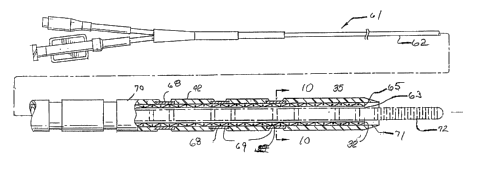
Figs. 9 and 10 schematically illustrate the embodiment of the
invention in the form of a catheter 61. In this embodiment, catheter shaft
62 has an inner lumen 63 defined by an inner tubular element or lining 64
which is preferably formed of lubricous material such as fluoropolymers,
e.g. Teflon° and polysulfones and hydrophilic polymers such as
polyvinypyrrolidone, polyethylene oxide and acrylate-based polymers. A
tubular member 65 is disposed about tubular lining 64 and is formed of at
least one braided layer 66. A plurality of the strands 67 making up each
of the braided layers are insulated electrical conductors which are
electrically connected to electrodes 68. As with the previously discussed
11
~~.54'~'~~
WO 94/16619 PCT/US94/010551~
embodiments, the electrodes 68 are preferably arranged in pairs 69 to
facilitate bipolar mode operation. While not shown in the drawing, a
plurality of braided layers may be desireable with more than eight
electrode pairs 69. Some of the strands 67 in each of the layers may be
formed of nonconducting materials such as nylon. An outer jacket 70
extends the length of the shaft 62 and the portion of the jacket extending
beyond the distal end of the braided tubular member 65 is tapered to
provide a nontraumatic flexible distal tip 71. As in the previously
described embodiments, the outer jacket 70 overlaps the edges of the
electrodes 68 to avoid exposing a sharp metal edge when advancing the
catheter through a patient's blood vessel. A guidewire 72 (shown in
phantom? is slidably disposed within the inner lumen 63.
The catheter 61 may also be used to direct diagnostic or
therapeutic fluids to distal locations within a patients coronary
vasculature. For example, fluids containing cardioplegic materials such as
iced saline, solutions of KCI, lidocaine, procaineamide hydrochloride and
the like can be delivered to areas of the patient's heart which are
suspected to be the origin of or conduct the aberrant signals. If the
arrhythmia stops upon the delivery of the cardioplegic agent, then the
operator is assured that the artery or vein through which the agent is
delivered leads toward or away from the area of the patient's heart which
needs to be ablated in order to terminate the arrhythmia.
When using an approach to the patient's heart through the
femoral artery or femoral vein, it is frequently helpful to utilize one or
more guiding catheters to guide the intravascular sensing devices of the
invention to the coronary artery ostium or the coronary sinus ostium.
Such guiding catheters frequently have specially shaped distal tips to
facilitate the seating thereof within the desired coronary ostium or
12
WO 94/16619 ~ ~ ~ PCT/LTS94101055
coronary sinus ostium. The use of guiding catheter eliminates the need to
direct the distal tip of a catheter or a guidewire of the invention into the
desire ostium.
The electrodes on the distal portions of the sensing devices
are typically gold bands with widths of about 0.5 mm. The distance
between the electrodes of an electrode pair is typically about 1 mm and
the distance between electrode pairs is typically about 7-8 mm.
The overall length of the intravascular devices of the
invention depend upon the site of introduction into the patient's peripheral
vasculature but may range from about 80 to about 300 cm, typically
about 135 cm for delivery through the femoral artery or vein and about
90 cm for delivery through the brachiocephafic artery or internal jugular
vein. The flexible distal portions of the intervascular sensing devices are
about 10 to about 50 cm in length and are configured to be readily
advanceable through a patient's coronary arteries or cardiac veins. The
outer diameter of the catheter form of the sensing device should be less
than about 0.055 inch ( 1.4 mm) and preferably about 0.035 inch (0.89
mm) and the inner lumen thereof is about 0.012 to about 0.022 inch
(0.3-0.56 mm) in diameter to facilitate the reception and advancement of
a guidewire therethrough. The distal portion of the guidewire is about 15
to about 40 cm in length and about 0.008 to about 0.022 inch (0.2-0.56
mm) in outer diameter to facilitate advancement through blood vessels
having native diameters of less than 1 mm, frequently less than 0.75 mm.
The distal coil on the guidewire is about 2 to about 10 cm in length and is
formed of wire about 0.0003 to about 0.006 inch (0.0076-0.15 mm) in
diameter. It is preferably formed of platinum to facilitate fluoroscopic
observation thereof within the patient, but it may be formed in whole or
13
21~~'~'~4
WO 94/16619 PCT/LTS94/01055~
in part with other material such as stainless steel, titanium, palladium,
niobium, iridium, rhodium and alloys thereof.
To the extent not previously described, the materials of
construction of the various guidewire and catheter parts may be formed
of conventional materials. The electrical conductors may be electrical
grade copper wire about 0.005 inch (0.127 mm) in diameter which are
provided with a thin insulated jacket or coating of polyimide or other
suitable insulator. The outer jacket may be a thermoplastic fluoropoiymer
such as THV which is available from 3M Corporation. The core wire of
the guidewire may be formed of stainless steel or a superelastic NiTi type
alloy, the latter exhibiting a stable austenite phase at body temperature.
Preferably, the NiTi alloy exhibits a stress induced transformation from the
stable austenite to a lower strength martensite phase. Upon release of
the stress, the alloy returns to the austenite phase. Proximal and distal
sections of the core member may be formed of different materials so as
to provide a stronger proximal section for greater pushability and a more
flexible distal section to facilitate passage through tortuous coronary
anatomy. Manufacturing techniques used in making catheters and
guidewires for angioplasty procedures may be used in the making of the
intravascular devices of the invention. An alternative
embodiment of the invention in the form of a guidewire 80 is shown in
Figs. 1 1 which is similar to that shown in Fig. 3-8 except that only a
single pair of electrodes 81 and 82 are shown on distal portion 83. The
shaft 84 has a proximal portion 85 which is formed in part of a hypotube
86. A core member 87 extends through the inner lumen of the hypotube .
86 and is electrically isolated from the hypotube 86 by insulating jacket
88. The distal portion of the core member 87 extends out the distal end
of the hypotube 86 as indicated in the drawing. The distal electrode 82 is
14
WO 94/16619
PCTIUS94101055
electrically secured to the core member 87 by solder 89 and the proximal
electrode 81 is secured by solder 90 to electrical conductor 91 which
may be an insulated wire or ribbon. The proximal end of the electrical
conductor 91 is secured by solder 92 to the hypotube 86 which is formed
of electrically conductive metal (stainless steel). The exterior surface of
the conductive metal tube 86 should be provided with an insulating jacket
or coating 93. The core member 87 and the conductive metal tube 86
are preferably secured together at one or more locations by an insulating
adhesive to facilitate the torqueabifity of the overall guidewire shaft.
Preferably, they are secured at least at the distal end of the metal tube
86.
A coil 94 is disposed about the distal portion of the core
member 87 proximal to the proximal electrode 81 and it is secured to the
core member by a suitable means 95. Such securing may be effected by
soldering, brazing, welding or suitable adhesive depending upon the
materials from which the coil 94 and the core member 87 are made. The
core member 87 and the coil 94 provide shapeabifity to the distal portion
83 of the guidewire 80 to facilitate its advancement into side branches of
a patient's vasculature.
An inner tubular member 96 may be disposed within the coil
94 in the distal section 83 to provide support to the electrode 81 and
inner tubular member 97 may be disposed within the coil 94 to likewise
provide support thereto to electrode 82. A suitable material is thin walled
polyimide tubing which is frequently mentioned as being suitable for use
in intravascular catheters.
Figs. 12 and 13 illustrate a catheter assembly 100 which
embodies an additional aspect of the present invention directed to an
intravascular catheter 101 for sensing electrical activity within a patient's
WO 94/16619 PCT/LJS94/01055
coronary or cardiac blood vessels. As shown in Fig 13 electrodes 102
are electrically connected to individual electrical conductors 103 which
are woven or wound to' form .the tubular shaft 104 of the catheter 101 .
All of the strands which are wound to form the shaft 104 need not be
conductors 103 as in the prior embodiments, and when there are more
than 16 electrodes and thus more than 16 electrical conductors, multiple
woven layers may be employed. The electrical conductors 103 are
typically electrical grade copper wires of suitable outer diameter such as
about 0.004 to about 0.01 inch (0.10-0.25 mm). The conductors 103
may be formed of other conducting materials such as silver, gold and
platinum. A suitable insulating material to coat the conductors 103 is
polyimide which minimizes cross talk and which can be applied in very
thin layers. As in the other embodiments of the invention the conductors
103 may be woven or merely wound, but preferably are woven.
The inner lumen 105 of the catheter 100 is configured to
slidably receive a guidewire to facilitate the advancement of the catheter
over the guidewire and preferably has at least in the distal portion thereof
a diameter about 0.002 to about 0.005 inch (0.051-0.127 mm) greater
than the guidewire which is to be disposed therein. For guidewire having
OD of about 0.016 to about 0.018 inch (0.41-46 mm), the inner lumen
97 would be about 0.018 to about 0.023 inch (0.46-0.58 mm). The OD
of the catheter may range from about 0.03 to about 0.1 inch (0.76-2.54
mm) but preferably is about 0.03 to about 0.05 inch (0.076-1.27 mm,
particularly 0.035 to about 0.040 inch (0.89-1.02 mm).
The proximal portion 106 of the catheter 100 makes up -
about 70 to about 95% of the total length of the catheter with the
intermediate portion 107 and the distal portion 108 which has the sensing
electrodes 102 being the remainder. Preferably the catheter 100 has
16
WO 94/16619 PCTlUS94101055
decreasing stiffness from the proximal portion 106 to the intermediate
portion 107 and the distal portion 108 to facilitate the advancement of
the catheter 100 within the patient's vasculature. The exterior surface of
the catheter 100 and the surface defining inner lumen 105 are formed of
lubricous materials or hydrophilic materials which become lubricous when
contacting aqueous based fluids. Polysulfones and polyfluoroalkanes are
examples of suitable lubricous polymers and polyvinypyrrolidone,
polyethylene oxide and acrylate-based polymers of examples of suitable
hydrophilic polymers.
The proximal end of the catheter 106 may be provided with
a multiple arm adapter 109 as shown in Fig. 12 with one arm 110 which
is configured to receive a syringe for delivering fluid into the inner lumen
and a second arm 111 which is provided with an electrical connector 112
which is electrically connected to the electrical conductors 103. The
central arm 1 12 facilitates entry of a guidewire (not shown) into the inner
lumen 105.
Once the arrhythmogenic site or conductive pathway causing
an arrhythmia is located by detecting the electrical activity, The guidewire
may be removed and means can be advanced through the inner lumen
105 of a catheter of the invention to occlude an arterial passageway
which feeds the arrhythmogenic site or conductive pathway so as to
terminate the arrhythmia.
Fig. 14 schematically represents the output from a plurality
of electrode pairs 120 and 121 on separate intravascular devices (not
shown) disposed in different, generally parallel coronary blood vessels,
e.g. a coronary artery and the companion vein to a nearly planar wave
front approaching on end to the intravascular devices. The bipolar
response 122 and 123 to the wave front 124 from each electrode pair is
17
215 4'~'~ 4
WO 94/16619 PCT/US94/01055
shown adjacent thereto, and as indicated, all of the responses are
essentially identical, except for the time-of-occurrence, because the wave
front 124 reaches all of the electrodes at the same angle. Changes in
tissue properties adjacent the catheters may retard the passage of the
wave front and may distort the shape of the output.
Fig. 1 5 schematically represents the responses 130 and 131
from a plurality of electrode pairs 132 and 133 on separate intravascular
devices (not shown) disposed in different generally parallel coronary blood
vessels, as in Fig. 14, but the wave front 134 originates from an
arrhythmogenic site between and in close proximity to the catheters. The
wave front 134 is circular (idealized) and the size and polarity of the
responses to the expanding wave front varies according to the angle of
incidence.
The time of occurrence and the directional information in the
aforementioned schematic drawings may be used to determine the origin
of the ectopic beat.
The present invention has been described herein in terms of
certain preferred embodiments but various modifications and
improvements may be made to the present invention without departing
from the scope thereof.
18