Note: Descriptions are shown in the official language in which they were submitted.
~ WO94/18218 2 I S S 1 ~ 6 PCT~S94/01156
-- 1 --
METHODS AND APPARATUS FOR DNA SEOUENCING
1. INTRODUCTION
Considerable interest has been developing in
the past few years to sequence the entire human genome
(i.e., all of the genetic material in a human cell).
The task, however, is enormous because it involves the
sequencing of at least 3,000,000,000 base pairs, an
effort which is likely to take ten or more years and
cost $3,000,000,000 if undertaken using conventional
technology (1993 Edgington, Bio/Technology 11:39-42,
which is incorporated herein by reference).
The Committee on Mapping and Sequencing the
Human Genome of the National Research Council in their
1988 report entitled, Mappinq and Sequencinq the Human
Genome (which is incorporated herein by reference),
stated that, "No foreseeable technology will be able
to automate DNA sequencing comprehensively." The
present invention is a method and apparatus for
comprehensively automating this effort with
substantial improvements in speed and cost. The
invention is applicable to the sequencing of genetic
material from any source, human or otherwise.
2. BACKGROUND OF THE INVENTION
2.1. DNA AND RNA
Deoxyribonucleic acid (DNA) is the primary
genetic material of most organisms. Ribonucleic acid
(RNA) is the primary genetic material in certain
viruses. Additionally, a form of RNA known as
messenger RNA (mRNA) is found in all cells and
comprises copies of portions of the primary genetic
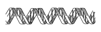
information found in the DNA. In its natural state,
DNA is found in the form of a pair of complementary
chains of nucleotides which are interconnected as a
double helix (see Fig. 1). A nucleotide in turn is
WO94/18218 21~ ~18 6 PCT~S94101156
L ' ~
composed of a nitrogenous base (see Figs. 2 and 3),
which identifies the nucleotide, linked by an N-
glycosidic bond to a five-carbon sugar. RNA differs
from DNA in that in DNA the nucleotide sugar is
deoxyribose, while in RNA, the sugar is ribose. A
phosphate group serves to link the nucleotides
together, forming the backbone of a single strand of
DNA (see Fig. 2). Normally, the nitrogenous base is
one of the following: adenine, guanine, thymine and
cytosine (respectively denoted A, G, T, and C), or
uracil (U) in place of thymidine in RNA (see Fig. 3).
The order of the four nucleotides, A, G, T and C, in
the chain is often referred to as the sequence of the
DNA and can be specified simply by setting down the
symbols A, G, T and C in the order in which these four
nucleotides appear in the DNA strand.
The two chains (or strands) of a DNA double
helix are held together by hydrogen bonding between
the nitrogenous bases of their individual nucleotides.
This hydrogen bonding is specific in that adenine in
one strand must pair with thymine (or uracil in RNA)
in the other strand, and guanine with cytosine. The
sequence of bases in one strand of DNA is thus
complementary to the sequence on the other strand.
A DNA chain has polarity: one end of the
chain has a free 5'-OH (or phosphate) group (termed
"the 5' end") and the other a free 3'-OH (or
phosphate) group ("the 3' end"). By convention, the
nucleotide sequence is written or read left-to-right
in the direction from the 5' end to the 3' end. The
two strands of a DNA double helix have opposite
polarities. Thus the 5' end of one strand pairs with
the 3' end of the other strand and the complementarity c
of the two strands is revealed by comparing one strand
WO94118218 _ 3 _ ~18~ PCT~S94/01156
read in the 5' to 3' direction with the other strand
read in the 3' to 5' direction.
Genetic information is encoded in the
particular sequence (order of occurrence) of
nucleotides along a DNA molecule and DNA sequencing is
the process of determining that order in a particular
DNA molecule.
2.2. ENZYMES USED IN DNA SEOUENCING
Two classes of enzyme activity which have
been employed in certain methods used to sequence DNA
are DNA polymerase and exonuclease activity.
A DNA polymerase is an enzyme that has the
ability to catalytically synthesize new strands of DNA
15 in vitro. The DNA polymerase carries out this
synthesis by moving along a preexisting single DNA
strand ("the template") and creating a new strand,
complementary to the preexisting strand, by
incorporating single nucleotides one at a time into
the new strand following the base-pairing rule
described above.
In contrast to polymerase activity,
exonuclease activity refers to the ability of an
enzyme (an exonuclease) to cleave off a nucleotide at
the end of a DNA strand. Enzymes are known which can
cleave successive nucleotides in the single DNA strand
of a single-chain DNA molecule, working from the 5'
end of the strand to the 3' end; such enzymes are
termed single-stranded 5' to 3' exonucleases. Other
enzymes are known which perform this operation in the
opposite direction (single-stranded 3' to 5'
exonucleases). There also exist enzymes which can
cleave successive nucleotides from the end of a single
strand of a double-stranded DNA molecule. These
enzymes are termed double-stranded 5' to 3' or 3' to
WO94/18218 2i~$~ 4 _ PCT~S94/01156
5' exonucleases, depending on the direction in which
they proceed along the strand. Exonucleases are also
characterized as being distributive or processive in
their action. Distributive exonucleases dissociate
from the DNA following each internucleotide bond
cleavage, whereas processive exonucleases will
hydrolyze many internucleotide bonds without
dissociating from the DNA.
2.3. SEOUENCING OF DNA
Approaches to DNA sequencing have varied
widely. Use of these enzymes or other chemical
methods, as described below, has made it possible to
sequence small portions of the human genome. Despite
these successes, most of the human genome remains
unexplored. Of the 3,000,000,000 base pairs in the
human genome, only about 20 million base pairs have
been sequenced (GenBank~ Release 74 - December 1992).
2.3.l. SEQUENCING LADDER METHODS
Many techniques for sequencing DNA have
involved generating fragments of labeled DNA, the
lengths of which are sequence-dependent, and
separating the fragments according to their lengths by
electric field-induced migration in a gel, so as to be
able to discern the DNA sequence from the appearance
of the separated fragments. Such a pattern of
sequence-dependent fragment lengths is known as a
sequencing ladder. The fragments can be generated by
either: (a) cleaving the DNA in a base-specific manner
(see Fig. 4), or (b) synthesizing a copy of the DNA
wherein the synthesized strand terminates in a base-
specific manner (see Fig. 5).
The ~-Gilbert technique for sequencing
(Maxam and Gilbert, 1977, Proc. Natl. Acad. Sci. USA
W094/18218 ~ PCT~S94/01156
74:560, which is incorporated herein by reference)
involves the specific chemical cleavage of DNA.
According to this tec-hn;que, four samples of the same
labeled DNA are each subjected to a different chemical
reaction to effect preferential cleavage of the DNA
molecule at one or two nucleotides of a specific base
identity. By adjusting the conditions to obtain only
partial cleavage, DNA fragments are thus generated in
each sample whose lengths are dependent upon the
position within the DNA base sequence of the
nucleotide(s) which are subject to such cleavage.
Thus, after partial cleavage-is performed, each sample
contains DNA fragments of different lengths each of
which ends with the same one or two of the four
nucleotides. In particular, in one sample each
fragment ends with a C, in another sample each
fragment ends with a C or a T, in a third sample each
ends with a G, and in a fourth sample each ends with
an A or a G. The fragments so generated are then
separated from one another by electric field-induced
migration in a polyacrylamide gel. The four
individual sets of fragments produced by cleavage
using chemical reactions of different specificity are
run side-by-side, in separate lanes of the gel. The
DNA fragments are then visualized, and sequence is
determined by the observing the position in the gel of
the generated fragments.
Fig. 4 schematically depicts the
visualization of DNA fragments that are generated by
cleaving the labelled DNA having the sequence 5'-
AAGTACT-3'-label. The fragments from the four samples
are run side-by-side in the four lanes of the gel
identified by G, A+G, C, T+C where G identifies the
sample in which all the fragments end with guanine
nucleotides, A+G identifies the sample in which all
W094/18218 ~6 PCT~594/0ll56
the fragments end with either an adenine or a guanine
nucleotide, C identifies the sample in which all the
fragments end with a cytosine nucleotide, and T+C
identifies the sample in which ali the fragments end
with either a thymine or a cytosine nucleotide. The
distance the fragments migrate in the gel is a
monotonic function of their length. Thus, after the
migrating fragments are visualized, the order of the
nucleotides in the labelled DNA molecule can be read
directly from the vertica~ position of the fragments
in the gel. The fragments that end with adenine that
appear in the A+G lane, and the fragments that end
with thymine that appear in the T+C lane, can be
distinguished from the fragments in the same lanes
that end with guanine and cytosine, respectively, by
noting that the fragments that end with guanine and
cytosine also appear at the same vertical position in
the G and C lanes, respectively.
In the DNA of many organisms, a significant
fraction of the cytosines are methylated in vivo at
the 5 position to give 5-methylcytosine. Such
methylation is involved in the regulation of gene
expression and in genetic imprinting. Church and
Gilbert (1984, Proc. Natl. Acad. Sci. USA 81:1991-
1995; incorporated herein by reference) and Saluz andJost (1987, "A Laboratory Guide to Genomic Sequencing,"
BioMethods, Vol. 1, Birkhauser, Boston; incorporated
herein by reference) devised a modification of the
Maxam and Gilbert chemical cleavage method to provide
a means for directly determining the position of 5-
methylcytosine in genomic DNA. In this method,
genomic DNA is chemically cleaved, then completely
digested with a restriction enzyme and separated by
gel electrophoresis, resulting in a complex mixture of
superimposed sequencing ladders. The DNA bands
WO94/18218 PCT~S94/01156
~ - 7 - ~
forming the rungs of the sequencing ladder are next
transferred and cross linked to a nylon membrane. A
specific ladder from the mixture is then recognized by
hybridizing the membrane with a labeled
oligonucleotide probe which uniquely recognizes the
sequence immediately adjacent to a particular
restriction site. Frommer et al. (1992, Proc. Natl.
Acad. Sci. USA 89:1827-1831, which is incorporated
herein by reference) have recently developed an
alternative genomic DNA sequencing method wherein
cytosines in the sample DNA are converted to uracil by
bisulfite treatment which leaves 5-methylcytosine
unmodified. Comparison of the sequence of modified
and unmodified DNA reveals the positions in the
sequence of 5-methylcytosine. Such genomic sequencing
methods can only be carried out with genomic DNA. The
methylation pattern is lost during gene cloning in
microorganisms in vivo, and during DNA copying or
amplification in vitro.
The plus/minus DNA sequencing method (Sanger
and Coulson, 1975, J. Mol. Biol. 94:441-448, which is
incorporated herein by reference) involves: (a) use of
DNA polymerase to generate complementary 32P-labeled
DNA oligonucleotides of different lengths; (b) (the
"minus" system) in four separate reaction vessels,
reaction of one half of the generated DNA with DNA
polymerase and three out of the four nucleotide
precursors; and (c) (the "plus" system) in four
separate reaction vessels, reaction of the remaining
half of the generated DNA with DNA polymerase and only
one of each of the four nucleotide precursors. Each
reaction mixture generated in steps (b) and (c) is
subjected to a denaturing polyacrylamide gel
electrophoresis. The generated fragments are
separated from one another by migration in the
WO94/18218 ` PCT~S94/01156
~ S ~ ~ 8 -
polyacrylamide gel; the shorter the fragment, the
greater the migration. After visualization of the DNA
in the gel by detection of its label, the sequence of
the DNA can be determined by observing the position in
the gel of the generated fragments.
The dideoxy method of sequencing was
published in 1977 by Sanger and his colleagues (Sanger
et al., 1977, Proc. Natl. Acad. Sci. USA 74:5463,
which is incorporated herein by reference). In
contrast to the method of Maxam and Gilbert which
relies on specific chemical cleavage to generate
fragments with lengths which are sequence-dependent,
the Sanger dideoxy method relies on enzymatic activity
of a DNA polymerase to synthesize fragments with
lengths that are sequence-dependent. The Sanger
dideoxy method utilizes an enzymatically active
fragment of the DNA polymerase termed E. coli DNA
polymerase I, to carry out the enzymatic synthesis of
new DNA strands. The newly synthesized DNA strands
consist of fragments of sequence-dependent length,
generated through the use of inhibitors of the DNA
polymerase which cause base-specific termination of
synthesis. Such inhibitors are dideoxynucleotides
which, upon their incorporation by the DNA polymerase,
destroy the ability of the enzyme to further elongate
the DNA chain due to their lack of a suitable 3'-OH
necessary in the elongation reaction. When a dideoxy
nucleotide whose base can appropriately hydrogen bond
with the template DNA is thus incorporated by the
enzyme, synthesis of the growing DNA strand halts.
Thus DNA fragments are generated by the DNA
polymerase, the lengths of which are dependent upon
the position within the DNA base sequence of the
nucleotide whose base identity is the same as that of
the incorporated dideoxynucleotide. The fragments so
WO94/1~18 ~ PCT~594/01156
generated can then be separated in a gel as in the
Maxam-Gilbert procedure, visualized, and the sequence
determined.
For example, for the case of a template DNA
molecule having the sequence 5'-GCCATCG-3'-label, Fig.
5 depicts the visualization of the DNA fragments that
are generated by the dideoxy method after terminating
synthesis at each of the nucleotides G, A, C and T.
Since the distance a fragment migrates in the gel is a
monotonic function of its length, the sequence of the
DNA molecule can be read directly from the gel after
the fragments are visualized.
Sanger and colleagues utilized an E. coli
DNA polymerase I fragment termed the Klenow fragment.
lS After the disclosure of the original Sanger dideoxy
technique, the enzyme used in most dideoxy sequencing
was the Klenow fragment. Other enzymes with DNA
polymerase activity that have been used in sequencing
include AMV reverse transcriptase and T7 DNA
polymerase (Tabor and Richardson, U.S. Patent No.
4,795,699, which is incorporated herein by reference).
DNA sequencing methods have been automated
to varying degrees. In the manual methods,
radioactive labels such as 32p are typically used to
identify the bands of the sequencing ladder by
autoradiographic imaging on X-ray film. Digital
imaging systems and pattern recognition software have
been developed by several groups for automatic
interpretation and data entry from such
autoradiographs tElder et al., 1986, Nucl. Acids Res.
14:417-424, which is incorporated herein by
reference). Real-time recording of the sequencing
ladder during gel electrophoresis was made possible by
positioning ~-emission detectors at the bottom of the
gel (EG&G Biomolecular ACUGEN~ Sequencer, Acugen~
W094/18218 ~5~ PCT~S94/01156 ~
q -- 10 --
;.''
System Report 88-106, EG&G Biomolecular), or by
employing fluorescent labeling techn; ques in
combination with real-time fluorescence detection
during electrophoresis. Smith et al. (1986, Nature
321:674, which is incorporated herein by reference)
disclose a method for partial automation of DNA
sequencing, which involves use of four different color
fluorophores bound to the primer (Smith et al., 1985,
Nucl. Acids Res. 13:2399-2412, which is incorporated
herein by reference) used for synthesis in one of four
reaction vessels, each contA;n;ng a different
dideoxynucleotide in the Sanger dideoxy method. The
reaction mixtures are combined and subjected to
electrophoresis, during which the separated DNA
fragments are identified by a fluorescent detection
apparatus, and the sequence information acquired
directly by computer. In an alternative approach, the
dideoxy nucleotide chain terminators have each been
chemically linked to different succinylfluorescein
fluorescent dyes which can be distinguished by their
fluorescent emission, allowing the four sequencing
reactions to be run in a single tube (Prober et al.,
1987, Science 238:336, which is incorporated herein by
reference). Japanese scientists and engineers are
participating in the development of a completely
automated DNA sequencing system, employing the Sanger
dideoxy method of sequencing (Endo et al., 1991,
Nature 352:89-90; Wada et al., 1987, Nature 325:771-
772, which are incorporated herein by reference).
Ladder-based sequencing methods are
currently the most widely utilized, and variations on
the Sanger method of generating the sequencing ladder
are used predominantly. The throughput and cost of
ladder-based sequencing methods are currently limited
by three major factors: (1) the number of resolvable
WO94/18218 ~ ~ PCT~S94/01156
bases in a single ladder, (2) the time required to
separate the fragments and generate the ladder, and
(3) the number of ladders which can be run in
parallel. Numerous efforts are presently underway to
further improve each of these aspects and to thereby
enhance the performance of ladder-based sequencing
methods, Conventional DNA sequencing gels are
typically -300-500 micrometers thick. With such gels
it is usually possible to obtain 300-500 bases of
sequence from a single sequencing ladder. The limit
depends on the ability to resolve a band containing
fragments which are N nucleotides long from those
containing fragments which are N+1 or N-l nucleotides
in length. Increased resolution can be achieved by
employing thinner gels, typically ~25-100 micrometer,
either in ultrathin slab gels (Kostichka et al., 1992,
Bio/Technology 10:78-81) or in capillary gels
(Drossman et al., 1990, Anal. Chem. 62:900-903, which
are incorporated herein by reference). It has
recently been demonstrated that such gels are capable
of resolving >1,000 bases, and further improvements
are projected to achieve -2,000 bases. One approach
to further increase the resolution of the gel is to
employ programmed pulse-field techniques (C. Turmel,
E. Brassard, R. Forsyth, J. Randell, D. Thomas, J.
Noolandi (1992) "Sequencing up to 800 bases manually
using pulsed field", IN: Genome Mapping & Sequencing,
Cold Spring Harbor Laboratory, ~bstract #112; C.
Turmel, E. Brassard, J. Noolandi (1992)
Electrophoresis (in press), which are incorporated
- herein by reference). Because ultrathin gels can be
cooled more efficiently, they can be operated at much
higher voltages per unit length, thereby reducing the
time required to effect the separation of the
sequencing ladder. Multiple capillaries can be run in
~094/18218 ~5~ 12 - PCT~S94101156 ~
. ..
parallel or a greater number of samples can be loaded
in slab gels to further increase throughput. Both
capillary and ultrathin slab gels have been
demonstrated to have some degree of reusability. In
order to achieve the improved performance offered by
ultrathin gels, it is necessary to reduce the number
of DNA molecules loaded onto the gel, which therefore
reduces the number of the DNA molecules in each band
or rung of the sequencing ladder. This requires more
sensitive detection methods which have included the
use of sheath-flow cuvette fluorescence techniques
(1991 Chen et al., SPIE Vol. 1435, Optical Methods for
Ultrasensitive Detection and Analysis: Techniques and
Applications, p. 161-167, which is incorporated herein
by reference), confocal fluorescence microscopy (1992
Mathies and Huang, "Capillary array electrophoresis:
an approach to high-speed, high throughput DNA
sequencing," Nature 359:167-169, which is incorporated
herein by reference), mass spectrometry (1990 T.
Brennan, J. Chakel, P. Bente, M. Field, "New Methods
to Sequence DNA by Mass Spectrometry," SPIE Vol. 1206,
New Technoloqies in cYtometry and Molecular BioloqY,
pp.60-77; 1990 T. Brennan, J. Chakel, P. Bente, M.
Field, "New Methods to Sequence DNA by Mass
Spectrometry," IN: A.L. Burlingame and J.A. McCloskey
(Eds.) Bioloqical Mass sPectrometry~ Elsevier,
Amsterdam, pp. 159-177, which are incorporated herein
by reference), and resonance ionization spectroscopy
(RIS)(1979 G.S. Hurst, M.G. Payne, S.D. Kramer, J.P.
Young, "Resonance ionization spectroscopy and one-atom
detection", Rev. Mod. Phys. 51:767-819; 1991 H.F.
Arlinghaus, M.T. Spaar, N. Thonnard, A.W. McMahon,
K.B. Jacobson, "Application of resonance ionization
spectroscopy for semiconductor, environmental and
biomedical analysis, and for DNA sequencing," SPIE
WO94/18218 ~ ~ PCT~S94/01156
- 13 - $~
~6~
Vol. 1435, Optical Methods for Ultrasensitive
~etection and Analysis: Techniques and ApPlications,
pp. 26-35; 1991 K.B, Jacobson, H.F. Arlinghaus, H.W.
Schmitt, R.A. Sachleben, G.M. Brown~ N. Thonnard, F.V.
Sloop, R.S. Foote, F.W. Larimer, R.P. Woychik, M.W.
England, K.L. Burchett, D.A. Jacobson, "An Approach to
the Use of Stable Isotopes for DNA Sequencing,"
Genomics 9:51-59, which are incorporated herein by
reference).
Another improvement which was developed from
the original genomic sequencing methods is known as
multiplex sequencing (Church and Kieffer-Higgins,
1988, Science 240:185-188, which is incorporated
herein by reference). In multiplex sequencing,
multiple sequencing reactions are pooled and
electrophoresed together in a single gel to generate
multiple superimposed sequencing ladders which are
then transferred and bound to a nitrocellulose
membrane. The membrane is then probed with an
oligonucleotide which is specific for only one of the
pools in order to reveal the-corresponding ladder. By
repeatedly stripping the membrane of probe and
rehybridizing with different oligonucleotides it is
possible to obtain the sequence from each of the
individual reactions. Although originally developed
using radioactive isotopes to label the probes and
therefore requiring lengthy autoradiographic exposures
in order to visualize the ladder, newer multiplex
sequencing protocols have been devised which employ
chemiluminescent detection of the probes (Gillevet,
- 1990, Nature 348:657-658, which is incorporated herein
by reference) or fluorescence detection (Yang and
Youvan, 1989, Bio/Technology 7:576-580, which is
incorporated herein by reference).
WO94/18218 ~S5~ PCT~S94/01156
: - 14 -
Mass spectrometry offers the potential of
further improving ladder-based sequencing by also
eliminating the electrophoresis step and replacing it
with mass separation of conventional sequencing
reaction mixtures using time-of-flight methods which
require only milliseconds. Matrix-assisted laser
desorption/ionization is currently being explored to
generate mass ions as large as -300,000 daltons
without fragmentation which might permit the
determination of ~600 bases. (1992 M.C. Fitzgerald,
G.R. Parr, L.M. Smith, "DNA Sequence Analysis by Mass
Spectrometry?" IN: Genome Mapping & Sequencing, Cold
Spring Harbor Laboratory, Abstract #113; 1992 G.R.
Parr, M.C. Fitzgerald, L.M. Smith, "Matrix-Assisted
Laser Desorption/Ionization Mass Spectrometry of
Synthetic Oligodeoxyribonucleotides," Rapid. Commu.
Mass. Spec. 6:369-372j which are incorporated herein
by reference.) Such an approach was described by
McCormick and Amendola as early as 1978 (in Theory,
Design and Biomedical Applications of solid State
Chemical Sensors, Cheung et al. (eds.), CRC Press,
West Palm Beach, pp. 219-250, which is incorporated
herein by reference).
2.3.2. SEOUENCING BY HYBRIDIZATION
A fundamentally different approach to
sequencing involves the determination of all of the
oligonucleotide sequences contained within a longer
sequence. The method is based on the ability of short
oligonucleotides to match or hybridize perfectly
through base-pairing with their complementary sequence
in another DNA molecule (Strezoska et al., 1991, Proc.
Natl. Acad. Sci. USA 88:10089-10093; Bains, 1992,
Bio/Technology 10:757-58, which are incorporated
herein by reference). Under appropriate conditions,
WO94/18218 ~ PCT~S94/01156
- 15 - SS~
8~
only perfect matches are formed and even single base
differences prevent successful hybridization. The
method can be practiced either with the sample DNA
immobilized on an appropriate solid support and the
hybridizing oligonucleotides in solution, or with the
oligonucleotide probes bound to the solid support and
the sample DNA in solution. By establishing which
oligonucleotides bind perfectly to the DNA, it is
possible to reconstruct the sequence of the DNA.
Repeat sequences in the sample DNA which are longer
than the length of the oligonucleotide probes employed
result in branch points in thè DNA sequence which must
be resolved by other methods, therefore limiting the
general utility of this method.
2.3.3. SEOUENCING BY MICROSCOPY
Beer and Moudrianakis (1962, Proc. Natl.
Acad. Sci. USA 48:409-416, which is incorporated
herein by reference) proposed a method for sequencing
single DNA molecules, wherein base-specific labeling
with heavy elements would allow subsequent electron
microscopic observation and identification of the
individual bases. Despite almost ten years of
subsequent effort, this approach was never
successfully reduced to practice. A proposal was made
by McCormick and Amendola (1978, in Theory, Design and
Biomedical Applications of Solid state Chemical
Sensors, Cheung et al. (eds.), CRC Press, West Palm
Beach, pp. 219-250, which is incorporated herein by
reference) to sequence single DNA molecules by
- transporting a linearly-extended DNA molecule using a
microminiature, iterative system of electrostatic
quadrupole lenses past a microminiaturized radial
array of electron guns and detectors. Such a design
was never realized in practice, but presaged the
WO94/18218 `~6 16 - PCT~S94/01156
development of the scAnn;ng tunneling microscope (STM)
by Binnig and Rohrer (1982, Phys. Rev. Lett. 49:57,
which is incorporated herein by reference) and its
application to sequencing. Binnig and Rohrer
themselves were the first to report images of DNA
molecules made with the STM (1984, in Trends in
Physiçs, J. Janta and J. Pantoflicek (eds.), European
Physical Society, The Hague, pp. 38-46, which is
incorporated herein by reference), and numerous
scientific publications have appeared in the past
decade purporting to show images of DNA (Driscoll et
al., 1990, Nature 346:294-296 and Dunlap and
Bustamante, 1989, Nature 342:204-206, which are
incorporated herein by reference). Many of these
reports have recently been shown to have likely been
artifacts of the highly ordered pyrolytic graphite
substrate (Clemmer and Beebe, 1991, Science 251:640-
642). Research effort continues on microscopy-based
approaches to sequencing, including a wider range of
techniques such as atomic force microscopy (Hansma et
al., l991, J. Vac. Sci. Technol. B 9:1282-1284, which
is incorporated herein by reference) and near-field
microscopy (1992 E. Betzig, J.K. Trautman "Near-Field
Optics: Microscopy, Spectroscopy, and Surface
Modification Beyond the Diffraction Limit," Science
257-189-195, which is incorporated herein by
reference). X-ray diffraction t~chn;ques have also
been proposed for use in DNA sequencing (1991 J.W.
Gray, J. Trebes, D. Peters, U. Weier, D. Pinkel, T.
Yorkey, J. Brase, D. Birdsall, R. Rill, "Investigation
of the Utility of X-ray Diffraction in DNA Sequence
Analysis," DOE Human Genome Program, Report of the
Second Contractor-Grantee Workshop, February 17-20,
1991, Santa Fe, New Mexico, P56, page 80, which is
incorporated herein by reference).
WO94/18218 ~SS PCT~S94/01156
- 17 - -~ 86
2.3.4. BASE-AT-A-TIME METHODS
Nucleotide Incorporation
Melamede (U.S. Patent No. 4,863,849, which
is incorporated herein by reference) discloses an
automatable process for determining the nucleotide
sequence of DNA and RNA involving the determination of
whether a specific one of the four nucleotides is
incorporated by polymerase at the nucleotide residue
3' of the primer terminus.
Nucleotide Cleavage
In an early approach, Cantor et al. (1964,
Biopolymers 2:51-63, which is incorporated herein by
reference) mathematically analyzed the kinetics of
exoenzyme digestion of linear polymers and proposed
the use of exonuclease under non steady-state
conditions to sequentially remove terminal nucleotides
from a population of RNA molecules. By monitoring the
evolution of the resulting mononucleotides with time,
they calculated that it should be possible to
determine the base sequence of oligomers up to 25
nucleotides.
SYnchronization
The experimental difficulty with such an
approach is that there is no means available for
synchronizing the hydrolytic action of the individual
exonuclease molecules. The cleavage of an
internucleotide bond is a stochastic process and there
is therefore a time distribution for cleavage of each
bond rather than a discrete time interval. The
consequence is that each individual DNA chain is
hydrolyzed at a slightly different rate. Even though
all of the DNA molecules start with identical terminal
nucleotides, they quickly evolve to a mixed population
WO94/18218 ~6 PCT~S94101156 ~
~5 ;. - 18 -
.,
having different terminal nucleotides as some chains
are degraded more slowly or rapidly than others. The
ability to derive sequence information by such
terminal cleavage is therefore limited by the point at
which the terminal nucleotides have become
sufficiently random within the population of DNA
molecules to mask the signal from those ch~;n~ which
are still synchronous.
One solution to this synchronization problem
would be to devise an exonuclease wherein
internucleotide bond cleavage is triggered by a very
short light pulse. The hydrolytic action of the
exonuclease would thus be synchronized to an external
source. Enzymes with similar properties are known in
the art. Photoreactivating enzyme (EC 4.1.99.3
Deoxyribodipyrimidine photolyase) binds to W-induced
dimers formed between adjacent pyrimidines in DNA,
cleaving the cyclobutane dimers upon absorption of
near-W light. Photoinitiated nuclease activity has
not been described, but a DNA-binding protein, the trp
repressor, has been successfully converted to a site-
specific nuclease by chemical modification (Chen and
Sigman, 1987, Science 237:1197-1201, which is
incorporated herein by reference) and oligonucleotides
have been chemically modified to incorporate
photosensitizers which permit the photoinduced
cleavage of DNA (Le Doan et al., 1987, Nucleic Acids
Res. 15:7749-7760; Praseuth et al., 1988, Proc. Natl.
Acad. Sci. USA 85:1349-53, which are incorporated
herein by reference).
An alternative solution to the
synchronization problem would be to avoid the issue
altogether by going to the limit of a single DNA
molecule, rather than sequencing a population of
molecules. In a lecture given at the dedication of
~ W094/18218 ~S$ PCT~S94/01156
- 19 - ~8~
Jadwin and Fine Halls, Princeton University, March 17,
1970, Freeman Dyson outlined a proposed method for
rapidly sequencing single DNA molecules one base at a
time (1992 Dyson, From Eros to Gaia, Pantheon Books,
New York, p. 155, which is incorporated herein by
reference). Dyson proposed to isolate a single DNA
molecule, attach one end to a solid support, and
extend the molecule under the influence of an electric
field in vacuo . Single nucleotides are then removed
one by one in sequence from the loose end of the
chain, ionized and directed into a mass spectrometer
which sorts the nucleotides into four channels labeled
adenine, cytosine, guanine and thymine. Counters in
each channel automatically record the sequence in
which the nucleotide arrives. Jett et al. (U.S.
Patent No. 4,962,037; 1989, J. Biomolecular Structure
and Dynamics 7(2):301-309; 1989, Book of Abstracts,
Sixth Conversation in Biomolecular Stereodynamics,
SUNY at Al h~ny, June 6-10, 1989, p. 157; and Davis et
al., 1991, GATA 8(1):1-7, which are incorporated
herein by reference) disclose a quite similar method
for DNA or RNA sequencing, involving the sequencing of
single nucleic acid molecules containing nucleotides
tagged with a fluorescent dye, which molecules are
suspended in a flow stream and subjected to
exonuclease digestion to liberate single nucleotides
sequentially. The nucleotides are transported by the
moving flow stream in an orderly train and identified
by fluorescent detection methods. Shera et al. (U.S.
Patent No. 4,793,705; 1990, Chemical Physics Letters
174(6):553-557, which are incorporated herein by
reference) and Soper et al. (1991, Anal. Chem. 63:432-
437, which is incorporated herein by reference)
describe single molecule detection systems, for use,
WO94/18218 ~ ~ PCT~S94/01156
e.g., in detecting a fluorescently labeled nucleotide.
2.3.5. REVIEWS
Current methods, prospects for automation,
and novel methods of DNA sequencing are reviewed by
Mar~in and Davies (1986, Bio/Technology 4:890-895), by
Bains (1990, Bio/Technology 8:1251-1256) and by
Hunkapiller et al., (1991, Science, 254:59 which are
incorporated herein by reference).
2.4. RNA SEOUENCING METHODS
RNA sequencing methods are also known.
Zimmern and Kaesberg (1978, Proc. Natl. Acad. Sci. USA
75:4257-4261, which is incorporated herein by
reference) disclose the use of AMV reverse
transcriptase with dideoxy-nucleotides to sequence
encephalomyocarditis virus RNA. Mills and Kramer
(1979, Proc. Natl. Acad. Sci. USA 76:2232-2235, which
is incorporated herein by reference) describe the use
of QB replicase and the nucleotide analog inosine for
sequencing RNA in a chain-termination mech~n;~m.
Direct chemical methods for sequencing RNA are also
known (Peattie, 1979, Proc. Natl. Acad. Sci. USA
76:1760-1764, which is incorporated herein by
reference). Other methods include those of Donis-
Keller et al. (1977, Nucl. Acids Res. 4:2527-2538),
Simonesits et al. (1977, Nature 269:833-836), Axelrod
et al. (1978, Nucl. Acids Res. 5:3549-3563), and
Kramer et al. (1978, Proc. Natl. Acad. Sci. USA
75:5334-5338, which are incorporated herein by
reference).
WO94/18218 - 21 - ~ PCT~S94/01156
2.5. SINGLE-MOLECULE DETECTION
Interest in analytical techniques for
optical detection and measurement of spectral
properties of single atoms and molecules goes back
more than two decades. Initial success was achieved
with very dilute atomic and molecular beams in vacuo
where background emissions and scattering are most
easily minimized. A more challenging problem has been
the detection of single molecules in condensed phases.
2.5.1. IN VACUO
Initial success in the optical detection of
single atoms was reported by Greenless et al. (1977,
Opt. Commun. 23:236-239, which is incorporated herein
by reference). Further improvements in optical
trapping and laser cooling extended these results to
other atomic and ionic systems (Pan et al., 1980j Opt.
Lett. 5:459-461; 1980 Neuhauser et al., Phys. Rev. A,
22:1137-1140, which are incorporated herein by
reference). By 1986 quantum jumps in single trapped
and laser-cooled barium and mercury-ions were reported
by Nagourney et al. (1986, Phys. Rev. Lett. 56:2797)
and by Bergquist et al. (1986, Phys. Rev. Lett.
57:1699-1702, which are incorporated herein by
reference). Nagourney reported that the blinking of
the fluorescence from a single ion was clearly visible
to the naked eye (Robinson, 1986, Science 234:24-25,
which is incorporated herein by reference).
2.5.2. IN LIOUID
- In liquids, fluorescent detection of single
molecules is made more difficult by background
fluorescence from other molecules present in the
excitation volume, and by both Rayleigh and Raman
scattering from the solvent molecules in the
WO94/18218 ~ ~ ~S~ - 22 - PCT~S94101156
excitation volume. One approach to minimizing the
background is therefore to reduce the excitation
volume. Hirschfeld (1977, SPIE: Multidisciplinary
Microscopy 104:16-20, which is incorporated herein by
reference) employed the use of attenuated total
reflection illumination to limit the dimensions of the
excitation volume to the depth of penetration of the
evanescent wave field in the sample (~A/20). Single
molecules of ~-globulin were fluorescently tagged by
binding to one molecule of polyethyleneimine (~20,000
MW) with ~80 fluorescein isothiocyanate groups
attached (Hirschfeld, U.S. Patent No. 4,166,105, which
is incorporated herein by reference) and detected by
spreading at a concentration of ~1 molecule/100 ~2 and
recording photon counts collected by a microscope
objective which was scanned across the sample
(Hirschfeld, 1976, Applied optics 15:2965-2966, which
is incorporated herein by reference). Hirschfeld
(U.S. Patent No. 3,872,312, which is incorporated
herein by reference) also applied spatial filtering
either to the excitation light source or to the
fluorescent emission as a means of limiting the
excitation volume.
Another approach to minimizing excitation
volume is to employ hydrodynamically focused flow in a
sheath-flow cuvette in combination with focused laser
excitation and spatial filtering of the fluorescent
emission. Dovichi et al. (1983, Science 219:845-847,
which is incorporated herein by reference) used such
an approach to detect ~35,000 molecules of the
fluorescent dye Rhodamine 6G, and projected
improvements which would permit single molecule
detection. Subsequent refinement of the t~chn;que
(Dovichi et al., 1983, SPIE: Laser-based
Ultrasensitive Spectroscopy and Detection V 426:71-73;
W094/18218 - 23 - ~ PCT~S94/01156
Trkula et al., 1984, in Analytical Chemistry Symposium
Series, Vol. 19, pp. 53-55; Dovichi et al., 1984,
Anal. Chem. 56:348-354; Zarrin and Dovichi, 1985,
~ , .
Anal. Chem. 57:2690-2692; Nguyen et al., 1987, J. Opt.
Soc. Am. B 4:138-143; Mathies and Stryer, 1986, in
Applications of Fluorescence in the Biomedical
Sciences, Alan R. Liss, Inc., pp. 12g-140, which are
incorporated herein by reference) resulted in the
successful detection of single molecules of the
highly-fluorescent protein phycoerythrin containing 34
bilin chromophores by both Nguyen et al. (1987, Anal.
Chem. 59:2158-2161) and Peck et al. (1989, Proc. Natl.
Acad. Sci. USA 86:4087-4091, which are incorporated
herein by reference). Yet further improvements
resulted in single fluorophore detection for single
molecules of Rhodamine 6G in ethanol by Soper et al.
(1991, Anal. Chem. 63:432-437, which is incorporated
herein by reference). The additional use of pulsed
laser excitation with time-correlated single photon
counting detection to further eliminate the background
from prompt scattering permitted the detection of
single molecules of Rhodamine 6G in water by Shera et
al. (1990, Chem. Phys. Lett. 174:553-557, which is
incorporated herein by reference).
Rigler and Widengren (1990, in Bioscience,
B. Klinge & C. owmar (eds.), pp. 180-183, which is
incorporated herein by reference) used the diffraction
limited Gaussian beam waist of a laser focused in a
drop of dilute aqueous dye solution to define an
excitation volume of only -6 femtoliters. Using
~ continuous laser excitation and autocorrelation of the
fluorescence, they were able to detect the diffusion
of single Rhodamine 6G molecules through this
excitation volume.
3S
WO94/18218 ~ ~5~ PCT~S94/01156
- 24 -
2.5.3. IN SOLID
Detection of single molecules in a solid
matrix is similar to detection in liquid, with the
added complexity that the individual molecular sites
provide heterogeneous local environments which affect
the optical properties of the molecule. An approach
similar to that of Rigler and Wedengren was employed
by Orrit and Bernard (1990, Phys. Rev. Lett. 65:2716-
2719, which is incorporated herein by reference) to
detect the fluorescence from single molecules of
pentacene in ultrathin p-terphenyl crystals. Further
refinements of the method by Ambrose and Moerner
(1991, Nature 349:225-227; 1991, J. Chem. Phys.
95:7150-7163; 1991, SPIE: Optical Methods for
Ultrasensitive Detection and Analysis: Techniques and
Applications 1435:244-250, which are incorporated
herein by reference) have permitted the recording of
the fluorescence excitation spectra of individual
pentacene molecules by scanning the p-terphenyl
crystal with the tightly focused laser beam.
2.6. NATIVE NUCLEOTIDE FLUORESCENCE
In the base-at-a-time single DNA molecule
sequencing method proposed by Jett et al. (U.S. Patent
No. 4,962,037; 1989, J. Biomolecular Structure and
Dynamics 7(2):301-309; 1989, Book of Abstracts, $ixth
Conversation in Biomolecular Stereodynamics, SUNY at
Alh~ny, June 6-10, 1989, p. 157; Davis et al., 1991,
GATA 8(1):1-7; Soper et al., 1991, SPIE: Optical
Methods for Ultrasensitive Detection and Analysis:
Techniques and Applications 1435:168-178, which are
incorporated herein by reference) it is repeatedly
noted that the individual free and bound bases found
in DNA have intrinsic fluorescence quantum yields <10-3
at room temperature, thus necessitating the use of
WO94/18218 - 25 ~ ~S~O PCT~S94/011~6
tagged nucleotides wherein fluorescent dyes are
covalently attached to the individual bases. The
method further requires that these dye-tagged
nucleotides first be efficiently and accurately
incorporated into a;synthetic copy of the DNA template
by a suitable polymerase, and then subsequently
cleaved with high efficiency by a suitable
exonuclease. The considerable, if not insurmountable,
difficulty in explicitly defining a compatible set of
dyes, linker chemistry, polymerase and exonuclease
have thus far prevented the successful reduction to
practice of their approach, despite a considerable
research effort which has been funded at a level in
excess of $1 million per year for many years (1988
Jett et al., "Advanced Concepts for Base Sequencing in
DNA", In: The Human Genome Initiative of the U.S.
PePartment of Enerqy, DOE/ER-0382, p. 33; 1990 Jett et
al., "Advanced Concepts for Base Sequencing in DNA",
In: Human Genome 1989-90 Proqram Report, DOE/ER-0446P,
p. 94; 1991 Davis et al., "Rapid DNA Sequencing Based
Upon Single Molecule Detection", S21, p. 24 and Soper
et al., "Single Molecule Detection of Nucleotides
Tagged with Fluorescent Dyes", P87, p. 103, In: DOE
Human Genome Program: Report of the Second Contractor-
Grantee Workshop, U.S. Dept. of Energy; 1991 Parker,"Los Alamos, Firm Join in Gene Mapping", Albuquerque
Journal, Friday, March 22, 1991; 1992 Jett et al.,
"Rapid DNA Sequencing Based on Fluorescence Detection
of Single Molecules", In: Human Genome 1991-92 Program
RePort, DOE/ER-0544P, p. 129; 1992 Harding and Keller,
- "Single-molecule detection as an approach to rapid DNA
sequencing", Trends in Biotechnology, 10:55-57, which
are incorporated herein by reference).
However, conditions under which the native
nucleotides have quantum yields equivalent to highly
W094/18218 ~ 26 - PCT~S94/01156
fluorescent dyes are known in the prior art. For
example, B0rresen (1967, Acta Chemica Scand. 21:920-
936, which is incorporated herein by reference)
reports a quantum yield of fluoréscence of 0.93 for
guanosine excited at 286 nm in 1:9 (v/v)
water:methanol 0.01 N H2S04 at 147-K. The major
factors contributing to the significant increase in
quantum yield are protonation of the base, increase in
the viscosity of the solvent and decrease in
temperature. A detailed investigation of the effects
of both solvent and temperature on the fluorescence
quantum yield of adenine was conducted by Eastman and
Rosa (1968, Photochem. Photobiol. 7:189-201, which is
incorporated herein by reference). They also observed
a correlation between quantum yieid and the viscosity
of the solvent with the highest quantum yields
recorded from the most viscous solvent tested,
glycerol. The authors also noted that an even more
viscous solvent matrix, polyvinyl alcohol, yielded
observable fluorescence at room temperature. They
concluded that the most cohesive and rigid solvent
matrix (i . e ., the solvent with the maximum extent of
intrasolvent hydrogen bonding) produced the highest
quantum yield by restricting the mobility of the
nucleotide, thereby reducing the opportunity for
internal conversion and non-radiative deexcitation.
Guéron et al. (1974, in Basic Principles in Nucleic
Acid Chemistry, Paul O.P. Ts'0 (ed.), pp. 311-398,
which is incorporated herein by reference) recorded a
three order of magnitude increase in the quantum yield
of fluorescence for neutral TMP (thymidine
monophosphate) in ethylene glycol:water as the
temperature was decreased from room temperature to
77 K.
WO94/18218 - 27 - ~SS~ PCT~S94/01156
2.7. FLUORESCENT NUCLEOTIDE ANALOGS
Intermediate between the enhancement of
native nucleotide fluorescence described supra and the
use of dye-tagged nucleotides as proposed by Jett
(U.S. Patent No. 4,962,037 which is incorporated
herein by reference) in which fluorescent dyes are
covalently attached to nucleotide bases by means of
linker arms, is the use of fluorescent nucleotide
analogs (1975 Leonard and Tolman, In: Chemistry,
Bioloqy, and Clinical Uses of Nucleoside Analogs, A.
Bloch (ed), Annals of the New York Academy of
Sciences, Vol. 255, p. 43-58, which is incorporated
herein by reference). Such analogs are obtained by
simple chemical modifications of the basic ring
structure of the purine or pyrimidine core of the
native nucleotides, or by different chemical
substitutions on the rings. The resulting nucleotide
analogs are similar in size and shape to the native
nucleotides, in contrast with the considerable
additional mass and steric constraints involved in
linkers and covalent attachment of dyes. As a natural
consequence of this general conservation of molecular
size and shape among the nucleotide analogs, many are
substrates for a variety of DNA or RNA polymerases and
can be incorporated into polynucleotides. Similarly,
many exonucleases can remove such nucleotide analogs
when they have been incorporated into polynucleotides.
For those analogs where the chemical modifications do
not seriously disrupt the normal hydrogen bonding
pattern of native nucleotides, the fidelity of such
~ enzymatic incorporation of analogs is maintained.
Some nucleotide analogs are highly fluorescent in
aqueous solution at room temperature, in contrast with
the native nucleotides. In other cases, the
fluorescence of nucleotide analogs increases with
WO94118218 PCT~S94/01156 ~
.. ..
~ ~ ~6 - 28 -
decreasing temperature, as observed for native
nucleotides, but with increased quantum yields of
fluorescence over their native nucleotide
counterparts.
Ward et al. have studied the fluorescence
properties of a number of nucleotide analogs, as well
as their incorporation into and removal from
polynucleotides (1969 J. Biol. Chem. 244:1228-1237;
1969 J. Biol. Chem. 244:3243-3250; 1972 J. Biol. Chem.
247:705-719; 1972 J. Biol. Chem. 247:4014-4020, which
are incorporated herein by reference). The analogs
investigated included formycin, 2-aminopurine, 2,6-
diaminopurine, and 7-deazanebularin.
3. SUMMARY OF THE lNV~N'l'lON
The present invention is a method and
apparatus for automated DNA sequencing. As used
herein, the term "DNA" or "deoxyribonucleic acid"
shall be construed as collectively including DNA
containing classical nucleotides, DNA containing one
or more modified nucleotides (e.g., dye-tagged
nucleotides contA; n; ng a chemically or enzymatically
modified base, sugar, and/or phosphate), DNA
contAin;ng one or more nucleotide analogs, and
combinations of the above, except where clearly or
expressly defined otherwise. As used herein, the term
"nucleotide" shall be construed as collectively
including all of the forms of nucleotides described
supra, except where clearly or explicitly stated
otherwise. In general, the method of the invention
comprises the steps of cleaving from a single DNA
strand the next available single nucleotide on the
strand, transporting the single nucleotide away from
the DNA strand and identifying the single cleaved
nucleotide. Preferably, the method of the invention
WO94118218 - 29 _ SS~ PCT~S94/01156
comprises the steps of: a) using a processive
exonuclease to cleave from a single DNA strand the
next available single nucleotide on the strand; b)
transporting the single nucleotide away from the DNA
strand; c) incorporating the single nucleotide in a
fluorescence-enhancing matrix; d) irradiating the
single nucleotide to cause it to fluoresce; e)
detecting the fluorescence; f) identifying the single
nucleotide by its fluorescence; and g) repeating steps
a) to f) indefinitely (e . g ., until the DNA strand is
fully cleaved or until a desired length of the DNA is
sequenced). In the preferred embodiment of the
present invention, the nucleotide is not bound to a
fluorescent molecule (e.g., fluorescein and other
~5 dyes); rather the fluorescence of the nucleotide
itself is detected.
In an a~ternative embodiment of the
invention, the natural fluorescent activity of a
nucleotide analog cleaved from the DNA is detected;
such analogs include but are not limited to 2-
aminopurine, formycin, 2,6-diaminopurine, 7-
deazanebularin. In another alternative embodiment of
the invention, the fluorescence of dye-tagged
nucleotides is detected. In yet another embodiment of
the invention, sequencing is accomplished by detecting
the fluorescence of various combinations of native
nucleotides, nucleotide analogs and dye-tagged
nucleotides.
Advantageously, the DNA strand is positioned
in a flowing aqueous solution and the cleaved
- nucleotide is transported from the DNA strand by the
flowing solution. The flowing solution is then
- injected into a flowing sheath solution such as
propane that is immiscible with the aqueous solution.
The flowing solutions are then cooled to a temperature
.
W094/18218 ~ ~ PCT~S94/01156
in the range of 170 to 85 K, vitrifying the a~ueous
solution but not the sheath solution, before the
nucleotide is irradiated. This greatly enhances the
natural fluorescent activity of the nucleotide.
Specific apparatus for implementing the
invention comprises a cleaving station, a transport
system and a detection station. The cleaving station
preferably includes equipment for the extra~tion and
purification of DNA from cells. Such equipment
advantageously comprises a sample chamber for sorting
and isolating cells as well as a culture chamber.
Following known methods, an appropriate culture medium
can be introduced into the culture chamber to cause a
cell to undergo DNA replication but to arrest the cell
replication cycle in metaphase at a point where the
cell can be disrupted and the chromosomes of the cell
released. The equipment also comprises a microchannel
leading from the culture chamber to additional
chambers for isolating the individual chromosomes.
The chromosome chambers, in turn, are connected to the
cleaving station by a microchannel.
The cleaving station itself is a
progressively narrower capillary channel with means
for immobilizing the DNA strand in an aqueous solution
that flows through the channel and means for
introducing a processive exonuclease into the channel
at the downstream end of the strand. Illustratively,
the DNA strand is immobilized by attaching its
upstream end to a substrate and suspending the
substrate along the central axis of the capillary
channel. For example, the substrate can be a
microsphere that is suspended in the entrance to the
channel by a focused laser beam. Advantageously, the
exonuclease is introduced through a side channel that
~ WO94118218 - 3l ~ ~S PCT~S94/01156
l ~6a
enters the capillary channel at a point where the
downstream end of the DNA strand is suspended.
The transport system illustratively includes
means for flowing an aqueous solution past the
suspended DNA strand so as to entrain the nucleotides
as they are cleaved and means for injecting the
nucleotide-bearing aqueous solution into a flowing
sheath solution. Advantageously, the system also
includes means for hydrodynamically focusing the
aqueous solution within the flowing sheath solution as
well as means for cooling the aqueous solution and the
sheath solution to a temperature of about 170 to 85
K, thereby vitrifying the aqueous solution and
providing a fluorescence-enhancing matrix for
nucleotide detection.
The detection station comprises a source of
radiation which preferably is a high repetition rate
pulsed laser for stimulating natural fluorescence from
the nucleotides, a detection system for detection of
fluorescence from the nucleotides, and means for
identifying the nucleotide from the detected
fluorescence. Advantageously, the nucleotide is
identified by a best fit comparison of features of the
time-resolved spectrum of the detected fluorescence
with previously recorded spectra of the four
nucleotides.
4. DESCRIPTION OF THE FIGURES
These and other objects, features and
advantages of the invention will be more readily
apparent from the following detailed description of
the invention in which:
Fig. l is a schematic illustration of a
double-strand of DNA;
WO94/18218 ~ ~ - 32 - PCT~S94/01156
Fig. 2 is a schematic illustration of a
segment of DNA;
Fig. 3 is a schematic illustration of the
chemical structure of the nucleotides;
Figs. 4 and 5 illustrate prior art
sequencing methods;
Fig. 6 is a flow chart depicting an
illustrative embodiment of the method of the present
invention;
Fig. 7 is a block diagram of a preferred
embodiment of the invention;
Fig. 8 depicts certain details of the
embodiment of Fig. 7;
Fig. 9 depicts certain other details of the
embodiment of Fig. 7;
Figs. lO and 11 depict alternatives to
various aspects of the apparatus of Fig. 9;
Fig. 12 depicts an alternative to the
apparatus of Fig. 8;
Fig. 13 depicts an alternative to the
apparatus of Figs. 8 and 9;
Fig; 14 provides an illustration useful in
understanding a portion of the disclosure; and
Fig. 15 depicts certain details of the
25 apparatus of Figs 8 and 9.
5. DETAILED DESCRIPTION OF THE I-NV~N'1'10N
In general, the method of the invention
comprises the steps of cleaving from a single DNA
30 strand the next available single nucleotide on the
strand, transporting the single nucleotide away from
the DNA strand and identifying the single cleaved
nucleotide. As shown in Fig. 6, a preferred r
embodiment of the method of the present invention
35 comprises the steps of:
W094/18~1~ ~ PCT~594/01156
a) using a processive exonuclease to
cleave from a single DNA strand the next available
single nucleotide;
b) transporting the single nucleotide away
from the DNA strand;
c) incorporating the single nucleotide in
a fluorescence-enhancing matrix;
d) irradiating the single nucleotide in
said matrix to cause the single nucleotide to
fluoresce;
e) detecting the fluorescence;
f) identifying the single nucleotide by
its fluorescence; and
g) repeating steps a) to f) indefinitely.
As shown in Fig. 7, apparatus for
implementing this method preferably comprises a
cleaving station 50, a transport system 70 and a
detection station 90. The cleaving station
illustratively comprises a sample chamber 52 for
sorting and isolating cells, a culture chamber 56 and
sample chambers 60 for sorting and isolating
chromosomes. The cleaving station further comprises a
progressively narrower channel 62 for immobilizing in
a flowing aqueous solution the DNA strand that is to
be sequenced and a microscope 65 for observing the
preparation of cells, chromosomes and DNA molecules.
The aqueous solution is supplied to channel 62 from a
pressurized reservoir 40 and a metering valve 41,
permitting independent adjustment of the flow rate of
the aqueous solution. Further details of the cleaving
station and its operation are discussed in Sections
5.1 through 5.2.4.
The cleaving station is fabricated from a
variety of materials including, but not limited to,
single crystal silicon, quartz, glass, metal, ceramic
WO94/18218 ~ ~$$ ~ PCT~S94/01156
or plastic. The apparatus materials must be
biocompatible with respect to the intended use. A
further consideration is that materials or coatings
for the channels are such that the DNA strand or the
nucleotides do not adhere.
In a particular embodiment, the features of
the micromachined body of the cleaving station are
fashioned by a combination of lithographic techniques
and selective etching. The lithographic methods
include photolithography, electron beam lithography or
direct ion beam or laser beam etching. Both isotropic
and anisotropic etching are employable, and also
microscopic abrasive etching. Electrodes are also
fashioned in the base material by using standard
semiconductor fabrication methods. Once the
microchannel network is fabricated in the base
material, an optically transparent cover material is
hermetically sealed to the base by use of suitable
adhesives (including W-curable adhesives) or by
silicon fusion or anodic bonding t~r-hn;ques in the
case of silicon/glass bonding. In such an embodiment,
the entire micromachined device is mounted on the
stage of a microscope such that the cells, chromosomes
and DNA molecules which are manipulated in the device
are directly observed in real-time. A microscope such
as the model MPM 800 Microscope Photometer from Carl
Zeiss, Inc., Thornwood, New York, which employs quartz
optics throughout is desirable. In one embodiment,
the microscope uses low level epi-illumination to
minimize photodamage to the sample materials, and
therefore uses an intensified high sensitivity
charged-coupled device SCCD) detector (e.g., ~Ar~r~tsu
Photonics model C2400-87, Bridgewater, New Jersey)
with video display of the image.
~ W094118218 - - ~ PCT~S94/01156
~.~
Transport station 70 comprises a first
microchannel 72 which is an extension of channel 62, a
second suL~ou,lding microchannel 75, a nozzle 80 and a
common exit microchannel 76. The first microchannel
72 guides the flowing aqueous solution to nozzle 80
from the point at which the nucleotides are cleaved
from the DNA strand into the flowing solution. Nozzle
80 injects the nucleotide-bearing stream into the
center of a coaxial sheath solution flowing in the
same direction in a second surrounding microchannel
75. Preferably, the sheath solution is immiscible
with the aqueous solution. Illustratively, the sheath
solution is propane or propane mixed with ethane. The
sheath solution is under sufficient pressure so as to
be in the liquid state. All flows are laminar to
prevent turbulence which would disrupt the sequential
order of the nucleotides entrained in the aqueous
sample stream. Careful attention is paid to the shape
of all flow channels and to all transitions so as to
maintain laminar flow throughout.
Advantageously, the aqueous solution exiting
microchannel 72 is then hydrodynamically focused to a
stream diameter of approximately one micron and the
solutions are then rapidly chilled by a refrigeration
system 85 to a temperature of about 170 to 85K
thereby vitrifying the aqueous solution. The
surrounding sheath solution remains liquid and non-
viscous in this temperature range. As a result, the
vitrified aqueous solution continues to be transported
to the detection station 90 by the surrounding liquid
sheath. Further details of the transport system and
its operation are set forth in Fig. 8 below and are
described in Section 5.3.
Detection station 90 comprises a source 92
of electromagnetic radiation, a detector system 94,
W094118218 ~ ~S~ ~G 36 PCT~S94/01156
and a computer 96. Source 92 is preferably a high
repetition rate pulsed laser such as a frequency-
tripled, 76 MHz modelocked Ti:Sapphire laser.
Detection system 94 is preferably a fast readout
synchroscanned streak camera. Further details of the
detection station are set forth in Figs. 9-ll below
and are described in Section 5.4.
5.l. OBTAINING DNA FOR SEOUENCING
The DNA to be sequenced in the present
invention can be obtained in purified form by any
method known in the art. Any cell or virus can
potentially serve as the nucleic acid source. The DNA
may be obtained by standard procedures known in the
art from cloned DNA, from amplified DNA, or directly
from the desired cells or tissue samples (see, for
example, Maniatis et al., 1982, Molecular Cloning, A
Laboratory Manual, Cold Spring Harbor Laboratory, Cold
Spring Harbor, New York, pp. 86-96 and 280-81, which
is incorporated herein by reference). By way of
example but not limitation, high molecular weight DNA
can be isolated from eukaryotic cells by detergent
lysis of cells followed by proteinase K digestion,
phenol extraction, dialysis, density gradient
centrifugation, and dialysis (see, e.g., id. at pp.
280-281). For the DNA thus obtained, the
concentration of the DNA should be determined (e.g.,
by OD2~ measurement), and the DNA should be
appropriately diluted prior to introduction into the
apparatus for sequencing so that only a single
molecule (or no molecule, in which case introduction
will be repeated) is introduced per each event.
If it is desired to amplify any of the
isolated DNA or a specific portion thereof, prior to
sequencing, polymerase chain reaction (PCR) can be
WO94/18218 ~ PCT~594/01156
employed (U.S. Patent Nos. 4,683,202, 4,683,195 and
4,889,818; Gyllenstein et al., lg88, Proc. Natl. Acad.
Sci. USA 85:7652-7656; Ochman et al., 1988, Genetics
120:621-623; Loh et al., 1989, Science 243:217-220,
which are incorporated herein by reference).
In the processing of the DNA, care should be
taken to avoid the introduction of nicks or breaks
into the DNA molecule (e.g., by inadvertent exposure
to endonucleases, undesirable shearing events, etc.).
In addition, processing should be chosen so as to
yield a DNA molecule compatible with the specific
sequencing method employed according to the instant
invention. For example, if a DNA molecule is to be
immobilized (see Section 5.1.2, infra) by a method
comprising linkage of the DNA molecule by reaction
with its single-stranded 5' overhang, a
single-stranded 3' overhang, or blunt-end, processing
events can be chosen accordingly to produce such sites
so they are available for reaction e.g., the DNA can
be reacted with the appropriate enzyme (restriction
enzyme, exonuclease, etc.) to produce such a reactive
site. Furthermore, care should be taken to ensure the
availability of the appropriate DNA terminus for
binding with the exonuclease (see Section 5.2.1,
infra) chosen for sequential liberation of nucleotides
according to the instant invention.
In an alternative embodiment of the instant
invention, an RNA molecule can be obtained and
sequenced as provided herein. Procedures for
isolation and purification of RNA are well known in
the art (see e.g., Maniatis et al., supra, pp. 187-
196). In such an embodiment, the exonuclease used for
sequential liberation of nucleotides must have an RNA-
dependent processive exonuclease activity. For
purposes of convenience of description, the invention
WO94/18218 ~ ~ - 38 - PCT~S94/01156 ~
~,
shall be described herein in terms of DNA but is to be
construed as applicable to RNA, except where such is
clearly or expressly made inapplicable.
In any of the specific embodiments of the
invention wherein fluorescence is detected of
sequentially cleaved nucleotide analogs and/or dye-
tagged nucleotides, rather than that of solely
stAn~rd nucleotides, the DNA is processed to
incorporate such analogs and/or dye-tagged nucleotides
prior to sequencing. This is accomplished, for
example, by synthesizing a DNA copy of the DNA or RNA
to be sequenced, using the appropriate DNA polymerase
or reverse transcriptase, respectively, in the
presence of the desired nucleotide analog(s) and/or
dye-tagged nucleotide(s). Such nucleotide analogs, as
used herein, are construed to mean analogs which are
not labeled by covalent attachment of specific tags or
dyes.
In one embodiment of the instant invention,
DNA is isolated, purified, and processed manually
prior to introduction into an apparatus for automated
sequencing as described hereinafter. In an
alternative embodiment, procedures for such isolation,
purification and processing are automated. In this
latter embodiment, for purposes of clarity but not
limitation, the automated steps can be divided into
those detailed in the subsections below. It should be
understood that the specific procedures described in
the subsections infra are exemplary, and are subject
to modification in accordance with knowledge common in
the art. In particular embodiments, all, none, or one
or more of the steps described infra may be automated.
For example, the sequencing apparatus may carry out an
automated method commencing with the confinement and
immobilization of DNA (see Section 5.1.2), or the
WO94/18218 ~ PCT~S94/011~6
- 39 - ~
microdissection of a chromosome, or chromosomal
separation, or lysis of a single cell, or cell
sorting, or cell culture, etc. Any of these
procedures can also be performed manually, following
procedures known to the skilled artisan.
5.1.1. OPTIONAL PROCEDURES FOR ISOLATION,
PURIFICATION AND PROCESSING OF DNA
Sources of Cells:
The DNA to be sequenced can be derived from
any type of cell including bacterial cells, yeast
cells, insect cells, plant cells or animal cells.
Additionally, the DNA or RNA can be isolated from
viruses. In a specific embodiment, the complete
genome of an organism is sequenced according to the
present invention. It is envisioned that the details
of sample introduction and cell manipulation will vary
from cell type to cell type, with the goal to provide
a small sample of isolated single cells in suspension
for further processing. In cases where the cells are
already in suspension (e.g., blood or cells from
suspension culture), further processing will likely be
unnecessary. If the sample is a piece of solid tissue
or contains aggregates of cells, preprocessing of the
sample with enzymes (e.g., by slight trypsin
digestion) and/or chemicals can be used to disrupt the
intercellular matrix and release cells into
suspension, which suspension can then be diluted to
the appropriate cellular concentration.
In a preferred aspect, the cells are
introduced into sample chamber 52 of Fig. 7 consisting
of a small depression in a substrate at the edge of a
transparent cover plate. The cells are typically
suspended in a buffered physiological saline solution.
The volume of the sample chamber is typically on the
order of one microliter. Leading from the sample
094/18218 ;~ PCT~S94/01156
l i _ 40 _
s~
chamber under a transparent cover is a micromachined
channel 53, the width of which is slightly larger than
the typical diameter of the cells in the sample.
Animal cells are typically 1-30 ~ in diameter while
plant cells are typically 10-100 ~ in diameter.
The cells in the sample chamber can be
viewed in real-time by the microscope system 65 which
is outfitted with an infrared single-beam gradient
optical trap (U.S. Patent No. 4,893,886 by Ashkin and
Dziedzic, which is incorporated herein by reference).
Such a device permits the non-destructive selection,
manipulation and transport of single cells and
subcellular particles. As described in Buican et al.,
1989, SPIE: New Technologies in Cytometry 1063:190-
197 and Ashkin et al., 1987, Science 235:1517, whichare incorporated herein by reference, a finely focused
laser beam can exert sufficient radiation pressure
upon a biological particle to suspend it against
gravity and to move it laterally. To a good
approximation, this can be explained by considering
the particle as a transparent, spherical particle with
an internal refractive index higher than the
surrounding medium (see Bakker et al., 1991, Cytometry
12:479-485, which i6 incorporated herein by
reference). Due to the change in the momentum of the
incident photons at the point of refraction, a net
radial force acts upon the particle in a direction
toward the beam axis; thus, appearing to attract the
particle toward the beam axis. Various alternative
configurations that utilize counter-propagating laser
beams may also be used to effectuate trapping.
However, the net effect is to manipulate the particle
by controlling the intensity and position of the laser
beams that define the trapping region. Based on this
approach, Buican et al. have demonstrated an
WO94/18218 ~ PCT~S94/011~6
- 41 - ~
instrument or micro-robot capable of analyzing,
separating, and further processing selected biological
particles.
In an alternative embodiment, the sample
chamber can also incorporate an electrode (not shown),
preferably provided with a micromachined guard screen
to prevent direct contact of the cells with the
electrode. Additional electrodes (not shown)
incorporated further along the microchannels in the
device allow for the application of an electric field
to the sample fluid. Typical field strengths would
be in the range of 1-lO volts/cm. Application of this
field causes the cells to migrate single-file into the
exit capillary channel 53.
Cell Sorting:
The target cell for sequencing is identified
by visual inspection by a human operator using the
microscope 65. Alternatively, real time computer
image processing techniques similar to those used in
high-resolution leukocyte analyzers (see Preston, K.,
Jr., 1987, Applied optics 26:3258-3265, which is
incorporated herein by reference) can be used to
automate this step. The target cell is confined in
the optical trap and translated along the exit channel
53 to the cell isolation chamber 56. Such techniques
are already known in the art (Buican et al., 1987,
Applied Optics 26:5311-5316, which is incorporated
herein by reference).
In an alternative embodiment, a bifurcation
- 54 in the capillary channel allows for the sorting or
selection of specific cells for sequencing. As a cell
migrates toward the bifurcation under the influence of
an applied electric field, it can be identified in the
microscope. At the bifurcation, the cell is
WO94/18218 2 1 5 5 1 8 6 - 42 - PCT~S94/01156 ~
., .
selectively diverted into one branch or the other of
the capillary channel by applying an electric field
along only the selected branch. This low speed
fluidic sorting of single cells permits the direct
isolation of one or more specific target cells for
sequencing from the small initial sample. Such
devices are known in the art (see e.g., U.S. Patent
No. 4,676,274 by J.F. Brown, which is incorporated
herein by reference).
In yet another alternative embodiment, the
cell sorting step can be eliminated by using a
micropipette to select a single target cell and
introducing it into the cell isolation chamber 56
directly.
Cell Culture Chamber:
Once the desired target cell has been
isolated in chamber 56, an appropriate culture medium
is introduced into the chamber through another
microchannel 57 (see e.g., U.S. Patent No. 4,676,274
by J.F. Brown, which is incorporated herein by
reference). The culture medium is designed to cause
the cell to undergo DNA replication, but to arrest the
cell cycle in metaphase once the chromosomes have
condensed. This is accomplished by standard methods
(see e.g., Gasser and Laemmli, 1987, Exp. Cell Res.
173:85-98, which is incorporated herein by reference)
that incorporate drugs such as colcemid into the
culture medium. For example, in a specific
embodiment, cells at a concentration of no more than 2
or 3 X l05/ml culture medium are exposed to demecolcine
(0.06-0.15 ~g/ml) (Colcemid, Sigma, St. Louis, MO) for
12-16 hours, in order to arrest them in metaphase.
The process of metaphase arrest can be directly
monitored with the microscope system 65.
WO94/18218 PCT~S94/01156
- 43 - ~ ~
~8~
Alternatively, cells can be cultured and
arrested in bulk, and then either sorted within the
instrument or micropipetted as above.
Cell Disru~tion:
Once the cell has been arrested in
metaphase, the cell membrane is disrupted. A number
of methods can be employed to effect membrane
disruption including chemical and physical techniques
or their combination. For example, solutions
formulated to lyse the cell can be introduced through
a microchannel. Such solutions can be hypoosmotic to
cause swelling of the cell, and might include enzymes,
detergents, solvents and other cofactors to promote
'5 gentle disruption of the cell membrane. These lysing
solutions can also include various enzyme inhibitors
(e.g., for nucleases or proteases, such as Trasylol
(FBA Pharmaceuticals), phenylmethyl sulfonylfluoride,
EDTA) to preserve the intact state of the chromosomes
once the cells have been disrupted. For example, such
a lysing solution can consist of 1% Triton X-100, 10%
glycerol, 2% Trasylol (FBA Pharmaceuticals), and 20 mM
EDTA in phosphate-buffered saline.
Physical means of cell disruption which can
be used include but are not limited to a highly
focused laser pulse which can be provided by using the
microscope system 65 to focus the laser beam (see Tao
et al., 1987, Proc. Natl. Acad. Sci. USA 84:4180-4184;
Steubing et al., 1991, Cytometry 12:505-510, which are
incorporated herein by reference). Another simple
method which can be used is to incorporate a pair of
electrodes in the culture chamber and to use a short,
high voltage pulse (typically ~lkV/cm and ~l msec
duration) to disrupt the membrane as is done in the
technique of electropermeabilization (see e.g.,
WO94118218 ~ ~ 5 18 ~ 44 _ PCT~S94/011~6
Zimmerman, 1986, Rev. Physiol. Biochem. Pharmacol.
105:175-256, which is incorporated herein by
reference).
$eparation of Chromosomes:
Once the cell has been disrupted and the
chromosomes released, the chromosomes are separated
into individual compartments. A microchannel 58
leading from the cell culture chamber has a cross
section slightly larger than the diameter of a
chromosome. Using the single-beam gradient optical
trap, individual chromosomes are captured and
transported via microchannel 58 to a chromosome
isolation chamber 60. Such techniques are already
known in the art (Buican et al., 1989, SPIE: New
Technologies in Cytometry 1063:190-197, which is
incorporated herein by reference).
In an alternative embodiment, an electric
field is applied (~1-10 V/cm) via suitably
incorporated electrodes (not shown), to induce the
chromosomes to migrate into microchannel 58 single-
file, much as is done in the initial step of cell
sorting. The individual chromoso~es are visualized by
the microscope system as they proceed along the
microchannel. This step can also be automated by
using computer image analysis for the identification
of chromosomes (see Zeidler, 1988, Nature 334:635,
which is incorporated herein by reference).
Bifurcations in the channel are similarly used in
conjunction with selectively applied electric fields
to divert the individual chromosomes into small
isolation chambers 60. Once individual chromosomes
have been isolated, the sister chromatids are
separated by either a focused laser microbeam and
W094/18218 ~SS~ PCT~S94/01156
- 45 - ~
optical tweezers, or mechanical microdissection to
provide two "identical" copies for sequencing.
Alternatively, chromosomes are prepared in
bulk, and then either sorted within the instrument or
micropipetted into individual chambers.
Chromosome Fraqmentation:
If desired, the DNA in the individual
chromosomes (especially the larger ones) can be
fragmented to allow for parallel sequencing of
individual chromosome fragments or to sequence a
smaller portion of the DNA or merely to simplify the
handling of large DNA molecules. Such fragmentation
is accomplished by methods known in the art, e.g. by
restriction endonuclease digestion or cleavage by
sequence-specific reagents or, in preferred aspects,
by laser microbeam irradiation (see, e.g.,
Monajemh~ch; et al., 1986, Exp. Cell Res. 167:262-265,
which is incorporated herein by reference) or
mechanical microdissection (see, e.g., Ludecke et al.,
1989, Nature 338:348-350, which is incorporated herein
by reference).
Microdissected fragments from individual
chromosomes can then be separated into individual
chambers 60 using a single-beam gradient optical trap,
as has been demonstrated by those skilled in the art
(Seeger et al., 1991, Cytometry 12:497-504, which is
incorporated herein by reference).
Chromosome Unfoldinq:
After each chromosome or chromosome fragment
has been isolated in a separate chamber 60 of the
device, the chromatin structure is then unfolded by
removing the chromosomal proteins. This is
accomplished by methods known in the art. For
W094/18218 2 l~S i~ - 46 - PCT~S94/01156 ~
~.,
i) ~
example, solutions containing (2 mg/ml) dextran
sulfate (or 2 M NaCl) and (0.2 mg/ml) heparin have
been shown to selectively remove the histones, leaving
extended loops of DNA attached to the nuclear scaffold
(see Paulson and Laemmli, 1977, Cell 12:817-828;
Adolph et al., 1977, Cell 12:805-816, which are
incorporated herein by reference). The nuclear
scaffold is then disrupted to release the chromosomal
DNA molecule, e.g., by exposure to thiol reagents or
by chelating copper ions from the metalloprotein which
stabilizes the scaffold (see, Lewis and Laemmli, 1982,
Cell 29:171-181, which is incorporated herein by
reference). For example, the thiol reagents
~-mercaptoethanol (1 mM-140 mM), dithiothreitol, OP t3
mM), or neocuproine (3 mM) can be used to dissociate
histone-depleted chromosomes (id.). High salt
concentrations (e.g., 1 M NaCl) can also be used to
disrupt the folded chromosome structure (see, Yanagida
et al., 1986, in Applications of Fluorescence in the
Biomedical Sciences, Taylor, L. et al. (eds.), Alan R.
Liss, Inc., New York, pp. 321-345, which is
incorporated herein by reference). Illustratively,
such solutions are introduced into chambers 60 via
microchannels 61.
In order to avoid mechanical breakage of the
DNA during protein extraction and subsequent
manipulation, especially for very high molecular
weight DNA, the DNA can be condensed into compact
aggregates known as ~ (psi) DNA by incubation with
threshold concentrations of polymers such as
polyethylene glycol (Laemmli, 1975, Proc. Natl. Acad.
Sci. USA 72:4288-4292, which is incorporated herein by
reference). Dilution of the polymer below the
critical concentration restores the extended DNA
strand for subsequent sequencing.
WO94/18218 ~ PCT~S94/01156
The process of chromosomal unfolding is
monitored by the microscope system 65 (see, e.g.,
Hiraoka et al., 1987, Science 238:36-41; Yanagida et
al., 1986, in Applications of Fluorescence in the
Biomedical Sciences, Taylor, D.L., et al. (eds.), Alan
R. Liss, Inc., New York, pp. 321-345; Richards, 1989,
Nature 338:461-462; Smith et al., 1989, Science
243:203-206; Schwartz et al., 1989, Nature 338:520-
522; Morikawa et al., 1981, J. Biochem. 89:693-696;
Matsumoto et al., 1981, J. Mol. Biol. 152:501-516;
Yanagida et al., 1983, C.S.H. Symp. Quant. Biol.
47:177-187; Hirschfeld, 1976, Applied optics 15:2965-
2966). In one specific embodiment, this is
accomplished by use of a fluorescent dye that is
noncovalently (e.g., by intercalation, 1992 Glazer and
Rye, Nature 359:859-861, which is incorporated herein
by reference) bound to the DNA. Care, however, must
be taken to ensure that the absorption spectrum of the
fluorescent dye does not overlap with either the
absorption or emission spectra of the native
nucleotides which would thus interfere with nucleotide
identification during sequencing. Furthermore, the
dye used must be bound in such a way so as not to
interfere with or prevent the processive exonuclease
reaction during sequencing (see Section 7.2, infra).
Alternatively, the fluorescent dye is removed from the
DNA prior to sequencing by passing an appropriate
solvent or competitive binding agent over the DNA once
positioned.
5.1.2. IMMOBILIZATION AND MANIPULATION OF DNA
In a preferred embodiment, to facilitate
separation of the DNA substrate from the
exonucleolytically released nucleotides, the DNA is
confined in an extended conformation in a
WO94/18218 PCT~S94101156 ~
215~8~
progressively narrower capillary channel 62 and the
terminus of the DNA distal from the exonuclease
binding site is immobilized. This is carried out by
various methods known in the art. However, whatever
the specific method used, care must be taken to leave
available the appropriate binding site for exonuclease
binding (depending on the specificity of the
exonuclease) and to ensure that the method does not
interfere with exonuclease digestion or disrupt the
sequential order of the released nucleotides.
For example, in a preferred embodiment, a
strand of DNA 66 is immobilized by linkage to a
microsphere 68 which is manipulated by a single-beam
gradient optical trap 69 shown in Fig. 8 (see U.S.
Patent No. 5,079,169 by Chu and Kron, which is
incorporated herein by reference). Numerous methods
exist in the art for attaching the DNA to a
microscopic bead. Covalent chemical attachment of the
DNA to the bead can be accomplished by using standard
coupling agents, such as water-soluble carbodiimide,
to link the 5'-phosphate on the DNA to amine-coated
microspheres through a phosphoamidate bond. Another
alternative is to first couple specific
oligonucleotide linkers to the bead using similar
chemistry, and to then use DNA ligase to link the DNA
to the linker on the bead. Oligonucleotide linkers
can be employed which specifically hybridize to unique
sequences at the end of the DNA fragment, such as the
overlapping end from a restriction enzyme site or the
"sticky ends" of bacteriophage lambda based cloning
vectors, but blunt-end ligations can also be used
beneficially. Homopolymer linkers may also find
utility in certain applications. By employing oligo-
dT coupled to the bead, it will be possible to
hybridize to the poly-A tail found in mRNA as a means
.
W094/18~18 ~ PCT~594/01156
for directly sequencing mRNA isolated from cells
(supra). Yet another method for coupling DNA to beads
would employ specific ligands attached to the end of
the DNA to link to ligand-binding molecules attached
to the bead. For example, a terminal transferase can
be used to incorporate such a ligand onto the end of
the DNA, oligonucleotide linkers already cont~;n;ng an
appropriate ligand can be ligated to the DNA, or
oligonucleotides capable of forming a stable triple-
helix with a target duplex DNA can be synthesized toincorporate an appropriate ligand. Possible
ligand-binding partner pairs include but are not
limited to biotin-avidin/streptavidin, or various
antibody/antigen pairs such as digoxygenin-anti
digoxygenin antibody (1992 Smith et al., "Direct
Mechanical Measurements of the Elasticity of Single
DNA Molecules by Using Magnetic Beads," Science
258:1122-1126, which is incorporated herein by
reference). In one particular embodiment in which the
DNA contains the appropriate single-stranded telomeric
recognition site, telomere terminal transferase
(Greider et al., 1987, Cell 51:887-898, which is
incorporated herein by reference) can be used to
incorporate a biotinylated nucleotide at the 3' end of
the DNA which can then be bound to avidin immobilized
on the bead. (In this embodiment, a 5' to 3'
exonuclease would then be used for sequencing, since
the 3' end would be the "tethered" end.) In another
embodiment, calf thymus terminal transferase (Kato et
al., 1967, J. Biol. Chem. 242:2780, which is
incorporated herein by reference) can be used to
incorporate a ligand-linked nucleotide onto the 3' end
of any DNA molecule with a free 3' hydroxyl group. In
still another approach, a DNA-binding protein can be
coupled to the bead by chemistries well known in the
WO94118218 PCT~S94/01156 _
art and in such a fashion that the DNA-binding site is
unperturbed. DNA containing the recognition sequence
for the DNA-binding protein can thereby be coupled to
the bead.
As an alternative to coupling to preexisting
microscopic beads, bead-like structures, herein
referred to as "optical handles," can be chemically
synthesized at the end of a DNA molecule in order to
provide a particle with dimensions and refractive
properties appropriate for manipulation by an optical
trap. Such particles can be as small as lO nm and
will have a refractive index as high as possible, but
at least greater than the surrounding solvent. It
will be recognized by those skilled in the art that
the ability to target the synthesis of an optical
handle to a specific nucleotide sequence provides a
means for identifying and purifying a unique DNA
fragment for sequencing from a complex mixture. For
example, the total genomic DNA from a single cell can
be prepared as described supra while under observation
by the microscope system 65. An optical handle is
then synthesized in situ at a unique DNA sequence,
such as a single copy gene, by methods described
infra. Alternatively, an optical handle can be
synthesized at the site of a repetitive element in the
DNA (e.g., an ALU sequence) which will provide
predictable sites distributed throughout the genome.
Upon completion of the synthesis of the optical
handle, the handle will become visible in the
microscope 65, and the unique fragment with the
attached handle can be isolated from the remaining DNA
by means of the single beam gradient optical trap 69
for subsequent sequencing. Such a process is
extremely difficult to accomplish using pre-formed
beads and linking technology, due to the very low
W094/18218 - 51 - ~ PCT~S94/01156
diffusivity of both the bead and the high molecular
weight DNA, and therefore the very low probability of
successfully coupling a bead to the unique target.
Synthesis of such particle-like structures at the end
of a DNA molecule can be accomplished by methods known
in the art, including the sequential addition of
branched oligonucleotides (Urdea et al., W0 90/13667
which is incorporated herein by reference) or by
modification of the t~chnigues for the synthesis of
starburst dendrimers (Tomalia and Wilson, EP 247629
A2, which is incorporated herein by reference).
In general, the first step in the synthesis
of an "optical handle" at a specific target sequence
requires the binding of a bifunctional binding agent
to the target sequence. The first functionality of
the binding agent is to provide a means for uniquely
recognizing and binding to the target DNA sequence.
Target sequence recognition can be accomplished by
means such as hybridization of a region of single-
stranded DNA to a complementary oligonucleotide,
hybridization of a duplex region of DNA with an
oligonucleotide capable of forming a triple helix with
the target sequence, complexing with a DNA binding
protein which specifically recognizes and binds to the
target sequence, or other means known in the art.
Further means for covalently linking the DNA binding
oligonucleotide or protein to its target sequence can
then be employed as an additional step to prevent
dissociation of the target sequence. Such methods
might include photochemical crosslinking (1987 LeDoan
et al., Nucleic Acids Res. 15:7749-7760, which is
incorporated herein by reference). The second
functionality of the binding agent is to provide a
means for binding or linkage to initiate the first
cycle of growth of the optical handle. Such
WO94/18218 21~518 ~ PCT~S94101156 ~
; - 52 -
.
functionality, for example, can be provided by a
biotin group attached to the DNA recognition and
binding functionality in such a fashion as to be
capable of binding to streptavidin. Adding
streptavidin to the bifunctional binding agent already
complexed with its target DNA sequence then results in
a unique ternary complex composed of one molecule each
of target DNA, bifunctional linker-biotin, and
streptavidin. Streptavidin contains four binding
sites for biotin with two each on opposite sides of
the streptavidin protein molecule (1989 Weber et al.,
Science 243:85-88; 1989 Hendrickson et al., Proc.
Natl. Acad. Sci. USA 86:2190-2194, which are
incorporated herein by reference). Only one of these
sites will be occupied by binding to the biotin group
of the bifunctional linker, leaving the three other
sites available for binding.
The next step in the growth of the optical
handle is to introduce a second linker comprised of
two biotin molecules joined by a spacer (1989 Ahlers
et al., Thin Solid Films 180:93-99, which is
incorporated herein by reference3. The spacer couples
the two biotin groups in such a manner that each
biotin is fully capable of binding to streptavidin,
but the length and rigidity of the spacer is selected
so that both biotin groups cannot bind to the same
streptavidin molecule. Addition of biotin-spacer-
biotin to the complex will result in the binding of
one, two or three such molecules to the previously
unoccupied sites in the single streptavidin molecule,
providing one, two or three exposed biotin groups for
subsequent binding to additional streptavidin
molecules. By alternating the addition of
streptavidin and biotin-spacer-biotin with washing
steps in between to remove any unbound reagents, the
WO 94118218 r ~ PCT~S94/01156
- 53 - ~S
s~8~
optical handle is synthesized as an exponentially
growing complex of cross-linked streptavidin
molecules. A sufficient number of cycles is carried
out to provide an optical handle of sufficient size
and optical properties for manipulation by optical
tweezers. The properties of streptavidin can be
modified using genetic engineering t~c~n;ques in order
to improve its properties for the formation of such
optical handles (1990 Sano and Cantor, Proc. Natl.
Acad. Sci. USA 87:142-146; 1991 Sano and Cantor,
Bio/Technology 9:1378-1381, which are incorporated
herein by reference). It will also be apparent to
those skilled in the art that many other chemistries
can be employed to achieve similar results.
It will also be recognized by those skilled
in the art that methods similar to those described
supra for the synthesis of "optical handles" can be
adapted for the in situ synthesis of "magnetic
handles" to produce microscopic magnetic particles
attached to the end of a specific target DNA molecule
for manipulation by magnetic rather than optical
forces (1992 Smith et al., Science 258:1122-1126,
which is incorporated herein by reference).
For the sequencing of an initially double-
stranded DNA molecule, several approaches are
possible. In one method shown in Fig. 14, single
beads 401, 402 are coupled or synthesized on both ends
of the duplex molecule, one per single strand 411,
412. Two independent optical traps 421, 422 are then
used to capture the beads, one per trap. The chemical
conditions in the microchamber can then be adjusted to
denature the duplex by methods well known in the art,
and the individual single strands can be separated by
manipulation of the optical traps and transported into
separate microchambers for subsequent independent
W094/18218 2 ~S ~ ~ 8 6 54 PCT~S94/01156 ~
sequencing. Alternatively, the duplex DNA molecule
can be extended to its full contour length by
separation of the optical traps and positioned
orthogonal to the flow axis of the microchannel 62. A
double-strand specific exonuclease which recognizes
the unlinked free end of the duplex DNA is then used
to cleave nucleotides from one strand of the duplex.
The orthogonal flow or the application of an electric
field will transport the single nucleotides away from
the DNA without interference from the growing single-
stranded region on the opposite strand. Upon
completion of the digestion of the first strand, an
appropriate single-strand specific exonuclease can be
employed to cleave the individual nucleotides from the
remaining single strand which is now extended from the
bead colinear with the flow axis in the microchannel
either by bulk liquid flow or by the application of an
electric field.
Multiple-beam gradient optical traps can be
similarly employed for the manipulation and
positioning of microspheres with DNA attached as
described supra.
In yet another specific embodiment, the DNA
molecule is immobilized by linkage using any of the
methods described supra to a microscopic mechanical
support such as a glass microneedle which can be
positioned in the flow stream. Kishino and Yanagida
(1988, Nature 334:74-76, which is incorporated herein
by reference) have demonstrated similar methods for
the attachment of single actin filaments to glass
microneedles. Another specific variation on this
approach is to attach the DNA by any of the methods
described supra directly to the wall of the
microchannel 62.
WO94tl8218 55~ PCT~S94/01156
Yet another means for immobilizing the DNA
molecule for sequencing would be to confine it to a
microchannel which has a small enough cross section to
retard the mobility of the DNA under the influence of
flow or an electric field, without appreciably
reducing the mobility of a single, released nucleotide
(i.e., comparable to adjusting the effective pore size
in a gel matrix). As the chromosome is progressively
unfolded, an applied electric field is used both to
separate the extracted proteins from the DNA, and to
extend and confine the DNA molecule in a progressively
narrower capillary channel 62 (see Holzwarth et al.,
1987, Nucl. Acids Res. 15:10031-10044; Richards, 1989,
Nature 338:461-462; Smith et al., 1989, Science
243:203-206; Schwartz et al., 1989, Nature 338:520-
522; Bustamante, 1991, Annu. Rev. Biophys. Biophys.
Chem. 20:415-446, which are incorporated herein by
reference). The walls of the microchannel will be
made of appropriate materials or chemically modified
to eliminate both electroendosmosis and nucleotide
adsorption by methods known in the art, such as
coating with a monolayer of non-cross-linked
polyacrylamide (Hjertén, 1985, J. Chromatog. 347:191-
198, which is incorporated herein by reference).
The purpose of this stage of the process is
to fully extend the linear chromosomal DNA molecule
and to confine it physically to a narrow microchannel
to prevent the DNA from becoming "tangled". At this
point the single isolated DNA molecule from the
chromosome is ready for sequencing.
.
5.2. EXONUCLEASE DIGESTION
Exonuclease digestion of the isolated single
DNA molecule is then carried out by use of a DNA-
specific exonuclease (deoxyribonuclease) to generate
WO94/18218 ~ ;- PCT~S94/011~6
~S5 1~ ~ 56 -
from the DNA single nucleotides in sequential order.
These nucleotides are then identifi~d as described
infra .
5. 2 .1. PROCESSIVE EXONUCLEASES FOR
USE IN THE INVENTION
Once the DNA molecule has been fully
extended and confined in a narrow capillary channel, a
highly processive exonuclease is introduced,
preferably through a side channel 63 near one end of
the DNA molecule. Such an exonuclease preferably
exhibits the following properties:
- The enzyme is strictly exonucleolytic,
with no endonucleolytic activity.
~ The enzyme is highly processive (i.e.,
once a single exonuclease molecule has
bound to the terminus of a single DNA
molecule, it can completely hydrolyze
that strand without dissociating from
the DNA).
- The enzyme removes only
mononucleotides, not dinucleotides or
oligonucleotides.
- The enzyme has a high turnover number
to maximize the sequencing rate.
- The enzyme is highly stable to
facilitate handling, improve
processivity and allow digestion at
elevated temperatures to increase the
turnover number.
- The rate of phosphodiester bond
cleavage by the enzyme is independent
of which nucleotide is being removed or
the sequence adjacent to that
nucleotide. This results in more
WO 94/18218 C~Ss,~ PCT/US94/01156
uniform generation of nucleotides for
detection.
- The enzyme does not require nucleotide
cofactors or an energy source.
- The activity of the enzyme is
controllable by a simple ion cofactor
such as Mg++ to allow for controlled
initiation of hydrolysis.
In addition, the enzyme chosen must have a
specificity (e.g., 5' to 3' or 3' to 5', single-
stranded or double-stranded) that is compatible with
the DNA substrate being sequenced.
Processive exonucleases suitable for use in
the present invention include but are not limited to
the following:
- Exonuclease I from E. coli (Brody et
al., 1986, J. Biol. Chem. 261:7136-
7143), which is a 3' to 5' single-
stranded DNA exonuclease.
- Lambda (~) exonuclease (Little, 1967,
J. Biol. Chem. 242:679-686; Carter et
al., 1971, J. Biol. Chem.
246:2502-2512; Little, 1981, in Gene
Amplification and Analysis, Vol. 2.,
Chirikjian and Papas (eds.),
Elsevier/North-Holland, New York, pp.
135-145), which is a 5' to 3' double-
stranded DNA exonuclease.
- Exonuclease VIII from E. col i ( Joseph
and Kolodner, 1983, J. Biol. Chem.
258:10418-10424, which is incorporated
herein by reference), which is a 5' to
3' double-stranded DNA exonuclease.
In addition to any other processive
exonucleases identified in the art, processive
WO94/18218 21~ 5 18 ~ PCT~S94/01156 ~
~ - - 58 -
exonucleases for use in the present invention can be
obtained by genetic engineering so as to modify known
exonucleases to optimize desirable properties.
The level of processivity of an enzyme
needed for use in the present invention will depend on
the size of the DNA desired to be sequenced, but
generally, the greater the processivity, the more
preferred is the enzyme, all other properties being
equal. For example, if a DNA molecule is 100
nucleotides in size, the exonuclease need only have a
processivity of 100 bases (i.e., not dissociate until
100 bases have been hydrolyzedj. However, since the
method and apparatus of the present invention are
generally more efficient the longer the DNA strand
being sequenced, an exonuclease of much greater
processivity (e.g., one which remains bound for at
least thousands of bases) is preferred. Any method
known in the art (see, e.g., Thomas and Olivera, 1978,
J. Biol. Chem. 253(2):424-429; Das and Fujimara, 1980,
Nucl. Acids Res. 8:657-671; Becerra and Wilson, 1984,
Biochem. 23:908-914; Joannes et al., 1985, Biochem.
24:8043-8049, which are incorporated herein by
reference) can be used to determine whether an
exonuclease is processive or not processive (i.e.,
distributive) or to determine the degree of
processing.
Suitable reaction conditions for exonuclease
digestion are known in the art. For example, suitable
reaction conditions for Exonuclease I are as follows
(Brody et al., 1986, J. Biol. Chem 261:7136, which is
incorporated herein by reference): 67 mM Tris buffer
(pH 8.5 at room temperature), 6.7 mM MgCl2, 10 mM 2-
mercaptoethanol, at 37 C. Suitable reactions
conditions for ~ exonuclease are as follows (Thomas
and Olivera, 1978, J. Biol. Chem. 253:424-429, which
WO94/18218 ~ PCT~S94/01156
- 59 - ~S5
is incorporated herein by reference): 67 mM
glycine/KOH (pH 9.6), 3 mM MgCl2, at 37 C. Suitable
reaction conditions for Exonuclease VIII are as
follows (Joseph and Kolodner, 1983, J. Biol. Chem.
258:10411-10417, which is incorporated herein by
reference): 20 mM Tris-HCl(pH 8.0), 10 mM MgC12, lo mM
2-mercaptoethanol, at 37 C.
The reaction conditions employed to carry
out exonuclease digestion in the method of the present
invention can vary widely from those set forth above,
in accordance with knowledge common in the art.
In an embodiment of the invention in which
the sample of DNA is known to contain or may contain
modified bases such as methylated bases (e.g. 5-
methylcytosine), the processive exonuclease chosen foruse should not be inhibited in its activity by such
modified bases.
In yet other embodiments of the invention in
which fluorescent nucleotide analogs or dye-tagged
nucleotides are incorporated into the DNA molecule to
be sequenced, the processive exonuclease chosen for
use should not be inhibited in its activity by such
modified bases, including possible combinations of
native nucleotides, fluorescent nucleotide analogs and
2s dye-tagged nucleotides in the same DNA molecule.
While highly processive exonucleases are
preferred for use in the present invention,
distributive exonucleases can also be utilized with
less efficiency. In the worst case, without enzyme
recycling, one molecule of exonuclease will be
consumed for each nucleotide sequenced, adding
significantly to the cost of sequencing. The speed of
sequencing will also be reduced because of the time
required for each successive exonuclease binding
event. In addition, the dissociated exonuclease must
WO94/18218 -` PCT~S94/01156 _
` 2~55~8~ - 60 - ~
either be removed from the flow stream without
disturbing the sequential order of those single
nucleotides already released, or have optical
properties (e.g., absorption, fluorescence, etc.) such
that the presence of nuclease molecules does not
interfere with the detection and discrimination of
individual nucleotides.
In an aspect of the invention involving the
sequencing of RNA, a processive exoribonuclease is
employed (McLaren et al., 1991 J. Mol. Biol. 221:81-
95, which is incorporated herein by reference).
Processive exoribonucleases suitable for use in the
present invention include but are not limited to the
following:
- Polynucleotide phosphorylase from
Micrococcus lysodeikticus (Klee and Singer, 1968, J.
Biol. Chem. 243:923-927, which is incorporated herein
by reference) which is a 3'-5' exoribonuclease.
- Ribonuclease II from E. coli (Nossal
and Singer, 1968, J. Biol. Chem. 243:913-922, which is
incorporated herein by reference) which is also a 3'-
5' exoribonuclease.
Suitable reaction conditions for exonuclease
digestion are known in the art. For example, suitable
reaction conditions for polynucleotide phosphorylase
are as follows (McLaren et al., 1991, J. Mol. Biol.
221:81-95, which is incorporated herein by reference):
50 mM Tris-HCl (pH 7.4), 10 mM K2HP04, 7mM MgCl2 at
37 C. Suitable reaction conditions for ribonuclease
II are as follows ((McLaren et al., 1991, J. Mol.
Biol. 221:81-95, which is incorporated herein by
reference): 20 mM Tris-HCl (pH 7.9), 100 mM KCl, 4 mM
MgCl2, 100 ~M dithiothreitol (prepared in situ) at
37 C.
WO94/18218 - 61 ~ S~O PCT~S94~011~6
In a preferred aspect of the invention,
after confinement and immobilization of the substrate
DNA, the following steps are carried out in the stated
order: appropriate reaction components, with the
exception of a required cofactor, are introduced to
achieve the desired reaction conditions; exonuclease
is introduced and allowed to bind to the DNA; excess
unbound enzyme is then removed; and the required
cofactor is then introduced to initiate digestion.
These steps are described more fully infra.
Temperature and pH of the reaction can be
varied in order to optimize the reaction rate for
sequencing efficiency. In order to slow the rate of
nucleotide cleavage in certain applications, the
temperature at nozzle 80 may be as low as 0 C, or just
above the freezing point of sample solution 71. In
yet other applications where the highest possible rate
of nucleotide cleavage is desired or when thermostable
exonuclease is employed, the temperature at nozzle 80
may be as high as l00-C, or just below the boiling
point of sample solut7on 71. A suitable temperature
control element 73 (Fig. 8) is used to achieve the
appropriate temperature.
5.2.2. BINDING OF ENZYME
After confinement and immobilization of DNA
as discussed supra, a suitable amount of exonuclease
is introduced, e.g. via a capillary channel, and
allowed to bind to the substrate DNA by incubation for
an appropriate time period. For example, such
incubation can be carried out for one minute at 37 C,
or such other time and temperature as may readily be
- determined by one skilled in the art. In a preferred
embodiment, excess unbound enzyme is then removed so
as to minimize interference of residual unbound enzyme
WO94/18218 PCT~S94/01156 _
2 ~5S ~8~ - 62 -
with the flow and detection of the sequentially
released nucleotides. In a preferred aspect, the
excess enzyme is removed by flowing buffer solution
over the immobilized DNA-exonuclease complex, or in
another embodiment, electrophoretically removing the
~Yc~ss unbound exonuclease, by applying an electric
field across the reaction chamber containing the
immobilized DNA-exonuclease complex.
In an alternative embodiment, a single
exonuclease molecule can be bound to the DNA under
appropriate conditions prior to introduction of such a
stable DNA-exonuclease complex into microchannel 72.
5.2.3. COFACTOR "TRIGGERING" OF REACTION
In a preferred aspect of the invention,
exonuclease digestion commences after removal of
residual enzyme by introduction of a required enzyme
cofactor into the reaction sample. Such a cofactor is
preferably a divalent cation such as magnesium. Thus,
for example, an amount of MgCl2 necessary for
exonuclease activity is withheld during the binding of
enzyme and removal of excess enzyme (e.g., by
chelation with EDTA), but is then introduced in order
to start exonuclease digestion and resultant
sequential release of nucleotides.
It will also be recognized by those skilled
in the art that exonucleases whose catalytic activity
cannot be completely regulated as described supra can
also be utilized in the present invention. In such
cases, the exonuclease is bound to the DNA under
conditions of minimal catalytic activity (e.g, low
temperature or non-optimal pH) and the resulting DNA-
exonuclease complex is prepared and positioned for
sequencing as rapidly as possible so as to minimize
the number of nucleotides which are cleaved during
WO94/18218 ~ S ~ PCT~S94/01156
these steps. Once positioned, the DNA-exonuclease
complex is then exposed to optimal conditions for
exonuclease activity by increasing the temperature
using heater 73 or by altering chemical conditions of
the sample stream 71.
5.2.4. ~TARTING EXONUCLEASE DIGESTION
In a preferred aspect, care is taken during
the processing of DNA prior to exonuclease digestion
so as to avoid defects in the DNA such as breaks,
nicks, apurinic or apyrimidinic sites, or chemical
modifications that may interfere with enzyme activity
and/or the uniform sequential release of nucleotides.
However, since such defects can sometimes be
~5 unavoidable, in the event that nucleotide release
halts prematurely, either due to the presence of such
a defect or due to dissociation of the enzyme or loss
of activity, exonuclease digestion can be restarted by
carrying out the following steps: a washing/removal
step (to remove interfering impurities, the cofactor
"trigger", etc.) (which can be accomplished, e.g.
electrophoretically), introduction of reaction
components (i.e., the appropriate buffer, reducing
agent) minus cofactor, introduction and binding to the
DNA of a fresh sample of exonuclease, removal of
excess unbound enzyme, and re-introduction of the
necessary cofactor (e.g., MgCl2). The exonuclease
reaction should then commence, but if the DNA defect
is such as to prevent such "restarting", sequencing
should be attempted on a fresh molecule of substrate
DNA.
5.3. TRANSPORT SYSTEM & MATRIX ENTRAINMENT
In general, there are two distinct classes
of transport system which can be used with the present
WO94/18218 2 1~Sl8~ - 64 - PCT~S94/01156
invention. The preferred embodiment is a transport
system which provides a continuous transport of single
nucleotides for detection by the detection station 90.
A second class of transport system allows for the
discontinuous transport and detection of single
nucleotides. In both classes of transport system,
means are provided for incorporating the single
transported nucleotides in a fluorescence-enhancing
matrix.
5.3.l. CONTINUOUS TRANSPORT & DETECTION
Fig. 8 depicts in schematic form an enlarged
cross-section of a preferred embodiment of the
continuous transport system 70 of the present
invention. As indicated previously, the transport
system comprises first microchannel 72, second
microchannel 76 and nozzle 80. A single molecule of
DNA 66 is attached to a microscopic bead 68 which is
held in a single-beam gradient optical trap 69 and
positioned along the central axis of microchannel 62
in such a manner that the other end of the DNA
molecule with a single bound molecule of exonuclease
67 is positioned at the mouth of microchannel 72 at
the nozzle 80 as indicated. Nucleotides 64 are
cleaved from DNA strand 66 at the nozzle 80 in
microchannel 72; and the nucleotides are detected by
detection station 90.
Within the first microchannel 72, each
nucleotide is processively removed from the DNA
molecule and is separated from the DNA. Separation is
conveniently and advantageously accomplished by
reliance on laminar flow of aqueous solution 71 in
microchannel 72. In an alternative embodiment,
separation can be achieved by the application of an
electric field along the first microchannel 72. The
WO94118218 - 65 - ~ PCT~S94101156
position of the site of exonuclease action can be
maintained constant with respect to the nozzle 80 by
moving the optically-trapped bead 68 toward the nozzle
at a rate equal to the rate of decrease in the length
of the extended DNA molecule 66 due to exonuclease
action.
After exiting the nozzle 80 of first
microchannel 72, the flowing aqueous solution 71
carrying the nucleotides is introduced along the
central axis of a laminar sheath fluid 77 flowing in
the same direction. The sheath fluid is immiscible
with the aqueous solution and provides a barrier
between the aqueous solution and the walls of the
second microchannel 76 to prevent absorption or
adsorption of the entrained nucleotides 64. The
desirable properties for a sheath fluid for use in the
present invention are as follows:
- Non-polar;
- Immiscible and chemically inert with
the aqueous, nucleotide-cont~in;ng sample stream,
including any polymer-forming, fluorescence-en~ancing,
or stability-enhancing agents as described infra;
- Optically transparent and non-
fluorescent in the ultraviolet (-240-300 nm) and near
ultraviolet (-300-450) regions where nucleotides
absorb and fluoresce respectively;
- Pure with respect to contaminants which
might degrade or interfere with the fluorescence of
the nucleotides;
- Liquid and non-viscous at the point of
introduction of the nucleotide-containing sample
stream under only slight or moderate pressure;
- For those embodiments involving cooling
of the nucleotides, the sheath must remain liquid and
WO94/18218 ~ ~SS ~ - 66 - PCT~S94/01156
non-viscous at the low temperature at which
fluorescence is detected; and
- Ideally, the composition of the sheath
is selected so as to match the refractive index of the
sample stream in order to avoid scattering at their
interface.
Typical compounds useful as the sheath fluid
are propane (B.P. -44.S C, M.P. -189.9 C), a mixture
of propane and ethane (B.P. -88.6 C, M.P. -182.8 C),
or other similar hydrocarbons. Ultrapure propane for
use in the present invention can be obtained in 99.99%
purity from Specialty Products & Equipment, Houston,
Texas. Residual impurities are principally n-
butane/isobutane (<30ppm), methane (<20ppm) and
propylene (<lOppm). Such sheath fluids are inserted
into the microchannel 76 at port 78 under sufficient
pressure to be in the liquid state. For example, pure
propane is liquid at approximately 10 atmospheres
(-130 PSIG) at room temperature. As schematically
illustrated in Fig. 7, sheath fluid 77 is provided
from a pressurized reservoir 98 by means of a metering
valve 99 which is used to adjust the rate of flow of
sheath fluid 77 into inlet port 78 of microchannel 75.
Advantageously, throughout this system the pressure
and temperature of the sheath solution are maintained
in the range where the sheath solution is a liquid.
Illustratively, the microchannels are
cylindrical in cross section and formed in a suitable
substrate. Other cross sections are permissible, so
long as all flows are laminar. Methods for
fabricating such microchannels and nozzles are known
to those skilled in the art, including but not limited
to the methods of Ohki et al. (U.S. Patent No.
4,983,038), Sobek et al. (U.S. Patent Application
Serial No. 08/012,066, for "Flow Cells, Thin-Film
W094tl8218 ~ S PCT~S94/01156
- 67 - ~
Windows and Methods for Their Manufacture", filed
concurrently herewith), and Little (1984, Rev. Sci.
Instrum. 55:661-680, which are incorporated herein by
reference).
The stream of sheath fluid 77 is inserted
into microchannel 76 at port 78 at a constant laminar
flow rate. Cleaved nucleotides 64 are entrained in
the aqueous solution 71 at the point of exonuclease 67
and inserted by nozzle 80 at a constant laminar flow
rate along the central axis of the stream of sheath
fluid in such a manner that the combined flow 82 is
also lA~;n~r. The combined laminar flow 82 is
regulated by a third metering valve 91 at the
downstream end of the detection station 90.
In the preferred embodiment shown in Figs. 7
and 8 the flow rate of the sheath fluid is greater
than the flow rate of the aqueous solution. As a
result, hydrodynamic pressure reduces the stream of
aqueous solution from a cross-section 84 to a cross-
section 85. This phenomenon is conventionally called
hydrodynamic focusing. Since the aqueous solution is
essentially incompressible, the effect of the
hydrodynamic pressure is to increase the flow rate of
the aqueous solution until it matches that of the
sheath fluid (see, for example, F. Zarrin et al.,
1985, Anal. Chem. 57:2690-2692; Howard M. Shapiro,
1988, Practical Flow Cytometry, Alan R. Liss, Inc.,
Chap. 4; Van Dilla et al. (eds.), 1985, Flow
Cytometry: Instrumentation and Data Analysis, Chap. 3,
Academic Press, which are incorporated herein by
reference). Illustratively, hydrodynamic focusing
achieves a 10-to-1 reduction in diameter of the
aqueous solution with a 100-to-1 increase in the speed
of flow of the aqueous solution relative to the sheath
fluid (see, for example, H.B. Steen, 1990,
W094/18218 PCT~S94tO1156 ~
215~18~ - 68 -
"Characteristics and Flow Cytometers," in Flow
Cytometry and Sorting, M.R. Melamed et al. (eds.), pp.
11-25, which is incorporated herein by reference).
With reference to Fig. 8, hydrodynamic focusing takes
5 place in the region between lines 86 and 87.
Downstream of line 87 the flow rates of the aqueous
solution and the sheath solution are the same.
In the embodiment where the cleaved
nucleotides are separated from the DNA by application
10 of an electric field (supra), the interaction of the
sheath flow 77 with the aqueous nucleotide-containing
flow is as described by Cheng et al. (1990, Anal.
Chem. 62:496-503, which is incorporated herein by
reference).
5.3.2. FLUORESCENCE ENHANCING MATRIX
After the aqueous solution 71 is focused and
before the point at which nucleotide detection occurs,
it is solidified into a solid, fluorescence-enhancing
20 matrix 88 by, for example, vitrification,
polymerization or polymerization and cooling. In
addition, the nucleotides may also be oriented in the
stream to enhance detection of their fluorescence
(infra). As will be apparent, orientation must be
25 accomplished before the aqueous solution solidifies.
Typically an electrostatic field is used to orient the
nucleotides. Illustrative apparatus for applying such
an electric field comprises an array of electrodes 89
surrounding the microchannel in the region where
30 hydrodynamic focusing occurs.
Illustrative apparatus for solidifying the r
aqueous solution is a microminiature cryogenic
refrigeration system 85 which comprises a
countercurrent heat exchanger, an expansion nozzle,
35 and a reservoir for the liquified refrigerant gas.
W094/18218 ~ PCT~S94/01156
Cooling is obtained through the Joule-Thomson effect
from the e~r~ncion of high-pressure gas. For example,
when nitrogen or argon is used at 200 to 400 atm, the
refrigeration system 85 can attain temperatures
between 77 and 87 K. See, for example, W.A. Little,
1984, "Microminiature Refrigeration," Rev. Sci.
Instrum. 55:661-680; and W.A. Little, 1990, Advances
in Cryogenic Engineering, Vol. 35, R.W. Fast (ed.),
Plenum Press, New York, pp. 1305-1314, which are
incorporated herein by reference. The refrigeration
system 85 is arranged with respect to the flow
channels so as to establish a very steep temperature
gradient between the nozzle 80 and the point of
fluorescence excitation in the detection station 90.
The temperature at the nozzle 80 is the temperature
desired for the exonuclease reaction. Typically this
temperature is 37 C. The temperature at the point of
fluorescence detection is the temperature of maximum
enhancement of native nucleotide fluorescence which
will typically be in the range of 85-170-K. In the
preferred embodiment, the distance between the nozzle
80 and the point of fluorescence excitation is the
minimum distance achievable, and typically is in the
range of 1-3 cm, preferably 1 cm. Refrigerator
designs which can provide such steep temperature
gradients are known in the art (see Little supra ) .
Fig. 15 depicts an exploded view of an
illustrative embodiment of a flow cell 500 which
incorporates the foregoing elements of transport
system 70 and provides access to the nucleotide-
containing matrix 88 for purposes of detection and
identification at detection station 90. The flow cell
is made by joining together sheets of material in
which are defined the various channels of the
transport system 70, the refrigeration system 85 and
WO94/18218 2 ~ ~ 5 ~ 8 6 70 _ PCT~S94/01156 ~
other elements. Advantageously, these features aredefined in the sheets using conventional
photolithographic techn;ques such as are used in the
manufacture of integrated circuits. Details
concerning the use of such t~c-h~;ques to fabricate a
refrigeration system in semiconductor materials such
as silicon and non-conducting materials such as glass
are set forth in the W.A. Little papers cited
immediately above. Details concerning the use of such
t~chn;ques to fabricate a flow cell in semiconductors
or in non-conductors are set forth in Sobek et al.,
U.S. Patent Application filed concurrently herewith.
Flow cell 500 comprises three sheets 510,
520, 530, illustratively made of glass. Refrigeration
system 85 is implemented in the upper sheet 510. As
shown in the case of sheet 510, the refrigeration
system comprises a gas inlet 511, a gas outlet 512, a
countercurrent heat exchanger 514, an expansion
capillary 516 and a reservoir 518. For purposes of
illustration, no attempt has been made to illustrate
the depth of the channels in the heat exchanger or
expansion capillary. Cooling is effected by supplying
a high pressure gas to inlet 511 which passes through
the countercurrent heat exchanger to the expansion
capillary where it expands and cools. It then enters
reservoir 518 and the cooled vapor passes back up the
heat exchanger to outlet 512, precooling the incoming
gas. A temperature gradient is thereby established
between the cooled reservoir and the warm gas inlet
and outlet. For simplicity the refrigeration system
is operated in an open cycle with the pressurized gas
being supplied from a high pressure tank.
Microchannel 76 is implemented in sheets
520, 530. The upper half of the channel is defined in
sheet 520 and the lower half in sheet 530.
_ WO94/18218 PCT~S94/01156
- 7~ SS~8
Also shown in Fig. 15 are nozzle 72 and
heating element 73 which have been withdrawn from the
opening of microchannel 75 for purposes of
illustration, lines 86 and 87 which identify the
region where hydrodynamic focusing takes place, and
electrodes 89 which are used to orient the nucleotides
in matrix 88. Additional electrodes (not shown) may
also be used across the top and bottom sheets.
Radiation from radiation source 92 is
directed to matrix 88 in microchannel 76 through the
bottom of sheet 530 by an appropriate external lens
(not shown). Similarly, the fluorescent emission from
individual nucleotides 64 in matrix 88 is collected by
the same external lens. Optical access to matrix 88
in the detection station may also be provided on the
opposite side of flow cell 500 by imaging through the
glass refrigerator 510 and the upper sheet of the flow
channel 520. Optical access to matrix 88 in the
detection station may also be provided through sheets
520, 530 by means of one or more waveguides 95 defined
in the sheets.
The entire flow cell 500 is contained within
a stainless steel vacuum dewar in order to thermally
isolate the flow cell from the ambient environment
(1982 Yakushi et al., Rev. Sci. Instrum. 53:1291-1293,
which is incorporated herein by reference). The dewar
is provided with optical windows positioned over the
flow cell so as to provide optical access to the
device for purposes of fluorescence detection of the
nucleotides 64 and manipulation of the DNA molecule 66
by means of the optical trap 69 operating on the
optical handle 68. In order to m~;m; ze the light
collection efficiency of the objective lens, a high
numerical aperture is desired. A high numerical
aperture is also desired to form the optical trap 69.
W094/18218 PCT~S94/01156
~ ~S~ ~ 72 -
It is therefore desirable to minimize the working
distance between the objective lens and the matrix 88.
This can be achieved by using a minimal thickness
window in the vacuum dewar, minimizing the vacuum gap
between the inside surface of the window and the
surface of the flow cell 500, and minimizing the
thickness of the sheet 530 in which microchannels 75
and 76 are formed, and minimizing the depth of
microchannel 76 itself. In an alternative embodiment
of the invention, the flow cell 500 and the vacuum
dewar are both fashioned from glass as a single
monolithic structure.
By using photolithographic tech~;ques it is
possible to make the flow cell with microminiature
'5 dimensions. For example, microchannel 76 may have a
cross-section on the order of 100 micrometers or less
and a length of only a few centimeters. Similarly the
channels of the heat exchanger may have cross-sections
of 250 micrometers or less and the entire longitudinal
length of the refrigeration system along microchannel
76 may be one or two centimeters.
In those embodiments where the individual
nucleotides are vitrified into a solid, glassy matrix
by rapid cooling, the sample stream may include
various chemical agents to facilitate vitrification by
raising the glass transition temperature above that
for pure water and/or to provide a more rigid and
cohesive matrix for incorporation of the nucleotides
and fluorescence enhancement. The glass transitions
and other phase and mech~n; cal properties of such
glass forming solutions are well known in the art for
agents including methanol, glycerol, ethylene glycol,
polyethylene glycol, propylene glycol, polyvinyl-
pyrrolidone, sucrose, glucose, and dimethylsulfoxide
(Luyet and Kroener, 1966, Biodynamica 10:33-40;
W094/l82l~ ~00 PCT~594101156
Kroener and Luyet, 1966, Biodynamica 10:41-45; Kroener
and Luyet, 1966, Biodynamica 10:47-52; Luyet and
Rasmussen, 1967, Biodynamica 10:137-147; Luyet and
Rasmussen, 1968, Biodynamica 10:167-191; Rasmussen and
MacKenzie, 1968, Nature 220:1316-1317; Rasmussen and
MacKenzie, 1971, J. Phys. Chem. 75:967-973; and
MacFarlane et al., 1986, Cryo-Letters 7:73-80, which
are incorporated herein by reference).
Another manner of solidifying the aqueous
solution is by polymerization. Polymerization may be
accomplished in two main ways. First, the
polymerizing compounds may be incorporated in the
aqueous solution if they do not interfere with the
action of the exonuclease. Polymerization can then be
initiated either by irradiation of the aqueous
polymer-containing solution upon exiting the nozzle 80
(1993 Misawa et al., Macromolec. in press, which is
incorporated herein by reference), or by incorporation
in the sheath liquid 77, of a chemical initiator which
can diffuse into the hydrodynamically-focused sample
stream. Second, a modified transport system 70 can be
employed which incorporates a second sheath flow as
depicted in Figure 12. Multiple sheath flow devices
are already known in the art (Fox and Coulter, 1980,
Cytometry 1:21-25; Steinkamp et al., 1973, Rev. Sci.
Instrum. 44:1301-1310, which are incorporated herein
by reference). With such a dual-sheath transport
system, it is possible to employ polymerizing
compounds which are incompatible with the exonuclease.
30 Such compounds are dissolved in an appropriate solvent
which is miscible with the aqueous sample stream 71
and introduced under laminar flow in an inner sheath
microchannel 76A. During a first stage of
hydrodynamic focusing, the polymerizing compounds
35 diffuse into the nucleotide-containing sample stream.
WO94/18218 2 15 ~ ~ 8 6 74 _ PCT~S94/01156 ~
An immiscible outer sheath is then introduced through
an outer microchannel 76B to provide a second stage of
hydrodynamic focusing. Polymerization can be
initiated as described supra. In both cases, the
polymerization conditions are selected to accomplish
two goals. The first goal is to ensure that
insufficient polymerization occurs in the region
upstream of line 87 so as not to interfere with
hydrodynamic focusing. The second goal is to ensure
that sufficient polymerization has occurred by
detection station so to solidify the nucleotide in the
solid matrix 88'.
Representative compounds useful in
polymerizing the aqueous solution include, for
'5 example, polyvinyl alcohol, polymethylmethacrylate
(PMMA), polyacrylamide, or silica glass (1988 S. Luo
and K. Tian, "Low Temperature Synthesis of Monolithic
Silica Glass from the System Si(OC2H5)4-H2O-HCl-
HOCH2CH2OH by the Sol-Gel Method," J. Non-Crystalline
Solids 100:254-262, which is incorporated herein by
reference).
In yet another embodiment of the present
invention, the fluorescence-enhancing matrix is
achieved in two steps, by first polymerizing the
nucleotide containing sample stream as described
supra, and then subse~uently cooling the polymerized
sample stream with a temperature gradient as also
described supra.
In any of the possible methods which might
be utilized to solidify the nucleotide-containing
sample stream, the outer sheath will remain fluid and
non-viscous while the viscosity of the core will
increase substantially during cooling and/or
polymerization. It is well known in the art that when
immiscible li~uids with different viscosities are
W094/18218 ~ PCT~S94/01156
forced to flow through a channel, the more viscous
liquid tends to concentrate in the center (Stockman et
al., 1990, Nature 348:523-525; Karagiannis et al.,
1988, Polymer Engineering and Science 28:982-988; 1991
Brauner, Int. J. Multiphase Flow 17:59-76, which are
incorporated herein by reference). The flow
properties of the instant invention are therefore
known to be stable (1992 Brauner and Maron, Int. J.
Multiphase Flow, 18:123-140, which is incorporated
herein by reference).
The incorporation of the individual
nucleotides into a solid matrix provides a number of
distinct and important advantages for the subsequent
steps of rapidly and accurately detecting and
identifying each individual nucleotide.
Hydrodynamically-focused flow cells known in the prior
art have employed miscible sheath and sample solutions
wherein the sample analyte is soluble in both liquids.
Typically the outer sheath liquid is either water or
the same aqueous buffer solution that is used to
introduce the sample. Even though it is possible to
hydrodynamically focus the sample stream to a cross-
section on the order of -1 micrometer or less (i.e.,
the flow lines for the inner stream are reduced to
that dimension), analyte molecules entrained in the
flow are not restricted in their diffusion and
therefore increase the effective diameter of the
actual sample stream over the theoretically predicted
diameter. The magnitude of this diffusional
broadening of the sample stream is dependent on the
diffusion constant of the analyte molecule, the
temperature and viscosity of the solution, and the
residence time in the flow from the point of mixing of
the sample and sheath streams. Such diffusional
broadening can substantially limit the effectiveness
W094/18218 ~i^ PCT~S94/01156
~ 155 18~ ~ 76 -
of hydrodynamic focusing and requires the excitationof a significantly larger volume in order to insure
that a single analyte molecule is contained within
that volume, as has been observed in the prior art by
Nguyen et al. (1987, J. Opt. Soc. Am. B 4:138-143,
which is incorporated herein by reference). As a
consequence, Rayleigh and Raman scattering as well as
background emission from any fluorescent impurities
are increased, degrading the performance of the
detection system.
In the novel design of the present
invention, the sample and sheath liquids are
immiscible and the single nucleotides are insoluble in
the sheath (e.g., charged nucleotides are insoluble in
a non-polar propane sheath). As a consequence, the
nucleotide is limited in its lateral diffusion to
remain within the sample liquid, and hydrodynamic
focusing can therefore achieve theoretically predicted
cross sections.
The solidification by cooling and/or
polymerization of the hydrodynamically-focused,
nucleotide-containing sample stream confers yet
additional advantages. A single analyte molecule in
solution undergoes rotational diffusion which
25 constantly changes the orientation of the molecule.
As a consequence, for a molecule which is undergoing
repeated cycles of fluorescence, the photons are
emitted into 4~ stearadians. Efficient collection of
emitted photons is crucial for detection and
30 discrimination of single nucleotides. A single, high
numerical aperature objective lens of the type P
typically used to collect the fluorescent emission
from sheath flow cuvettes might have a collection
efficiency of only -10% (Wu and Dovichi, 1989, J.
Chromatog. 480:141-15S, which is incorporated herein
W094/18218 ~ PCT~S94/01156
by reference). Nine out of ten photons are simply
missed. More elaborate optical collection schemes
have been devised, including the incorporation of a
concave retroreflective mirror to effectively double
the collection efficiency of a single objective lens
(Nguyen et al., 1987, J. Opt. Soc. Am. B 4:138-143),
the addition of a planoconvex lens to the flow cell
(Fox and Coulter, 1980, Cytometry 1:21-25),
ellipsoidal flow chambers (Skogen-Hagenson et al.,
1977, J. Histochem. Cytochem. 25:784-789), spherico-
ellipsoidal flow chambers (Watson, 1989, Cytometry
10:681-688), as well as other methods (Watson, 1985,
Br. J. Cancer 51:433-435; Leif and Wells, 1987,
Applied optics 26:3244-3248, which are incorporated
herein by reference), but none of them have proved
practical.
In the novel design of the present
invention, the single nucleotides are oriented by
electrodes 89 and incorporated into a solid matrix as
described supra, which limits the diffusion of an
individual nucleotide such that it can be considered
to be in a fixed and known orientation during the time
required for fluorescent detection and identification
(infra). A molecule can only absorb a photon if the
instantaneous electric field vector of the incident
light is parallel to the internal molecular dipole.
For a molecule with a fixed and known orientation, it
is therefore possible to choose the alignment and
polarization of the incident beam for maximal
efficiency of excitation.
Similarly, a molecule with a fixed and known
orientation in the excited state will emit a
- fluorescent photon in a particular vector direction
with respect to its internal dipole, allowing one to
position a collecting lens with a sufficiently large
W094/18218 21~ i8 ~ - 78 - PCT~S94/01156 ~
angle of acceptance in such a position and orientation
with respect to the molecule so as to efficiently
collect all of the emitted photons. Essentially the
fluorescence is restricted to a cone of emission
rather than a sphere, greatly enhancing the efficiency
of photon collection, which in turn results in
superior photon counting statistics (infra) and
increased speed and accuracy of sequencing.
Another major factor which affects the
photon counting statistics is the photostability of
the nucleotides. Fluorescent organic compounds such
as nucleotides have finite photobleaching half-lives,
and cannot undergo an infinite number of cycles of
fluorescent excitation and emission. In order to
maximize the number of detected photons per nucleotide
and thereby the accuracy of the nucleotide
identification, conditions which provide the maximum
photostability are desired. The instant invention
provides such conditions by incorporating the
nucleotide in a rigid, solid matrix which greatly
restricts the translational, rotational and
vibrational degrees of freedom of the molecule and
provides isolation from other nucleotides, impurities,
and photodecomposition products. Such solid matrices
are known in the prior art to enhance the quantum
yield of fluorescence while at the same time reducing
the quantum yield of photobleaching for organic dyes
(Avnir et al., 1984, J. Phys. Chem. 88:5956-5959;
Gromov et al., 1985, J. Opt. Soc. Am. B 2:1028-1031;
Rodchenkova et al., 1986, Opt. Spectrosc. (USSR)
60:35-37; Bondar et al., 1987, Opt. Spectrosc. (USSR)
62:798-800; Reisfeld, 1987, Journal de Physique 48:C7-
423-426; Reisfeld et al., 1988, SPIE: French-Israeli
Workshop on Solid State Lasers 1182:230-239, which are
incorporated herein by reference). Cooling of the
W094/18218 _ 79 _ ~ S~ PCT~S94/01156
nucleotide-cont~;n;ng matrix provides other advantages
for photostability by further reducing the
translational, rotational and vibrational degrees of
freedom of the nucleotide and greatly reducing or
eliminating any thermal processes which might
contribute to photoinstability.
In certain embodiments, it will be
advantageous to introduce other chemical compounds in
the nucleotide-containing sample flow to yet further
enhance both florescence and photostability, and to
enable the discrimination of the different nucleotides
by virtue of their fluorescence properties. Such
compounds might include acids or bases which will
alter the pH to produce protonated or deprotonated
forms of the nucleotides, or triplet state quenching
compounds to reduce or eliminate the time a nucleotide
might spend in the triplet state, thereby improving
the speed of sequencing. It will also generally be
advantageous to deoxygenate all solutions and reagents
which come in contact with the individual nucleotides,
to further reduce possible sources of photooxidation.
Compounds which can scavenge free radicals can
similarly be added to the nucleotide-containing sample
stream to reduce radical-initiated photoinstability.
In those cases where any of these chemical additives
(supra) are incompatible with the desired activity of
the nuclease, they can be introduced by means of a
dual-sheath flow cell (Figure 12) as described for
incompatible polymerizing agents (supra). In another
embodiment of the present invention, chemical agents
are included in the sample flow which cause the
chemical and/or photochemical conversion of individual
- nucleotides entrained in the flow into compounds with
superior fluorescence properties for detection and
discrimination. An example of such photochemical
WO94/18218 PCT~S94/01156
215S18~ 80 -
conversion would include but not be limited to the
conversion of guanines in the presence of diols when
irradiated at wavelengths below 300 nm at temperatures
between 140-190K (1979 J.P. Morgan and P.R. Callis,
"Photochemistry and Photophysics of Guanine-Containing
Dinucleotides," Photochem. Photobiol. 29:1107-1113,
which is incorporated herein by reference.) It will
be recognized by those skilled in the art that any
chemical additive described supra must also be
compatible with the fluorescent detection of the
nucleotides.
5.3.3. DISCONTINUOUS TRANSPORT & DETECTION
The second class of transport system
provides for the discontinuous transport and detection
of individual nucleotides as illustrated in Figure 13.
In such transport systems, the individual nucleotides
generated by the processive exonuclease are entrained
in a hydrodynamically focused flow by any of the
methods described supra, and are then deposited by a
nozzle 300 in a thin continuous liquid film 305 or as
discrete droplets on a transparent support 310. The
liquid film or droplets are then solidified on the
solid support by cooling the support and/or
polymerization to provide the fluorescence-enhancing
matrix as described supra. Finally, the film is
transported by movement of support 310 through a
detection station 90 where it is irradiated by a
radiation source 92; and the resulting fluorescence is
detected by detection system 94 and identified by
computer 96 as described in more detail below.
The support can take the form of any surface
geometry which can be moved with respect to the output
nozzle 300 so as to allow for the deposit of the
nucleotide containing liquid stream in such a manner
WO94/18218 PCT~S94/01156
~ - 81 - ~ S~
~,~
that the position of deposit of each nucleotide 308 is
both unique and known (Merrill et al., 1979, J.
Histochem. Cytochem. 27:280-283, which is incorporated
herein by reference). Uniqueness requires that
individual nucleotides are deposited on the surface
with sufficient distance between each nucleotide and
any other nucleotide so as to be isolated in an
optically-resolvable volume element during the
subsequent step of nucleotide detection and
identification infra . The sequential position of
deposit of each nucleotide 308 must be known with
sufficient accuracy so as to be able to position the
volume element containing each nucleotide with respect
to the excitation volume of the detection system
15 infra. Although a random pattern of nucleotide
deposition on a surface of arbitrary geometry is
possible, preferred embodiments employ regular
patterns of deposition on simple surface geometries.
Examples include linear deposition on a moving tape
(Schildkraut et al., 1979, J. Histochem. Cytochem.
27:289-292), deposition in a spiral pattern or
concentric circles on a rotating disk, deposition by
translational movement in a two-dimensional grid on a
rectilinear surface (Stovel and Sweet, 1979, J.
Histochem. Cytochem. 27:284-288, which are
incorporated herein by reference), or by deposition in
spiral or circumferential tracks on the cylindrical
surface of a rotating drum.
5.4. DETECTION STATION
In accordance with the invention, individual
nucleotides in solid, fluorescence-enhancing matrix 88
- are identified at detection station 90 by stimulating
and detecting their natural fluorescence. At room
temperature in aqueous solution, individual native
.
WO 94/18218 ;~ 8 ~ 82 - PCT/US94/01156
nucleotides found in DNA have intrinsic fluorescence
quantum yields less than lo-3 making detection of a
single nucleotide inherently difficult if not
impossible. Low guantum yields are largely due to
efficient non-radiative deexcitation pathways for a
nucleotide under these conditions. Upon exciting the
nucleotide with an appropriate wavelength of light, it
is more probable that the nucleotide will return to
the ground state by such a non-radiative internal
conversion process, accounting for the weak
fluorescence observed and reported in the literature.
Accordingly, other proposed fluorescence techniques
for the rapid sequencing of single large fragments of
DNA are typically based upon the prior labeling of
each nucleotide with specific tags or fluorescent dyes
that have large fluorescence quantum yields, typically
~0.9. See, for example, U.S. Patent No. 4,962,037
issued to Jett et al., which is incorporated herein by
reference.
In accordance with the present invention,
the nucleotides are contained in a solid matrix 88
that enhances the fluorescence of the nucleotides to
levels near or comparable to those of fluorescent dyes
used for tagging. For example, B0rresen reported a
quantum yield of fluorescence for guanosine in l:9 v/v
water:methanol O.Ol N H2S04 at 147-K of 0.93 (1967,
Acta Chemica Scand. 21:920-936, which is incorporated
herein by reference). Placing the individual
nucleotides in an appropriate solid matrix has the
effect of limiting the internal degrees of freedom of
motion of the nucleotide, increasing the probability
of fluorescent emission and therefore quantum yield.
Advantageously, the composition and temperature of the
nucleotide-containing matrix is selected to both
m~r;~;ze the fluorescence of the nucleotides and to
W094/18218 - 83 - ~ PCT~S94/01156
facilitate the differentiation of one type of
nucleotide from another as described infra.
The four native nucleotides found in DNA
have first excited singlet states which may be
populated by irradiation with ultraviolet light,
typically between 240-300 nanometers. In accordance
with the invention, the wavelength of an irradiating
laser beam is matched to the maximum in the nucleotide
excitation spectrum that yields the strongest
fluorescence or highest quantum yield. For example,
adenine at 77K has a quantum yield at an excitation
wavelength of 282 nm that is twice the value for a
wavelength of 270 nm. Fluorescent emission from the
first singlet state to the ground state results in
fluorescence in a wavelength band of 300-450 nm for
native nucleotides (see, for example, Gueron et al.,
1974, "Excited States of Nucleic Acids", in Basic
Principles in Nucleic Acid Chemistry, Vol. 1, Academic
Press, New York, pp. 311-98, which is incorporated
herein by reference).
In operation, a laser beam optically excites
each nucleotide that has been cleaved from the DNA
strand. In a preferred embodiment, each nucleotide is
excited repeatedly during its transit time through the
laser beam by using temporally short excitation pulses
from a mode-locked laser. The fluorescence or
transition from the excited state to the ground state
is then detected by a suitable detector.
Advantageously, the spectroscopic emission has a
characteristic fluorescence band and decay half-life
that may be used to identify each type of nucleotide.
Various detectors such as streak cameras or
- multichAnnel time-correlated single-photon counting
systems and the like are used to detect the time-
resolved fluorescent emission spectrum.
W094/18218 PCT~S94/01156
2 1~S 1~ ~ 84 -
As shown in Fig. 9, solid matrix 88 is
carried by sheath fluid 77 into an excitation region
lO0 in detection station 90. There, a highly focused
laser beam 105 intersects the solid matrix at right
angles. More particularly, the waist of laser beam
105 is centered on the axis of the solid matrix. In
this manner, a probe volume of about a few tenths of a
picoliter to as little as one femtoliter results. The
extremely small probe volume is desirable to maximize
the ratio of the fluorescence signal from the
nucleotide to the background Rayleigh and Raman
scattering or fluorescence from cont~ nt molecules
in the probe volume. Since the background signal
scales linearly with probe volume, the background
signal can be decreased by decreasing the probe volume
while the fluorescence signal of a single nucleotide
contained in the probe volume remains constant.
The detection of nucleotides in a continuous
film 305 or in discrete droplets on a support 310 is
similar to the detection of nucleotides in matrix 88.
As indicated in Fig. 13, film 305 (or droplets) is
moved through a beam of radiation from a radiation
source 92 and fluorescence is detected by a detector
system 94. However, in this case the cross-sectional
dimension of film 305 (or of the droplets) is almost
certain to be much greater than the one micron
diameter of matrix 88. In such case it will be
advantageous to scan the laser beam transversely to
the direction of motion of the film (or droplet)
through the detection station. Such scanning motion
can readily be implemented by directing the laser beam
at a rotating mirror such that the reflected beam
sweeps across the path of the moving film.
The intensity of each laser pulse is
appropriately chosen so as to insure saturation of the
WO94118218 PCT~S94/01156
- 85 - ~
fluorescence excitation as individual nucleotides
traverse laser beam 105. Excitation intensity which
exceeds that required for saturation of fluorescence
is undesirable from the standpoint of minimizing
photobleaching and avoiding 2-photon processes. Laser
power is adjusted by varying the angle of a half-wave
plate 113 with respect to polarizer 112, or by other
appropriate means. Advantageously, the laser
polarization is also chosen so as to minimize Rayleigh
and Raman scattering into the detector field of view.
This is accomplished by appropriately rotating
polarizer 112.
The shape and dimensions of excitation
region 100 are chosen so as to provide uniform
irradiation of solid matrix 88 as it passes through
laser beam 105. It is desirable to avoid or m;n;~;ze
any irradiation of the nucleotide-containing matrix 88
upstream of the point of fluorescence detection so as
to avoid or minimize undesirable photobleaching of
those nucleotides which are next to be detected. For
a simple Gaussian focused beam, the l/e2 intensity
occurs at a beam waist wO and the intensity as a
function of the radius from the central beam axis is
given by I(r)=I(0)exp(-2r2/wO2). For example, if solid
matrix 88 is approximately 1 ~m in diameter and laser
beam 105 is focused by lens 125 to a waist diameter of
approximately 5 ~m, then the matrix will be within the
>90~ intensity region of the beam. In more complex
geometries, lens 125 is a cylindrical lens which
focuses laser beam 105 to an ellipsoid whose major
axis is coaxial with the central axis of solid matrix
88.
Lens 125 may be a refracting microscope
objective or a reflecting objective such as those made
by Ealing Electro-Optics, Holliston Massachusetts.
W094118218 PCT~S94/011~6 ~
21S~
Laser beam 105 is generated by radiation
source 92 which preferably is a mode-locked laser llO.
In a preferred embodiment, laser llO comprises an
argon ion pumped, mode-locked Ti:sapphire laser whose
5 output is frequency tripled to provide tunable femto-
or picosecond pulses over the wavelength range of 240-
300 nm at a mode-locked rate of 76 MHz. Suitable
argon and mode-locked Ti:sapphire lasers are available
as models INNOVA 420 and MIRA 900 respectively from
lO the Laser Products Division of Coherent, Inc., Palo
Alto, California. Devices suitable for generating
second and third harmonic output from the Ti:sapphire
laser are available using appropriate thickness beta
barium borate (BB0) crystals (typically l.0-2.0 mm) in
15 two Model Ti:Sapphire Autotracker II units with a
Model BC-BHlO00/TS polarization rotation assembly
between them, which are available from INRAD in
Northvale, New Jersey. In an alternative embodiment,
laser llO comprises a mode-locked Nd:YAG laser
20 (~ = 1064 nm) whose output is frequency-tripled to
pump a laser dye such as Coumarin 500 in a dye laser
whose output is frequency-doubled to produce tunable
picosecond, ultraviolet excitation pulses in the
wavelength range of 240-300 nm and at a repetition
25 rate of 76 MHz. Preferably, mode-locked laser llO is
tuned at a wavelength of -260 nm, with an average
power of 1-2 mW and a pulse width of about l psec.
Suitable Nd:YAG lasers, mode-lockers, third harmonic
generation devices, Coumarin 500 tunable dye lasers,
30 and second harmonic generation devices are available
as models ANTARES 76-S, 468-ASE, 7950, 701, and 7049 J
respectively, from the Laser Products Division of
Coherent, Inc., Palo Alto, California.
In those alternative embodiments of the
35 present invention described supra in which fluorescent
W094/18218 - 87 - ss~ YCT~S94/01156
nucleotide analogs, dye-tagged nucleotides or various
combinations of native nucleotides, fluorescent
nucleotide analogs, and/or dye-tagged nucleotides are
incorporated into the DNA to be sequenced, it will be
obvious to one skilled in the art that the laser
excitation source will need to be modified from that
described supra so as to provide optimal excitation
wavelengths for the types of nucleotides employed.
Fluorescent nucleotide analogs and dye-tagged
nucleotides typically have excitation maxima in the
near UV or visible range, unlike native nucleotides.
In general, such wavelengths are easier to generate
with available laser technology than the deeper W .
In the most complex situation, four discrete laser
sources may be required to provide optimal excitation
for four different types of nucleotides.
Time-correlated single photon counting
(TCSPC) is used to detect the individual nucleotides
(see, for example, O'Connor et al., 1984, Time-
Correlated Single Photon Counting, Academic Press, NewYork; Rigler et al., 1984, "Picosecond Single Photon
Fluorescence Spectroscopy of Nucleic Acids," in
Springer Series in Chemical Physics, Vol. 38, pp. 472-
476, which are incorporated herein by reference).
With time-correlated single photon counting, the delay
in the arrival time of a single fluorescent photon
after a very short laser pulse is measured. By
repeating this process many times in rapid succession,
it is possible to accumulate a large statistical
sample of single fluorescent photon events from which
the fluorescent half-life of the nucleotide can be
determined. Those skilled in the art will realize
- that single photon counting inherently provides
greater noise immunity than other detection techniques
(1972 H.V. Malmstadt, M.L. Franklin, G. Horlick,
WO94/18218 ~ PCT~S94/01156 ~
21~ & - 88 -
"Photon Counting for Spectrophotometry," Anal. Chem.
44:63A-76A, which is incorporated herein by
reference).
In a preferred embodiment, the full time-
resolved emission spectrum of each individualnucleotide is recorded by employing a streak camera
150. This arrangement provides a measurement of the
3-D contour of the fluorescence intensity versus time
and wavelength. At the time solid matrix 88 is
irradiated by laser beam 105, a signal 137 is
generated indicating the onset of a laser pulse.
Illustratively, signal 137 is generated by inserting a
beam-splitter 115 into the path of laser beam 105 so
as to split off an auxiliary laser beam 107. Beam 107
is incident on a fast photodiode 120 which produces an
output signal that is supplied to discriminator 122.
Discriminator 122 is set to generate an output signal
137 representing the occurrence of an excitation pulse
from laser 110 only when the number of photoelectrons
incident on photodiode 120 exceeds a threshold value,
thereby eliminating false detection.
Fluorescence emission 130 from the
nucleotide is collected by a high numerical aperture
lens 145, spatially and spectrally filtered, directed
through a prism 170, or other dispersive element such
as a monochromator, and focused onto a photocathode
175. It will be recognized that lenses 145 and 125
can be a common lens which both focuses the exciting
laser beam 105 and collects fluorescent emission 130
as is commonly practiced in epifluorescence
microscopy. If such a common lens is used, an
additional dichroic beam splitter (not shown) must be
included to combine and separate the two optical
paths. Prism 170 disperses incident photons,
deviating the path of the photons along the x-axis
W094/18218 ~ S~ PCT~S94101156
- 89 - ~ ~
according to their wavelength. Wavelengths outside of
the fluorescent emission band of the nucleotides are
excluded by such means.
Signal 137 is used to synchronize the mode-
locked frequency of the laser with a sinusoidalvoltage generator 180 to trigger high voltage sweeps
across orthogonal electrode pairs, one pair of which
is shown as electrodes 182A and B in Fig. 9 and the
other pair of which is at right angles thereto.
Advantageously, the sweep frequency is such that only
a single sweep takes place between successive laser
pulses. The single photo-electron emitted when the
single fluorescent photon strikes photo-cathode 175 is
accelerated in the high vacuum inside the streak tube
by extraction grid 177 and experiences a unique
electrical field that is a function of the time of
emission of the single photon after the laser pulse.
As a result, the single photo-electron strikes
microchannel plate 185 at a point along the y-axis
proportional to its emission time. Accordingly, the
spatial coordinates of the photoelectron incident on
micro-channel plate 185 are representative of the
delay time and wavelength of each detected photon.
These coordinates are digitized by digitizer 190 and
provided to computer 96.
As long as the nucleotide remains within
excitation region loO, the nucleotide goes through
repeated cycles of excitation and emission. For each
fluorescent photon that is detected, the time of
detection is converted to a spatial coordinate along
the y-axis and the wavelength is converted to a
spatial coordinate along the x-axis. These spatial
- coordinates are digitized by digitizer 190 and
provided to computer 96. As a result, for a large
number of detections, a histogram is developed which
WO94/18218 2 ~ ~ 5 18 ~ PCT~S94/01156 ~
records the number of photons detected in appropriate
time intervals after irradiation and appropriate
wavelengths. For each of the four nucleotides, these
histograms are characteristic.
Accordingly, to identify each nucleotide,
the histogram that is generated for each detected
nucleotide is compared with the previously recorded
reference histograms of each of the four nucleotides.
To this end, the previously recorded reference
histograms are stored in computer 96; and as each
histogram of a detected nucleotide is generated, it is
compared by computer 96 with the stored histograms.
More particularly, when the nucleotide has
completed the transit of the laser beam, as evidenced
by a reduction below a predetermined threshold in the
rate of total photon counting indicative of the
nucleotide-free matrix between successive nucleotides,
the histogram that has just been recorded in memory is
processed by computer 96 while a second bank of memory
is used to record the next histogram. Various
computer algorithms, including neural networks, may be
employed to identify the best match of the sampled
single nucleotide histogram to the reference
histograms. In addition, the recorded histogram of
the single nucleotide may be archived for off-line
analysis by transferring the data to a high capacity
disk drive, such as a write-once read many (WORM)
optical drive. In making comparisons of the recorded
single nucleotide histogram with reference histograms
of the different native and modified nucleotides
(e.g., 5-methylcytosine), it will also be possible to
perform a comparison with reference histograms for
known fluorescent contaminants which are present in
the system. Such histograms can be previously
obtained by recording the transit of single
WO94/1~18 ~5~ PCT~S94/01156
- 9 1 - ' ~
fluorescent contaminant molecules through the
excitation volume when no nucleotides are present in
the system. By recording histograms on the blank
solid matrix and sheath it is possible to develop
fingerprints for contaminants which are characteristic
of the system, and to thereby increase the accuracy of
nucleotide identification. Such information about
fluorescent contaminants in the blank solid matrix and
sheath can also be utilized to refine purification
schemes designed to further reduce or eliminate such
cont~r;n~nts from the system. For certain fluorescent
contaminants, an alternative means of eliminating
their contribution to the background will be to
irradiate the sample and/or sheath liquids with
ultraviolet light of sufficient intensity and duration
to bleach the contaminants upstream of their point of
introduction into microchannels 62 or 77. Such
bleaching of fluorescent contaminants may be
accomplished by using the 254 nm line of a mercury
lamp or other similar sources.
For optimal use in the present application,
the streak camera has several features. The streak
camera window 152 must be highly transmissive in the
near ultraviolet region of fluorescent emission by
native nucleotides between 300-450 nm. Windows of MgF2
provide ideal transmission characteristics, but
windows of sapphire, fused silica or W-transmitting
glass may also be employed. Alternatively, a
windowless streak tube may be employed, requiring a
vacuum to be maintained between the photocathode 175
and at least the next preceding optical element in the
collection system such as prism 170.
The photocathode 175 should have the highest
possible quantum efficiency for photons in the range
of 300-450 nm. Bialkali photocathodes with quantum
WO94/18218 ~ ~CT~S94/01156
efficiencies exceeding 25% are available commercially
(Hamamatsu Photonics, Bridgewater, New Jersey). Even
higher quantum efficiencies are possible using
specifically designed super lattice photocathodes
(Howorth et al., 1989, SPIE: New Methods in Microscopy
and Low Light Imaging 1161:189-196, which is
incorporated herein by reference). In those
emho~;ments of the present invention which employ
fluorescent nucleotide analogs and/or dye-tagged
nucleotides, it will be obvious to those skilled in
the art that the specific properties of the streak
camera described supra will need to be modified to
accomodate the fluorescent emission properties of
these nucleotides. In general, the emission bands for
such nucleotides will be in the visible range.
As indicated above, it is desirable to
synchronize the mode-locked laser with the sweep
frequency of the streak tube, such that a single sweep
is recorded between successive laser pulses. It is
also desirable that the return sweep be deflected by
an orthogonal synchronous sweep to provide blanking.
Such synchroscan streak cameras with synchronous
blanking have been developed (Tsuchiya et al., 1986,
SPIE: High Speed Photography, Videography, and
Photonics IV 693:125-133, which is incorporated herein
by reference) and commercial units which can be
synchronized to the 76 MHz mode-locked frequency of
the Ti:sapphire laser are available from Hamamatsu
Photonics, Bridgewater, New Jersey as Model C1587
Universal Streak Camera with M1955 Synchroscan Unit
and M2567 Synchronous Blanking Unit.
Since single photoelectrons are being
recorded by the streak camera, a microchannel plate
electron amplifier 187 is incorporated in the streak
tube to provide for single photon counting. Such
W094/l8218 ~ PCT~594/0ll56
photon-counting streak cameras have been developed
(Urakami et al., 1986, SPIE: High Speed Photography,
Videography, and Photonics IV 693:98-104, which is
incorporated herein by reference), and will preferably
employ a two-stage, proximity-focused microchannel
plate with 6 micrometer diameter channels.
The coordinates of the amplified electron
pulse must be rapidly digitized at the output anode of
the microchannel plate. In conventional streak
cameras, a phosphorescent screen is typically employed
to convert the electrons back to photons which can
then be recorded by photographic film or by a charge-
coupled device (CCD) or other video camera. For use
in the current invention, the streak camera preferably
is able to record the time-resolved fluorescent
emission spectrum of a single nucleotide, digitize the
spectrum and transfer it to a computer for
identification of the nucleotide, and record the
spectrum of the next nucleotide in sequence. At a
sequencing rate of 100 nucleotides per second, each
nucleotide will spend lo msec transiting the
excitation volume of the focused laser beam. At a 76
MHz repetition rate for the mode-locked Ti:sapphire
laser, each single nucleotide will be excited 760,000
times. When the single nucleotide has completed the
transit of the excitation volume, the accumulated
time-resolved spectrum is compared to reference
spectra to identify the nucleotide. Accordingly, the
computer has 10 msec to perform this identification,
during the collection of the spectrum of the next
sequential nucleotide, if the sequencing is to be done
in real time.
Two methods for high-speed readout and
digitization of the streak camera output are possible.
In one embodiment, the output phosphor screen of the
W094/18218 PCT~S94/01156
2`~aS~ 18~ ~ 94 ~
microchannel plate of the streak tube is replaced with
a two-dimensional array of discrete output anodes.
Each anode is in turn connected to a pulse
discriminator and pulse counter which are capable of
operating at the data rates required. The output of
the pulse counter is used to increment the content of
the appropriate address in fast solid-state memory
corresponding to the delay time and wavelength of that
anode, thereby accumulating the digitized time-
resolved spectrum. Ultrafast, two-dimensional,
multianode microchannel plate devices have been
developed (Kume et al., 1988, Applied optics 27:1170-
1178, which is incorporated herein by reference) and
can be incorporated into streak tubes. An alternative
method for high-speed readout of the streak tube is to
directly replace the output phosphor with a two-
dimensional charge-coupled device (CCD) which can be
read out at appropriate rates. Such streak cameras
have also been developed (Cheng et al., 1978, J. Appl.
Phys. 49:5421-5426; 1991 J.M. Bernet, G. Eumurian, C.
Imhoff, "Streak cameras applied to spatial
chronometry," SPIE Vol. 1539 Ultrahiqh-and Hiqh-SPeed
PhotoqraPhy, VideoqraPhy. and Photonics, pp. 89-99,
which are incorporated herein by reference).
In an alternative embodiment shown in Fig.
10, the delay in arrival time of the fluorescent
photon is measured directly. The excitation system is
the same as that of Fig. 9. During a single
excitation cycle, a single nucleotide absorbs a photon
and undergoes a transition to the first excited
singlet state. Simultaneously, photodiode 220 which
monitors the excitation pulses of laser beam 105 by
monitoring laser beam 107 is triggered. The output
from photodiode 220 is fed to a discriminator 222
which is set to record the occurrence of an excitation
W094/18218 ~ PCT~S94/01156
pulse from mode-locked laser 110 only when the number
of photoelectrons incident on photodiode 220 exceeds a
threshold value, eliminating false detection. Upon
the detection of an excitation pulse, the output of
discriminator 222 is provided to delay means 235, and
the output of delay means 235 is provided as a start
signal 237 to trigger a time-to-amplitude converter
(TAC) 240. Converter 240 is essentially a ramp
generator.
Radiation from the irradiated matrix 88 is
collected by a lens 245 and directed through a filter
247 and a polarizer 248, and a vertical slit to a
microchannel plate (MCP) 250. Filter 247 has a
transmissive peak centered near the wavelengths of
fluorescence of the nucleotides. Scattered light,
both Rayleigh and Raman, which are instantaneous and
present only while laser beam 105 irradiates the
sample, is collected by lens 245. Most of these
scattered photons are filtered by wavelength filter
247 since the wavelengths of most of these photons
differ from those of fluorescence from the
nucleotides. Emitted fluorescence photons collected
by lens 245 are spatially filtered by vertical slit
249 and detected by micro-channel plate 250.
Scattered photons not blocked by filter 247
may be discriminated from the longer lived
fluorescence photons by techniques such as time-gated
detection and polarization filtering. Time gating
exploits the temporal difference between Rayleigh and
Raman scattering and the fluorescence signal.
- Employing a fast MCP detector, such as MCP 250, it is
possible to determine whether detected photons are
coincident or delayed with respect to excitation
pulses from laser beam 105. Photons that are
coincident with the excitation pulses are due mainly
W094/18218 ~ PCT~S94/01156 _
tQ Rayleigh and Raman scattering while florescent
photons are delayed due to the longer half-lives of
the nucleotides. By gating or time-gating a window to
observe and measure only those photons which are
delayed with respect to the excitation pulses from
laser beam 105, the scattered light can be completely
rejected.
If the nucleotides are oriented in solid
matrix 88 in substantially the same direction,
scattered radiation may also be reduced by
polarization filtering since the fluorescence photons
from the nucleotides are polarized along one
substantially fixed direction while the scattered
light is randomly polarized. Polarizer 248 is aligned
with the polarization of the fluorescence from the
nucleotides. As a result, it passes the fluorescence
photons but blocks that portion of the scattered light
which is not polarized in the direction of the
polarizer.
Depending on the quantum efficiency of MCP
250, some fraction of photons collected by MCP 250
will emit a photoelectron and cause a signal 252 to be
generated. Signal 252 is passed through a constant
fraction discriminator (CFD) to eliminate background
counts, and the output of the CFD is applied to TAC
240 to terminate its time-to-amplitude conversion. As
a result, TAC 240 outputs a signal 242 proportional to
the time delay between the moment of excitation and
the moment of emission of the fluorescent photon from
the excited nucleotide. Signal 242 is then digitized
by an analog-to-digital (A/D) converter 260 and stored
in the appropriate time channel of a multichannel
analyzer (MCA) 265.
In an alternative mode of operation, it is
possible to take advantage of the regular timing of
W094/18218 ~ PCT~S94/01156
the pulses from laser 110 and use signal 252 to
initiate the time-to-amplitude conversion and signal
137 to terminate the conversion. In this case, signal
242 is inversely proportional to the time delay
between emission and excitation. Advantageously, by
operating TAC 240 in this inverted mode, the dead time
associated with resetting the electronics is
minimized, providing the maximum counting rate (G.R.
Haugen, B.W. Wallin and F.E. Lytle (1979) Rev. Sci.
Instrum. 50:64-72, which is incorporated herein by
reference).
Identification of the nucleotides is similar
to the procedures followed in the embodiment of Fig.
9. As long as the nucleotide remains within
excitation region loO, the nucleotide goes through
repeated cycles of excitation and emission. For each
fluorescent photon that is detected, the time of
detection is recorded in MCA 265. As a result, for a
large number of detections, a histogram is developed
which records the number of photons detected in
appropriate time intervals after irradiation. For
each of the four nucleotides, these histograms have
characteristic decay times. Accordingly, to identify
each nucleotide, the histogram developed by MCA 165 is
compared by computer 96 with previously recorded
reference histograms of each of the four nucleotides.
During each excitation cycle (about 13 nsec)
of mode-locked laser llo, a single nucleotide will
only be capable of emitting a single fluorescent
photon. For a sample flow velocity of about 0.5
mm/sec, the transit time through a 5 ~m excitation
region is lo msec. For a pulse repetition rate of 76
MHz, in lOmsec, a nucleotide will be excited a total
of approximately 760,000 times.
WO94/18218 PCT~S94/01156 _
2~S18~ - 98 -
Advantageously, the transit time is
determined in combination with the repetition rate of
the mode-locked laser and the fluorescent lifetimes of
the nucleotides under the conditions of measurement to
be approximately twice the photo-bleaching half-life
of the single nucleotides under those same conditions.
This provides, on average, the maximum possible number
of collected photons per nucleotide, thereby providing
the maximum statistical accuracy for nucleotide
identification. Other flow rates and repetition rates
may be also be used if fewer collected fluorescent
photons are desired, resulting in an increase in the
speed of sequencing and a reduction in the accuracy.
Such a high-speed scAnn;ng mode of sequencing may
prove useful in searching for genes in anonymous
regions of genomic DNA. Considerable sequence error
can be tolerated in some classes of sequence analysis
(l99l States and Botstein, Proc. Natl. Acad. Sci. USA,
88:5518-5522, which is incorporated herein by
reference).
The average number of photons that can be
obtained by repeatedly exciting the nucleotide is the
quantum yield (Q) x the total number of excitations
per nucleotide, limited by the quantum yield of
photobleaching. The photostability of the nucleotides
is generally A>G>>T>C. The purines are generally much
more stable than the pyrimidines. The primary
photoproducts which will result in the loss of
fluorescence from the pyrimidines under the conditions
of excitation employed in the present invention are
reversible photohydrates (1976 Fisher and Johns,
In:Photochemistry and Photobleachinq of Nucleic Acids,
S.Y. Wang (ed.) Academic Press, New York, Vol. l, p.
169-224, which is incorporated herein by reference).
For expected quantum yields of photobleaching for the
W094/18218 ~ S PCT~S94/01156
_ 99 _ . ~p
four native nucleotides, it is anticipated on average
that between 5 x 10⁴ and 8 x 10⁵ photons will be
emitted by each nucleotide during a 10 msec transit
time through laser beam 105. The statistical variation
in the number of emitted photons will of course be the
Poisson distribution P(n,~ )ne~n/n! , where ~ is
the average photon count expected, and P(n,~) the
probability that n photons are emitted by the
nucleotide.
For an estimated collection efficiency of
10%, between 5,000 and 80,000 photons should be
detected for each nucleotide. As with any detection
system, the sensitivity is ultimately limited by the
signal-to-noise ratio. Based on experience with
similar time-correlated fluorescence detection
systems, it is anticipated that fewer than one photon
from background emission will be detected during the
10 msec transit time. Such a low background count is
attributed to the utilization of pulsed laser
excitation, time-gating and single photon counting.
For each laser excitation pulse, the delay
time for each photon is measured by correlating the
arrival of the photon to signal 137 which is locked to
the repetition rate of mode-locked laser 110. The
time resolution is ultimately limited by the pulse
width of laser beam 105 as well as the transit time
spread of MCP 150 and jitter time of TAC 140. Typical
jitter times are on the order of a few psec. With
micro-channel plates having a transit time spread of
approximately tens of psec, it is contemplated that
the photon delay time can be measured to within 50
psec. Those skilled in the art will readily note that
the pulse width of laser beam 105 (about 1 psec) does
not affect the resolution because it is substantially
less than the transit time spread and jitter time.
WO94/18218 PCT~S94/01156 _
` 2i5~ oo -
In an alternative embodiment, detection
station 90 may employ fiber optics or other optical
waveguides in place of lenses to illuminate and
collect emitted photons from solid matrix 88. As
illustrated in Fig. ll, excitation pulses from laser
beam 105 are coupled by a lens 225 into a single mode
optical fiber 270 that is optimized for transmission
near a wavelength of 260 nm. End 272 of optical fiber
270 is disposed orthogonal to sheath flow stream 77
such that laser beam 105 uniformly illuminates solid
matrix 88. Similarly, multi-mode optical fibers 280
are positioned around sheath flow stream 77 in order
to maximize the collection solid angle of the
fluorescence emission. Appropriate lenses such as
self-focusing (SELFOC) or gradient refractive index
(GRIN) lenses (not shown) may also be used to improve
the collection efficiency of fibers 280.
Fibers 270 and 280 are fixed into a plate
290 having perpendicular grooves fashioned into the
surface of the plate. With a hole of the appropriate
diameter drilled through plate 290 at the point of
intersection of the grooves, micro-channel 76 is
inserted through the hole. In this manner, each
nucleotide may be observed during its transit in the
micro-channel as it traverses through the intersection
point of fibers 280 and 290.
Alternatively, the function of one or more
of fibers 270, 280 may be achieved using optical
waveguides fabricated in the flow cell through which
sheath flow stream 77 and solid matrix 88 flow. See
Sobek et al., U.S. Patent Application filed
concurrently herewith, which is incorporated herein by
reference.
When employing fibers, each fiber preferably
terminates in either a multiple-anode, micro-channel
WO94/18218 ~5$ PCT~S94/01156
- l0l - ~3~
plate or a fast photodiode. It is anticipated that
collection efficiency as high as 50% may be realized
with the above design. Note that fibers 280 may be
judiciously selected, such as by the appropriate
selection of fiber material, to spectrally block Raman
scattering from reaching the micro-channel plate.
The choice of fiber for the fluorescence
collection is not affected by any requirement that the
fibers preserve the modal structure of the collected
light. High numerical aperture fibers may therefore
be used for this purpose so long as their ends are
close enough to the point of observation to subtend a
solid angle equal to or larger than its corresponding
numerical aperture.
In another embodiment, multichannel TCSPC is
used to record the full time-resolved emission
spectrum in a manner analogous to the streak camera
supra. In a similar manner as above, dispersive
elements such as prisms, gratings, holograms and the
like, redirect the emitted photons to a linear array
of fibers according to their wavelength. Each fiber
is coupled to a micro-channel plate which generates
fast timing pulses using a constant fraction
discriminator. Each discriminator provides two
coincident timing pulses, one to serve as a
multiplexed stop signal and one to route the time-
amplitude conversion to the appropriate memory segment
of the multi-channel analyzer. Accordingly, the
fluorescence decay rate for the discrete wavelengths
are acquired simultaneously in separate segments of
the MCA memory on a statistical time-sharing basis.
Other methods of multichannel TCSPC are
known in the art and may be employed to measure the
emission spectra of each nucleotide. (D.J.S. Birch,
A.S. Holmes, R.E Imhof and J. Cooper (1988) Chem.
2l5~l8~
WO94/18218 ` PCT~S94/01156
- 102 -
Phys. Lett. 148:435-444; D.J.S. Birch, A.S. Holmes,
R.E. Imhof, B.Z. Nadolski (1988) "Multiplexed time-
correlated single photon counting," SPIE Vol. 909
Time-Resolved Laser Spectrosco~Y in BiochemistrY, pp.
8-14; J.R. Knutson (1988) "Fluorescence Detection:
Schemes to Combine Speed, Sensitivity and Spatial
Resolution," SPIE Vol. 909 Time-Resolved Laser
s~ectroscoPy in BiochemistrY, pp. 51-60: and J.M.
Beechem, E. James, L. Brand (1990) "Time-resolved
fluorescence studies of the protein folding process:
New instrumentation, analysis, and experimental
approaches," SPIE Vol. 1204 Time-Resolved Laser
Spectrosco~Y in Biochemistry II, pp. 686-698; 1991
Courtney and Wilson, Rev. Sci. Instrum., 62:2100-2104,
which are incorporated herein by reference).
In conventional time-correlated single
photon counting, multiple photons are emitted during
each excitation pulse, with only the first photon
emitted by the sample detected and timed. Because of
the inherent "dead time" required to reset the
electronics, there results a bias in the counting
statistics for photons emitted at short times after
excitation when operating at high counting rates.
Typically to avoid this problem, which is commonly
known as "pulse pile-up", single photon counting
instruments operate only at a fraction of the maximum
data collection rate. For example, it is not uncommon
that the ratio of the number of detected photons to
the number of excitation cycles to be as low as 0.001.
In the present invention, there is only one
nucleotide present in the detection region at any
given instant. As such, a maximum of one photon is
emitted for each excitation pulse or cycle, completely
eliminating pulse pile-up. It is therefore possible
to operate at the limit set by the counting rate of
W094118218 ~ PCT~S94/01156
- 103 ~ ~SS
~6~
the electronics. For excitation rates that exceed the
capabilities of the electronics, it will be necessary,
however, to employ some type of rate reduction scheme,
such as described in W. R. Laws et al., 1984, Rev.
Sci. Instrum. 55:1564-1568, which is incorporated
herein by reference.
It is known in the art that under certain
physical and chemical conditions, native nucleotides
have a significant intersystem crossing rate. This
means that a nucleotide which has been excited to the
first singlet state by absorption of a photon of
appropriate wavelength will cross to the spin-
forbidden triplet state with some frequency. The
lifetime of the triplet state is significantly longer
than the lifetime of the singlet state. Relaxation
from the triplet state can occur by both radiative
(phosphorescence) and non-radiative mechanisms. If a
nucleotide is in the triplet state, it is not able to
undergo repeated cycles of fluorescence until it has
returned to the ground state, which slows the rate of
accumulation of photon counts, thereby reducing the
sequencing rate.
In the current invention, it is highly
desirable to reduce the frequency of intersystem
crossing in order to obtain the highest possible
sequencing rate. Chemical and physical conditions for
fluorescence measurement in the solid matrix are
therefore selected with this feature in mind. In
addition, methods can be employed to reduce the time
spent in the triplet state by incorporation of
- suitable chemical quenching agents, or by active
optical quenching.
Chemical quenching of phosphorescence is
well known in the art. Suitable quenching agents may
be selected based on the known photophysical
WO94/18218 PCT~S94/01156
~5~g~ - 104 -
,.,.~
properties of the nucleotides. Such a quenching agent
will have a first singlet energy level which is equal
to the first triplet energy level of the nucleotide.
It is thereby possible for the energy of the triplet
state nucleotide to be transferred by a non-radiative
mech~n;sm to the first singlet state of the quencher,
thereby returning the nucleotide to the ground state.
The quenching agent is further chosen such that it
rapidly undergoes a non-radiative deexcitation from
the first singlet state to the ground state. Other
desirable properties of the chemical quenching agent
are that it be chemically inert with respect to
reaction with the nucleotide, photostable, non-
fluorescent and non-absorbing in the wavelength region
for nucleotide excitation and fluorescent emission.
An alternative means for quenching the
triplet state of the nucleotide is to employ an
optical quenching mechanism. In optical quenching,
each laser excitation pulse is followed immediately by
a second pulse of similar duration and at a wavelength
which corresponds to the energy level of the first
triplet state. If the single nucleotide is excited by
the first pulse to the first singlet state, the second
pulse will not be absorbed and the fluorescent
emission of the nucleotide will not be disturbed. If,
however, the nucleotide has crossed to the triplet
state, the second pulse which is matched in energy to
the triplet state will cause stimulated emission from
the triplet state, returning the nucleotide to the
ground state. No fluorescent photon will be recorded
during that excitation cycle, but the nucleotide will
be available for excitation by the next pulse. In
effect, every time the nucleotide crosses to the
triplet state, it is stimulated back to the ground
state by the secondary pulse.
WO94/1~18 - 105 - ~6' PCT~594101156
While the various embodiments described
above have been based on irradiating the nucleotide
with pulses having a photon energy equivalent to the
energy of the first singlet energy state, other means
of excitation are contemplated. For example, it is
expected that two photon excitation may be utilized as
described in S. A. Williams, 1989, "Polarized One- and
Two-Photon Fluorescence Excitation Spectroscopy On
Selected Nucleic Acid Bases", Doctoral Thesis, Montana
State University, which is incorporated herein by
reference. Rather than exciting the nucleotide with a
single photon having an energy equal to the first
singlet energy state, two photons coincident in time
and each having half the singlet state energy may be
used to excite the nucleotide. Desirably, this means
of excitation requires photons in the wavelength range
of 480-600 nm rather than the 240-300 nm range, which
because of the wavelength dependence reduces the
background from fluorescent impurities and Raman and
Rayleigh scattering. Suitable laser sources are
available by frequency doubling the output of a
femtosecond or picosecond Ti:sapphire laser (Nebel and
Beigang, l99l, Optics Lett. 16:1729-1731, which is
incorporated herein by reference) such as the MIRA 900
Dual Femto and Picosecond Modelocked laser with Model
4500 Frequency-doubling assembly from Coherent Laser
Group, Palo Alto, California.
In considering the accuracy of the current
sequencing method, it is necessary to analyze the
photon counting statistics. In the simplified case
where discrimination of nucleotides is based solely on
the difference in measured fluorescent half-life, the
precision in lifetime measurement is approximated by
l/~N where N is the number of detected photons.
Therefore l00 detected photons are required for a -10%
WO94/18218 ` 2 ~S ~ _ 106 - PCT~S94/01156
precision in lifetime. Similar estimates are obtained
from a Monte Carlo simulation of the sampling of the
emitted photons. For two nucleotides which differ in
fluorescent half-life by only 0.4 nsec, (e.g., 1.1 vs.
1.5 nsec), recording of only 50 photons results in a
-2~ error in ide~tification, whereas 100 recorded
photons produces no errors in 200 trials (<0.5%
error).
It will be recognized by one skilled in the
art that a critical requirement for base-at-a-time
single-molecule DNA sequencing is the ability to
detect and discriminate individual nucleotides. This
is best accomplished by recording the time-resolved
fluorescence emission spectra of each successive
nucleotide, and comparing it with previously measured
reference spectra for each of the nucleotides. The
accuracy with which a nucleotide can be detected and
discriminated will depend on the number of fluorescent
photons which can be recorded from that nucleotide.
The upper limit on the number of possible photons
which can be recorded will be dependent on the time
integrated fluorescent emission under the conditions
of measurement (1976 Hirschfeld, Applied Optics
15:3135-3139, which is incorporated herein by
reference). For purposes of the present invention it
will therefore be desirable to provide measurement
conditions which maximize both photostability and the
quantum yield of fluorescence for each of the
nucleotides which will affect the rate and efficiency
of photon detection.
6. ALTERNATIVES
In the preferred embodiment of the present
invention, operating parameters are provided which
allow for the accurate detection and discrimination of
W094/18218 - 107 - ~ PCT~5941~1156
each of the native nucleotides. It will be recognized
that there are many alternative embodiments where, for
various reasons (e.g., increasing the rate of
sequencing or simplifying the instrumentation), these
ideal conditions cannot be achieved. Nonetheless, it
is possible to practice numerous variations of the
present invention which still allow practical
sequencing.
In general, there are three categories of
nucleotides which can be employed in the practice of
the present invention. Native nucleotides are the
preferred form, which provide the only opportunity for
direct genomic sequencing and further eliminate
possible sources of error, time and expense involved
lS in the incorporation of non-native nucleotides into
synthetic templates for sequencing. The second class
of nucleotides are the fluorescent nucleotide analogs
as described supra (Section 2.7), while the third
class involves covalent attachment of fluorescent
chromophores to nucleotides by means of linkers as
explored by Jett et al., (U.S. Patent No. 4,962,037).
It must be recognized that in the latter two cases, it
is necessary to first synthesize a copy of the DNA to
be sequenced using an appropriate polymerase which is
able to incorporate the nucleotide analogs or the dye-
tagged nucleotides. Furthermore, it is necessary to
employ an exonuclease which can cleave such synthetic
templates containing nucleotide analogs or dye-tagged
nucleotides.
In addition to methods which utilize only
- native nucleotides, nucleotide analogs, or dye-tagged
nucleotides, there are four general possibilities for
using combinations of these nucleotides: native
nucleotides plus nucleotide analogs, native
nucleotides plus dye-tagged nucleotides, nucleotide
WO94118218 ~8~ 108 - PCT~S94/01156
analogs plus dye-tagged nucleotides, and native
nucleotides plus nucleotide analogs plus dye-tagged
nucleotides. Within each of these four categories,
all possible combinations are possible (e.g., 3 native
plus l analog, 2 native plus 2 analogs, l native plus
3 analogs, etc.). The ability to combine various
classes of nucleotides overcomes many of the
difficulties encountered by others in attempting to
incorporate dye-tagged nucleotides exclusively (1992
Harding and Keller, Trends in Biotechnology lO:55-57,
which is incorporated herein by reference).
Further possibilities are provided by multi-
pass sequencing, wherein the sequence is derived by
sequencing the same strand multiple times. In each
separate pass, information is obtained about one or
more nucleotides by changing the operating parameters
of the instrument and/or by employing different
combinations of detectable nucleotides. The final
sequence is obtained by combining information from
such multiple passes. This method is further enhanced
and extended by including the sequence of the
complementary DNA strand. The exact combinations
required for multi-pass sequencing will depend on
whether: (a) the nucleotide can be uniquely
discriminated from the other three nucleotides, (b)
the nucleotide can be discriminated as either a purine
or pyrimidine, (c) the nucleotide can be detected as a
nucleotide, or (d) the nucleotide cannot be detected
at all. It will be obvious to those skilled in the
art that there are many combinations of these
conditions for detection and discrimination which will
allow sequencing to be carried out by the present
invention. Several general examples are provided
below for illustration, but they are not meant to
limit the scope of possible combinations.
W094/18218 ~ PCT~S94/01156
For example, if only one of each of the
complementary pairs of the nucleotides can be
discriminated (e.g., A and C) and their complements
(e.g., G and T) can be detected as nucleotides but
cannot be discriminated, then sequencing of both
complementary strands will provide sufficient
information to reconstruct the full sequence as
illustrated below. This is independent of whether the
nucleotides are native, analogs, dye-tagged or any0 combination thereof.
5'-ACGTTCAG-3'
3'-TGCAAGTC-5'
5'-ACXXXCAX-3'
3'-XXCAAXXC-5'
In a case where only one nucleotide can be
discriminated and the other three are detectable as
nucleotides, at least three and preferably four
separate sequences will need to be combined to
reconstruct the final sequence. The ability to
discriminate a different nucleotide in each separate
pass can be accomplished by adjusting the operating
parameters of the nucleotide-containing matrix 71
and/or the operating parameters of the detection
station 90 and/or by incorporating a different
discriminateable nucleotide into a separate copy of
the DNA template for each separate pass.
Even in cases where one or more nucleotides
cannot be detected, it will be possible to sequence if
the rate of cleavage of the exonuclease employed is
sufficiently uniform. With a uniform generation of
single nucleotides, the arrival time of the next
nucleotide in the excitation region 100 can be
predicted. Nucleotides which are not detectable will
therefore show up as gaps in the sequence. Such gaps
can then be filled in either by sequencing the
complementary strand, if the nucleotide which is
W094/18218 2 ~S~ - llO - PCT~S94/01156
complementary to the undetectable nucleotide is itself
detectable and discriminateable, or if the
undetectable nucleotide can be made detectable and
discriminateable in a subsequent pass by any of the
methods indicated supra.
It is apparent that many modifications and
variations of this invention as hereinabove set forth
may be made without departing from the spirit and
scope thereof. The specific embodiments described are
given by way of example only and the invention is
limited only by the terms of the appended claims. As
used in the claims, the term "DNA" or
"deoxyribonucleic acid" shall be construed as
collectively including DNA containing native
nucleotides, DNA contA; n; ng one or more modified
nucleotides (e.g., dye-tagged nucleotides containing a
chemically or enzymatically modified base, sugar,
and/or phosphate), DNA contA;n;ng one or more
nucleotide analogs, and combinations of the above
unless expressly stated otherwise. As used in the
claims, the term "nucleotide" shall be construed as
collectively including native nucleotides, nucleotide
analogs, modified nucleotides (e.g., dye-tagged
nucleotides cont~; n ing a chemically or enzymatically
modified base, sugar and/or phosphate), and
combinations of the above, unless stated otherwise.