Language selection

Search

Patent 2155428 Summary

Third-party information liability

Some of the information on this Web page has been provided by external sources. The Government of Canada is not responsible for the accuracy, reliability or currency of the information supplied by external sources. Users wishing to rely upon this information should consult directly with the source of the information. Content provided by external sources is not subject to official languages, privacy and accessibility requirements.

Claims and Abstract availability

Any discrepancies in the text and image of the Claims and Abstract are due to differing posting times. Text of the Claims and Abstract are posted:

  • At the time the application is open to public inspection;
  • At the time of issue of the patent (grant).
(12) Patent: (11) CA 2155428
(54) English Title: INTRAOCULAR INSERT FOR IMPLANTATION IN THE EYE
(54) French Title: ELEMENT D'INSERTION INTRAOCULAIRE POUR IMPLANTATION DANS L'OEIL
Status: Expired
Bibliographic Data
(51) International Patent Classification (IPC):
  • A61F 2/16 (2006.01)
(72) Inventors :
  • GROSS, JOSEPH (Israel)
  • LIPSHITZ, ISAAC (Israel)
(73) Owners :
  • VISIONCARE OPHTHALMIC TECHNOLOGIES, INC. (United States of America)
(71) Applicants :
  • BARISH, BENJAMIN J. (Israel)
(74) Agent: BULL, HOUSSER & TUPPER LLP
(74) Associate agent:
(45) Issued: 2004-09-14
(86) PCT Filing Date: 1994-01-31
(87) Open to Public Inspection: 1994-08-18
Examination requested: 2001-01-29
Availability of licence: N/A
(25) Language of filing: English

Patent Cooperation Treaty (PCT): Yes
(86) PCT Filing Number: PCT/US1994/000749
(87) International Publication Number: WO1994/017756
(85) National Entry: 1995-08-03

(30) Application Priority Data:
Application No. Country/Territory Date
08/013,387 United States of America 1993-02-04

Abstracts

English Abstract


An intraocular insert for implantation in the interior
of a human eye, characterized in that the inset includes a
positive lens (15) to face the anterior side of the eye, and
a negative lens (14) in alignment with and spaced behind
the positive lens to face the posterior side of the eye.


Claims

Note: Claims are shown in the official language in which they were submitted.


-7-

CLAIMS

1. An intraocular insert for implantation in the interior of a
human eye having an anterior side and a posterior side, including a
positive lens carried by the insert to face the anterior side of the
eye and a negative lens carried by the insert in alignment with and
spaced behind said positive lens to face the posterior side of the
eye, characterized in that said insert includes:
a body member supporting said positive lens and negative lens, said
body member defining a closed cavity, said closed cavity being filled
with a fluid.

2. The intraocular insert according to claim 1 and wherein said
fluid comprises a gas.

3. The intraocular insert according to claim 1 and wherein said
fluid comprises air.

4. The intraocular insert according to claim 1, wherein said
cavity is of cylindrical configuration.

5. The intraocular insert according to claim 1, wherein said
positive lens and negative lens are separate elements fixed to the
body member at the opposite ends of said cavity.

6. The intraocular insert according to claim l, wherein said
positive lens is integrally formed with said body member at one end of
said cavity, and said negative lens is fixed to the body member at the
opposite end of said cavity.


-8-

7. The intraocular insert according to claim 1, wherein the
outer periphery of the anterior face of said converging lens is
substantially flush with the anterior face of the body member.

8. The intraocular insert according to claim 1, wherein the
outer periphery of the posterior face of the negative lens is
substantially flush with the posterior face of the body member.

9. The intraocular insert according to claim 1, wherein the
anterior face of said positive lens projects forwardly of the anterior
face of the body member.

10. The intraocular insert according to claim 1, wherein said
body member includes a soft lens carrying said positive lens and
negative lens in alignment with the central axis of the soft lens.

11. The intraocular insert according to claim 10, wherein said
positive lens and said negative lens are mounted at the opposite ends
of a cylindrical lens holder carried by said soft lens.

12. The intraocular insert according to claim 1 wherein the body
member is made of a transparent material.

13. The intraocular insert according to claim 1, wherein said
closed cavity is a throughgoing bore.

14. The intraocular insert according to claim 1, wherein said closed
cavity is integrally formed at one end with one of said lenses.


Description

Note: Descriptions are shown in the official language in which they were submitted.


WO 94/17756 ~ PCT/US94/00749
INTRAOCULAR INSERT FOR IMPLANTATION IN THE EYE
The present invention relates to an intraocular
~ insert for implantation in the interior of the human eye to
replace the human crystalline lens.
Macular degeneration is a disorder in which the
central retinal area (the macula) degenerates, e.g., because
of age (age-related macular degeneration, or AMD), diabetic
retornopathy, ocular vascular accidents, retinal dystrophies
as for example cone dystrophy, central nervous system (CNS)
diseases, etc. These disorders in the nnacular area cause
difficulty in vision such that the afflicted person is
unable to read without special telescop.Lc or microscopic
eyeglasses that create a magnification of the object on the
retina. However, when an outside telescope is used, the
visual field is very narrowly restricted, and therefore the
afflicted person has to move his or her head back and forth
to follow the lines being read.
An object of the present invention is to provide a
novel intraocular insert for implantation in the interior of
the human eye particularly for use by persons suffering from
macular degeneration diseases.
According to the present invention, there is
provided an intraocular insert for impl~3ntation in the
interior of a human eye, characterized :in that the insert
includes a positive lens carried by the insert to face the
anterior side of the eye; and a negative lens carried by the


WO 94/17756 PCT/US94/00749
- 2 -
insert in alignment with and spaced behind the converging
lens to face the posterior side of the eye.
An intraocular insert constructed in accordance
with both the positive lens and negative lens mounted in the ,
interior of the eye increases the visual field that the
patient enjoys. Moreover, it obviates the need of using an
outside telescope, and therefore the need for the patient to
move the head back and forth when scanning lines being read.
A further advantage in the above intraocular device to be
implanted in the eye, to replace the human crystalline lens,
is that it enables the patient also to use outside
magnification (e.g., spectacles or contact lenses) in
combination with the intraocular insert to achieve higher
magnification than possible by using just magnifying
spectacles or contact lenses alone.
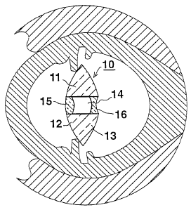
With reference first to Fig. 1, there is
illustrated a horizontal section of a human eye, including
one form of intraocular insert, generally designated 10,
constructed in accordance with the present invention. The
means for fixing the insert 10 in the eye are not described
herein, as many such means are known for mounting artificial
intraocular lenses and can be used for fixing the
intraocular insert 10.
The intraocular insert 10 includes a body member '
11, of generally convexo-convex or convexo-plano
configuration; that is, its front or anterior face 12 facing
the anterior side of the human eye is of convex


WO 94/17756 ~ ~ ~ PCT/US94100749
- 3 -
configuration, and similarly its rear o:r posterior face 13
facing the posterior side of the human ~~ye is of convex (or
planar) configuration.
The body member 11 is formed with a central
'S cylindrical bore 14 extending through its anterior face 12
and its posterior face 13.
A positive-power or convex lens 15 is fixed within
bore 14 at the anterior side of body me~~nber 11, and a
negative-power or convex lens 16 is fixed within the bore at
the posterior side of the body member. 'The negative lens 16
is thus aligned with the positive lens 15 but is spaced
rearwardly of the positive lens by the cavity defined by
bore 14. The two lenses 15 and 16 thus define a Galilean
telescopic system commonly used in opera glasses.
Such a telescopic system, when incorporated in an
intraocular insert implanted into the human eye in place of
the natural crystalline lens, increases the visual field
that the patient enjoys, thereby enabling the patient to
read fine print without the use of an outside telescope.
Thus, the normal eye movements in the reading process are
preserved, and the patient does not need to move his or her
head from one side of the line to the other in order to
read, as generally required when using external telescopic
spectacles.
The two lenses 15 and 16 may be made of the same
material as presently used for making intraocular lenses,
such as transparent plastic (e. g., methyl methacrylate),


WO 94/17756 PCT/US94/00749
4 -
~.~ ~'~ _
glass, sapphire or the like. The body member 11 may be of
the same transparent rigid material. The cavity 14 between
the two lenses 15 and 16 may be filled with a fluid, such as
air, a gas, or a suitable liquid such as water. ,
Fig. 2 illustrates an intraocular insert,
generally designated 20, similar to insert 10 of Fig. 1, and
also including a body member 21 formed with a central
cylindrical cavity 24 covered at its front side by a
positive lens 25 facing the anterior side of the eye, and at
its rear side by a negative lens 26 facing the posterior
side of the eye. In Fig. 2, however, the positive lens 25
is integrally formed with the body member 21, whereas the
negative lens 26 a.s formed as a separate element and is
fixed, as by an adhesive or a weld, in the rear part of the
cylindrical cavity 24 of the body member.
It will be seen that in the constructions of both
Figs. 1 and 2, the outer periphery of the anterior face of
the positive lens (15, 25) is substantially flush with the
anterior face of the body member 11; and similarly, the
outer periphery of the posterior face of the negative lens
(16, 26) is substantially flush with the posterior face of
the body member 11, 21.
Fig. 3 illustrates an intraocular insert,
generally designated 30, also including a body member 31
formed with a central cylindrical bore 34 closed at the
anterior end by a positive lens 35 and at the posterior end
by a negative lens 36. In this case, however, the negative


WO 94/17756 PCT/US94100749
_ 5 _ ~I~~~~~
lens 36 is mounted to the end of a cylindrical lens holder
37 so that it extends rearwardly of the posterior face of
the body member 30 and thereby produces a larger space
between it and the positive lens 35. Such an arrangement
increases the magnification of the intraocular insert.
In all other respects, the ini:raocular insert 30
illustrated in Fig. 3 is constructed anc~ operates in the
same manner as described above with respect to Figs. 1
and 2.
Fig. 4 illustrates an intraocular insert,
generally designated 40, including a body member 41 in the
form of a soft lens formed with a centr~~l cavity in the form
of a throughgoing bore 43 coaxial with ithe central axis of
the soft lens. A cylindrical lens holder tube 44 is mounted
to the anterior side of the soft lens 4'1 within its bore 43,
and carries a positive lens 45 facing the anterior side of
the eye. A negative lens 46 is mounted within bore 43 to
face the posterior side of the eye. As seen in Fig. 4, the
anterior face of the positive lens 45 projects forwardly of
the anterior face of the soft lens 41, whereas the negative
lens 46 is substantially in coaxial ali~~nment with the soft
lens. This produces a relatively large cavity between the
two lenses 45, 46, thereby increasing t:he magnification of
the intraocular insert.
The soft lens 41 is preferably made of a silicone,
whereas lenses 45 and 46, as well as the cylindrical lens
holder 44, are made of transparent glass or plastic. The


WO 94/17756 PCTIUS94/00749
- 6 -
center cavity of holder 44, between the two lenses 45, 46,
may be filled with any suitable fluid, e.g., air, a gas or
transparent liquid., -In all other respects, the intraocular
insert 40 illustrated in Fig. 4 is constructed and operates
in the same manner as described above.
In the embodiment of Fig. 4, the body member 41 is
preferably a soft lens, but could be a hard lens material,
such as of glass, plastic or sapphire. Preferably the cavity
defined by the cylindrical lens holder 44 is filled with
air, but could be filled with another inert gas or inert
liquid.
While it is contemplated that all the elements of
the intraocular insert would be implanted as an assembly at
one time, it is conceivable that the intraocular insert
could include a body member formed with a central cavity
implanted in the interior of the human eye, and the lenses
attached to the body member during or after its
implantation. The intraocular insert could also include more
than two lenses, combination lenses, holographic lenses,
etc. Many other variations, modifications and applications
of the invention will be apparent.

Representative Drawing
A single figure which represents the drawing illustrating the invention.
Administrative Status

For a clearer understanding of the status of the application/patent presented on this page, the site Disclaimer , as well as the definitions for Patent , Administrative Status , Maintenance Fee  and Payment History  should be consulted.

Administrative Status

Title Date
Forecasted Issue Date 2004-09-14
(86) PCT Filing Date 1994-01-31
(87) PCT Publication Date 1994-08-18
(85) National Entry 1995-08-03
Examination Requested 2001-01-29
(45) Issued 2004-09-14
Expired 2014-01-31

Abandonment History

There is no abandonment history.

Payment History

Fee Type Anniversary Year Due Date Amount Paid Paid Date
Application Fee $0.00 1995-08-03
Maintenance Fee - Application - New Act 2 1996-01-31 $50.00 1996-01-05
Registration of a document - section 124 $0.00 1996-07-11
Maintenance Fee - Application - New Act 3 1997-01-31 $50.00 1997-01-03
Registration of a document - section 124 $100.00 1997-02-10
Maintenance Fee - Application - New Act 4 1998-02-02 $50.00 1997-12-31
Maintenance Fee - Application - New Act 5 1999-02-01 $75.00 1998-12-23
Maintenance Fee - Application - New Act 6 2000-01-31 $75.00 2000-01-06
Request for Examination $200.00 2001-01-29
Maintenance Fee - Application - New Act 7 2001-01-31 $75.00 2001-01-29
Maintenance Fee - Application - New Act 8 2002-01-31 $75.00 2002-01-31
Registration of a document - section 124 $100.00 2002-09-04
Maintenance Fee - Application - New Act 9 2003-01-31 $75.00 2003-01-14
Maintenance Fee - Application - New Act 10 2004-02-02 $125.00 2004-01-21
Final Fee $150.00 2004-07-05
Maintenance Fee - Patent - New Act 11 2005-01-31 $125.00 2005-01-04
Maintenance Fee - Patent - New Act 12 2006-01-31 $125.00 2006-01-23
Maintenance Fee - Patent - New Act 13 2007-01-31 $125.00 2006-12-19
Maintenance Fee - Patent - New Act 14 2008-01-31 $250.00 2008-01-25
Maintenance Fee - Patent - New Act 15 2009-02-02 $450.00 2009-01-15
Maintenance Fee - Patent - New Act 16 2010-02-01 $450.00 2010-01-14
Maintenance Fee - Patent - New Act 17 2011-01-31 $450.00 2011-01-20
Maintenance Fee - Patent - New Act 18 2012-01-31 $225.00 2012-01-19
Maintenance Fee - Patent - New Act 19 2013-01-31 $225.00 2013-01-14
Owners on Record

Note: Records showing the ownership history in alphabetical order.

Current Owners on Record
VISIONCARE OPHTHALMIC TECHNOLOGIES, INC.
Past Owners on Record
BARISH, BENJAMIN J.
GROSS, JOSEPH
LIPSHITZ, ISAAC
VISIONCARE
Past Owners that do not appear in the "Owners on Record" listing will appear in other documentation within the application.
Documents

To view selected files, please enter reCAPTCHA code :


To view images, click a link in the Document Description column. To download the documents, select one or more checkboxes in the first column and then click the "Download Selected in PDF format (Zip Archive)" or the "Download Selected as Single PDF" button.

List of published and non-published patent-specific documents on the CPD .

If you have any difficulty accessing content, you can call the Client Service Centre at 1-866-997-1936 or send them an e-mail at CIPO Client Service Centre.


Document
Description 
Date
(yyyy-mm-dd) 
Number of pages   Size of Image (KB) 
Representative Drawing 1998-07-16 1 17
Claims 2004-01-12 2 60
Cover Page 1996-01-15 1 16
Abstract 1994-08-18 1 53
Description 1994-08-18 6 221
Claims 1994-08-18 3 70
Drawings 1994-08-18 2 64
Representative Drawing 2004-04-16 1 19
Cover Page 2004-08-11 1 45
Assignment 1995-08-03 21 721
PCT 1995-08-03 6 257
Prosecution-Amendment 2001-01-29 1 42
Prosecution-Amendment 2001-11-15 2 45
Assignment 2002-09-04 2 88
Correspondence 2002-10-23 1 17
Fees 2003-01-14 1 35
Assignment 2003-03-10 3 122
Prosecution-Amendment 2003-07-15 3 104
Prosecution-Amendment 2004-01-12 5 155
Fees 1998-12-23 1 34
Fees 2004-01-21 1 27
Fees 2002-01-31 1 37
Fees 1997-12-31 1 39
Fees 2000-01-06 1 33
Fees 2001-01-29 1 37
Correspondence 2004-07-05 1 31
Fees 2005-01-04 1 34
Fees 2006-01-23 1 30
Fees 2008-01-25 1 26
Fees 1997-01-03 1 27
Fees 1996-01-05 1 35