Note: Descriptions are shown in the official language in which they were submitted.
WO 94/18913 215 5 7 26 PCT/US94/01484
FUSION STABIEIZATION CHAMBER
BACKGROUND OF THE INVENTION
The present invention relates to the field of neurosurgery, and pro-
vides a device which facilitates the implantation of bone into the spine
following removal of vertebrae, and which also facilitates the fusion of
the implanted bone with the surrounding bone. The invention also in-
cludes a method of performing spinal surgery, and in particular, of
stabilizing the spine following removal of one or more vertebrae.
Cancer or trauma or degenerative changes can cause parts of the
human vertebrae to develop outgrowths or ridges that can touch the spinal
cord and cause pain and/or paralysis. Neurosurgeons have developed means
of treating such conditions, by removing part of the vertebrae, and,
where appropriate, replacing the removed bone with something else. The
removal of all or part of a vertebra is called a "corpectomy" or a "ver-
tebrectomy". In some cases, one can replace the bone removed by corpec-
tomy with bone taken from another site on the body of the patient; in
other cases, one can obtain bone from a "bone bank". Given the right
conditions, the new bone material will fuse to the bone surrounding the
corpectomy site, and can become for practical purposes a part of the
patient's body. To achieve the desired fusion, one must stabilize the
spine so that the bone has time to fuse. The fusion process can take
from six weeks to six months.
In performing spinal surgery, one can approach the spine either from
the front (anterior) or rear (posterior) sides. The posterior approach
has the disadvantage that since the vertebrae lie on the anterior side of
WO 94/18913 ~15 5 7 2 ~ PCT/US94/01484
the spinal cord, the surgeon must navigate past the spinal cord before
reaching the vertebrae, and must take special care not to disturb the
spinal cord. Conversely, with the anterior approach, the surgeon does
not encounter the spinal cord while en route to the vertebrae. The pres-
ent invention concerns the anterior appr~oach.
The prior art contains many systèms for stabilizing various parts of
the spine following surgery. The development of such systems has made it
possible to treat certain lesions of the spine aggressively, instead of
simply immobilizing them in a brace. The typical external immobilizing
device of the prior art comprises the halo vest. The typical internal
immobilizing device comprises the Caspar plate, described below.
The Caspar plate system, named after Dr. Wolfhard Caspar, comprises
a means for stabilizing the spine after anterior spinal surgery. The
Caspar system includes a set of plates which one attaches to the remain-
ing vertebrae surrounding the corpectomy site. In the Caspar procedure,
one screws a plate directly onto the spine, the screws approaching within
about one or two millimeters of the spinal cord. The Caspar system pro-
vides immediate stabilization of the spine following a corpectomy, and in
other cases where the spine has become unstable following an accident.
The Caspar system also eliminates the need for wearing the very cumber-
some halo vest, and eliminates the need to undergo a separate surgical
procedure from the rear.
However, the Caspar system also has disadvantages. It requires a
large inventory of expensive equipment, including screws and plates of
all sizes. The latter expense can represent a formidable obstacle to
many medical institutions. Also, one needs to insert the screws through
the spine, engaging the posterior cortex. Although one can monitor the
WO 94/18913 215 5 7 ~ ~ PCT/US94/01484
position of the screws with an appropriate real-time viewing apparatus,
the procedure carries the potential risk of spinal cord injury or lacera-
tion of the vertebral artery. When a competent surgeon performs the pro-
cedure, these complications rarely occur, but other complications such as
loosening of the screws and persistent instability may develop. More-
over, the difficulty of the procedure discourages many surgeons from even
attempting the anterior plating procedure.
The Synthes cervical spine locking plate constitutes another
anterior plating system of the prior art. In the Synthes system, one
inserts a second screw into the head of the anchor screw, thus creating a
second affixation of the plate to the vertebrae. Many regard the Synthes
system as easier, safer, and faster to use than the Caspar plate system,
because the anchor screw does not penetrate the posterior cortex and be-
cause one therefore does not need to monitor the precise position of the
screw during insertion. However, the Synthes locking plate has less ver-
satility than the Caspar plate, as it provides the ability to fuse only
two to three levels of the cervical spine.
Both the Caspar and Synthes systems also have the disadvantage that
they do not work well in patients with osteoporosis, rheumatoid
arthritis, ankylosing spondylitis, and other conditions of poor bone
growth or metabolic bone disease.
- Both the Caspar and Synthes systems have additional disadvantages
inherent with the use of screws. First, as mentioned above, screws do
become loose. If one uses the screws as the primary means of affixing
the stabilizing device to the spine of the patient, loosening of the
screws represents a major problem. Moreover, the use of screws presents
a technical challenge to the surgeon. Correct screw placement requires
experience, as well as a large inventory of expensive equipment, as well
2~5~ ~
WO 94/18913 PCT/US94/01484
as imaging devices for monitoring the position of such screws. Also,
with screw-based systems of the prior art, the surgeon must create a
large opening in the patient, so as to view the screw along its shaft.
Such an opening creates additional risks to the patient, such as the risk
of injury to vascular structure and to nearby nerves.
In addition to the problem of how to stabilize the spine immediately
after performing a corpectomy, vertebral surgery poses problems relating
to the replacement of the removed bone. Some systems of the prior art
require the use of a bone strut to replace the diseased bone segments
removed in surgery. This bone grafting material costs a great deal, and
sometimes one cannot obtain enough material when performing multiple ver-
tebrectomies. Furthermore, bone graft material, usually taken from
cadavers, has typically been sterilized by radiation, a process believed
to weaken or destroy the strength and osteoconductive properties of bone.
While it is possible to use other means of sterilization, such as
ethylene oxide or freeze drying, it usually turns out that the best bone
graft material comes from the patient, because the patient's own bone
will likely fuse more rapidly than bone obtained elsewhere. Unfortunate-
ly, harvesting such bone consumes substantial time, involves substantial
pain to the patient, and presents other risks, such as risk of infection
at the harvest site, hemorrhage, and peripheral nerve injury.
The present invention overcomes the disadvantages of the prior art
systems described above. First, the invention provides a device which
surgeons can learn to use very easily, and which they can insert without
intraoperative fluoroscopy or other means of accurately monitoring the
position of a device within the body. Most neurosurgeons can use the
device of the present invention with instruments already in their posses-
~ WO 94/18913 2 ~ 5 5 7 2 6 PCT/US94/01484
s
sion.
Secondly, the invention provides an adjustable device which can fita large range of patients. This feature eliminates the need to keep a
large inventory of parts in order to accommodate every possible patient.
Thirdly, the device allows one to use the patient's own cancellous
bone which one removes during the vertebrectomy, possibly with the addi-
tion of further cancellous bone material from an external source. In any
event, the invention reduces or eliminates the need to obtain a pelvic
bone autograft from the patient.
The device of the present invention also reduces or eliminates the
problem of loosening of screws, which can occur with the plating systems
of the prior art, and which clearly can cause substantial pain and ex-
pense.
SUMMARY OF THE INVENTION
The fusion stabilization chamber of the present invention includes a
pair of hollow members, both of which may have a rectangular or slightly
trapezoidal cross-section. One of the hollow members slides within the
other. Thus, the chamber comprises two telescoping hollow members. Each
hollow member includes at least one barrel vault at one end, each barrel
vault comprising threaded means for receiving a screw. The barrel vaults
are arranged in a mutually oblique manner, such that the screws inserted
into the vaults also lie along mutually oblique lines. The hollow
members preferably comprise enclosures defined by four walls formed of a
metal mesh. The hollow members may also include means for locking the
members in a desired position relative to each other.
In using the stabilization chamber described above, the surgeon
WO 94/18913 21 5 5 7 2 6 PCT/US94/01484
first removes the diseased portion of vertebra in the usual manner. The
surgeon measures the length of the corpectomy site (the length of the
space to be filled), and adjusts the length of the chamber accordingly.
One may fasten the locking means so that the telescoping chamber main-
tains its desired position. Then, the surgeon fills the chamber with
bone material, such as bone chips obtained from the corpectomy operation
itself, or bone material from other sources, and inserts the chamber into
the corpectomy site. The surgeon gently taps the device into place, so
that it fills most of the corpectomy site, i.e. the space formerly oc-
cupied by the removed vertebra. The chamber does not extend all of the
way towards the spinal cord, due to the retaining action of a pair of
stabilizing plates.
The surgeon then drills holes in the surrounding bone, using the
barrel vaults as guides for the drill bit. The surgeon then inserts the
screws through the barrel vaults and fastens them to the bone. Due to
the orientation of the barrel vaults, the screws lie along mutually
oblique paths, reducing the likelihood that the device will become dis-
lodged.
In an alternative embodiment, one can provide threaded holes in the
stabilizing plates also, so that additional screws can pass directly
through the stabilizing plates and into the surrounding bone.
The present invention therefore has the primary object of providing
an improved method and apparatus for performing spinal surgery, and in
particular, for stabilizing the spine following removal of one or more
vertebrae.
The invention has the further object of providing a device which
promotes bone fusion in addition to providing stabilization of the spine.
~ WO 94/18913 21~ ~ 7 2 6 PCT/US94/01484
The invention has the further object of simplifying the surgical
process of stabilizing the spine after performing a corpectomy.
The invention has the further object of reducing the cost and com-
plexity of the equipment needed to practice spinal surgery.
The invention has the further object of reducing the time required
for a surgeon to learn to stabilize the spine following a corpectomy.
Persons skilled in the art will recognize other objects and ad-
vantages of the invention, from a reading of the following brief descrip-
tion of the drawings, the detailed description of the invention, and the
appended claims.
BRIEF DESCRIPTION OF THE DRAWINGS
Figure 1 provides a side elevational view of the fusion stabiliza-
tion chamber of the present invention.
Figure 2 shows an end view of the stabilization chamber of the pres-
ent invention.
Figure 3 provides a perspective view of the fusion stabilization
chamber.
Figure 4 shows a top view of the stabilization chamber.
Figure 5 provides a diagrammatic view showing the fusion stabiliza-
tion chamber inserted into a corpectomy site.
Figure 6 provides a perspective view of an alternative embodiment of
the invention, wherein additional screws pass directly through the
stabilizing plates.
WO 94/18913 PCT/US94101484 ~
2155~ 8
DETAILED DESCRIPTION OF THE INVENTION
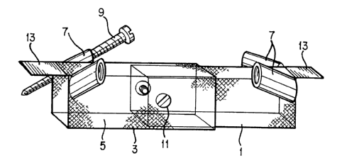
Figures 1-4 show the physical structure of the fusion stabilization
chamber of the present invention. The chamber includes first hollow mem-
ber 1 and second hollow member 3. Both hollow members have a slightly
trapezoidal cross-section, as illustrated in the end view of Figure 2.
Figure 2 exaggerates the trapezoidal shape of the cross-section; in prac-
tice, the width of the member might increase by one millimeter for each
15 mm of depth, but one could use other dimensions. Thus, by "slightly
trapezoidal", one means that the members are nearly rectangular in cross-
section, except for the variation in width described above. The trape-
zoidal cross-section helps to maintain the chamber in position within the
corpectomy site. One inserts the narrower portion of the hollow member
into the body cavity first, with the wider portion oriented towards the
outside. Thus, the chamber tends to become wedged in its place within
the corpectomy site; once pushed in, it becomes difficult to pull out.
Although the preferred embodiment includes the trapezoidal cross-section,
one can also form the chamber with a perfectly rectangular cross-section,
within the scope of the invention.
The first hollow member 1 slides within the second hollow member 3.
The members 1 and 3 preferably have walls formed of metal mesh 5. One
prefers walls having openings which permit bone growth from the adjacent
vertebrae, through the interior of the chamber. However, the walls can
have a different construction. They can even comprise solid metal, as
bone can fuse to metal. In the latter case, the chamber could be empty.
In the preferred embodiment, the chamber has two pairs of barrel
vaults 7, arranged at the opposite ends of the hollow members. One can
vary the number of barrel vaults, within the scope of the invention. The
~l~S72~
O 94/18913 PCT/US94/01484
barrel vaults comprise threaded cylinders through which screws 9 pass.
Figure 1 shows that the screws form an angle of about 30~ relative to the
top longitudinal axis of the chamber. Figure 4 shows that the screws
also form an angle of about 10- relative to the sides of the chamber.
One can vary these angles; one should not consider the invention limited
to particular angles. In general, one selects angles which enable the
screws to pass through the greatest possible thickness of bone, above and
below the corpectomy site, and to provide an angle which, from the per-
spective of the surgeon, facilitates insertion of the screws without the
need to make a larger or additional incision.
As shown in the Figures, the barrel vaults comprise mutually oblique
members. The screws become self-locking in the barrel vaults. One can
also provide an adjustable hexagonal head screwdriver to facilitate
tightening of the screws from any angle.
Locking screw 11 holds the first and second hollow members in place.
The locking screw thus permits adjustment of the size of the chamber.
One slides the hollow members until the chamber has the desired length,
and then fixes the selected length by tightening the locking screw.
Figure 5 provides a diagram of the fusion stabilization chamber in-
serted into a corpectomy site. The figure shows vertebrae 15, the spaces
17 between adjacent vertebrae representing intervertebral discs. Each
vertebra includes an outer bony layer, or cortex 27, which surrounds can-
cellous material 29 inside. Figure 5 also shows spinal cord 19, and the
structures adjoining the spinal cord, including the posterior longitudi-
nal ligament 21, the ligamentum flavum 23, and the posterior spinous pro-
cesses 25. As shown in the figure, one has removed several vertebrae,
and has inserted the chamber into the resulting empty space.
Stabilizing plates 13 extend from both hollow members, as shown in
WO 94/18913 215 5 7 2 6 PCT/US94/01484
the Figures. The stabilizing plates serve several purposes. First, as
illustrated in Figure 5, the stabilizing plates keep the chamber at an
appropriate depth, preventing the chamber from touching spinal cord 19 or
the ligaments surrounding it. By making the depth of the chamber less
than the depth of the adjacent vertebrae, one prevents the chamber from
coming too close to the spinal cord.
Secondly, the stabilizing plates tend to distribute the bending
loads experienced by the chamber, and divert part of these loads away
from the screws. As the vertebrae flex back and forth, the stabilizing
plates tend to oppose some of the vertebral movement, and absorb some of
the tension, thereby tending to prevent the screws from loosening or
breaking.
Thirdly, the stabilizing plates help to rigidify the joints formed
between the ends of the chamber and the respective adjacent vertebrae.
Keeping these joints rigid facilitates the growth of blood vessels from
the adjacent vertebrae, through the holes in the chamber walls, and into
the bone material within the chamber.
Figure 6 shows, in a perspective view, an alternative embodiment
wherein a third screw passes through a threaded hole in each stabilizing
plate, in addition to the pair of screws inserted through the associated
barrel vaults. Figure 6 shows additional screw 10 inserted through the
stabilizing plate on the right-hand side. The figure does not show the
corresponding additional screw on the other side, in order to show the
hole in the stabilizing plate, but in practice a similar additional screw
10 would normally bP provided. However, one should consider each screw
as optional, since it is possible to affix the chamber to the adjacent
bone using fewer than all of the available screws.
215S726
WO 94/18913 PCT/US94/01484
11
One would use the embodiment of Figure 6 in cases where the bone has
become weakened. In rare cases, one might even attach the chamber only
with the stabilizing plate screws, without any barrel vault screws. In
all of the embodiments wherein one provides a threaded hole in the
stabilizing plate, the holes should have low "profiles", so that the
material defining the plate does not project significantly beyond the
plane of the plate.
In using the chamber of the present invention, the surgeon begins by
performing a corpectomy in the conventional manner. Immediately after
removal of one or more vertebrae, the surgeon measures the length of the
corpectomy site with calipers, and adjusts the length of the chamber to
make it conform to the length of the corpectomy site. One adjusts the
length of the chamber by pulling the hollow members 1 and 3 away from
each other or pushing them together, as needed. Then one tightens the
locking screw 11 to fix the length (and thus the volume) of the chamber.
Next, the surgeon fills the chamber with bone. The bone can com-
prise bone chips obtained from the vertebrae removed in the corpectomy
procedure, or it can comprise cancellous bone obtained from another site.
One might also use a biocompatible osteogenic polymer.
In a variation of the latter step, the surgeon may place bone chips,
obtained from the corpectomy, into the chamber, while the corpectomy pro-
gresses. However, in this case, one would still need to adjust the cham-
ber to fit the corpectomy site, and one would also need to insure that
the bone has substantially filled the volume of the chamber after adjust-
ment of the size of the chamber.
The surgeon then inserts the bone-filled chamber into the corpectomy
site, and gently taps it into place, such that the stabilization plates
13 come to rest on the vertebrae immediately adjacent to the corpectomy
WO 94/18913 2j~SS 7 2 ~ PCT/US94/01484
12
site. The chamber should fit tightly within the corpectomy site. One
may take a lateral spine X-ray to insure that the chamber has seated it-
self properly in the corpectomy site.
Next, the surgeon drills holes into the adjacent vertebrae, using an
appropriate drill, such as a 2mm twist drill. The barrel vaults 7 form
guides for the drill bit, and thereby determine the direction of the
holes. The orientation of the barrel vaults unambiguously determines the
orientation of the holes. The holes therefore make the same angles as
the barrel vaults, relative to the axes of the chamber.
The surgeon then threads the screws 9 into the barrel vaults 7. The
barrel vaults direct the screws along the correct path. Due to the in-
teraction of the heads of screws 9 with the barrel vaults, the barrel
vaults also insure that the screws 9 become inserted to the correct
depth. When tightened, the screws 9 tend to draw the adjacent vertebrae
towards the chamber. Note also that the screws pass twice through the
cortex of the vertebrae. In other words, each screw has a length suffi-
cient to pass through the cortex 27 at one surface of the vertebra, then
through the cancellous material 29 at the core of the vertebra, and again
through the cortex as the screw exits the vertebra. Fastening the screws
in this manner minimizes the likelihood that the screws will become dis-
lodged.
Following the tightening of the screws, one can take a lateral X-ray
to verify proper placement of the screws. If all is correct, one can
then close the wound in the conventional manner.
The present invention has many advantages, as outlined below:
1. The fusion stabilization chamber does not rely on screws as the
sole means of stabilizing the spine following surgery. Due to the trape-
~5572~
WO 94/18913 PCT/US94/01484
13
zoidal cross-section of the chamber, the chamber becomes firmly wedged
within the corpectomy site even before attachment of the screws.
2. The surgeon can learn to insert the fusion stabilization chamber
much more quickly than devices of the prior art. Since the barrel vaults
automatically determine the direction and depth of the screws, the
surgeon will be less likely to make mistakes while using the present in-
vention, and the invention therefore is less intimidating to the surgeon
than devices of the prior art. In particular, the oblique direction of
the screws lessens the potential damage to the spinal cord. Moreover,
most neurosurgeons can use the fusion stabilization chamber with
instruments already in their possession.
3. The oblique direction of the screws has the added benefit that
it increases the compression effect, by drawing vertebrae above and below
the chamber into firm contact with the chamber. Such compression speeds
fusion of the bone.
4. The oblique direction of the screws has the additional advantage
of reducing the required size of the surgical incision, because the
surgeon can reach deeply into adjacent vertebrae, using the screws, with-
out exposing those vertebrae.
5. Because of the ease and manner of insertion of the device, the
surgeon need not use intraoperative fluoroscopy, or other monitoring
means, while inserting the device.
6. The present invention eliminates the need for a large inventory
of stabilization plates and screws for fitting different sizes of verte-
brae. One can construct the present invention in two or three basic
sizes, which together fit virtually all possible corpectomy sites, due to
the telescoping feature of the chamber. Thus, the invention reduces the
cost of maintaining an inventory of materials. Moreover, due to the
WO 94/18913 2~5S7 2~ 14 PCT/US94/01484
simple structure of the fusion stabilization chamber, one can manufacture
it relatively inexpensively.
7. One can make the fusion stabilization chamber of strong titanium
metal mesh which allows bone to grow from end to end and from side to
side. One can easily fill the chambèr with the patient's own cancellous
bone mixed with hydroxyapatite crystals and/or other biocompatible syn-
thetic bone substitutes known to increase the rate of bone formation.
Thus, the present invention reduces the need to harvest bone from other
sites on the patient's body.
8. The structure of the fusion stabilization chamber provides
stability through all three degrees of freedom of movement.
In an alternative embodiment, one can replace the locking screw with
a screw device located inside the chamber and extending along the entire
length of the chamber. Thus, the latter screw device would comprise a
type of jack. Turning the latter screw would vary the overall length of
the jack, which is equivalent to varying the length of the chamber. With
this arrangement, one need not adjust the length of the chamber before
inserting it into the corpectomy site. Instead, one would first insert
the chamber, and then turn the screw to adjust the jack, until the cham-
ber becomes long enough to occupy the entire space. The above-described
screw device would then comprise the means for locking the hollow members
into a fixed position relative to each other, and could be used instead
of, or in addition to, locking screw 11. One would use a bevel gear, or
equivalent mechanical device, for adjusting the jack while the chamber is
in position. The latter alternative should be considered within the
scope of the present invention.
In another alternative embodiment, one can coat the outside of the
~lSS726
WO 94/18913 PCT/US94/01484
chamber with an osteoconductive substance, such as hydroxyapatite, or the
like, to promote fusion of the chamber to the surrounding bone. This
coating can be instead of, or in addition to, the filling of the chamber
with bone material. The invention should be considered to include the
latter alternatives.
The chamber used in the present invention can have various cross-
sections. The invention is not limited to the rectangular or trapezoidal
cross-sections discussed above, but can include other shapes. For ex-
ample, one could form the chamber with a circular cross-section, in which
case the chamber would have the general shape of a cylinder.
The present invention is also not limited to a chamber having
straight walls. Instead, the chamber could be curved along its length.
In this way, one can make the chamber fit the curvature of the spine. In
the latter case, both hollow members would be curved, so that they could
slide back and forth within each other, while maintaining the desired
curvature. This embodiment would be useful for a corpectomy which spans
a relatively large number of vertebrae.
While the above description illustrates the preferred embodiments of
the invention, one can vary the invention in still other ways. For ex-
ample, as noted above, one can vary the structure of the walls of the
chamber. While one prefers a chamber having holes, such as provided by a
metal mesh, one could use an empty box having solid walls. The position
and number of barrel vaults can also vary. These and other modifica-
tions, which those skilled in the art will recognize, should be consid-
ered within the spirit and scope of the following claims.