Note: Descriptions are shown in the official language in which they were submitted.
21~~'~~5
Docket No. 1653 CANADA
VASCULAR sUTURING APPARATUS
BACKGROUND
i. Technical Field
The technical field relates generally to surgical
suturing instrumentation and, more particularly, to a
surgical suturing apparatus and a method for suturing
vascular tissue sections together.
2. Description of Related Art
During many surgical procedures it is often
necessary to join or even rejoin portions of vascular
tissues or vessels to form an anastomosis. Various methods
of joining vascular tissues to create an anastomosis are
used, such as, for example, suturing, stapling or clipping
the ends of the vessels together. Additionally, various
stents may be used to join the vessels together and create
the anastomosis. Where vessels are joined open end to open
end it is termed an "end to end" anastomosis. However, in
certain surgical procedures it is often desirable to join a
free open end of one vessel to an incision in the side of
another vessel to create an "end to side" anastomosis or
even an incision in the side of another vessel to form
"side-to-side" anastomosis.
In some instances suturing of vessels is
preferred over stapling or clipping the vessels. Due to
the small size of the vessels, a very small suturing needle
is used having a length of suture material attached thereto
to suture the vessels together. The suturing needle is
typically grasped by a needle holder and passed through one
vessel and then the opposite vessel. The procedure is
repeated to thread or impart a series of stitches to the
vessels to suture them together.
Because of the extremely small size of the
suturing needle used, typically on the order of ten
2
thousands of an inch in diameter, handling problems may
arise while manipulating the suturing needle through the
vascular tissues. For example, upon piercing a vessel, the
needle must be pushed through the vessel, released by the
needle holder at one end of the needle and subsequently
grasped at the opposite end of the needle to draw the
needle and suture through the vessel thus requiring the
release of the needle and suture during the procedure.
Release of the needle is often undesirable and may pose
problems in regaining control of the needle. To avoid this,
it may become necessary to use two needle holders, one
positioned on either side of the vessel, to continually
grasp the needle, thereby requiring two hands to perform
the operation. Additionally, precise control of the needle
is often difficult when using typical needle holders. The
small size of the needle also makes it difficult and time
consuming to recover if dropped during the surgical
procedure.
As noted above, the vascular sections to be
sutured are typically extremely small. Suturing of such
vascular tissue sections is often performed under
magnification by equipping the surgeon with special
magnifying glasses. The use of these magnifying glasses,
while enlarging the view of the vascular tissues and
needle, reduces the field of view within which the surgeon
has to operate. Thus, as the needle and suture material
are passed through the vessels and pulled to draw the
suture material through, it often becomes necessary to move
the needle holder and needle from the field of view. This
may require the surgeon to look away from the field of view
containing the vessel sections to be sutured and may
present problems in repositioning the needle within the
magnified field of view to form another stitch in the
vascular tissues and increases the time required to suture
the vessels together. Even when magnifying glasses are not
being used, the surgeon needs to look at the needle as it
is moved away from the surgical site to pull the length of
suture through the vessel and is passed to his other hand
3
to be re-gripped. Due to the minute size of the vessels,
it takes time for the surgeon to refocus on the surgical
site to once again pass the needle and suture through the
vessel. This repeated change of focus is time consuming
and can place a strain on the surgeon's eyes.
Thus, it would be advantageous to have a surgical
suturing apparatus and a method of suturing vessels which
are particularly suited to suturing vascular tissues in
anastomosis procedures. It would further be advantageous
to have a surgical suturing apparatus which is operable
with a single hand and is capable of maintaining precise
and constant control of the needle as it is passed from one
needle holding arm of the apparatus to another to avoid
release of the needle during the suturing operation. It
would also be advantageous to have a surgical suturing
apparatus which is capable of suturing vascular tissue
sections together with limited hand, apparatus and needle
movement in order to maintain the entire suturing operation
within a restricted field of view.
SOMMARY
The disclosed surgical apparatus for suturing
vascular tissue sections and includes a first arm having a
first needle receiving recess and a first needle engaging
member or blade mounted for movement with respect to the
first arm. A second arm is provided and is mounted for
movement with respect to the first
arm and has a second needle receiving recess, the second
arm having a second needle engaging member or blade mounted
for movement with respect to the second arm. There is also
provided a reciprocating mechanism connected to the first
and second needle engaging members and movable for
alternately moving the first and second needle engaging
members into and out of the first and second needle
receiving recesses to secure a surgical needle therein.
The reciprocating mechanism is movable between a first
position advancing the first needle engaging member into
engagement with the surgical needle and a second position
CA 02157745 2004-12-03
4
advancing the second needle engaging member into engagement
with the surgical needle. A ramming member or lever is
operatively associated with one of the arms for
automatically ramming the reciprocating mechanism between
the first and second positions upon full closure of the
first arm against the second arm.
Preferably, the reciprocating mechanism includes a
toggle wheel rotatably affixed the arms and having first and
second ramming surfaces such that an initial closure of the
arms forces the ramming member into engagement with the
first ramming surface to move the toggle wheel to the first
position. A subsequent closure of the arms forces the
ramming member into engagement with the second ramming
surface to move the toggle wheel to the second position.
In accordance with another embodiment of the
present invention there is provided an apparatus for
suturing vascular tissue sections comprising: a) a pair of
needle holding tweezer like arms movably interconnected for
movement between open and closed positions; and b) a
securing mechanism operatively associated with the pair of
arms and engagable with a surgical needle to secure the
surgical needle alternately within each arm of the pair of
arms in response to closure of the arms.
There is also disclosed a method of threading a
suture through a vascular tissue section which includes the
step of providing an apparatus having a first arm with a
first needle receiving recess arid a second arm with a second
needle receiving recess, the first and second arms movable
toward and away from each other. The method also includes
the steps of holding a surgical needle within the second
needle receiving recess, positioning the first and second
arms about a first vascular tissue section to be sutured,
moving the second arm toward the first arm such that the
surgical needle pierces the first vascular tissue section d
CA 02157745 2004-12-03
4a
and enters the first needle receiving recess and releasing
the surgical needle from the second needle receiving recess
and holding the surgical needle within the first needle
receiving recess. The method also includes the step of
moving the first arm away from the second arm to draw a
suture affixed to the surgical needle at least partially
through the first vascular tissue section.
BRIEF DESCRIPTION OF THE DRAWINGS
Various embodiments are described herein with
reference to the drawings, wherein:
FIG. 1 is a perspective view of a vascular
surgical suturing apparatus with arms in an open position
30
2 I ~'~'~ 4
and holding a surgical suturing needle within one of the
arms:
FIG. 2A is a side cross-sectional view of the
suturing apparatus taken along line 2A-2A of Fig. 1:
5 FIG. 2B is an enlarged side view, partially shown
in section, depicting the reciprocating mechanism of the
suturing apparatus of Fig. 1:
FIG. 3 is a cross-sectional view taken along line
3-3 of Fig. 2B:
FIG. 4 is a perspective view of the toggle wheel;
FIG. 5 is a perspective view of the caroming lever
for moving the toggle wheel:
FIG. 6A is a side cross-sectional view similar to
Fig. 2A depicting the arms moved to a closed position:
FIG. 6B is an enlarged side view, partially shown
in section, of the reciprocating mechanism position
corresponding to the position of Fig. 6A:
FIG. 6C is an enlarged side cross-sectional view
of the distal end of the suturing apparatus corresponding
to the position of 6A:
FIG. 7A is a side view of the vascular surgical
suturing apparatus of Fig. 1, partially shown in section,
illustrating initial actuation of the reciprocating
mechanism in a clockwise direction:
FIG. 7B is an enlarged side view, partially shown
in section, of the reciprocating mechanism position
corresponding to the position of Fig. 7A:
FIG. 8 is a view similar to Fig. 7B, illustrating
further actuation of the reciprocating mechanism:
FIG. 9A is a view similar to Fig. 8, illustrating
still further actuation of the reciprocating mechanism:
FIG. 9B is an enlarged side cross-sectional view
of the distal end of the suturing apparatus of Fig. 1
corresponding to the reciprocating mechanism position of
Fig. 9A:
FIG. l0A is a view similar to Fig. 9A,
illustrating a final position of the reciprocating
mechanism;
21~7~4~
6
FIG. 10B is a view similar to Fig. 9B
corresponding to the position of Fig. 10A:
FIG. 11A is a cross-sectional view taken along
the line 11A-11A of Fig. 9B;
FIG. 11B is a cross-sectional view taken along
the line 11B-11B of Fig. 9B;
FIG. 12A is a cross-sectional view taken along
the lines 12A-12A of Fig. lOB;
FIG. 12B is a cross-sectional view taken along
the lines 12B-12B of Fig. 10B;
FIG. 13 is a side view of the vascular surgical
suturing apparatus, partially shown in section,
illustrating the surgical needle positioned in the opposing
arm;
FIG. 14 is an enlarged side view, partially shown
in section, of the reciprocating mechanism illustrating
initial actuation of the reciprocating mechanism in an
opposite (counterclockwise) direction;
FIG. 15 is a view similar to Fig. 14,
illustrating further actuation of the reciprocating
mechanism;
FIG. 16A is a perspective view of a pair of
vascular tissue sections to be end to end sutured as viewed
within a limited field of view under magnification;
FIG. 16B is a view similar to Fig. 16A with the
ends of the vessels everted in preparation for suturing;
FIG. 16C is a sectional view of a distal end of
the surgical suturing apparatus of Fig. 1 and the pair of
everted vessels ready for suturing;
FIG. 16D is a view similar to Fig. 16C
illustrating the piercing of the everted edges of the
vascular sections by the surgical needle:
FIG. 16E is a view similar to Fig. 16D
illustrating the surgical needle having been passed to an
opposing arm and the suture being drawn through the everted
vessel edges;
2~~'~74~
7
FIG. 16F is a view similar to view 16E
illustrating the surgical needle being passed back to the
first arm.
FIG. 16G is a view similar to FIG. 16D
illustrating the arms being closed again about the everted
vessel ends to pierce the vessels and form another stitch;
FIG. 16H is a view of the vessels sutured
together to form an end to end anastomosis:
FIG. 16I is a view taken along lines I-I of FIG.
16H;
FIGS. 17A and 17B are enlarged views of an
alternate suturing apparatus distal end and associated
needle engaging member configuration;
FIGS. 18A and 18B are enlarged views of another
alternate distal end and needle engaging member
configuration;
FIGS. 19A and 19B are enlarged views of a further
alternate distal end and needle engaging member
configuration for use with a suturing needle having a hole
at least partially therethrough;
FIG. 19C is a side plan view of a double pointed
suturing needle having a hole at least partially
therethrough;
FIGS. 20A and 20B are enlarged views of yet
another alternate distal end and needle engaging member
arrangement;
FIG. 21 is an enlarged view of an alternate
suturing apparatus distal end slotted for ease in
manipulation of a length of suture affixed to a surgical
needle;
FIGS. 22A and 22B are enlarged views of another
alternate distal end and needle engaging member
configuration designed for easy loading of a surgical
needle:
FIGS. 23A and 23B are enlarged views of an
alternate easy load style distal end and needle engaging
member configuration; and
21~'~745
8
FIGS. 24A and 24B are enlarged views of still
another alternate easy load style distal end and needle
engaging member configuration.
DESCRIPTION OF PREFERRED EMBODIMENTS
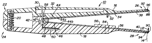
Referring to Fig. 1, there is depicted a vascular
suturing apparatus 10 which is particularly suited to
facilitate suturing vascular tissue sections or vessels
together. Further, apparatus 10 is specifically designed to
repeatedly pass a small surgical needle, having an
associated length of suture material attached thereto, such
as, for example, surgical needle 12 and suture 14, through
vascular tissue sections while maintaining precise control
of needle 12 during all phases of the suturing operation.
Apparatus 10 generally includes a first or upper
arm 16 movably connected to a second or lower arm 18. Arms
16 and 18 are movable toward and away from each other in
order to repeatedly pass needle 12 therebetween in a manner
described in detail hereinbelow. Preferably, apparatus 10
is approximately 7 inches long. Apparatus 10 is preferably
designed to handle surgical needles 12 having a length of
approximately .1 to .5 inches and a diameter of
approximately ten thousandths to .025 of an inch which are
particularly suited for vascular surgery. Preferably the
diameter of the surgical needle and the diameter of the
suture are substantially the same to prevent fluid leakage
from the vessel between the needle hole and suture.
Note that the use of the terms "upper" and
"lower" herein refers to the orientation of the instrument
in Figure 1. Clearly, if the orientation changes, these
designations will likewise change.
In many instances, certain ergonomic and
operational advantages may be obtained by biasing a
surgical suturing instrument in an initially open position.
Thus, in this embodiment there is provided a spring 20
which is affixed to proximal ends 22 and 24 of first and
second (or upper and lower in the orientation of Fig. 1)
arms 16 and 18, respectively. Spring 20 biases apart
~~5774~
9
distal ends 26 and 28 of first and second arms 16 and 18,
respectively, in order to facilitate positioning arms 16
and 18 about vascular tissue sections. The biasing action
of spring 20 also aids in pulling suture 14 through the
tissue sections upon opening of arms 16 and 18.
Apparatus 10 further includes holding structure
to secure needle 12 within either distal end 26 and 28 of
arms 16 and 18, respectively. The holding structure allows
surgical needle 12 to be initially held within one of the
arms and, upon closure of the arms, to be subsequently
passed to the opposite arm. To facilitate single handed
use of apparatus 10, a cam actuating lever 30 is provided
to automatically actuate the holding structure upon closure
of the arms. Cam actuating lever 30 is affixed to an
extension arm 32 formed on arm 16. Extension arm 32 is
sufficiently stiff to prevent depression of cam actuating
lever 30 before arms 16 and 18 have been closed against the
bias of spring 20. Thus, after such closure of arms 16 and
18, continued pressure on arms 16 and 18 depresses cam
actuating lever 30 to automatically actuate the holding
structure to release the surgical needle 12 from one arm
for transfer to an opposite arm as described in more detail
hereinbelow.
Referring now to Fig. 2A, in order to hold or
secure needle 12 within arms 16 or 18, there are provided a
pair of needle engaging members or clamping blades 34, 36
which are longitudinally movable within arms 16 and 18. A
first needle engaging blade 34 is slidably disposed within
first arm 16 while a second needle engaging member or
second blade 36 is slidably disposed within second arm 18.
Distal ends 38 and 40 of first and second blades 34 and 36,
respectively, are dimensioned and configured to engage an
edge of needle 12 and securely hold needle 12 within
recesses 88, 90 formed in first and second arms 16 and 18.
To prevent damage to a tip or needle point of surgical
needle 12 and to prevent the suture carrying end from
sliding through arm 18, there are provided a pair of plates
92 and 94 formed in distal ends 26 and 28, respectively,
CA 02157745 2004-12-03
each of which include a reduced recess area 96 and 98 to
prevent surgical needle 12 from sliding completely through
recesses 88 and 90 in arms 16 and 18, respectively. Arms 16
and 18 are formed with longitudinally extending channels 100
and 102 which extend from distal ends 38 and 40 to
reciprocating mechanism 42. Channels 100 and 102 intersect
recesses 88 and 90 to allow members 34 and 36 to engage an
edge of surgical needle 12 disposed in the respective
10 recess .
As shown in Fig. 2A, needle engaging members 44,
46 are slightly bowed at regions 44a, 44b, respectively,
thereby automatically adjusting to needles of various
diameters. That is, as the needle is clamped, the needle
engaging member buckles at a predetermined location so that
the spring force applied to the needle is constant,
regardless of the needle diameter. The needle engaging
member is slightly buckled even when the needle is not
clamped to ensure that the increased buckling occurs in the
same area whenever the needle is clamped.
While the preferred blade engagement structure
includes V-notches 106 and 108, it will be appreciated by
those skilled in the art the various other configurations at
blade distal ends 38 and 40 may be provided to securely hold
needle 12 within arms 16 and 18. Thus, alternate structure
in either arms 16 or 18 or alternate structure in needle 12
itself such as, for example, notches in an edge of needle
12, or holes completely therethrough, may be provided to
accept corresponding engagement structure formed on blades
34 and 36. Thus, for example, a double-pointed surgical
needle, or surgical incision member, having suture
attachment structure intermediate the points may be
utilized. One exemplary example of a surgical incision
member is disclosed in U.S. Patent No. 5,569,301 entitled
SURGICAL INCISION MEMBER.
To repeatedly pass needle 12 between arms 16 and 18, a
reciprocating mechanism 42 is affixed adjacent
11
proximal ends 44 and 46 of first and second engaging
members 34 and 36, respectively. Reciprocating mechanism
42 alternately advances and retracts engaging members 34
and 36, within arms 16 and 18, respectively, thereby
alternately engaging the members 34 and 36 with needle 12.
Reciprocating mechanism 42, along with first and second
engaging members 34 and 36, provide the aforementioned
holding structure for securely and alternately holding
needle 12 within needle receiving recesses formed in arms
16 and 18.
Cam actuating lever 30 actuates reciprocating
mechanism 42 automatically upon full closure of arms 16 and
18. In order to prevent inadvertent release of surgical
needle from arms 16 or 18 prior to closure of arms 16 and
18, there is provided a lock member 48 which is flexibly
mounted within a recess 50 in lower arm 18. Lock member 48
is engagable with reciprocating mechanism 42 to prevent
movement of reciprocating mechanism 42, and thus release of
needle 12, when arms 16 and 18 are in a open position,
i.e., when cam actuating lever 30 has not been depressed.
As noted hereinabove, apparatus 10 is
particularly suited to suturing small vessels or vascular
tissue sections when viewed under magnification or within a
restricted field of view. Further, suturing of vessels
requires an extremely small needle 12 and suture 14,
typically on the order of ten thousands of an inch in
diameter. Thus, in order to ensure precise positioning and
transfer of needle 12 between arms 16 and 18 upon closure
there are provided a pair of alignment pins 52 which are
mounted on first arm 16. Upon closure of arm 16 towards
arm 18, alignment pins 52 enter into guide holes 54 formed
in lower arm 18. The engagement of alignment pins 52 within
guide holes 54 is sufficiently precise to ensure accurate
alignment of needle 12 within the recesses 88, 90 formed in
distal ends 26 and 28 of arms 16 and 18, respectively.
Additionally, to prevent the vascular tissue sections from
being compressed or crushed during the suturing operation,
there is provided a spacer block 56 which ensures that
21~774~
12
distal ends 26 and 28 of arms 16 and 18, respectively, do
not touch upon closure. However, spacer block 56 does
allow arms 16 and 18 to close sufficiently such that needle
12 may be transferred therebetween.
Referring now to Fig. 2B, actuation of the
reciprocating mechanism 42, is caused by cam actuating
lever 30 extending through an aperture 58 formed in arm 16.
Reciprocating mechanism 42 preferably includes a rotatable
toggle wheel 60 which is provided to alternately advance or
retract needle engaging members 34 and 36 in response to
depression of cam actuating lever 30 upon full closure of
arms 16 and 18, i.e. in response to movement of extension
arm 32 with respect to arm 16. Toggle wheel 60
additionally provides recesses (described below) for
engagement with lock member 48.
As noted hereinabove, first and second arms 16
and 18 are movably connected together and configured to be
operated in a tweezer-like manner if desired. Referring to
Fig. 3, arms 16 and 18 are provided with a transverse pivot
pin 62 for connecting and pivotally moving arms 16 and 18
with respect to each other. Pivot pin 62 is connected to
arms 16 and 18 at a position intermediate proximal ends 22,
24 and distal end 26, 28 of arms 16 and 18, respectively.
Thus, pressure on arms 16 and 18 distally of pivot pin 62,
to close distal ends 26, 28, stretches spring 20, while
release of arms 16 and 18 allows spring 20 to bias distal
ends 26 and 28 to an open or spaced apart position.
While the preferred position of pivot pin 62, and
thus the pivot point of arm 16 and 18, is intermediate the
ends of apparatus 10, pivot pin 62 may be positioned at
proximal ends 22 and 24. Further, although the preferred
method of moving arms 16 and 18 is by pivotal motion,
parallel movement of arms 16 and 18 is also contemplated.
Parallel movement of arms 16 and 18 is especially desirable
when using relatively straight surgical needles and may be
accomplished in several ways. For example, arms 16 and 18
could be mounted with respect to each other to both move
perpendicular to their respective longitudinal axes.
21~7'~4~
13
Alternately, arms 16 and 18 could be mounted to move or
slide parallel to their respective longitudinal axes to
advance and retract their distal ends. When arms 16 and 18
slide relative to each other, it is preferable to have the
distal faces of the arms open to a needle holding recess to
facilitate transfer of a surgical needle or surgical
incision member therebetween.
Pin 62 further serves as a pivot point for toggle
wheel 16 which is rotatable in the clockwise or
counterclockwise direction about pin 62. Preferably,
proximal ends 44 and 46 of needle engaging members 34 and
36, respectively, are affixed to toggle wheel 60 by means
of pins 64 and 66, respectively (see Fig. 3). Thus,
rotation of toggle wheel 60 on pivot pin 62 alternately
advances and retracts members 34 and 36 within arms 16 and
18, respectively.
Referring to Figs. 2A and 4, toggle wheel 60 is
provided with a first angled caroming surface 68 having a
first ledge 70 which, when engaged by cam actuating lever
30, translates to a clockwise rotation of toggle wheel 60
and thus a distal advancement of needle engaging member 34
and a proximal retraction of needle engaging member 36.
Similarly, a second angled caroming surface 72 and second
ledge 74 are provided, such that when engaged by cam
actuating lever 30, toggle wheel 60 is rotated in a
counterclockwise direction to advance member 36 and retract
member 34.
Toggle wheel 60 is provided with lock notches 76
and 78 which correspond to the distalmost advancement of
first and second needle engaging members 34 and 36,
respectively. Thus, when toggle wheel 60 is rotated
counterclockwise to a position where lock member 48 engages
lock notch 78, engaging member 36 is locked into an
advanced or distalmost position to securely hold needle 12
within arm 18. Likewise, when toggle wheel 60 is rotated
clockwise to a position where lock member 48 engages lock
notch 76, needle engaging member 34 is locked into an
advanced or distalroost position to secure needle 12 within
14
arm 16. Thus, lock member 48, in conjunction with lock
notches 76 and 78, prevents release and transfer of needle
12 when arms 16 and 18 are not fully closed. Additionally,
toggle wheel 60 is provided with a knock off pin 80 to aid
lock member 48 in entering lock notches 76 and 78, the
operation of which is described in more detail hereinbelow.
Referring now to Figs. 2B and 5, cam actuating
lever 30 is provided to automatically perform the dual
sequential functions of unlocking toggle wheel 60 from
engagement with lock member 48 and rotating toggle wheel
60. Cam actuating lever 30 generally includes a base
portion 82 which is preferably affixed to arm extension
member 32. Alternatively in a non-automatic version, base
82 may be separately movable with respect to extension
member 32. Cam actuating lever 30 includes a flexible
release leg 84 which is affixed to base 82 and which is
provided to cam lock member 48 out of lock notchs 76 or 78
and thus allow toggle wheel 60 to be rotated. Cam
actuating lever 30 also includes a flexible toggle leg 86
formed parallel to release leg 84. Toggle leg 86 is
engagable with angled caroming surfaces 68 and 72 and ledges
70 and 74 in order to rotate toggle wheel 60.
Referring initially to Figs. 1 and 2A, the
operation of vascular suturing apparatus 10 will now be
described. As noted hereinabove, apparatus 10 is
particularly suited to repeatedly pass surgical needle 12
back and forth between arms 16, 18 automatically upon full
closure of the arms, i.e. closure of arms 16 and 18 with
respect to one another and subsequent closure of arm
extension 32 with respect to arm 16. In the initial
position, distal ends 26 and 28 of arms 16 and 18,
_ respectively, are biased to an open position by spring 20.
Needle 12, having an associated length of suture material
14 attached thereto, is positioned and held within distal
end 28 of second (lower) arm 18 by needle engaging member
36. Alignment pins 52 are spaced from guide holes 54 and
extension arm 32 is in an unbiased state holding caroming
lever 30 away from toggle wheel 60 in the initial position.
21~~~4~
To facilitate transfer of needle 12 between arms 16 and 18,
needle 12 preferably has a radius of curvature which is
substantially equal to the distance between either needle
recess, described hereinbelow, and the pivot point, i.e.,
5 pin 62, of apparatus 10. In this manner the radius of
curvature of surgical needle 12 matches the arc defined by
the closure of arms 16 and 18.
Referring now to Fig. 2B, still in the initial
position, toggle wheel 60 is in a counterclockwise most
10 position with lock member 48 in engagement with lock notch
78 of toggle wheel 60 to prevent rotation. Thus, surgical
needle 12 is locked into second (lower) arm 18. It will
be noted that a portion of angled caroming surface 68 of
toggle 60 is oriented at a position slightly proximal of a
15 center line c of cam actuating lever 30, and, as best shown
in Fig. 3, toggle leg 86 is spaced above toggle wheel 60
while release leg 84 is spaced above lock member 48. When
arms 16 and 18 are in an open position, center line c, and
thus release leg 84, are not in alignment with lock member
48. Thus any inadvertent depression of cam actuating lever
prior to closure of arms 16 and 18 will not result in
release of lock member 48 from lock notch 78.
Referring now to Fig. 6A, to actuate vascular
suturing apparatus 10, pressure is applied to first and
25 second arms 16 and 18 at a point distal of pivot pin 62 in
the direction of arrows X to cause jaws 16 and 18 to come
to a closed position where distal ends 26 and 28 are in
close cooperative alignment. Arms 16 and 18 are closed
against the bias of spring 20 which now assumes a stressed
30 state. As best shown in Fig. 6B, upon closure of arms 16
and 18, the center line C of cam actuating lever 30 and
thus of release leg 84 is rotated into alignment above lock
member 48. Note at this point, due to the relative
stiffness of extension arm 32, arm 32 has not moved in
relation to arm 16.
Referring to Figs. 6B and 6C, when toggle wheel
60 is in a counterclockwise most rotation with lock member
48 engaging lock notch 78, (corresponding to needle 12 held
215745
16
in the upper arm 16 as viewed in the orientation of Fig.
6C), needle engaging blade 36 is in a distally advanced
position such that distal end 40 of member 36 engages an
edge of surgical needle 12. Engaging member 34 of lower
arm 14 is in a proximalroost position with its distal end 38
disengaged from surgical needle 12.
Referring now to Fig. 7A, continued depression on
arm extension 32 of upper arm 16 causes arm extension 32 to
move because arms 16 and 18 are in abutment. This movement
of arm extension 32 moves cam actuating lever 30 attached
thereto through aperture 58. As cam actuating lever 30 is
moved downwardly through aperture 58, release lever 84
contacts lock member 48 to move it downwardly and out of
engagement with lock notch 78 (see Fig. 7B). Thus, toggle
wheel 60 is released for rotation. After lock member 48
has been disengaged from lock notch 78, cam actuating lever
30 performs its second function as toggle lever 86 of lever
30 contacts caroming surface 68 and ledge 70 of toggle wheel
60 to initiate rotation in a clockwise direction. As noted
hereinabove, toggle lever 86 will engage angled caroming
surface 68 due to the positioning of a portion of angled
caroming surface 68 slightly proximal of a center line C of
cam actuating lever 30.
Further pressure on extension member 32 as shown
by arrow Y in Fig. 8 causes toggle lever 86 to further
rotate toggle wheel 60 in a clockwise direction. Clockwise
rotation of toggle wheel 60 begins to advance upper needle
engaging member 34 in a distal direction and retract lower
needle engaging member 36 in a proximal direction.
Note that when caro actuating lever 30 is
depressed release leg 84 moves lock member 48 out of lock
notch 78, and holds it down and away from toggle wheel 60.
To allow lock member 48 to spring back up and into
engagement with lock notch 76 upon complete rotation of
toggle wheel 60, knock off pin 80 is provided to move
flexible release leg 84 away from lock member 48. Thus, as
shown, when toggle wheel 60 is rotated in a clockwise
~I5774~
17
direction, knock off pin 80 moves into abutment with a
lower end of release leg 84.
As cam actuating lever 30 is continuingly
depressed (by movement of arm extension 32), with reference
to Fig. 9A, toggle leg 86 rotates toggle wheel 60 in a
clockwise direction thereby causing knock off pin 80 to
move release leg 84 away from and off of lock member 48.
Thus as toggle wheel 60 is rotated, lock member 48 now
rides along, and is held down by, a lower circumferential
surface 104 of toggle wheel 60. Needle engaging members 34
and 36 continue to be advanced and retracted respectively.
Referring to Fig. 9B, and as noted above, when jaws 16 and
18 are in a closed position, surgical needle 12 is held
within recesses 88 and 90 in distal ends 26 and 28,
respectively. Plates 92 and 94 having reduced recess areas
96, 98 prevent surgical needle 12 from sliding out of
recesses 88 and 90. Thus upon rotation of toggle wheel 60
to cause member 34 to advance and member 36 to retract,
surgical needle 12 is securely contained within jaws 16 and
18 although neither of the needle engaging members 34 are
in engagement with surgical needle 12. This non-engagement
of the needle engaging members 34, 36 is illustrated in
Figs. 11A and 11B as the V-shaped caroming edges 106, 108,
respectively are out of contact with surgical needle 12.
Referring to Fig. 10A, it can be seen that
further depression of arm extension 32 and attached cam
actuating lever 30 results in toggle wheel 60 rotating to a
fully clockwise or final position. As noted above, release
lever 84 is held out of alignment with lock member 48 by
means of knock off pin 80. Thus, as toggle wheel 60 is
rotated to the final position, lock notch 76 assumes a
position directly above lock member 48 and lock member 48,
being spring biased, moves upward within recess 50 to
engage lock notch 76. Thus, toggle wheel 60 becomes locked
out from further rotation. Any further depression of cam
actuating lever 30 at this time will cause no further
rotation of toggle wheel 60.
215'~74~
18
In Fig. 10B toggle wheel 60 is in a clockwise
position, upper needle engaging member 34 is in a
distalmost position while lower needle engaging member 36
is in a proximalmost position. Thus, distal end 38 of
needle engaging member 34 engages an edge of surgical
needle 12 while distal end 40 of member 36 remains
disassociated from surgical needle 12. This is shown in
Figs. 12A and 12B where V-shaped caroming edge 106 of needle
engaging member 34 cams or wedges surgical needle 12 within
respective recess 88 and V-shaped caroming edge 108 of
needle engaging member 36 is moved to its proxiroalmost
position spaced from surgical needle 12.
Thus, in this manner, surgical needle 12 has been
transferred from arm 18 to arm 16 while containing needle
12 within recesses 88 and 90, thereby accomplishing the
transfer of needle 12 from arms 18 to 16 without risk of
release or escape of needle 12.
As shown in Fig. 13, pressure is then released
from arms 16 and 18, resulting in spring 20 biasing arms 16
and 18 into an open position. Arms 84 and 86 of lever 30
also spring back to their starting positions. Needle
engaging member 36 is disengaged from surgical needle 12
while member 34 is engaged and securely holds surgical
needle 12 within recess 88 in arm 16. Lock member 48
engagement with lock notch 76 securely locks toggle wheel
60 against rotation to secure surgical needle 12 in arm 16.
It should also be noted that depression of cam actuating
lever 30 will not cause release of surgical needle 12 as
release lever 84 is not in alignment with lock member 48
and thus cannot disengage and free toggle wheel 60 for
rotation.
Referring now to Figs. 13-15, to reverse the
sequence, i.e. pass surgical needle from arm 16 back to arm
18, arms 16 and 18 are again moved to the closed position
and continued pressure on arm 16 causes extension arm 32 to
move cam actuating lever 30 through aperture 58. At this
stage, at least a portion of caroming surface 72 of toggle
wheel 60 is located distal to center line C of cam
19
actuating lever 30 and thus of toggle leg 86. Thus, upon
depression, as cam actuating lever 30 moves through
aperture 58, it again performs the dual sequential
functions of causing release leg 84 to contact and
disengage lock member 48 from lock notch 76 and cause
toggle leg 86 to engage angled cam surface 72 and ledge 74
to initiate rotation of toggle wheel 60 in a
counterclockwise direction.
Counterclockwise rotation of toggle 60 retracts
needle engaging member 34 out of engagement with surgical
needle 12 and advances needle engaging member 36 into
engagement with surgical needle 12 in a manner similar to
that described above. Knock off pin 80 also cams release
leg 84 away from lock member 48 (see Fig. 15). Thus upon
complete counterclockwise rotation of toggle wheel 60, lock
member 48 will again engage lock notch 78 thereby securing
toggle wheel 60 and locking surgical needle 12 back within
recess 90 in arm 18. Arms 16 and 18 are then released to
return to this open position. The apparatus, reciprocating
mechanism and needle 12 are thus returned to the original
position shown in Figs. 2A and 2B. In this manner surgical
needle 12 may be repeatedly and passed back and forth
between arms 16 and 18 upon closure of arms 16 and 18.
Thus, surgical needle 12 is under the total and precise
control of an operator during an entire suturing operation
without risk of needle 12 being released. It can be
appreciated from the discussion above that closure of arms
18 and 16 due to pressure thereon, automatically
reciprocates needle securing members 34, 36 to transfer
control of surgical needle 12 to the respective arm 16 and
18. Thus, by merely squeezing arms 16 and 18 together,
surgical needle 12 is automatically transferred from arm 18
to arm 16 without additional effort or manipulations on the
part of the user.
Figs. 16A through 16I illustrate the use of
vascular suturing apparatus 10 to attach a pair of vascular
tissue sections. The operation of apparatus 10 is best
described in terms of suturing open or free ends of vessels
21~~~4~
to form an end-to-end anastomosis procedure. It will be
appreciated by those skilled in the art that a similar
procedure and operation of apparatus 10 is readily
applicable to suture an open end of a vascular tissue
5 section to an incision in a side of a second vascular
tissue second to form an end to side anastomosis or to
suture the sides of vascular tissue sections to form a side
to side anastomosis.
In order to facilitate discussion of the
l0 anastomosis procedure, the operation of vascular suturing
apparatus 10 will be described solely in terms of arms 16
and 18, and their respective distal ends 26 and 28 along
with surgical needle 12 and associated length of suture
material 14. However, it will be appreciated that the
15 working operations of vascular suturing apparatus 10 e.g.
the passing of the surgical needle 12 between arms 16 and
18, are performed in the manner described above.
Fig. 16A shows a pair of vascular tissue sections
or vessels A and B. As noted above, suturing of vascular
20 tissue sections is typically accomplished under
magnification or within a reduced field of view. Thus the
following operation will be described as being performed
within a restricted space or field of view indicated by
circular line D. Ends A1 and B1 of vascular tissue
sections A and B, respectively, are prepared in known
fashion to ensure that clean and undamaged tissues are
sutured together.
In order to form surfaces through which surgical
needle 12 can readily be inserted, ends A1 and B1 are
preferably everted or spread open in known fashion to
create everted edges A2 and B2 in vessels A and B,
respectively, (Fig. 16B).
Vessels A and B are approximated to bring everted
edges A2 and B2 into an abutting relationship as shown in
Fig. 16C. At this point, vascular suturing apparatus l0 is
brought within the field of view by manipulating distal
ends 26 and 28 of arms 16 and 18, respectively, adjacent
everted edges A2-B2. As shown, preferably surgical needle
215~~~5
21
12 and associated length of suture material 14 initially
contained within arm 18 are positioned adjacent one side of
everted edges A2-B2 while distal end 26 of arm 16 is
positioned adjacent an opposite edge of everted edges A2-
B2.
Arms 16 and 18 are closed together to insert
needle 12 through everted edges A2-B2 and enter into distal
end 26 on the opposite arm 16 (Fig. 16D). At this point
continued operation of vascular suturing apparatus 10
causes control of surgical needle 12 to be automatically
transferred from arm 18 to arm 16 in the manner described
above. Additionally, as arms 16 and. l8 are closed about
everted edges A2-B2, spacer block 56 (described above with
respect to Figs. 1 and 2A) maintains a working gap E
between arms 16 and 18 to prevent undesired compression or
possible crushing of everted edges A2-B2 of vascular tissue
sections A and B.
Upon opening of arms 16 and 18, (Fig. 16E)
surgical needle 12 is securely held within arm 16 and is
drawn through the everted edges A2-B2 of vessels A and B
along with a portion of length of suture material 14. In
this manner, vessels A and B have been pierced and thereby
have a suture stitch formed therein.
Referring to Fig. 16F, once length of suture
material 14 has been at least partially drawn through both
everted edges A2-B2, length of suture material 14 may be
tied off to form a single stitch in edges A2-B2.
Alternatively, arms 16 and 18 may be closed in a
manner described hereinabove to repass or transfer control
of surgical needle 12 from arm 16 back to arm 18 and thus
reposition the point of surgical needle 12 to again pierce
tissue and form another stitch. Thereafter, jaws 16 and 18
may be opened with needle 12 securely held within jaw 18
and again positioned on opposite sides of everted edges A2--
B2 to form another stitch (Fig. 16G). Thus, continued
repetition of the above described procedure will form a
series of overlapping stitches through everted edges A2-B2
as best illustrated in Figs. 16H and 16I.
CA 02157745 2004-12-03
22
Thus, it is possible in the above described manner
to suture or pass a length of suture material through a
vascular tissue section by positioning a surgical needle
held within a first arm adjacent the vascular tissue section
to be sutured and closing the first arm adjacent a second
arm. The needle may then subsequently be transferred to the
second arm and opened to draw the surgical needle and length
of suture material through the tissue section. This
procedure may be repeated to perform a series of stitches in
a single vascular tissue section or to join two or more
tissue vascular tissue sections together, for example, in
side to side or end-to-end in anastomosis procedures. The
above described operation occurs automatically upon closure
of the arms and no further manipulation on the part of the
operator are required to transfer the surgical needle from
one arm to another.
While the discussion above contemplates piercing
two vascular tissue sections upon a single closure of
apparatus 10, it is well within the knowledge of those
skilled in the art to suture vascular tissue sections by
piercing a single vascular tissue section with needle 12 at
a time and drawing suture material 14 therethrough. Thus,
in extremely delicate procedures it is possible to insert a
portion of length of suture material 14 within only a single
vascular tissue at a time to suture a pair of vascular
tissue sections together.
The above description of surgical suturing
apparatus 10, its method of operation, and the various
methods of suturing vascular tissues best illustrate the
preferred embodiments and methods associated with vascular
suturing apparatus 10. However, as noted above, a double
pointed surgical needle, or surgical incision member as
described in U.S. Patent No. 5,569,301 may be utilized which
will allow suturing in both directions without having to
repass a single pointed surgical needle and suture back to
an opposing arm to form another stitch. Further, as will be
21~~~4~
23
appreciated by those skilled in the art, various alternate
arm distal end configurations along with alternate blade or
needle engaging member configurations may be provided to
facilitate suturing of various vascular tissues. The
following alternate arm and needle engaging member
configurations and embodiments are suitable for use in
vascular suturing apparatus 10 and will therefore be
described merely in terms of the arm and blade interactions
with surgical needle 12.
Figs. 17A and 17B illustrate an alternate
embodiment of a distal end arm configuration 110 and needle
engaging member 112. Arm distal end 110 includes an
enlarged bore 114 for receipt of a surgical needle 12.
Bore 114 aids in positioning and transferring surgical
needle 12 between arms especially when pushed through tough
tissue sections which may cause deflection of surgical
needle 12. Bore 114 includes a V-shaped notch 116 at a
distalroost end which cooperates with a V-shaped caroming
edge 118 on needle engaging member 112. Thus, as shown in
Fig. 17B, upon distal movement of engaging member 112, V-
shaped caroming edge 118 cams surgical needle 12 within bore
114 against notch 116 to securely hold surgical needle 12
therein. As noted above, surgical needle 12 may be either
smooth sided or notched adjacent an edge to receive at
least a portion of V-shaped caroming edge 118 of needle
engaging member 112.
Figs. 18A and 18B illustrate an alternate arm 120
and blade or needle engaging member 122 configuration which
utilizes proximal retraction, rather than distal
advancement, of engaging member 122 to securely hold
surgical needle 12 against arm 120. Arm 120 includes a V-
shaped engagement notch 124 formed on the distal end while
needle engaging member 122 contains an elongated slot 126
for receipt of surgical needle 12 therein. Referring to
Fig. 18B, as blade 122 is retracted, surgical needle 12
disposed within slot 126 is caromed against and securely
held within notch 124 in arm 120.
215'~~4~
24
Figs. 19A and 19B illustrate yet another
alternate embodiment of an arm distal end 128 and needle
engaging member 130 configuration best suited for securing
a U-shaped, half-circle or otherwise relatively hollow
surgical needle 132 which preferably has engagement
structure in the form of an engagement hole 134 formed
therethrough. Surgical needle 132 (Fig. 19C) may have
various cross-sectional configurations while still having
suitable engagement structure in the form of hole 134. Arm
128 has a bore 136 formed therein and needle engaging
member 130 has a projecting tip 138 which preferably
corresponds to the interior shape of the surgical needle
132. Needle engaging member 130 is further formed with a
point or finger 140 formed on tip 138 and which is
specifically designed to engage the engagement structure or
hole 134 in surgical needle 132. Thus, upon positioning of
surgical needle 132 within recess 136, distal advancement
of needle engaging member 130 causes finger 140 to engage
hole 134 and securely hold surgical needle 132 within arm
128.
Referring now to Figs. 20A and 20B, an alternate
arm 142 and needle engaging member 144 is shown for
securely holding a round or otherwise preferably solid
cross-sectional surgical needle 12 which enables easier
loading. Arm 142 preferably includes an angled forward
edge 146 and a groove or slot 148 proximal to angled
forward edge 146. Needle engaging member 144 also includes
an angled forward edge 150 and a caroming member 152 formed
at a distalmost portion of angled edge 150. Thus,
referring to Fig. 20B, upon retraction of needle engaging
member 144 caroming member 152 slides past edge 146 and
forces surgical needle 12 into recess slot 148, thereby
securing surgical needle 12 within arm 142.
While the above described arm and needle engaging
member configurations include enclosed recesses or holes
through which surgical needle 12 may be perpendicularly
inserted, it may often be desirable to provide open ended
or easy loading structure which will allow surgical needle
21~774~
12 to be inserted parallel rather than perpendicular to the
arm structure. Turning first to the embodiment of Figs.
22A, 22B, arm 166 is preferably formed with a V-shaped
needle guiding recess 168 having a relatively round or
5 circular needle receiving portion 170 at the apex of the V.
Thus, advancement of arm 166 distally towards surgical
needle 12 will allow needle 12 to be inserted into recess
160 from the distal end of arm 166, i.e., parallel to its
longitudinal axis, rather than moving surgical needle 12
10 perpendicularly to arm 166 to enter an enclosed recess.
Needle engaging member 172 includes an angled surface 174
which, when advanced as shown in Fig. 22B, cams against
surgical needle 12 to firmly hold surgical needle 12 within
circular recess 170 of arm 166. It will particularly
15 appreciated that the easy load style of arm distal end
configurations and needle engaging member configurations
are particularly suited to parallel moving jaw structure
which may either move perpendicular to the longitudinal
axis of a surgical needle, that is, slide parallel to each
20 other or may move parallel to the longitudinal axis of a
surgical needle, i.e., that is, move perpendicular with
respect to each other.
Referring now to Figs. 23A and 23B, there is
disclosed a further alternate easy load style arm distal
25 end and needle engaging member configuration. Arm 176
preferably includes a single, distally extending hook 180
having an angled needle guiding front surface 182 and small
semi-circular recess 184 disposed distally of angled needle
guiding front surface 182. Additionally, a channel 186 for
receipt of needle engaging member 178 includes a pair of
angled caroming surfaces 188 and 190. Needle engaging
member 178 includes a distally extending caroming finger 192
having a dog leg connecting portion 194 which connects
finger 192 to the remainder of needle engaging member 178.
Dog leg portion 194 has caroming edges 196 and 198 which
cooperate with caroming edges 188, 190, respectively, in arm
176. As best shown in Fig. 23B, as needle engaging member
178 is retracted, caroming edge 196 abuts caroming edge 188
21~774~
26
to move finger 192 sideways forcing surgical needle 12 to
be firmly held within recess 184 in arm 176. Similarly,
distal advancement of needle engaging member 178 results in
abutting caroming edge 190 of arm 178 to engage cam edge 198
to again move finger 192 sideways away from recess 184
thereby releasing surgical needle 12 from area 176.
Figs. 24A and 24B illustrate a further alternate
embodiment of an easy load style arm and needle engaging
member configuration. Arm 200 preferably includes a V-
shaped needle guiding distal end 202 terminating in an
elongated slot or recess 204 for receipt of surgical needle
12 therein. Preferably, arm 200 has an elongated needle
engaging member channel 206 having angled edges 208 and
210. Needle engaging member 212 includes a dog leg end
portion 214 similar to that described with respect to the
embodiment disclosed in Figs. 23A and 23B and contains a
hook or recess edge 216 at the distalmost end thereof.
Thus, as shown in Fig. 24B, upon proximal retraction of
needle engaging member 212, a caroming edge 218 on needle
engaging member 212 engages angled edge 208 on arm 200 to
move needle engaging member 212 sideways thereby capturing
surgical needle 12 within recess 204 by curved finger or
hook 216. Similarly, distal advancement of needle engaging
member 212 within channel 206 causes a caroming edge 220 on
needle engaging member 212 to engage angled edge 210 on arm
200 to move hook 216 away from recess 204 thereby releasing
surgical needle 12 from arm 200.
Referring now to Fig. 21, an arm 154 is
illustrated which is particularly suited for use with a
single pointed surgical needle 12 having an associated
length of suture material 14 extending from an opposite end
of the point. Arm 154 is configured to manage and
manipulate suture material 14 such that it does not
interfere with the transfer of surgical needle 12 from an
opposing arm similar to arm 16. Preferably, arm 154
includes angular inwardly sloped projecting portion 156
having a recess 158 therethrough. Recess 158 has a slot
160 along one edge thereof to allow suture material 14 to
215~74~
27
pass therethrough. Additionally, projecting portion 156 is
further formed with a chamfered or channeling surface 162
which serves to guide suture material 14 through slot 160
and into recess 158. In this manner, when the surgical
needle 12 is passed to arm 154, the suture will be
prevented from being tangled. Additionally, the channeling
surface 162, by guiding the suture material through slot
160, keeps the suture out of the way of the needle engaging
member extending through slot 164. By forming the slot
through arm 154, a distal end of a needle engaging member
may abut surgical needle 12 to hold it within recess 158
or, alternatively, a side edge of needle engaging member
may cam against an edge of surgical needle 12 to hold it
within recess 158.
It will be understood that various modifications
may be made to the embodiments disclosed herein. For
example, as noted hereinabove, parallel movement along or
perpendicular to the arm axes is contemplated as well as
straight and/of double pointed surgical needles such as
surgical incision members. Further, other methods of
grasping a surgical needle within a single arm are
contemplated. Additionally, modifications within the skill
of those knowledgeable in the are may be made to the
caroming and reciprocating mechanisms to facilitate
automatic transfer of a surgical needle from one arm to
another. Therefore, the above description should not be
construed as limiting, but merely as exemplifications as
preferred embodiments. Those skilled in the art will
envision other modifications within the scope and spirit of
the claims appended hereto.