Note: Descriptions are shown in the official language in which they were submitted.
215~21~
CROSS-REFERENCE DATA
Disclosure document No 371,251 recorded 27
February 1995 before the U.S. Patent and Trademark Office,
is hereby incorporated by way of reference to the present
patent application.
FIELD OF THE INVENTION
The present invention relates to devices used by
medical practitioners in performing tissue sampling from
the uterine endometrium of non anesthesized patients.
10BACKGROUND OF THE INVENTION
Samples from the uterine wall are taken for a
number of medical reasons, and more particularly for:
emptying the uterine cavity, either following spontaneous
incomplete abortion, prior to hysteroscopy, or during
menorrhagia and metrorrhagia; detecting endometrial cancer;
extracting the uterine menstrual content; bacterial
culturing; etc.... There have been many apparatuses
developed for this, to be used without anaesthesia on the
patient.
20Prior art shows that it is known to provide a
collecting bottle or flask in which the samples are to be
deposited. A curette is axially inserted into the uterus
to capture and collect tissue samples from its inner wall,
or endometrium, by rotating the curette and gently scraping
25the uterine wall with the sharp edge bevelled end mouth
portion of the curette. This process is called curetting.
The fluids and/or tissues flow from the uterus through the
hollow curette and into the flask by means of a sample
intake tube. Negative pressure is applied inside the flask
30through the instrumentality of a vacuum pump assembly
connected to the flask or curette by means of a downstream
air tube. This negative pressure is what sucks in the
fluids and tissue from the uterus, into the curette,
through the inlet tube and in the flask. Once the flask
35has gathered enough tissue and/or fluids, both tubes are
removed from the flask. The flask cover is removed and a
21~821~
formaldehyde solution is poured through the flask top mouth
to fixate the sampled cells and sterilize microbiological
contaminants at the same time. The flask is then sealed at
its top and bottom mouths, to be forwarded to a laboratory.
U.S. patent n3,542,031 issued in 1970 to M.B.
TAYLOR shows such a system. The flask 26 has a closure 28
through which are inserted two rigid tubes 30 and 32. Both
tubes extend through the top closure plug 28, and open at
the upper portion of flask 26, near the bottom wall of
closure 28 into a mesh basket 38.
U.S. patent n3,774,613 issued in 1973 to J.R.
Woods is similar to the TAYLOR patent, in that both tubes
(i.e. the tissue inflow tube and the vacuum pump tube)
extend through the top closure plug and open at the upper
portion of the bottle.
Such apparatuses have a major problem: the fluid
flowing out of the sample intake tube usually reaches the
access port of the air outlet tube, because both tube
openings are so close to one another. When such fluid is
sucked in by the air outlet tube, this tube air outlet
become contaminated by the fluid.
This apparently simple task can be very dangerous
for the medical staff in charge of such an operation.
Indeed, blood and other bodily fluids may be contaminated
by potentially contagious viruses, such as the HIV virus
responsible for the deadly AIDS (Acquired Immuno Deficiency
Syndrome) disease. The medical support staff in charge of
cleansing the tubes and vacuum pump parts so infected, in
between sample collections from successive patients, thus
could expose themselves to these viruses. With the
worldwide spreading of the HIV virus, it is getting to be
a very important matter that all operations remain safe for
the medical staff accomplishing them to prevent them from
being contaminated in the midst of their job.
Another dangerous time period occurs when the
flask cover is removed to open the flask mouth, to pour
215821_~
with a funnel member the formaldehyde solution to fixate
the sampled material.
Another less dangerous, though more practical,
problem with the prior art devices resides in the
awkwardness of use of the sample collecting device by the
medical practitioner. Some prior art references, such as
U.S. patents n3,661,144 issued in 1972 to J.A. JENSEN and
n4,257,425 issued in 1981 to J.P. RYAN, disclose
collecting bottles rigidly linked with the curette. To
efficiently capture tissue samples from the uterine wall,
it is necessary to continuously rotate the curette back and
forth to gently scrape the uterine wall: in doing so, the
rigidly linked flask swings back and forth and the fluids
inside the flask are shaken, thus increasing the likelihood
that the fluids be prone to reach and get sucked into the
outlet pump tube. This, as explained before, is highly
undesirable, for it could expose the medical staff to
contaminated blood when cleansing the pump tube.
Moreover, since the amount of collected sample
material will decrease due to this loss to the outlet tube
from the flask, and since the load in the sample flask is
relatively small, the remaining load may become below the
minimum threshold level required for satisfactory anatomo-
pathology laboratory diagnosis.
The above-mentioned TAYLOR and WOODS patents have
provided flexible inlet tubes that allow better
manoeuvrability of the curette, but the proximity of their
inlet and outlet tubes where they are plugged on the
collecting bottle constrains them to provide a relatively
long inlet tube: otherwise, the medical practitioner
manipulating the inlet tube may accidentally hit the outlet
tube with possibly important consequences (due to the
contamination); if the outlet tube is securely fastened, it
may still be in the way of the user of the device and
therefore be cumbersome. Also, the considerable length of
the sample intake tube results in the need of a much
21~821 0
greater negative air pressure to counter the greater flow
resistance offered by the flexible tube. Moreover, the
chances of getting some tissue samples stuck in the inlet
tube increase with the length of the tube. It is therefore
undesirable to have a long inlet tube to overcome the
problem that the inlet and outlet tubes are plugged close
to one another on the collecting bottle.
OBJECTS OF THE INVENTION
It is the main object of this invention to
provide an uterine sample collecting device in which the
inlet curette-connected tube and the outlet vacuum pump
tube are relatively positioned so as to prevent, as much as
possible, any contamination of the interior of the outlet
pump tube, and consequently to prevent the contamination of
the medical staff coming in contact with the collecting
device.
A further object of the invention is to eliminate
the need to open the sample flask for pouring the sample
fixation solution.
An important object of the invention is to
substantially improve the efficiency, thoroughness, and
handling finesse of medical acts by medical practitioners
when performing uterine endometrium sampling.
An object is to facilitate the task of anatomo-
pathology laboratory practitioners in retrieving tissue
samples from the fixated and sterilized material in the
sample collecting flask.
SUMMARY OF THE INVENTION
In accordance with the objects of the invention,
there is disclosed a fluid sample collecting device for use
in sampling endometrial tissues, comprising: (a) an
elongated rigid fluid-tight pressure chamber, adapted to be
hand-held vertically in an operative condition and
including a top sample inlet and a bottom air outlet; (b)
a first elongated flexible sample conveying hose member
defining an inner end, sealingly mounted to said pressure
2 1 ~
chamber top inlet, and an outer end, adapted to be
sealingly mounted to a sampling curette; (c) a second
elongated flexible air hose member defining an inner end,
sealingly mounted to said pressure chamber bottom outlet,
and an outer end, adapted to sealingly fit a vacuum pump;
(d) a first elongated tubular member defining a top end,
sealingly mounted to said pressure chamber top inlet, and
a bottom sample outlet port, opening inside said pressure
chamber; and (e) a second elongated tubular member
defining a bottom end, sealingly mounted to said pressure
chamber bottom inlet, and a top air intake port, opening
inside said pressure chamber; wherein said bottom sample
outlet port and said top air intake port are spaced by a
vertical gap of a value representing a large fraction of
the total height of said vertically extending pressure
chamber.
Preferably, there is provided tactile cue means,
carried by said first hose member outer end and adapted to
be in alignment with the outer mouth of the curette, for
assisting the medical practitioner in continuously tracking
the orientation of the curette outer mouth inside the
uterus.
Said pressure chamber could include a large top
mouth releasably sealingly closed by a flat closure cap
member, said top sample inlet being mounted onto said
closure cap member; and the pressure chamber could consist
of a rigid bottle having a peripheral wall and a bottom
flat flooring, said top sample inlet and said bottom air
outlet being geometrically centred relative to said closure
cap member and to said bottom flooring, respectively,
wherein automatic vertical self-centering of said pressure
chamber occurs whenever said first hose member is raised to
hang said pressure chamber spacedly over ground.
Advantageously, said bottle is cross-sectionally
cylindrical; and is preferably cross-sectionally frusto-
conical, with said bottle top mouth being diametrally
2~ 5821 ~
larger than said bottle bottom flooring.
The invention also concerns a method of
collecting fluid sample from the endometrium of a uterus
with a collecting device; said collecting device of the
type comprising a rigid elongated pressure chamber
including a top sample inlet and a bottom air outlet, a
first flexible hose member defining an inner end sealingly
mounted to said pressure chamber top inlet and an outer end
sealingly mounted to a sampling curette, a second flexible
hose member defining an inner end sealingly mounted to said
pressure chamber bottom outlet and an outer end sealingly
adapted to be fitted to a vacuum pump assembly, a first
elongated tubular member defining a top end sealingly
mounted to said pressure chamber top inlet and a bottom
sample outlet port opening inside said pressure chamber,
and a second elongated tubular member defining a bottom end
sealingly mounted to said pressure chamber bottom inlet and
a top air intake port opening inside said pressure chamber,
said bottom sample outlet port and said top air intake port
being spaced by a vertical gap of a value representing a
large fraction of the total height of said elongated
pressure chamber; wherein said method comprises the
following steps: (a) grasping said first hose member and
raising said pressure chamber spacedly over ground; (b)
inserting the curette through the patient's cervix and into
her uterine cavity; (c) powering the vacuum pump, to
generate a negative pressure inside said pressure chamber;
(d) manipulating said first hose member outer end for at
least one of rotational, reciprocating, and tilting motions
of the curette, to bring the curette outer end mouth in
contact with various areas of the endometrial wall, wherein
endometrial tissue and fluid samples are captured,
collected, conveyed and deposited into said negative
pressure chamber; (e) monitoring fluid sample intake in
said negative pressure chamber to prevent build-up of said
fluid sample beyond a first threshold level located well
21582l 0
below said top air intake port; (f) withdrawing the
curette from the uterine cavity; and (g) carrying said
pressure chamber including the loaded tissue and fluid
sample to a medical facility laboratory, for detailed
anatomo-pathology analysis.
It is envisioned to add the following steps,
occurring after step (f) but before step (g): (fa)
inserting the curette outer half mouth portion vertically
into an elongated container enclosing a tissue fixating and
sterilizing solution, wherein the latter solution is
aspirated and deposited into said negative pressure chamber
and admixed with the collected endometrial fluid sample
(the curette is now sterilized on the inside as well as the
outside walls thereof, it can be released and discarded to
the medical waste bin together with the first hose member);
(fb) monitoring intake of said fixating and sterilizing
solution to prevent build-up of the admixed compound of
solution and fluid sample beyond a second threshold level
located well below said top air intake port; and (fc)
releasing said first and second hose members from said
pressure chamber.
The following step should also be added,
occurring after step (fc) but before step (g): sealing said
top sample inlet and said bottom air outlet with closure
plugs.
It is recommended to keep the flask in a vertical
position when releasing said first and second hose members.
Said top sample inlet is sealed first, and the flask can
then be reversed for an easier fluid-proof plugging of said
bottom air outlet.
BRIEF DESCRIPTION OF THE DRAWINGS
In the annexed drawings:
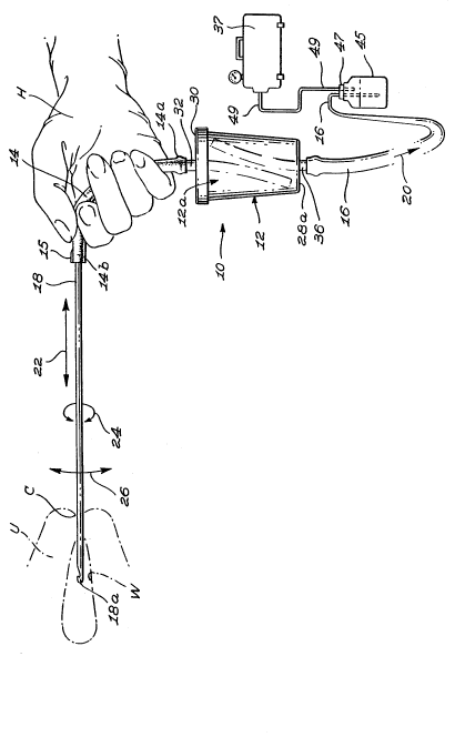
Figure 1 is a side elevation of the uterine
sample collecting device of the invention as held by a
medical practitioner's hand, showing in phantom lines the
cervix of the uterus and suggesting with arrows the manual
21~821~
movements of the curette;
Figure 2 is a partial side elevation of the
collecting device of figure 1, suggesting with arrows the
corresponding rotation of the curette for a given rotation
of the manoeuvring hose between the thumb and forefinger;
hand;
Figure 3 is a fragmented perspective view, at an
enlarged scale, of the curette of figures 1 and 2, and
associated outer end portion of the manoeuvring flexible
hose;
Figure 4 is a vertical sectional view at an
enlarged scale of the collecting flask of the device of
figure 1, with a load of uterine tissue and fluid sample
deposited inside the flask;
Figure 5 is a partly sectional, fragmented
elevation, at an enlarged scale, of the collecting flask of
the device of figure 1, suggesting that tissue fixating
fluid aspirated through the curette has been admixed with
the initial load of uterine tissue and fluid sample; and
Figure 6 is an elevation, at an enlarged scale,
of the collecting flask of the device of figure 1, the
flexible inlet and outlet tubes being removed from the
flask, the flask being ready for transport of the loaded
flask to a medical facility laboratory for detailed sample
analysis.
DETATTlT~n DESCRIPTION OF THE EMBODIMENTS
The present invention is a hand-held sample
collecting device for gathering tissue samples from the
uterine wall without anaesthesia.
Figure 1 shows, in phantom lines, the cervix C of
a woman's uterus U, the latter defining an inner wall W or
endometrium. A uterine sample collecting device 10 is
provided according to the invention, comprising a
collecting bottle or flask 12, having e.g. a capacity of
about 90 millilitres (ml), for collecting a certain
quantity of uterine tissue and fluids. Flask 12, which is
2 1 ~ 0
preferably made from a transparent plastic material,
consists of a main peripheral wall 28, a bottom flooring
28a, and a top open mouth 12a. A closure cap 30 releasably
seals the top mouth 12a. The wall of flask 12 is
preferably cross-sectionally circular, e.g. as illustrated
slightly frusto-conical in shape, with the top mouth 12a
being diametrally larger than the flooring 28a and the
closure cap 30 being complementarily discoid and
threadingly engaging by its inwardly threaded annular
flange 3Oa the outwardly threaded edge portion 12a' of
flask mouth 12a. However, other closure cap and flask
shapes - e.g. cross-sectionally quadrangular - and other
suitable cap to flask securing means - e.g. snap-fit
interlock - would not be excluded from the scope of the
invention.
For example, closure cap 30 could also include an
annular rim, not illustrated, being mounted thereto in
radially inwardly spaced fashion from the peripheral flange
3Oa; this annular rim being flexible and conical and
complementary to the flask mouth 12a, wherein friction fit
fluid-tight engagement can be achieved by progressive
engagement of flask mouth 12a into the annular conical gap
formed between cover flange 3Oa and said annular rim.
Closure cap 30 includes a central, integral,
transversely outturned nipple 32a, and a coextensive,
integral, transversely inturned nipple 32b, these nipples
circumscribing a through-channel 32 for fluid communication
into flask 12 through the body of cap 30.
One end 14a of a flexible - preferably
elastomeric - hose 14 is deformingly mounted sealingly
against the radially outer wall of outturned rigid nipple
32a, while the opposite end thereof 14b is engaged by the
inner end of an elongated, rigid (usually metallic)
conventional curette 18. The interior cap nipple 32b is
preferably diametrally enlarged relative to outer nipple
32a, so as to frictionally sealingly accommodate inside its
21~82~ ~
cavity the top end 34b of an elongated, semi-rigid tube 34.
The length of tube 34 is sized so that its bottom outlet
end 34a be located very close to but spacedly from flask
bottom flooring 28a. Flooring 28a includes a nipple
arrangement similar to that of closure cap 30, comprising
an inner, transverse, diametrally enlarged nipple 36b, an
outer, transverse, diametrally smaller nipple 36a, these
nipples circumscribing a through-channel 36 for air
communication through the centre of flooring 28a and into
flask 12.
Moreover, hose 14 is sized to be sufficiently
elongated to allow rotation of the curette end 14b of hose
14, with the thumb and forefinger of a practitioner's hand
H, while the remaining fingers of the same practitioner's
hand H grasp firmly the sample flask end portion 14a of
hose 14. That is to say, during rotation of hose end
portion 14b, a torsional load is applied lengthwisely of
hose 14, which will twist, since the opposite end portion
14a is not allowed to rotate. Accordingly, during rotation
- and similarly during reciprocating motion - of hose end
portion 14b, sample flask 12 will not rotate nor move fore
and aft, thanks to the elasticity of the material of
dampening hose 14. Usually, such a minimal length for
rubber hose would be at least twelve centimetres (12 cm).
A semi-rigid tube 38 is frictionally sealingly
engaged at its bottom end 38b into the cavity formed by
inner nipple 36b, and is of a length sized so that its top
inlet end 38a be located at the level of mouth 12a, very
close to but spacedly from the main discoid wall of closed
closure cap 30. Semi-rigid (or rigid) tubes 34, 38, should
be arcuate, as illustrated, so as to diverge from one
another inside flask 28 to axially clear one another (since
inner nipples 32b, 36b are coaxial).
Another flexible, preferably elastomeric, hose 16
is deformingly sealingly engaged at its inner end 16a
releasably around the radially outer wall of outer nipple
2158210
36a, and forms part of an air pump machine 37 by connecting
to a second collecting flask 45, through the top closure
plug 47 thereof. Flask 45 is connected by line 49 to air
pump machine, and is usually concealed into the pump
casing. Flask 45 may be of a conventional make, e.g. based
on the concept disclosed in U.S. patent No 3,542,031 to
Taylor, supra, and does not form part of the invention as
such. Flask 45 is conventionally used to shield pump
machine 37 from accidental contamination - however unlikely
- that could engage into tube 16. Hence, any contaminant
accidentally escaping from main collecting bottle 12,
through outlet port 36 and into downstream hose 16, would
deposit into buffer flask 45; the pump 37 would not be
reached by the contaminants. Since the medical
practitioner must periodically monitor this transparent
flask 45, any deposit
therein will prompt the practitioner to shut off the pump.
Arrow 20 in figure 1 suggests that air is sucked in from
flask 12 into hose 16. Accordingly, flexible hoses 14 and
16 fluidingly communicate with the interior of flask 12 via
tubes 34 and 38, respectively.
Hollow curette 18 conventionally has a free outer
end mouth 18a, for retrieving samples from the uterine
wall.
In operation, tissue sampling device 10 is to be
operated single-handedly, as shown in figure 1. Indeed,
with a single hand H, the medical practitioner firmly
grasps inner end portion 14a of flexible sample hose 14,
and with his thumb and forefinger engaging the hose portion
distal end portion 14b where it overlaps the inner end of
curette 18. By holding it there, the medical practitioner
has a very good control over the movements of curette 18,
as suggested in figure 1: thus, the medical practitioner
can manually perform with curette 18 any one or a
combination of the following movements: (a) an axial
displacement (suggested by arrow 22); (b) an axial
21~821~
rotation (arrow 24); or (c) a tilting motion of free end
18a (arrow 26). Flask 12 simply hangs from hose 14
in a vertical position; it will not rotate on itself as
curette 18 is being axially rotated, since a torsional load
will be enabled axially of flexible hose 14, and the same
will be true as curette 18 is endwisely tilted and/or
axially displaced, because curette 18 is linked to flask 12
by flexible hose 14, which allows curette 18 a relatively
good freedom of movement without moving flask 12.
10- That flask 12 remains immobile is important,
although not essential, in view of preventing leaks of
tissue sample material therefrom. Thus, it would not be
excluded from the scope of the method of use not to prevent
rotation of the flask when finger rotating the upper hose
1514. The three curette motions 22, 24, 26, can be performed
in a fine adjustment manual mode, with wrist and fingers
being solely involved, i.e. without any arm or forearm
motion being required.
Figure 2 shows how, with a twist of the thumb and
forefinger of hand H, about hose distal portion 14b,
curette 18 can be axially rotated to accomplish the
circular curettage of the uterine wall, i.e. the basic
function of curette 18. As discussed above, during this
rotation, a torsional load is applied axially of elastic
hose 14, so that distal hose portion 14b rotate, while
proximal hose portion 14a does not, because the other three
fingers of hand H anchor hose proximal end 14a to prevent
axial rotation of bottle 12. The reader will appreciate
the quantum leap in handling finesse achieved when
curetting is performed by a medical practitioner.
Figure 3 shows that free end 18a of curette 18
conventionally defines an elongated ovoidal bevelled end
portion forming a sample intake mouth, 18b, having sharp
edges that permit the user to scrape the superficial
surface of wall W of uterus U when axially rotating curette
18, without injury to the uterus during cervix ingress of
12
1 0
curette 18. Figure 3 further
21$~2`IO
shows that curette 18 defines an inner channel 18c merging
with aperture 18b, in which the tissue samples will engage
once dislodged from wall W by the edges of aperture 18b,
during the curettage process. It is in lumen 18c that the
uterine fluids are adapted to flow from uterus U to intake
hose 14, before loading into flask 12 via tube 34 by
depositing against flooring 28a.
Preferably, and as illustrated in particular in
figure 3, a tactile cue means 15 is provided, e.g. a short
axial end bulge, about hose outer end portion 14b. Tactile
cue means 15 is in axial alignment with the ovoidal cutting
edge mouth 18b of curette 18, to assist the medical
practitioner in continuously feeling with his forefinger
the relative orientation of the sharp cutting edges 18b of
the opposite end of curette 18.
Figures 4 and 5 show collecting flask 12. Flask
12 defines a main body 28, being slightly frusto-conical,
threaded at its upper end (corresponding to upper end 12a
of flask 12) to be complementarily releasably engaged in a
fluid-tight fashion by a threaded sealing closure cap 30.
In an alternate embodiment of the invention, not
illustrated, inner tube 34 is integrally mounted to
inturned nipple 32b of the flask closure cap 30 at its
inner end 34b (which thus merges with nipple 32b); and
inner tube 38 is integrally mounted to inturned nipple 36b
of the flask flooring 28a at its outer end 38b (which thus
merges with nipple 36b).
As suggested by the arrows in figure 4, the air
pump 37 is serially connected to downstream hose 16 via
assembly 45-49 (fig 1) will create a negative pressure
chamber inside flask 28. Because of this negative pressure
in chamber 28, fluid tissue samples from curette 18 and
from hose 14 will be biased downwardly through tube 34 to
deposit onto floor 28a, and this deposited fluid tissue
material T will progressively build up and raise in level
21~g2IO
over flooring 28a to a given level T1 located well below
top tube mouth 12a.
To gather uterine tissue and fluids in flask 12,
curette 18 must first be inserted inside uterus U axially
through cervix C. By powering the vacuum pump, air is
drawn up in flask 12 through inner tube 38, through outlet
through-channel 36 and into outlet hose 16 up to the pump,
thus creating a negative pressure in flask 12. This
negative pressure will cause a suction in first inner tube
34, transmitted in inlet through-channel 32, sample intake
hose 14 and curette 18 to suck in all the desired fluids
and tissue samples which have been scraped off - and
collected from - the uterine wall W surface by curette free
end 18a during the curettage process. The uterine fluids
flowing in from first inner tube 34 are thus gathered in
collecting flask 12. It is of course highly undesirable
that the fluids reach the vacuum hose, for several reasons:
a) the tissue to be analyzed will be difficult to collect,
since often, the sample volume is minimal and easily
wasted; and most importantly,
lb) the medical staff would have to manipulate this blood-
stained material, which would expose them to being
contaminated by one or more viruses in the uterine fluids,
some of which could be very dangerous such as the HIV
virus. It is therefore vitally important to keep the
uterine fluids away from the top mouth 38a of second inner
tube 38. Such fluid level monitoring of fluid T will
easily be performed in real time fashion, simply by having
the walls of flask 12 being made transparent, for see-
through capability by the medical practitioner.
To accomplish this, it is necessary that flask 12
stay vertical at all times during the fluid collecting
operation. Indeed, since the air intake port 38a is very
close to top cap 30, if flask 12 is kept in an upright
position with top cap 30 at the upper end, then flask 12
21~8210
would have to be almost full for the uterine fluids to
reach the level of top mouth 38a of second inner tube 38
and be sucked in. It is thus up to the medical
practitioner in charge of the collecting operation to
monitor collection of sample fluid and to stop filling
flask 12 well before the fluid level inside flask 12
reaches the level of air intake port 38a. For example, the
level of fluid in figure 4 being less than half the total
inner volume of flask 12, e.g. 30 % thereof, would be an
approximately fairly safe level, which would constitute
enough fluid to accomplish the medical testings - i.e.
approximately 30 millilitres (ml) of fluid tissue sample
for the suggested 90 ml total capacity of flask 12.
By holding collecting device 10 as suggested in
figures 1 and 2, i.e. leaving a certain free length of
inlet hose 14 between hand H and flask 12, the latter will
hang freely from inlet hose 14 and the gravity force will
automatically pull flask 12 to a natural vertical position,
given the freedom of movement conferred by flexible inlet
hose 14. Indeed, flask 12, either filled with uterine
fluid or not, simply hangs from sample intake hose 14 that
acts as an axial support, the weight of flask 12 being
uniformly and axially symmetrically distributed and hose 14
and air outlet hose 16 being coaxially attached thereon.
All the forces acting on flask 12 are thus either axial
forces or are counter-balanced by another force due to the
axially symmetrical distribution of the weight of flask 12
and the weight of hose 16.
Self-centering of hoses 14 and 16 continuously
occurs, in these circumstances, thanks to the geometrically
central positioning of closure cap nipple 32a and of
flooring nipple 36a. Therefore, when accomplishing the
fluid collecting operation, the person doing so does not
have to be concerned with adjusting the verticality of
flask 12, since it will automatically perform self-
centering to hang in an upright position.
16
2t58~1 0
Since first inner tube bottom end 34a is located
near flooring 12b of flask 12, it is thus positioned far
away from second inner tube top mouth 38a. This means
that, if the protocol of operation of the present sample
collecting device 10 is well followed, it would not be
possible that the fluid flow-ing out of first inner tube 34
could in any way reach the second inner tube free end 38a
and accidentally enter into tube 38.
Considering that vertical flask 12 will not
normally be brought into any significant relative motion,
since the curettage operation is performed by relative
motion of flexible hose 14 which dampens motion to flask
12, and since the incoming fluid should not reach into
second inner tube 38 towards the pump, no contamination of
the outlet hose 16 assembly should occur by the uterine
fluids. Thus, the medical staff will not have to expose
themselves to the (possibly contaminated) fluids when
cleansing said hose assembly 16.
once the sample collecting operation is finished,
curette 18 is released from the uterine cavity U, and
brought into a bottle containing a tissue fixating and
microbial sterilizing solution, e.g. a 10 % solution of
formaldehyde. From 20 to 30 ml of this formaldehyde
solution is then sucked through curette mouth 18b into
flask 12, with a slight negative pressure, since the vacuum
pump has remained activated. The formaldehyde solution
will fixate the sampled cells and will sterilize
microbiological contaminants in the uterine tissue fluids,
without the need for opening the flask 12. The level of
the fluid inside flask 12 relative to level Tl, will thus
rise by a certain height ~h, to a second level T2, as shown
in figure 5, e.g. to reach a level about half the total
volume of flask 12; but the fluid level must positively
remain well below the second tube air intake top mouth 38a.
After having introduced the formaldehyde solution
into flask 12, both sample intake hose 14 and air outlet
- 215R21~
hose 16 are forcibly detached from nipples 32a and 36a,
respectively, and thus from flask 12, making sure of
keeping the latter in an upright position during this
operation to prevent the fluid from flowing out from inlet
and outlet through-channels 32 and 36. No leak should
occur in these circumstances, provided the verticality of
the flask 12 is maintained at all times by the medical
practitioner, because the outlet port 38a remains above the
highest fluid level T2.
As shown in figure 6, a first and a second fluid-
tight caps 40 and 42 are then sealingly applied on lips 32a
and 36a, respectively. The first outlet to be plugged will
be the top nipple 32; then the flask 12 is overturned, to
plug the bottom nipple 36. The fluid, trapped inside flask
12, can then be conveyed safely without regard to
maintaining the verticality thereof, and forwarded to a
medical facility laboratory for the proper anatomo-
pathological examination to take place.
It is important to note that, at all times, the
fluid remains inside flask 12, and that at no time did the
medical staff come in contact with it. When it will have
reached the laboratory, it will already have been
sterilized by the formaldehyde solution so that all viruses
and other microbial components will have been killed.
It is understood that even though, during this
description, the implied use of the sample collecting
device 10 was to gather uterine samples, it could just as
well be used to empty the uterine cavity or any other
similar use; only the size and volume of the collecting
flask would need to be accordingly modified.
Alternately, if microbiological analysis is
needed for the sample material, this material should not be
fixated with formaldehyde, but rather with a non toxic
solution such as physiological serum. Again, the reader
will be able to appreciate the value of a sample containing
flask that remains continuously sealed during handling
18
215821~
thereof, with the micro-organisms being kept alive.
19