Note: Descriptions are shown in the official language in which they were submitted.
21S8874
APPARATUS AND METHOD FOR OCULAR TREATMENT
BACKGROUND OF THE INVENTION
1. Field of the Invention.
This invention relates to the medical treatment of ocular disease.
More particularly this invention relates to an electronic apparatus for the
application of electrical current to the eye for treating diseases thereof, for
example macular degeneration.
2. Description of the Prior Art.
Macular degeneration is a debilitating ocular disease having
hemorrhagic and exudative variants, both of which are susceptible to safe
and efficient treatment by the invention hereof. Treatment typically results
in amelioration of the ophthalmoscopic manifestations of the disorder, and
substantial restoration of central visual acuity.
It is proposed in Fedorov et al., U.S. Patent No. 5,147,284, to treat
diseases of the optic nerve and retina by the application of a pulsed
magnetic flux, the magnetic field induction being from 0.1 T to 0.25 T.
However the technique is invasive, requiring exposure of the posterior
portion of the eyeball and optic nerve and introduction of fhe inducer into
the orbit.
- It is proposed to treat glaucoma with the application of transcutane-
OUS electrical stimulation from Liss et al., U.S. Patent No. 4,614,193. Liss
et al. discloses the application of pulsed electrical current at a level less
than 4 milliamperes, the pulse trains occurring at 12 - 20 kHz, amplitude
modulated at 8 - 20 hz, and having a 3:1 duty cycle. Applying this
waveform through electrodes positioned on the temple and on the
ipsilateral hand, Liss et al. achieved an approximately 28% reduction in
intraocular pressure in the treated eye. To the knowledge of the inventors,
passage of electrical current through the eye (hereinafter "transocular
electrical conduction") has not been used in the art for the treatment of
macular degeneration.
215887~
SUMMARY OF THE INVENTION
It is therefore a primary object of the present invention to provide an
improved apparatus and method for the treatment of macular degeneration
and certain other ocular pathology.
it is another object of the present invention to provide a safe,
improved, noninvasive method for the restoration of vision by treating the
eye with transocular conduction of electrical current.
- These and other objects of the present invention are attained by a
direct current generator that produces low level direct current, electrodes
_
being placed on the closed eyelid and on or near the occiput of the sku!l.
The inventors have found that this waveform is particularly effective in the
treatment of macular degeneration, and is believed to benefit a variety of
other ocular disorders. While the principles under which the invention
produces its beneficial effects are not fully understood, and without
restriction to a particular theory of operation, it is suggested that
transocular electrical conduction as practiced in accordance with the
invention may restore cellular electrical balance by changing potentials
across cell membranes. This may alter the levels of certain ions and
molecules toward a desirable equilibrium. Other physiological effects are
believed to be produced: reduction of alkalinity proximate the passage of
electrical current and the production of low levels of hydrochloric acid;
attraction of oxygen to the region; localized vasoconstriction; reduction of
local hemorrhage; sedation; increased tonicity of loca! tissues; antisepsis;
production of desirable fibroplasia; and reduced neuromuscular irritability.
In accordance with one aspect of the invention rnacular degeneration
in a subject is treated by the steps of: placing a first electrode of a direct
current source in electrical contact with a closed eyelid of a subject;
placing a second electrode of the source in electrical contact with a site
remote from the eyelid of the subject; and causing-a direct current of 1 -
1000,uA to flow between the electrodes through the subject for about 10
minutes.
--2--
` 21S8879
In accordance with-another aspect of the invention the source is a
portable, battery powered constant direct current generator which is affixed
to the subject. The subject is thus enabled to ambulate while the direct
current is flowing therethrough.
In accordance with yet another aspect of the invention, the first
electrode is a positive electrode, and the second electrode is a negative
electrode. The remote site for the second electrode is the posterior neck
of the subject.
In accordance with still another aspect of the invention, the current
flowing between the electrodes is 200,uA, and is constant in magnitude.
BRIEF DESCRIPTION OF THE DRAWING
For a better understanding of these and other objects of the present
invention, reference is made to the detailed description of the invention
which is to be read in conjunction with the following drawings, wherein:
FIG. 1 is a side view of a subject receiving current to the eye that is
delivered in accordance with a first embodiment of the invention;
FIG. 2 is a rear view of the subject shown in FIG. 1;
FIG. 3 is a partially schematic side view of a subje~t receiving current
to the eye that is delivered in accordance with a second embodiment of
the invention; FIG. 4 is a schematic of a constant current source suitable
for delivering current to the eye in accordance with the invention; and
FIG. 5 is a schematic of another constant current source designed to
deliver a current through the eye in accordance with the invention.
DESCRIPTION OF THE PREFERRED EMBODIMENT
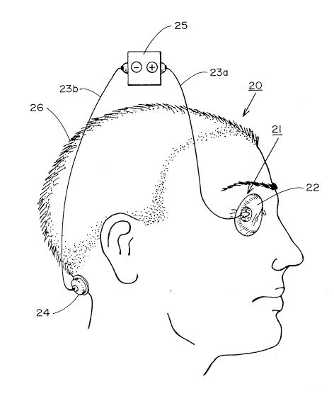
Turning now to the Drawing and to FIGS. 1 and 2 thereof, there is
shown a subject 20 having the closed lids of an eye 21 in contact with a
sponge electrode 22. The sponge electrode 22 is connected to the
positive output of a constant current generator 25 having a suitable power
source (not shown) connected thereto or incorporated therein. The
negative output of the generator 25 is connected to an occipital electrode
24, which is attached to the skin at the occiput, substantially in the midline.
~ 2ls887l
When the generator 25 is activated, a current loop is established that
extends in order from the generator 25 through the sponge electrode 22,
the eye 21, the cranium 26, and the second electrode 24. The loop is
completed at the generator 25.
In FIG. 4 there is shown an electronic circuit of a constant current
source that is included in a preferred embodiment of the invention. The
circuit is designed to deliver a constant current of between 5 ~A and 1 mA
to the eye for the treatment of ocular diseases, such as macular degenera-
tion. A current delivery of 200,uA is preferred. The unit is composed of
two blocks, a current source 402 and a timer 404. It operates as follows:
Upon closure of switch 405, power is applied to the circuit. Resistor
408 and LED 410 act as an indicator light for the power-on condition.
Switch 415 acts as a trigger for the timer. When this button is pressed, a
voltage is applied to pin 420 of integrated circuit 418 for a period of time
determined by the setting on potentiometer 422 and the values of resistor
424 and capacitor 426. This causes a voltage to be passed through the ~.optical isolator 430, which in turn causes a voltage to be applied to pin
431 of the optical isolator 430. LED 434 indicates that sw~tch 415 has
- been depressed, and that treatment has been initiated. Voltage regulator
432, which is preferably of type LM317, is such that it maintains a constant
voltage of 1.25 volts across its output pin 441 and adjust pin 443. From
Ohms law then, since pins 436 and 439 of U439 see a very high imped-
ance, the current through the electrodes 440 is 1.25/R Amperes, where R - -
is the net resistance across the output and adjust pins 441, 443 of the
voltage regulator 432 as determined by the resistance of the network of
resistors 445 - 447-and trimmer potentiometer 448. The magnitude of the
current is set by adjustment of the trimmer potentiometer 448. The current
-- - through the electrodes is independent of the resistance thereacross within
an operating range, and is monitored by the ammeter 450. Switch 454
allows the operator to read the voltage across the electrodes. Battery 456
supplies power to operate the timer circuit 404. Batteries 457-459 are
--4--
` 2l58874
included to provide a larger voltage reference across the electrodes so
that the constant current can be maintained over a larger range of
electrode resistance.
The constant current source can also be realized as explained with
reference to FIG. 5. This shown a schematic of another constant current
source designed to deliver a 5 - 1000,uA current through the eye. The
major-circuit components consist of a current source and a timer. When
momentary switch SW2 is turned on, a small current flowing through LED
012A in the optoisolator 502 turns on the SCR 012B which powers VCC
throughout the circuit. Holding the switch SW2 in an on position also
causes the reset net of the circuit to become activated. Turning the switch
SW2 to the off position (also momentary) switches the battery voltage to
the gate of the SCR 012B, thereby sl~utting it down. A tone will be emitted
from the unit whenever the switch is held in either position. D2, Z1 and Z2
form a circuit which will cause a continuation of the audio tone for
approximately one second upon startup of the unit in the event of a low
battery.
Pressing the start-treatment push-button SW3 activates flipflop U4A,
which turns on the constant current source, and turns on the reset line to
timer TMR1A, starting the clock. As the clock pulses, LED D1 will flash. ~ .
The clock pulses are counted by U1, U8, and U2, and every one hundred
counts of the clock-causes a-shift in the light bar 504, U3C and U3D
combine to reset flipflop U4A when the count reaches 100% of the
treatment time. This causes the constant current source to turn off, the
counters to reset, and flipflop U4B to set. This flipflop turns on timer
TMR1 Bj which in turns causes transistor Q1 to conduct. This causes the
audio alarm 506 to sound for about one second. U3B charges C2 through
- R8 until U4B resets. At this point the circuit is back to its original ready
state, where it remains until the start-treatment push-button SW3 is
3 o pushed, or the unit is switched off.
~ 21$8871
The constant current source VR1 is preferably an LM317 variable
voltage regulator, which by its design maintains a constant voltage
between its output pin 508 and its adjust pin 510 of 1.25 volts. This
constant voltage across a-fixed resist.ance causes a constant current to be
5 delivered through the electrodes to ground when the irripedance of the
entire path is low enough to allow that much current, given that the circuit
has a 9V voltage limitation. R1A allows the resistance to be adjusted in
order to vary the magnitude of the treatment current.
AM1 is a 100,uA DC ammeter. R4 and potentiometer R15 provide an
o adjustable shunt so that the meter can be adjusted to read correctly for ~-
any selected treatment current.
011 is an optical isolator which is connected to-a self powered
elapsed time meter M2. This meter will only log time when current is
passing through the electrode jacks 512a, 512b regardless of the state of
the unit. For example, if the unit is on, but the electrodes are not con-
nected to a patient, time will not be logged until the electrodes are
connected to the patient, or are otherwise placed in electrical connection.
Use of the invention will now be explained with reference to FIGS. 1
- - and 2. The patient 20 has an ocular disease such as macular degenera-
20 tion in the eye to be treated. It is believed that beneficial results areobtained in other ocular disorders. Analgesic medication should be
discontinued at least 4 - 8 hours prior to treatrnent, $o that the patient's
ability to perceive pain is not impaired. A constant current generator 25,
which can be realized as either the embodiment of FIG. 4, or of FIG. 5, is
25 connected to a patient 20. The positive and negative terminals are
connected by two wires 23a, 23b to sponge electrode 22 and occipital
electrode 24 respectively, and the constant current generator 25 is
- connected to a suitable source of power (not shown). The sponge
electrode 22 is placed on the closed eyelid, and the occipital electrode 24
30 placed at the back of the neck substantially in the midline. The contacts
are held in place by straps (not shown), which can be fastened by
--6--
`- . 2ls887g
VELCRO (a trademark). The straps are tight enough to prevent the eye
from opening during the treatment.
The patient 12 is requested to relax, and the unit then actuated to
begin treatment, which is preferably maintained for 10 minutes, after which
5 time the internal timer in the constant current generator 25 shuts down the
unit. The straps and contacts are then removed from the patient.
During treatment the patient 20 is preferably monitored for hypoten-
sion. It has been found that many patients experience a modest decline in
blood pressure during treatment, and it is believed that certain hyperten-
10 sive patients could benefit from the treatment described.
Treatment is repeated three times per week for at least twelve weeks.
Turning now to FIG. 3, there is shown an alternate embodiment of thesystem. A constant current generator 425, which can be either embodi-
ment described with reference to FIGS. 4 and 5, is constructed in suitable
miniature dimensions to be attached to a headband 427. The generator
425 is preferably powered by miniature batteries (not shown). This
embodiment is suitable for portable, or home use by patient 20 having
macular degeneration, and could-also be used for contro~!ing hypertension
in certain patients. The generator is connected to the patient 20 in the
20 same manner as described with reference to FIGS. 1 and 2. The patient
20 initiates treatment by depressing momentary switch 430. Treatment
then proceeds as described above.
Example 1
Patient MH, born February 18, 1916, complained of reduced central
25 visual acuity, and had a medical diagnosis of macular degeneration. She
was receiving medication for hypertension and for elevated cholesterol.
Prior to treatment the eyes were free of all other pathology. Pupillary
- responses were normal, and the best corrected visual acuity using the
- Snellen chart was O.D. 20/200, O.S. 20/200. Near visual acuity was best
30 corrected to 2M print.
2ls887l
Low visior~ -aids were prescribed to assist her adaptation to her visual
loss. She returned after 13 months with little change noted on her part.
Visual acuity was again measured-at the same level, 20/200 in each eye. A
contrast sensitivity test was done at the same time, and revealed that
5 visual acuity was in the 20/200 range-in both eyes. A visual field examina-
tion was normal for both form and blind spot.
One month later she began a series of transocular electrical conduc-
tion treatments. Using a constant direct current generator, 200 ~uA was
applied for 10 minutes, with the positive pole attached to the closed eyelid,
10 and the negative pole attached to the back of the neck. After thirteen
sessions, averaging three times a week, acuity was remeasured with
results as shown in table 1-l.
Table 1-1 - after 13 sessions
Right Eye Left Eye Both eyes
Snellen Chart 20/100 p!us 2 20/100 20/80 plus 2
Contrast sensitivity 20/70 20/70
At near, the patient could read 1 M print easily. The results after 20
sessions are given in table 1-2.
Table 1-2 - after 20 sessions
Rlght Eye Left Eye Both eyes
Snellen Chart 20/80 minus 1 20/80 minus 1 20/70
Contrast sensitivity 20/70 20/80
Treatment was discontinued. A progress evaluation was done two
months later with the result as shown in table 1-3
Table 1-3
Right Eye Left Eye Both eyes
Snellen Chart 20/80 20/80 minus 1 20/60
~ 2ls887~
Near vision acuity was now 0.8M. The same findings were again dupli-
cated using contrast-sensitivity testing.
Example 2
Patient JC was born December 15, 1917, and when first seen for
5 evaluation complained of inability to read and see clearly both at far and at
near. A diagnosis of macular degeneration was confirmed by two
ophthalmologists. He was currently taking medication for hypertension
and diabetes. His best corrected visual acuity was OD 20/50; OS 20/30.
The eyes were free of all other pathology.
He was treated in the manner described in Example 1. After six
treatments, he was re-evaluated with the results shown in table 2-1.
Table 2-1
Right Eye Left Eye
Snellen Chart 20/40 20/25
Blood pressure before and after treatment was 190/80 and 120/70
respectively. Contrast sensitivity done prior to treatment was as follows:
Table 2-2
OD OS
Aug. 31 20/70 20/50
Sept 18 20/40 20/30
A visual field examination on September 18 revealed a marked
constriction of the color field at near, measured with a one millimeter target
at 14 inches. There was also a three time enlargement of the blind spot
on the right eye and about- a four fold enlargement of the blind spot in the
left eye. The color fields were also markedly constricted in the left eye.
, 21~887~
Treatment was continued for another eight sessions. He was seen
for a progress evaluation on October 5, with the results as shown in Table
2-3.
Table 2-3
Right Eye Left Eye
Snellen Chart 20/30 20/25 plus 2
Contrast sensitivity 20/30 20/25
A visual field was repeated, and there was a marked improvement or
expansion in the color fields for both eyes. The blind spot had also
- reduced and become much more normalized in the right eye, and
10 although enlarged in the left eye, was reduced by 100% over the findings
since September 18.
Treatment continued, and he was seen again on October 26 for a
progress evaluation, with the results as shown in table 2-4:
Table 2-4
Right Eye Left Eye
Snellen Chart 20/30 plus 2 20/25 plus 2
Contrastsensitivity 20/25 20/25
The color field had continued to expand on visual field measurement. The
blind spot was still enlarged in both eyes.
Treatment continued, and he was seen after approximately 30
20 treatments on November 20, with the results as seen in table 2-5.
Table 2-~
Right Eye Left Eye
Snellen Chart 20/30 plus 3 20/25 plus 2
Contrast sensitivity 20/30 20/25
--10--
21$887~
The visual field was repeated, and the color field had not expanded to
what would be considered normal levels. The blind spot was only
enlarged by about 10% now in each eye.
Treatments continued through December 16, with all findings
5 remaining the same. In a progress evaluation on February 12 of the
following year, the results were as shown in table 2-6.
Table 2-6
Right Eye Left Eye
Snellen Chart 20/30 plus 1 20/25 plus 2
Contrast sensitivity 20/25 20/25
10 Subjectively, he reported much better vision both at far and near, could
read easily, drive safely, and reported that he was seeing things much
brighter and clearer at all distances, and at all times.
Example 3
Patient HD was born on April 2, 1919, and was first seen on June 6
with a history of macular degeneration, and central scoto~a with
metamorphosis. No other ocular pathology was noted, and the she was
on no medication.
Measurement of her central visual field revealed a large central
scotoma in each eye corresponding to the macular area. Her visual acuity
iS shown in table 3-~ -
Table 3-1
Right Eye Left Eye
Snellen Chart 20/100 20/70
Contrast sensitivity 20/100 20/80
Re-examination after her summer absence in the Fall revealed the results
25 shown in table 3-2.
21 $88 7q
Table 3-2
Right Eye Left Eye
Contrast sensitivity 20/100 - 20/200 20/70
She began a series of transocular electrical conduction treatments under
the conditions of Example 1, except that the treatments were performed
5 weekly for six weeks.
Re-examination on November 11 revealed the results shown in table
3-3.
Table 3-3
Right Eye Left Eye Both eyes
Snellen Chart 20/50 plus 2 20/40 plus 2 20/30
Contrast sensitivity 20/100 20/50
She reported that an ophthalmologist who saw her the previous week said
that her visual acuity was now stable, and that ocular hemorrhage had
diminished. She reported seeing much better at all distances and was
able to read comfortably and efficiently.
Example 4
Patient CR was born February 13, 1917, and was first seen on
December 9. A cataract had been removed from the right eye, and she at
that time had a cataract in the left eye. She had a diagnosis of senile
macular degeneration in both eyes. She was taking medication for
20 hypertension, iron for anemia, and occasionally a sleeping pill.
Her best corrected acuity was OD 4/200th's vision, OS 10/70th's
- vision, near acuity was 1.2M. Due to her poor vision, contrast sensitivity
testing was not possible. By placing a 2.5 power telescope in front of the
left eye, visual acuity could be improved to 20/40 minus 1.
--12--
~ 21~887~
Transocular electrical conduction treatment was begun under the
conditions of Example 1. After seven treatments her visual acuity was OD
20/400; OS 20/80 plus 1. Her near visual acuity was now 1 M with reading
glasses. A 2.5 power telescope in front of the left eye produced a visual
5 acuity of 20/40 plus 2. She reported much improved sùbjective visual
acuity both at far and near, and noticed that vitreous floaters had im-
proved.
After six more treatments she was re-evaluated on February 17 of the
next year. Visual acuity was OD 20/400; OS 20/70. Visual acuity through
10a 2.5 power telescope was almost 20/30.
In summary her acuity in each eye improved approximately 100%
over a course of 13 sessions.
Example 6
Patient HD from Example 3 was subsequently treated weekly over a
period of 12 weeks with pulsed electrical current using the Liss Cranial
Stimulator noted herein. The stimulator was operated at 400
microamperes for 10 minutes. The wave form consisted ~f "on:" periods of
pulse trains alternating with "off periods" at 500 hz and a duty cycle of 3:1.
The pulse trains occurred at a frequency of 15khz and were amplitude
2 o modulated at 15 hz. Results were as follows:
Table 6-1
Right Eye Left Eye
Before Treatment 20/50 -1 20/50 -2
After Treatment 20/40-1 20/40 -2
Exarriple 7
25Patient CT complained of bilateral vitreous floaters, and underwent
transocular electrical conduction treatment was performed according to the
- conditions of Example 1, except that treatments were undertaken thrice
2l5887~
weekly for a period of two weeks. After completion of the treatments the
patient reported that the floaters were no longer perceptible.
While this invention has been explained with reference to the
structure disclosed herein, it is not confined to the details set forth and this5 application is intended to cover any modifications and changes as may
come within the scope of the following claims: