Note: Descriptions are shown in the official language in which they were submitted.
215!235
- 1 -
S P E C I F I C A T I O N
TITLE
~~APPARATUS AND METHOD FOR
MEASURING COMPONENTS IN A BAG~~
HACRGROUND OF THE INVENTION
The present invention relates to a novel and
useful apparatus and method for non-invasively
analyzing l~.quid medium components in a bag.
Liquid compounds are often placed in bags
for various purposes. For example, the use of total
parenteral nutrients (TPN) which are eventually the
source of intravenous feeding are stored in
transparent or translucent flexible bags. TPN
compounds are commonly mixed in pharmacies using
commerciall~~ available compounders which accept three
or more TPN compounds and automatically mix these
compounds into an appropriate container such as an
intravenous (I. V. bag). Intravenous use of the bags
usually takes place at a later time in a hospital or
medical facility. Typical compounds include 70%
dextrose injection U.S.P., 10% Travasol (amino acid)
injection, Intralipid 20% fat I.V. emulsion, sterile
water, and moony others.
Presently, methods such as color coding are
relied upon to avoid making errors during the
compounding or mixing process. Different tubes
feeding the I.V. k>ag possess connectors of different
colors which correspond to the colors of the specific
mixing positions an the compounder. For example,
setting a red indicator on a compounder for 100
milliliters would deliver 100 milliliters from a
starting bottle connected to the tubing line which
possesses red connectors. However, there is no
CA 02159235 2005-03-21
_2_
assurance that the correct compound was initially connected to the red tubing
line.
Consequently, an incorrect connection of the tubing between bottles of
dextrose
solution and water, for example, may have dire consequences, such as death for
patients with sugar intolerance.
j Many of the TPN compounds are clear liquids. That .is to say, water,
amino acid injection, dextrose injection, and electrolytes are clear liquids
precluding
visual distinction among them. Furthermore, it is preferable to perform
identification
of TPN components non-invasively and rapidly to minimize potential
contamination
and to minimize analysis time by personnel.
An article entitled "Near Infrared Multi-Component Analysis of
Parenteral Products Using The InfraAlyzer 400" by John J. rose 71-78, dated
March-
April 1892. examined meglumine and meglumine diatrizoate in 30% diatrizoate
megluinine injections solutions using diffuse reflectance in the near infrared
region.
The best combinations of three or four wavelength filters from nineteen ( 19)
available
wavelength filters were selected using multiple regression statistical
methods. The
specific wavelengths were not identified.
An article entitled "The Spectrophotemetrie Absorbance of Intral~id"
by Cane et al., pp. 53-55, dated 1980. develops calibrations for Intralipid in
water in
concentrations from 2.5 to 40 mg/mi at six (6) visible wavelengths between 505
and
626.6 nanometers. Intralipid interferes with spectrophotometric analysis of
oxyhemoglobin, carboxyhemoglobin, and total hemoglobin.
A writing entitled, ''Simple Methods For The Quantitative
Determination of Procaine Hydrochloride In Parenteral Products" by Das Gupta
et al.,
pp. 408-410, dated July 1969. presents calibrations in the ultraviolet region
of
spectrophotometry. Specifically, the Das Gupta reference obtained calibrations
at 228
nanometers for buffered solutions of procaine hydrochloride in the 0-20
microgram/ml concentration range.
An article entitled "Nonodestructive NIR and NIT Determination Of
Protein, Fat, And Water In Plastic Wrapped Homo~eni:~ed Meat" by Tomas
Isaksson
et al., pp. 1685-1694, dated 1992. describes NIR measurements of proteins by
diffuse
reflectance in meat samples with and without plastic coatings. Samples were
placed in
a rubber cup prior to covering the meat sample with plastic laminant.
CA 02159235 2005-03-21
-3-
United States patents 4,800,279 and 5,002,397 describe methods and
devices for visible and near-infrared evaluation of physical properties of
samples.
United States patent 4,872,868 shows an. analyzer for collection bags
which provides an envelope that permits the insertion of reagent's test strips
and the
like.
United States patents 3,857,485 and 3,924,128 teach a method of
analyzing sample containers by liquid scintillation spectrometry which
utilizes light
transmission sealing means to prevent entry of ambient light or the escape of
light
from the photomultiplier tube detection devices.
United States patent 5,239,860 describes a sensor for continuously
measuring alcohol and gasoline fuel mixtures in a clear TeflonTM tube using a
predetermined optical path and electromagnetic radiation at a pair of
wavelengths
which are generated by rapidly switching currents through a light-source.
Thermopile
detectors are used to detect an increase in temperature due to light
transmitted through
the flowing gasoline/alcohol mixture.
An apparatus and method for identifying solutions in a translucent
transparent or semi-transparent plastic bag, such as parenteral nutrients, non-
invasively, qualitatively and quantitatively would be a notable advance in the
chemical analysis field.
SUMMARY OF THE Il\TSIEN'TION
In accordance with the present invention a novel and useful apparatus
and method for identifying parenteral nutrients is herein provided.
Accordingly, in one aspect of the present invention there is provided an
apparatus for measuring a component in a liquid medium within the chamber of a
flexible translucent container formed by a wall portion, .comprising:
a. spacer means for sequentially determining a plurality of optical
paths across the container wall portion and chamber, and the bag wall portion
alone,
said spacer means including a passage for electromagnetic radiation; and
CA 02159235 2005-03-21
-4-
b. a source of electromagnetic radiation capable of directing
electromagnetic radiation through said passage, through the wall portion of
the
container, and along any of said determined optical paths, said source of
electromagnetic radiation being capable of interaction with a component in the
bag
chamber and the wall portion of the bag; and
c. detector means for analyzing said electromagnetic radiation
after interaction with the components in the bag chamber and the wall portion
of the
bag.
According to another aspect of the present invention there is provided
a method of analyzing components in a liquid medium within the chamber of a
flexible bag comprising the steps of:
a. placing the bag in spacer means for determining the optical
path across the bag chamber, said spacer means including a passage for
electromagnetic radiation;
b. directing electromagnetic radiation from a source of
electromagnetic radiation to the components in said bag chamber for
interaction
therewith; and
c. analyzing said electromagnetic radiation with detector means
after interaction of said electromagnetic radiation with the components in the
bag
chamber.
According to yet another aspect of the present invention there is
provided an apparatus for detecting light scattering components in a liquid
medium
within the chamber of a flexible translucent bag formed by a wall portion,
comprising:
a. a fence element, said fence element including a passage for
electromagnetic radiation to the wall portion of the bag;
b. a source of electromagnetic radiation capable of directing
electromagnetic radiation through said fence element passage, said source of
CA 02159235 2005-03-21
-4a-
electromagnetic radiation being capable of interaction with a component in the
bag
chamber and the wall portion of the bag; and
c. detector means for analyzing said electromagnetic radiation
after interaction with the components in the bag chamber and the wall portion
of the
bag, said detector means receiving said electromagnetic radiation, after
interaction
with a component in the bag chamber, through said fence passage.
According to still yet another aspect of the present invention there is
provided an apparatus for non-invasively identifying components in a liquid
medium
within an interior of a flexible bag, the apparatus comprising:
a source of electromagnetic radiation capable of directing
electromagnetic radiation into the interior of the flexible bag, the source of
electromagnetic radiation capable of interaction with the components in the
flexible
bag interior and the flexible bag;
optical detector means located outside the flexible bag receiving the
electromagnetic radiation after interaction with the components in the
flexible bag
interior and the wall portion of the flexible bag and providing a signal
indicative
thereof; and
means for analyzing the signal to identify the components of the liquid
medium in the flexible bag.
The apparatus of the present invention employs spacer means for
supporting a flexible transparent or translucent bag and for determining the
optical
path across the bag chamber. The spacer means includes a passage for
electromagnetic radiation. The spacer means may take the form of a pair of
rigid
elements or fences and a mechanism for shortening or lengthening the distance
between the rigid fences. The rigid fences may support the bag against
vertical
movement and also be capable of exerting compressive force on the bag to a
precisely
determined dimension between the rigid fences. Such dimension would correspond
to
a particular optical path, which may include the bag wall alone or the bag
wall and the
bag filled with components in a liquid medium.
A source of electromagnetic radiation directs electromagnetic radiation
through the spacer passage and to the wall portion of the bag. The
2159235
- 5 -
source of electromagnetic radiation may produce
coherent light, ultraviolet radiation, x-rays,
infrared raf~iatiory broad band radiation e.g., a
tungsten sou~,rce, and the like.
Deaector means is also employed in the
present invention for analyzing the electromagnetic
radiation passed through the spacer passage and along
a determined. optical path which is through a dimension
of the bag chamber. In certain cases the optical path
may pass completely across the bag such that the
detector is receiving light which has been transmitted
through the bag. In other cases, the detector may be
placed on th.e same side of the bag as the source of
electromagnetic radiation and receive light which has
interacted with the contents of the bag by diffuse
reflectance. Further, the detector may receive light
by diffuse reflectance and/or by diffuse
transflectance, i.e., where light passes through the
bag and then is reflected back through the bag by a
diffuse reflector or mirror located on the opposite
side of the bag relative to the detector. It has been
found that near-infrared radiation is particularly
useful in detecting parenteral nutrients in a flexible
I.V. bag. In addition, specific wavelengths, rather
than a continuum of wavelengths, may be used as the
radiation sought for analysis to enable the use of
simpler and less expensive instrumentation comprised
of several discrete detectors, each covered by a
narrow wavelength filter. Fiber optics may also be
utilized to carry light to and from the I.V. bag.
Mathematical models may be employed to quantitatively
and qualitatively detect components within the I.V.
bag accurately and quickly.
21591235
- 6 -
Another adaption of the device of the
present invention produces a self referencing device
with respect: to the intensity. Specifically,
transmittanc:e measurements may be taken of the bag
alone, squeezed to eliminate a chamber and to expel
the liquid components, and along a determined optical
path of the bag filled with certain components.
Absorbance for a sample may be accurately determined
under the Be'er's Law relationship. Chemical
concentration can be directly related to an absorbance
difference c>btained from a spectral measurement using
two different path lengths. This technique minimizes
or eliminates common spectroscopic measurement
problems dues to contamination, changes in the
spectroscopic windows holding the sample, instrument
problems dues to temperature changes on internal
optical elements, the source of electromagnetic
radiation, a.nd the like.
It. may be apparent that a novel and useful
apparatus far analyzing components in a liquid medium
has been de~~cribec~.
It. is therefore an object of the present
invention to provide an apparatus and method for
analyzing liquid components in a flexible transparent
or translucent container or without invading the
integrity of the bag.
Another object of the present invention is
to provide a method and apparatus for analyzing
components in a liquid medium within a flexible
transparent or translucent container to prevent misuse
of such components> in treating patients in a medical
facility.
2159235
A further object of the present invention is to
provide a method and apparatus for analyzing components
in a liquid medium :Found in a transparent or translucent
plastic container wlz.ich is accurate and may include
qualitative a:~ well as quantitative measurements of the
components therein.
A further object of the present invention is to
provide a method and apparatus for analyzing components
in a liquid medium found in a transparent or translucent
flexible bag where such components are parenteral or
enteral nutrients typically used for intravenous feeding.
A further object of the present invention is to
provide a method and apparatus for analyzing components
in a liquid medium i=ound in a translucent flexible bag
which is capable of detecting light from a source after
interaction with the bag alone and after interaction with
the bag and the components in the bag.
Another object of the present invention is to
provide a method and apparatus for analyzing components
in a liquid medium within a flexible bag employing either
transmittance or dif=fuse reflectance techniques.
According to one aspect of the invention, there
is provided ar.. apparatus for measuring a component in a
liquid medium within the chamber of a flexible
translucent container formed by a wall portion
comprising: s~~acer means for sequentially determining a
plurality of optical. paths across the container wall
portion and chamber, and the bag wall portion alone the
spacer means including a passage for electromagnetic
radiation; and. a source of electromagnetic radiation
capable of dizectinc~ electromagnetic radiation through
the spacer means pa~~sage, through the wall portion of the
container, and. along any of the determined optical paths,
2159235
7a
the source of electromagnetic radiation being capable of
interaction with a component in the bag chamber and the
wall portion of the bag; and detector means for analyzing
the electromagnetic radiation after interaction with the
components in the b<~g chamber and the wall portion of the
bag.
According to another aspect of the invention,
there is provided a method of analyzing components in a
liquid medium within the chamber of a flexible bag
comprising the step: of: placing the bag in spacer means
for determining the optical path across the bag chamber,
the spacer means inc:iuding a passage for electromagnetic
radiation; directing electromagnetic radiation from a
source of electromagnetic radiation to the components in
the bag chamber for .interaction therewith; and analyzing
the electromagnetic radiation with detector means after
interaction of~ the electromagnetic radiation with the
components in the bag chamber.
Accc~rding to a further aspect of the invention,
there is provided a method of analyzing components in a
liquid medium within the chamber of a flexible bag in
which the com~~onent:~ are parenteral and enteral
nutrients.
According to yet a further aspect of the
invention, there is provided an apparatus for detecting
light scattering components in a liquid medium within the
chamber of a flexible translucent bag formed by a wall
portion comprising: a fence element, the fence element
including a passage for electromagnetic radiation to the
wall portion cf the bag; a source of electromagnetic
radiation capable of: directing electromagnetic radiation
through the fence element passage, the source of
_. ~~".electromagnetic radiation being capable of interaction
2159235
7b
with a component in 'the bag chamber and the wall portion
of the bag; and detE=~~tor means for analyzing the
electromagnetic radiation after interaction with the
components in the b<~g chamber and the wall portion of the
bag, the detector means receiving the electromagnetic
radiation, after ini~eraction with a component in the bag
chamber, through the fence passage.
According to yet another aspect of the
invention, there is provided an apparatus for non-
invasively idE~ntify_Lng components in a liquid medium
within an interior of a flexible gas, the apparatus
comprising: a source of electromagnetic radiation capable
of directing electromagnetic radiation into the interior
of the bag, the source of electromagnetic radiation
capable of interaction with the components in the bag
chamber and the bag; optical detector means located
outside the bag rece:iving the electromagnetic radiation
after interaction w~_th the components in the bag chamber
and the wall portion of the bag and providing a signal
indicative thereof; and means for analyzing the signal to
identify the components of the liquid medium in the bag.
The invention possesses other objects and
advantages es~~ecially as concerns particular
characteristics and features thereof which will become
apparent as tl-..e specification continues.
BRIEF DESCRIPTION OF THE DRAWINGS
FIG. 1 is a sectional view of a first
embodiment of the apparatus of the present invention.
A'~
21592 5
_$_
FIG. 2 is a sectional view of the apparatus
of Fig. 1 taken along line 2-2 of Fig. 1.
FIG. 2A is a sectional view taken along line
2A-2A of Figs. 2.
FIG. 2B is a sectional view of an I.V. bag
collapsed by the apparatus of the present invention.
FIG. 3 is a side sectional view of another
embodiment of the apparatus of the present invention.
FIG. 4 is a graphical representation with
experimental results described in Example 1.
FIG. 5 is a graphical representation of the
experimental results described in Example 2.
FIG. 6 is a graphical representation of
experimental results described in Example 3.
FIG. 7 is a graphical representation of an
experimental result described in Example 4.
FIG. 8 is a graphical representation of PCA
plot of scores described in Example 5.
FIGS. 9 and 10 are graphical representations
depicting predictions using PLS analysis described in
Example 4.
FIG. 11 is a graphical representation of a
PLS model utilizing data found in Example 4.
FIGS. 12,, 13, and 14, are graphical
representations of MLR methods applied to the data
shown in Table 2 of Example 4.
FIG. 15 is a graphical representation of a
step wise ML~R program utilizing the data of Table 1 of
Example 4.
For a better understanding of the invention
reference is made to the following detailed
description of they preferred embodiments which should
be referenced to t:he herein before described drawings.
CA 02159235 2005-03-21
-9-
DETAILED DESCRIPTION OF THE PREFERRED EMBODIMENTS
Various aspects of the present invention will evolve from the following
detailed description of the preferred embodiments thereof which should be
taken in
conjunction with the herein before described drawings.
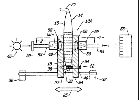
The invention as a whole is depicted in the drawings by reference
character 10. The apparatus 10 is shown in the drawings as including multiple
embodiments, denoted by the addition of an upper case letter. Referring to
Fig. 1,
apparatus lOA is depicted in which spacer means 12 is provided to hold a
flexible
transparent or semi transparent bag 14 in place. The term "translucent" is
used herein
to indicate a transparent, semi-transparent, or non- opaque bag. Bag 14
includes a
chamber 16 which is capable of holding components in a liquid medium 18.
Liquid
medium including such components 18 are passed through tube 20, shown
partially in
Fig. l, which is ultimately clamped or sealed when the bag 14 is filled. Bag
14 may
take the form of a plastic intravenous bag (LV. bag) formed of polyvinyl
chloride
(PVC), ethylene vinyl acetate (EVA), and like materials. However, other
containers
may be employed herein. The liquid medium and components 18 found within bag
14
may consist of mixtures of parenteral or enteral nutrients which may be
intravenously
fed to a patient. For example, such nutrients may include sterile water, 70%
dextrose
injection U.S.P., 10% TravasolTM (amino acid) Aminosyn or FreeAmine injection,
IntralipiaTM 20% fat LV. emulsion, potassium chloride, and the like,
individually or in
various combinations.
2159235
- 10 -
Spacer means 12 includes a pair of elements
22 and 24 which are shown in Figs. 1 and 2 as a pair
of solid fences placed in opposition to one another.
Fences 22 and 24 are slidingly supported by rods 26
and 28 which are supported to a surface by stands 30
and 32, depicted in phantom on Figs. 1 and 2. Rods 26
and 28 serve as a guide for fences 22 and 24. A
threaded screw 34 is fixed to fence 22 through a boss
36. Knurled wheel and bearing unit 38 is capable of
turning relative to boss 36. The turning of wheel 38
also rotates. threaded connect screw 34 which
threadingly engages a threaded bore 40 through fence
24. Thus, fence 22 is stationary relative to rods 26
and 28 whiles fence 24 is moveable thereto, manually or
by motor means, such as a solenoid, directional arrow
25. Unit 38 serves to measure or to stop the relative
movement of fencer 22 and 24, to determine the optical
path through. bag 14 or sequentially determine a
plurality of optical paths through bag 14. It should
be noted that fences 22 and 24 may be hingedly
attached to one another to determine the distance
therebetween;, like a clamshell.
With reference to Fig. 2A, it maybe observed
that such oP~tical path (O. P.) of components 18 within
bag 14 may be easily adjusted by the movement of fence
24 relative to fence 22. The O.P. may vary between
1.0 millimeter to 15 millimeters in many cases. In
certain instances, an empty bag 14 may be compressed
by fences 22 and 24 such that walls 42 and 44 touch
one another, Fig. 2B, eliminating chamber 16. This
configuration is useful in obtaining a reference value
for the bag without medium 18 in chamber 16. Inner
surfaces 42 and 44 of fences 22 and 24, respectively
2159235
- 11 -
also provide sufficient friction to prevent the
slippage of bag 14 downwardly between fences 22 and
24. Of course, other structures may be employed to
prevent the slippage of bag 14 within spacer means 12
such as a floor, or suspension device pulling upwardly
on bag 14 while bag 14 is within spacer means 12, and
the like.
Apparatus 10 further possesses a source of
electromagnetic radiation 46. Source 46 may take the
form of laser light, infrared radiation, ultraviolet
radiation, visible radiation, or any other
electromagnetic radiation found in the spectrum.
Light 46 is passed to bag chamber 16 through passage
48 in fence 22 and from chamber 16 of bag 14 through
passage 50 of fence 24. Electromagnetic radiation
from source 46 may be filtered by filter 52 and led to
passage way 48 by optical fiber or fiber bundle 54.
Fitting 56 directs radiation from fiber optical bundle
54 to collimating lens 58. Parallel rays of
electromagnetic radiation are then passed through bag
14 and liquid medium 18 containing various components
to converging lens 60. Fitting 62 directs the
electromagnetic radiation through optical fiber or
fiber optic bundle 64 to a detector 66 for analysis.
Detector means 66 in its broadest sense may take the
form of any suitable spectrophotometer, single or
multiple detectors, or sources being appropriately
filtered for the wavelength of interest, in
combination 'with a computer employing an appropriate
software program such as Gram 386, available from
Galactic Industries, Inc. of Salem NH.
With reference to Fig. 3, embodiment lOB of
the present invention is depicted. Apparatus 10B
2159235
- 12 -
possesses a pair of opposing elements or fences 68 and
70. As in Embodiment 10A, guide 72 may take the form
of a pair oi.° rods supported by stands on a surface a
hinged clamshell configuration or the like. Fence 68
is fixed to guide 72 while fence 70 is moved and the
distance bei:ween fences 68 and 70 is set by threaded
screws 74, in the same manner as threaded screw 34
found in embodiment 10A. Fence 68 includes a diffuse
reflector 7t> on one side of bag 14 containing liquid
medium 18 having various components therewithin. Of
course, difi_.use reflector may be formed integrally
with bag 14.. Diffuse reflector 76 may take the form
of a white c:eramin disk or other suitable reflector.
Light from :source 46, not shown, passes through outer
fiber or fixer bundle 78, from bag 14 containing a
liquid medium having various components 18, and
through fibE:r optic bundle 80, which is formed
concentricaT.ly with fiber optic bundle 78. Analysis
of the components within bag chamber 16 takes place by
interaction of tha_ electromagnetic radiation from
fiber optic bundle 78 by diffuse reflectance, by
diffuse transflectance, in conjunction with diffuse
reflector 7Ei, or a combination of both. The latter is
especially useful where liquid medium is murky to an
uncertain dE~gree. Such diffuse reflectance
measurements may be obtained simply by pressing an
I.V. bag fi7.led with light scattering material against
fence 70 without the use of fence 68. Again, fiber
optic bundlE~s 78 and 80 may be angularly disposed with
respect to Each other i.e., 30 degrees, to minimize
specular components of electromagnetic radiation
reflected from bag 14.
-13- 2159235
The general operation of embodiments 10A and
10B takes place by supporting bag 14 within spacer
means 12, which may include elements 22 and 24 of
embodiment 1.OA, or fences 68 and 70 of embodiment 10B.
Spacer mean; 12 is then adjusted to solely determine
the desired optical path within bag 14 or to
sequentially determine the optical path through a
plurality of bags such as bag 14. In general, where
liquid medium 18 is not perfectly clear, as in the
case of lipids, the optical path would be short. For
murky light scattering liquids, it is unlikely that
radiation will reach the far side of bag 14 and be
reflected back from diffuse reflector or mirror 76.
In such a case, the space between fences 68 and 70 is
not critical. The converse is true with clear liquid
medium 18. Electromagnetic radiation from source 46
is then directed through passages 48 and 50 of
elements 22 and 24, or simply directed through passage
82 of fence 70 of embodiment lOB. After interaction
with the comaponents in a liquid medium 18,
electromagnetic radiation is passed from bag 14 to
detector 66 for analysis by a suitable software
program in conjuncaion with a personal computer.
Where bag 14 is collapsed by fences 22 and 24,
electromagnetic radiation may be passed through bag 14
to obtain a reference reading for use with spectral
analyses of liquid in bag 14.
This spectroscopic method and apparatus of
the present invention is capable of identifying and
measuring many components in a liquid medium 18.
Colorless materials, such as parenteral nutrients have
distinctive spectral characteristics in regions
outside the visible electromagnetic spectrum (400-
215923 5
- 14 -
700nm). In particular, infrared (3000-25,OOOnm)
regions of the electromagnetic spectrum produces
distinctive spectral features arising from specific
molecular structures that are characteristics of the
compounds. Such features derive from molecular
vibrations of bonded atoms such as oxygen-hydrogen,
carbon-hydrogen, nitrogen-hydrogen, and oxygen-carbon.
Different types o1. carbon-hydrogen bonds can be
distinguished, such as those arising from terminal C-H
groups or CH:3 groups, i.e., fundamental molecular
vibrations. Similar features occur at multiples of
these fundamental frequencies (i.e., shorter
wavelengths) and, hence, commonly occurred in the
near-infrared. These are referred to as vibrational
overtones such as the first and second overtones of
carbon-hydrogen near 1700 and 1100 nm, respectively.
Several different fundamental vibrations can combine
to form a vibrational absorption at shorter
wavelengths called a combination mode, ie., such as
oxygen and h.ydrogE~n in molecular water near 1900nm.
Therefore, many regions of the electromagnetic
spectrum may be used to obtain useful spectral data.
While in the foregoing, embodiments of the
present invention have been set forth in considerable
detail for the purposes of making a complete
disclosure of the invention, it may be apparent to
those of skill in the art that numerous changes may be
made in such. detail without departing from the spirit
and principles of the invention. Further description
of the invention is contained in the following
examples.
Th.e following examples are described in
detail herein for the purpose of illustration of the
2159235
- 15 -
present invention, but are not deemed to limit the
scope of the invention herein.
EXAMPLE 1
The near infrared spectra of several
parental nutrient compounds, i.e., water, 70% dextrose
injection USP and 10% Travasol amino acid injection
was measured individually or in various combinations
to illustrate specaral characteristics. Such
compounds formed an optically clear solution, which
was placed in a fused quartz cuvette having an optical
path of one (1) millimeter. The spectral data were
acquired with a germanium detector found in a
spectrophotometer, Model 200, manufactured by Guided
Wave, Inc. of El L)orado Hills, California. Two (2)
one meter long, 50U micron core diameter, silica-clad
low-OH optical fibers and collimating lenses were
connected to the sspectrophotometer. Collimating
lenses were placed between one end of each fiber and
the cuvette. One fiber transmitted light from the
tungsten light source inside the spectrophotometer
through collimating lens and cuvette. The second
collimating lens received light passing through the
cuvette and focused the light into the second fiber,
which transmitted the light back to the monochrometer
and detector in the spectrophotometer. The absorbance
characteristics obtained are charted in Fig. 4. Water
reaches a maximum absorbance between 1400 and 1500
nanometers. The remaining components produce
additional changes on the long wavelength side of the
main water peak in the 1500-1800 nanometer region in
Fig. 4.
2159235
- 16 -
EXAMPLE 2
A polyvinyl chloride bag used for
intravenous feeding (IV bag) was filled with sterile
water, 70% dextrose injection, USP, and 10% Travasol
amino acid injection, which are typical parenteral
nutrients. These components formed an optically clear
solution. Utilizing the apparatus shown in Fig. 1,
spectral data were attained with a germanium detector
found in a spectrophotometer distributed by Guided
Wave, Inc. under the designation model 200. A pair of
one meter long, 500 micron core diameter, silica-clad
low-OH optical fiber and collimating lenses were
employed with the subject detector. The IV bag was
compressed to an optical path of 15 millimeters.
Distinguishing characteristics were uncovered in the
compounds within the I.V. bag in the 800 to 1100
nanometer region of an electromagnetic source of
radiation. Reference analysis was also performed on
an empty I.V. bag. Fig. 5 represents the results of
this analysis. EXAMPLE 3
The parenteral compounds of Examples 1 and 2
were placed in an I.V. bag with the addition of a
common 20% Intralipid intravenous fat emulsion (I. V.
fat emulsion). The final mixture of nutrients
included fat compounds occupying less than 50% of the
volume of the I.V. bag mixture. Fig. 6 shows that, in
spite of the light. scattering characteristics of the
milky solution found in the IV bag containing the fat
compounds, transmission may still be performed through
1-2 millimeters of an optical path of the bag. In
other words, the bag shown in Fig. 6 represents a
squeezing of the bag to a smaller optical path, (1.5
to 2 millimeters) than the optical path represented in
2159235
- 17 -
Fig. 5. It is estimated that 95% of the light passed
through thc~ bag in. this Example was scattered and lost
through the first millimeter of the optical path.
Various mixtures o~f 70$ dextrose injection USP, 20%
Intralipid I.V. fat emulsion, 10% Travasol, and water
are employed and are identified on Fig. 6. The
absorbance characteristics are clearly identifiable
for each mixture in which changes in the intensity of
the water peak near 1450 nm, effects due to amino
acids and dextrose in the 1500-1700 nm region, and
contribution from lipid near 1200 nm are identifiable.
As will be shown from diffuse reflectance spectral
data hereinafter in Example 4, these features can be
used to perform quantitative analysis of the mixtures
in the bag.
EXAMPLE 4
The apparatus shown in Fig. 3 was employed
to conduct diffuse: reflectant/transmittance
measurements through I.V. bags constructed of
polyvinyl chloride (PVC) filled with mixtures of
parenteral nutrients. The mixtures were composed of
20~ Intralipid I.V. fat emulsion, 10% Travasol (amino
acid injection solution), 70o dextrose injection
solution, and sterile water. Nutrients were measured
volumetrically with a graduated cylinder mixed, and
placed in one (1) liter PVC I.V. bags. The device
depicted in Fig. 3 was set to provide an optical path,
excluding the thickness of the I.V. bag material, of
about 15 millimeters through the solution in the
filled I.V. bag. The set screw spacer means 12, Fig.
1, was employed to compress the bag to this particular
optical path setting. Since these mixtures all
contained lipid and hence, scattered light suitable
2159235
- 18 -
for diffuse reflectance measurements, the space set
for the optical path was not critical to the
measurement. A spectrophotometer was employed,
similar to t:he spectrophotometer utilized in Example 1
using an Inc~aAs detector. The source of light was a
tungsten lamp. The light was delivered through a 6 mm
dia hole in a white Spectralon block available from
Labsphere, 7:nc., North Sutton, NH from a 20 watt
tungsten source. This block was attached to the
stationary fence '70 in Fig. 3. A bundle of 10
individual fibers of the type described in Example 1
were cementE:d into a small metal fitting and inserted
through the Spectralon block at 30 degrees to the hole
containing t:he tungsten light source. The end of the
fiber bundle fitting was coincident with the end of
the block in contact with the IV bag. Near infrared
spectra werE~ collE~cted between 1100 and 1650
nanometers. Fig. 7 depicts the results obtained where
the various mixtures used were clearly recognizable,
with the exception of the 75:25 Travasol and lipid
mixture, which, generally, is only slightly different
from the 75:25 water and lipid mixture.
The spectral differences shown in Fig. 7 can
be quantified with the commonly used method of
Principal Component Analysis (PCA). PCA is
essentially a pattern recognition procedure that can
assign one number to the entire spectrum employed in
the analysis,. PCA is accomplished by analyzing all
spectral data of all samples in determining the linear
combination of data that explains the largest
variation of spectral information. A different linear
combination is ne};t determined that shows the next
largest variation in the spectral data. The linear
2159235
- 19 -
combinations: are determined in this way. Each linear
combination is referred to as a FACTOR which provides
co-efficient: that multiply the data at each
wavelength. The product of this multiplication is
summed to dsaermine one number which is referred to as
a SCORE. Thus, by plotting SCORES from FACTOR I
against those from FACTOR II, samples can be
distinguished or identified. In mathematical terms,
FACTORS are a set of orthogonal eigenvectors whose
lengths represent the percentage of variation in
spectral data. SCORES are obtained by multiplying the
elements of each e~igenvector, referred to as a LOADING
(each of which is a co-efficient for the spectral data
at a specific wavelength) times the absorbance at that
wavelength, and summing the results. Essentially,
each sample is projected on each eigenvector and the
distance from the origin i.e., the intersection of all
eigenvector~; is thus measured. Other mathematical
methods exi~,t for the purposes of identification of
data, includling the computation of direction cosines,
factor analysis, and cluster analysis. Referring to
Fig. 8, a PC'A plot is illustrated using SCORES from
the first two FACTORS of the diffuse reflectance data
from the samples presented in Fig. 7. Table 1 herein
represents the volume fraction of the nutrients
employed in the preparation of Fig. 8:
2159235
- 20 -
TABLE I
10% STERILE 70% 20%
SAMPLE _TRAVASOL WATER DEXTROSE INTRALIPID
1 0.50 0.50
2 0. 50 0. 50
3 0.50 0.50
4 0.85 0.15
5 O.B5 0.15
6 0.85 0.15
7 0.75 0.25
8 0.75 0.25
9 0.'75 0.25
10 0.'75 0.25
11 0.75 0.25
12 0.75 0.25
13 0..25 0.25 0.25 0.25
14 0..25 0.25 0.25 0.25
These two f,~ctors account for 970 of the spectral
variation among all of the samples, each identified by
a sample nw:nber with a circle around the same. The
70% dextrose injection USP, water, and l0% Travasol
amino acid injection components are clearly
distinguished by the solid lines of Fig. 8. The 20%
intralipid fat emulsion content is also clearly shown
by the three dashed lines representing 15%, 25%, and
50% intrali~~id. The solid Travasol and water lines
lie closer to one another, but are significantly
spaced from the dextrose line in Fig. 8. Such a
relationship corresponds to spectral data in which
water and T:ravasol are more similar to each other than
dextrose. repeated measurements on the same IV bag
showed reasonable reproductibility, i.e., samples 7
and 8 and s~~mples 11 and 12 of Fig. 8.
P~~rtial-Least Squares (PLS) method was also
employed in the diffuse reflectance method for the
samples shown in Fig. 8. The results of this method
are shown in Figs. 9-10 of the present invention.
21 5 92 3 5
- 21 -
Partial-Least Squares (PLS) multivariate procedure is
commonly used to determine chemical or physical
property information from spectral data. Computer
programs such as L1NSCRAMBLER are available from Camo
of Norway. SPECTRACALC, and GRAMS/386 are available
from Galactic Industries, Inc., of Salem, New
Hampshire. Pirouette is available from Infometrics of
Redmond, Wa~.hington. GRAMS/386 was used in the
present analysis with a personal computer. PLS
incorporate~~ the benefits of PCA and attempts to
provide a model of. the data with as few a number of
FACTORS as is needed. A six FACTOR PLS model of the
diffuse reflectance data of Table I represents a
strong indication that mixtures in IV bags can be
analyzed nom-invasively. Figs. 9 and 10 show
predictions of 70°-<> dextrose injection and 20%
intralipid IV fat emulsion.
PL~S ana7.ysis of the one millimeter optical
path using transmission through cuvettes, shown in
Fig. 4 for samples without fat emulsion, also produced
excellent quantitative results. The prediction for
70% dextrose injecaion USP in the mixture from a five
FACTOR PLS model as presented in Fig. 11. The
standard error of calibration for the sample set was
5%. The PLS, model in Fig. 11 utilized all spectral
information from all samples as seen in Fig. 4.
Since it is often desirable to use a simple
system comprising only a few wave lengths for
quantitative analysis, a multi linear Regression
Method (MLR) can also be used to predict physical
properties of liquids and solids and analyze mixtures
from near infrared spectra. MLR was performed on the
one millimeter transmission spectra shown in Fig. 4.
2159235
- 22 -
The result :in calibration are presented in Figs. 12,
13, and 14, for 70~ dextrose injection USP, sterile
water, and :LOS Travasol amino acid injection,
respectivel~~. The data were generated by employing 2,
3, and 5 wa~~elengths between 1100 and 1800 nanometers,
respectivel~t. This data are shown in Table 2 as
follows:
TABLE II
70°~ DEXTRO:iE WATER 10% TRAVASOL
SAMPLE ACT._ PRED. ACT. PRED. ACT. PRED.
1 0.000 0.007 0.000 0.014 1.000 1.003
2 0.000 -0.005 1.000 0.977 0.000 0.072
3 1.000 0.986 0.000 -0.041 0.000 -0.061
4 0.000 0.008 0.500 0.468 0.500 0.475
5 0.500 0.522 0.500 0.517 0.000 -0.063
6 0.500 0.489 0.000 0.038 0.500 0.524
7 0.333 0.344 0.333 0.310 0.333 0.307
8 0.6fi7 0.681 0.166 0.128 0.166 0.241
9 0.1(>6 0.156 0.166 0.198 0.667 0.675
10 0.1(>6 0.162 0.667 0.653 0.167 0.149
11 0.700 0.697 0.300 0.306 0.000 0.052
12 0.2~i0 0.240 0.500 0.488 0.250 0.219
13 0.300 0.308 0.700 0.747 0.000 0.057
14 0.400 0.399 0.200 0.209 0.400 0.352
15 0.000 -0.007 0.300 0.320 0.700 0.683
Wavelen _c~th
nm c:oeff. Waveln. Waveln. Coeff.
Coeff.
156 6 L6.21 1528 3.5355 1533 -51.3572
1716 --5.374:1 1716 -104.7859 1778 56.0781
Offset --4.9432 1744 98.6863 1287 194.9018
Mult. 0.99378 Offs et -3.4804 1193 668.3302
R -
SEC 0.0347 Mult . R 0.99521 1194 455.3483
SEC 0.0306 Offset 9.8891
Mult.R 0.98888
SEC 0.0548
As may be observed, lengths of
wave 1566 and
1716
nanomet ers were for dextrose. The
employed
wavelengths water and
employed Travasol
for are also
indicated he indication is thata
on TablE~
2. T
simpler and less ve system ng several
expensi havi
2159235
- 23 -
discrete dei:.ectors, each covered by a narrow
wavelength :Filter or a device having means to switch
between several discrete wavelengths, could be built
to quantitai=ively predict the composition of mixtures
as these shown in Fig. 4.
A stepwise multiple linear regression
program was also 'used to determine a small set of
wavelengths which could be used to predict the
intralipid <ind 70% dextrose mixture in IV bags filled
with the samples identified in Table II, analyzed by
the embodiment of l0A of Fig. 1 (transmittance). The
optical path was set to a distance of approximately
1.6 mm. Aci:ual versus predicted values were plotted
as shown on Fig. 15. It should be noted that a
perfect calibration would plot on the diagonal l~.ne
found in Fic~. 15. Three (3) and five (5) wavelengths
were used to produce excellent fits with standard
errors of pi~ediction of 2.1 and 3.7% for the 20%
intralipid component and the 70% dextrose component in
the mixture,, respectively. The three (3) wavelengths
used for thc: 20% intralipid were 1606, 1536, and 1532
nanometers which .are listed in decreasing order of
importance. Similarly, the five (5) lengths employed
for the 70% dextrose were 1414, 1610, 1212, 1424, and
1200 nanomei~ers, also listed in order of importance.
Each mathem<~tical solution also included a constant
term.