Note: Descriptions are shown in the official language in which they were submitted.
2159706
WO 95/20969 ~ PCT/US95/01430
METHOD FOR PREVENTING KERATOCYTE LOSS
Back~round of the Invention
The present invention relates generally to the field of medicine. In
particular, the present invention is directed to methods of ~l~vellLillg keratocyte
loss after trauma to the cornea, in particular during and after corneal surgery.Corneal wound healing after superficial injury, including excimer laser
ker~tectomy, has been studied e~lellsivt;ly. Preliminary studies have shown that10 an early decrease in the density of keratocytes is followed by an increased number
of these cells in the underlying stroma and by production of collagen and
extracellular matrix [Fantes, F. et al., "Wound healing after excimer laser
keratomileusis (photorefractive k~laleclullly) in monkeys," Arch Ophthalmol.
108:665-675 (1990); Hirst, L.W. et al., "Colll~aldlive studies of corneal surface
15 injury inthe monkey and rabbit, " Arch Ophthalmol. 99: 1066-1073 (1981); Kenyon,
K.R. et al., "Prevention of stromal ulceration in the alkali-burned rabbit cornea by
glued-on contact lenses. Evidence for the role of polymorphnmlrle~r leukocytes
in collagen degradation," Invest Ophthalmol Vis Sci. 18:570-587 (1979)]. Ihis
stromal regrowth is related to subepithelial haze in the visual axis and results in a
20 lack of predictability of the refractive result after excimer laser keratectomy.
Stromal regrowth also induces instability of the refractive result, making
allllent with the laser more ~ifflrult
Removal of the corneal epithelium is l-~ce~ before pholul~rldctive
keratectomy. Several techniques, such as mrçh~nir~l removal alone, the use of
25 chrTnir~l~ (such as cocaine) to facilitate epithelial removal, or laser ablation have
been used for deepitheli~ tion. Results on how the absence of corneal
epithelium affects the stromal keratocytes are contr~-lictory. Initial studies
demo~ aled that metabolites, such as carbohydrates, are exch~n~ed belween the
epithelium and stroma [Herrm~nn, H. & ~irl~m~n, F.H., "Exploratory studies on
30 corneal metabolism," Bull Johns Hopkins Hosp. 82:225-259 (1948); He~ , H.
& ~ir~m~n, F.H., "Further e~ illlents on corneal metabolism in respect to
WO 95/20969 ' 59~1~6 PCT/US9~/01430
glucose and lactic acid," Bull JohnsHopkinsHosp. 82:260-272 (1948)]. Other
studies suggested that the epithelium infll-Pnres the cellular activation and metabolic
activities of stromal cells during wound healing [Dohlman, C.H., "The function
of the corneal epithelium in health and disease. The Jonas S. Friedenwald
5 Memorial Lecture," Invest Ophthalmol. 10:383-407 (1971); Johnson-Muller, B. &
Gross, J., "Regulation of corneal collagenase production: epithelial-stromal cell
interactions, " Proc Natl Acad Sci. 75:44174421 (1978)] . Although some reports
con~hlded that the loss of stromal cells was related to mPch~ni(~l trauma, otherstudies suggested that an aL~ ir removal of the epithelium does not prevent
10 changes in the stromal cells [Hel~ -, H. & Lebeau, P.L., "ATP level, cell
injury, and a~alc~ll epithelium-stromal interaction in the cornea," J Cell Biol.25:465-471 (1962); Nakayasu, K., "Stromal changes following removal of
epith~ lm inrat cornea," Jpn JOphthalmol. 32:113-125 (1988)]. Recent studies
using the excimer laser to pclfo"l, ablations of the cornea attributed the early15 keratocyte loss to the excimer laser ultraviolet irradiation itself [McDonald, M.B.
et al., "One-year refractive results of central photorefractive keratectomy for
myopia in the nonhllm~n primate cornea," Arch Ophthalmol. 108:40-47 (1990)].
The methods of the studies vary; some examine only imm~ te changes and others
only very delayed ch~nges, and still others utilized histologic studies at the light
20 microscopic level only. The experimental animals have included rats, rabbits and
guinea pigs, but rarely plil"ales. Information on the interactions bclwcell the
corneal epithelium and underlying stroma may improve underst~n~iing of wound
healing after refractive procedures such as phololcrldctive ke".l~cLQi"y.
U.S. Patent 5,104,787 (the entire disclosure of which is hereby incorporated
25 by reference) ~lesçrihe~ compositions and methods for preservation of eye tissue,
particularly corneal tissue, bclwcell removal from the donor and transplantation.
No other specific use for the corneal storage solutions is disclosed; in particular,
there is no suggestion of the use of the solutions in in vivo Ll-,dLlllellL~.
It is an object of the present invention to provide compositions and methods
30 for use in plc~elllillg keratocyte loss after trauma to the cornea, in particular during
and after corneal surgery.
Wo 95/20969 r2 I 5 9 7 0 ~ PCT/US95/01430
Su~ a~ y of the Invention
In accordance with the present invention, keratocyte loss after trauma to the
corneal epithelium is ~lcvc.l~d or reduced by applying to the corneal epitheliuma keratocyte ~l,aillLcl~nce solution (as hereinafter defined) over a period of time
sufficient to permit corneal epithelial wound healing. Pursuant to the invention,
the keratocyte m~inten~nre solution is applied to the cornea after trauma (for
example, removal of the epithelium prior to surgery, such as pholo.crldctive
kcldlc~;Lollly). Pu-~u~llL to one plcr~ d embodiment, the solution is m~int~in.od
over the cornea by means of a fluid bath. In a particularly ~refell~d embodimentof the invention, the solution is m~int~inPd in contact with the cornea by means of
an absorbent material effective for controlled release of the solution to the cornea.
Brief Description of the Drawin~s
The invention may be better understood with reference to the accolllpallyillg
drawings, in which:
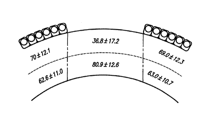
Fig. 1 .~,~resel~L~ a sch~m~tic section of the rabbit cornea showing
the number of keratocytes present in each of the studied areas;
Fig. 2 le~l~se"ls a sch~m~tic section of the monkey cornea showing
the number of keratocytes present in each of the studied areas.
Detailed Description of the Invention
Pursuant to the present invention, damage to the cornea following trauma
thereto resulting in at least some injury to the epithPlillm is plcvcllL~d or minimi7.od
by applying to the corneal epith~linm a keratocyte m~ lei~"re solution over a
period of time sufficient to permit corneal wound healing. The method of the
present invention is particularly useful in promoting healing after surgery which
involves removal or disruption of the corneal epithelium, including but not limited
to the following procedures: phoLo.crlactive k~..AIrcLc~llly, kelaLo-l-ileusis,
epikeratophakia, lamellar ker~tectomy, radial ke.aLuLu..ly, corneal transplantation,
and other intraocular surgical procedures. In addition, healing of the cornea ispromoted in accordance with the present invention following scraping of the
30 corneal epithelium for other reasons such as epithelial debri<l~ nt to improve
visibility during intraocular surgery) or any type of trauma res--lting in some injury
W0 95/20969 ? '~-~$9~ 0 6 PCT/U595/J1430
to the epithelium, such as ~aceration or abrasion of the eye (caused, for example,
by a superficial corneal foreign body).
For purposes of the present invention, a keratocyte mAi,)le,~A,~re solution is
defined as a solution which enhAnres keratocyte viability by mAintAining normal
5 physiologic metabolism. A fairly wide variety of solutions may be employed forthis purpose. To dt;L~ e rapidly whether a particular solution as hereinafter
described would be suitable for use in accordance with the present invention, the
behavior of cells from a culture of keratocyte cells [see Saika, S., "Ultrastructural
effect of L-ascorbic acid 2-phosphate on cultured keratocytes, " Cornea 11 :439-445
10 (1992)] brought into contact with the solution may be observed. Solutions clearly
not suitable for use as keratocyte mailllelldllce solutions cause the cells to become
enlarged, and llltimAtPly to rupture. On the other hand, keratocytes exhibit anormal, healthy a~ea,dllce even after several hours in contact with a suitable
keratocyte IllAil~ Ai~re solution.
A plillldly con~ider"tinn for forml-lAtion of a suitable keratocyte
"lai,lL~na~lce solution is the osmolarity thereof. In contact with low osmolarity
~y~OtOlliC) solutions, the keratocytes do not survive for very long periods of time.
Accor~ingly, a keratocyte mAi"lrnAI-~e solution in accordance with the present
invention will have an osmolarity of at least about 250 mOsm. Preferably, the
20 osmolarity of the solution is at least about 280 mOsm, and most preferably, at least
about 320 mOsm. One particularly useful manner in which to provide solutions
with high osmolarity is the inclusion therein of one or more non-immlln-~genic
macromolecules. Examples of such materials include, but are not limited to, the
following: glycosAminoglycans (e.g., chondroitin sulfate, le, IllAIAi~ sulfate,25 ~, IlIA~ sulfate, heparin sulfate, heparan sulfate, keratin sulfate, keratan sulfate
and/or hyaluronic acid); low or high molecular weight poly~Arch-Ari~lPc, such asdextran; dextran sulfate; polyvinyl pyrrolidone; polyethylene glycol; polyvinyl
acetate; hydro~yp,u~yl cellulose; hydro~y~,opyl,llethyl cellulose; carboxymethylcellulose; and carbo~y~,u~yl",ethyl cellulose. Neutral or ,le~d~ively-charged
30 macromolecules are particularly suitable for use in this regard. While the
co,l~enl,aLion of macrûmolecule may be varied over a fairly broad range as
WO 95/20969 . ~ ~ r PCT/US95/01430
S ~;9~
a~,oplial~ to provide a solution with the desired osmolarity, it is preselltly
prert;ll~d that the at least one macromolecule comprise about 0.1% to about 10%
by weight of the keratocyte l,lah~lellallce solution.
In addition, it has been clr~ i.lr(l that a suitable keratocyte m~ "re
5 solution must contain at least one of m~gnrcil-m and calcium, and preferably both.
While the present invention is not bound to any particular theory, it is believed that
the presence of at least one of m~"~ci"", and calcium is nrceS~ry to m~int~in
enzyme activity and proper keratocyte cell binding in the cornea during
reepithelialization. In addition, it is ~re~ d that the keratocyte lllahll~l~llce
10 solution comprise other ions (for example, Na+, K+, Cl-, etc.) which are generally
recognized in the art as useful in m~int~ining normal cell growth in, e.g., cellculture solutions.
It is further considered to be desirable to include a suitable buffer system
in the keratocyte m~i"lt?"~nre solution so as to provide a composition which hasa pH roughly comparable to physiological pH. In general, any of the art-
recognized buffer systems which are suitable to provide a pH in the range of about
6. 8 to about 7.6 would be useful for this purpose. Suitable buffer systems include,
but are not limited to, bicarbonate buffer systems, HEPES buffer systems and
lc;s thereof.
One class of materials suitable for use in accordance with the present
invention are a number of compositions which have heretofore been employed as
corneal storage media. As is well known in the art, corneal storage media
comprise nli~Lules of components selected from bal~nre~l salts, amino acids, energy
sources, antioxidants, burr~ g agents, cell membrane stabilizers,
glycosaminoglycans, deturgescents and antibiotics. Antibiotics and all~illlycoLics
in the media are useful for pl'eVC~lltillg infection (i.e., to ~u~l~le;,s growth of bacteria
or fungi), rather than for k~latO~;y~ m~i"le~"re and growth; accordingly, they are
considered an optional component of the pl~rell~d compositions for use in
accordance with the present invention. Similarly, for purposes of the present
invention various additional components conventionally included in the heretofore-
known corneal storage media (for example, antioxidants, cell membrane stabilizers,
c
wo 9s/~u969 ~ 6 l~CT/US95/01430
ATP precursors, and nlltri~nt cell supplements), may be included in accordance
with particular emborlim~nt~ of the present invention but are not ~lesell~ly
considered es~enti~l for m~ n~ e of keratocytes. One particularly useful
composition for purposes of the present invention is a corneal storage mP~ m
5 cont~ining chondroitin sulfate, available commercially from Chiron Ophth~lmirs,
Inc., Irvine, CA under the trade ~ ign~tion OptisolTM [~llfm~n, H.E. et al.,
"Optisol Corneal Storage Me~lillm, " Arch Ophthalmol 109, 864-868 (1991);
Lindstrom, R.L. et al., "Optisol Corneal Storage Medium," Am. J. Ophthalmol.
114, 345-356 (1992)].
Pl~sell~ly ~lc~lled for use in accordance with the present invention are the
corneal storage media as generally disclosed in the aforementioned U.S. Patent
5,104,787. As more fully described therein, in general these solutions contain an
aqueous nutrient and electrolyte solution, at least one glyco~minoglycan, at least
one de~ulgescell~ agent, at least one energy source, at least one buffer system, at
15 least one antioxidant, at least one antibiotic, at least one membrane stabilizing
component, at least one ATP precursor and nutrient cell supplements, all in
amounts s -fflri~nt to enh~nre cell viability and metabolism and wound healing.
Nutrient and electrolyte solutions, which are well defined in the art of tissue
c~-lh~ring, contain the essential mltri~nt~ and electrolytes at at least the minim~l
20 concell~lalions ~-~ce~ , y for cell maintenance and growth. In general, they contain
u~olg~llic salts (e.g., calcium, m~"~ "" iron, sodium and potassium salts of
carbonates, nitrates, phosphates, chloride and the like), essential and non-e~enti~l
amino acids, viL~ills and other essential nutrients. Suitable basal nutrient media
are commercially available, for example from Gibco Laboratories, Grand Island,
25 NY and Microbiological Associates, Walkersville, MD under the ~lesign~tions
TCl99 and Eagle's Minimal F.~nti~l Medium (MEM). As more fully explained
in U.S. Patent 5,104,787, these and similar lluLlicllL media (or llli~lul~s of one or
more thereof) serve as the basis for form~ ting corneal storage media as are
particularly suitable for use in accordance with the present invention. Of course,
30 for purposes of the present invention the l~lcsellce of at least one of m~g"Ps;''''~ and
calcium is considered e~enti~l; inclusion of other typical components of
wo 95120969 ~ i 706 CT/US95101430
conventional basal nutrient media useful in the mahlL~lldllce of cell growth (e.g.,
other salts, amino acids and viLa llhls, etc.) is ~-~;fel~cd.
Particularly p-~re..~:d compositions (as described in U.S . Patent 5,104,787)
for use in accordance with the present invention comprise the following: an
5 aqueous electrolyte solution (e.g., Minimal F.~çnti~l Medium and/or TCl99); a
glycos~min~glycan (e.g., standard or purified high or low molecular weight A, B
or C isomers of chondroitin sulfate, ~erm~t~n sulfate, dermatin sulfate, heparinsulfate, heparan sulfate, keratin sulfate, keratan sulfate and/or hyaluronic acid) in
a range of 0.01 mg/ml to 100 mg/ml; a d~lulgescel~l agent (e.g., low or high
10 molecular weight polysaccharide, such as dextran, dextran sulfate, polyvinyl
pyrrolidone, polyethylene glycol, polyvinyl acetate, hydro~y~l~yhllethyl cellulose,
carboxymethyl cellulose, carbo,~y~ro~ylmethyl cellulose, etc.) in a range of 0.01
mg/ml to 100 mg/ml; an energy and carbon source (e.g., glucose, py.uvdle,
sucrose, fructose and/or dextrose) in a range of 0.05 mM to 10 mM; a buffer
15 system (e.g., a bicarbonate buffer system, HEPES buffer, etc.) to m~int~in a
physiologic pH (desirably beLweell about 6.8 and about 7.6) in a range of 0.1 mMto 100 mM; optionally, an antioxidant (e.g., ascorbic acid, 2-mercaptoeth~nnl,
gl~lt~thione and/or alpha tocopherol) in a range of .001 mM to 10 mM; optionally,
membrane stabilizing agents (e.g., viL~ulli-ls A and B, retinoic acid and/or other
20 cofactors, ethanolamine, phosphoethanolamine, selenium and/or L dn~re.li) in a
range of 0.01 mg/ml to 500 mg/ml; optionally, for purposes of the present
invention, antibiotics and/or anLil.ly~;otic agents (e.g., amphotericin B, ge"~ in
sulfate, kalldlllycill sulfate, neomycin sulfate, lly~Ldlill, penicillin, tobl~llycill,
~Ll~Lolllycill sulfate) in a range of 0.1 ~ug/ml to 1 mg/ml; optionally, ATP
precursors (e.g., adenosine, inosine, adenine) in a range of 0.001 mM to 10 mM;
and optionally, lluL iellL cell supplements (e.g., cholesterol, L-hydroxyproline, d-
biotin, calciferol, niacin, para-aminobenzoic acid, pyridoxine HCl, vitamin B12,Fe(NO3)3 and/or non-essential amino acids) in a range of 0.001 mM to 10 mM.
As noted in U.S. Patent 5,104,787, the solutions are preferably serum free.
One particularly ~ler~l-ed solution according to U.S. Patent 5,104,787 has
the following composition: as aqueous m-tri~nt and electrolyte solution, Eagle's
WO 95/20969 ~ PCT/US95/01430
MinimAl F~entiAl Medium (MEM); as a glycosAminl)glycan, 2.5% chondroitin
sulfate; as a dt;~ul~escc;ll~ agent, 1% ~l~xtr~n; as an energy source, 110 mg/L
~yl~lv~ and 1000 mg/L glucose; as a buffer system, 2200 mg/L bicarbonate
buffer and 25 mM HEPES buffer; optionally, as antioxidant, 0.05 mM 2-
5 mercaptoethanol and 0.01 mg/L alpha-tocopherol; optionally, as antibiotic and/or
all~i,llycotic, 100 mg/L gellL~lllicin sulfate; as ATP precursors, 5 mg/L adenosine,
10 mg/L inosine and 10 mg/L adenine; and optionally, as mltriPnt cell
supplements, 0.2 mg/L cholesterol, 10 mg/L L-hydroxyproline, 0.01 mg/L d-
biotin, 0.1 mg/L calciferol, 0.025 mg/L niacin, 0.05 mg/L para-aminobenzoic
10 acid, 0.25 mg/L pyridoxine HCl, 1.36 mg/L vitamin B12, 0.5 mg/L ~e(NO3)3 and
0.1 mM non-e~sentiAl amino acids.
In accordance with the invention, the keratocyte main~ellallce solution may
be either periodically applied to or mAint~inPd continuously or substAntiAlly
continuously in contact with the cornea for a period of time sufficient to permit
15 corneal wound healing. While the amount of time required for healing (which, for
purposes of the present invention, refers in particular to closure of the epithelial
defect) will of course vary with the particular patient, it is generally a~,u~,iate
to mAintAin the solution in regular periodic or continuous contact with the cornea
for a period of time on the order of at least about 12 hours to about one week, and
20 preferably about 24 to about 72 hours.
In accordance with one embodiment of the present invention, a quantity of
the keratocyte ,..Ai.-l~ re solution snfflrient to subslA..I;Ally coat the surface of
the corneal epithelium (e.g., about 20 ~l to about 100 ,ul, preferably about 50 ,.41)
is periodically applied to the corneal surface at a rate snfflriPnt to m~intAin (or
25 subst~ntiAlly mAint~in) normal keratocyte metabolism. Such application may beeffected mAnllAlly (for example, by means of an ~;yed,u~er) or ~utom~tir-Ally (for
example, by automated dropper means). It has been ~ cl that application
of one or two drops of k.,la~ ;yL~ m~ r ~A.,re solution at a rate of once about
every S mimltes to once about every hour, preferably once about every ten mimlt.os
30 to once about every half-hour, and most preferably once about every fifteen
WO 95/20969 ~oa PCT/US95/01430
",i~ es, is sufficient to m~int~in (or subst~nti~lly m~int~in) normal keratocytemetabolism.
Pul~u~lL to one ~lcrellc;d embo-limPnt, the solution is m~int~inPcl over the
cornea as a fluid bath. This can be achieved, for example, by m~int~inin~ a
5 quantity of the solution over the cornea in a glass or plastic container. The
solution in the co"L~i"~r may be periodically repleni~hPd or replaced, preferably
on a daily basis.
In a particularly LJl~fell~d embodiment of the invention, the solution is
m~int~inPd in contact with the cornea by means of a piece of an absorbent material
10 (for example, in the form of a lens or a shield having roughly the dimensions of
a convention~l contact lens) effective for controlled release of the solution to the
cornea. While a variety of m~teri~l~ are suitable for this purpose, one particularly
~.rer~ d material colllplises a collagen shield in the shape of a contact lens. Such
collagen shields are available coll~nt,.;ially, for example from Chiron Ophth~lmirs,
15 Inc., Irvine, CA under the designation SurgiLensTM. The collagen shield is
suitably soaked in the keratocyte m~ AI~re solution for several ",i""~es to
absorb the solution. Thereafter, the solution-impregnated shield is applied to the
eye and m~int~inPd in place during the healing period. A particular advantage ofthis embodiment of the invention is that the collagen shield dissolves in situ over
20 a period of time which in many cases is sufficient to permit corneal healing.Collagen seems to be advantageous as a biomaterial for these purposes due to itsmPcll~nir~l and biological plop~ ies. One important therapeutic benefit appears
to be a reduction in ",Pch~l-ir~l trauma to the epithelium, due to lubrication and
protection of the corneal surface, secondary either to lid abnorm~litiPs or to normal
25 blinking. This may reduce pain and polellli~l~ the process of epithelial adhesion
to the underlying tissue.
In the course of developing the present invention, it was co"ri,."Pd that
stromal keratocytes are affected by the absence of overlying corneal epithelium.Keratocyte loss alters the normal course of corneal wound healing, and thus the
30 corneal clarity or refractive outcome. The ultrastructural effects of the absence of
epithelium on stromal keratocytes in rabbits and primates were further investig~tPd
W095/20969 i- 'S9~ ~G PCT/US95/01430
and ~ d and the effect of the absence of epithelium on the healing of
stromal incisions in rabbit cornea was e~r~min~d As a consequence of these
studies, a method to prevent the keratocyte loss caused by deepithelialization was
developed.
Rabbits and monkeys were used in these studies. The results obtained in
both species were comparable, although the acellular zone in the ~l~mlrl~otl stroma
24 hours after surgery appears to be greater in the rabbit. The importance of this
finding is that the plcsence of Bowman's membrane in the monkeys did not ~l~;vt;llL
rapid degeneldLion of the keratocytes after deepitheli~ tion.
The onset of the ultrastructural changes in keratocytes in response to
superficial corneal injury was early in the postoperative period. These cellulardegelleldLive ch~nges in the rabbit cornea were visible with tr~ncmicsion electron
microscopy as early as 15 ,.-i"~ s after scraping. Keratocyte changes in the most
anterior aspect of the stroma of rabbit cornea were observed within 30 mimltes of
epithelial removal; this short time course suggests that osmotic injury may account
for the cell damage. The changes observed here cannot be ascribed solely to
corneal swelling, as it has been reported that the absence of corneal endothelium
caused no a~al~;nL loss of stromal keratocytes even in the presence of intense
corneal edema [D~ hlm~n, C.H. et al., "The effect of the absence of corneal
epithelium or endothelium on the stromal keratocytes, " Invest Ophthalmol. 7:520-
534 (1968)]. At six hours after surgery, the corneal epithelium showed initial
signs of migration over the denuded stroma. Invasion of the corneal stroma by
polymorphonuclear leukocytes (PMNs) was also first a~alc:nl at this time. The
presence of PMNs in the stroma might be related to the reg~lltldLion of the
epithelium itself or be stim~ te~l by chemotactic factors liberated by the
dege~ g keratocytes. The timing of all of these fin-lin~ suggests an
interaction not only between the epith~ lm and keratocytes, but also between
epithelium and PMNs. Clinically, this may collLlibuL~ to melting of the underlying
stroma in per.cictent epithelial defects.
To e~ e for effects of epithelial removal on wound healing after
l~rlacLiv~ surgery, k~ldLoL(Jllly incisions in normal and deepithelialized rabbit
wo 95no96s ~ ~ 9~76 PCT/US95/01430
corneas were st~ cl At 24 hours po~LopelaLively the wounds in the denuded
corneas showed an intense infl~mm~tcry r~s~ollse and an absence of keratocytes.
Incisions in non-deepithelialized corneas showed only minim~l infl~mm~tion and
keratocytes present adjacent to the incision. Studies of ~imil~rly treated corneas
5 14 days postop~ldLively showed advanced wound healing in both corneas, but thedeepith~ li7~d cornea showed a larger epithelial plug and retraction of the stroma.
These results suggest that creation of an epithelial defect may alter wound
morphology. The inrl~lcetl retraction of the stroma might increase the wound gape,
and consequently increase the refractive effect of linear radial incisions.
In all animals shlt~ including the monkeys, epithelial denll-l~tion of the
cornea prior to refractive procedures in~nced death of anterior stromal keratocytes.
Since this occurs in monkeys, which have a Bowman's layer, it likely occurs in the
human cornea as well.
While the present invention is not bound to any particular theory, it is
believed that a metabolic interaction occurs between the epithelium and the
underlying stroma and its cells. The epithelium lc~l~s~ a barrier between the
tear film fluids and the t:llvi~ lllent within the corneal stroma. Exposing the
keratocytes to the p,ecollleal tear film appears to result in rapid dege"cl~Livechanges in these cells. The use of topical metabolic uuLliCllL~ may reduce the
deleterious effects of epithelial removal by m~ il,g a physiologically acceptable
stromal ellvhol~llent and facilit~ting normal metabolic processes. Denudation ofthe cornea followed by application of a composition in accordance with the present
invention limits keratocyte damage and speeds reepitheli~li7~tiQn. These
compositions are rich in cellular mltrient~ such as glucose, in which the tear film
may be relatively deficient. Also, the compositions are higher in osmolarity than
is the ~lccol~lcal tear film and many commercially-available tear film subsLi~uLt;s.
These fin~ing~ suggest that the compositions of the present invention may protect
keratocytes and modify corneal wound healing, possibly by ~c;ve~Lillg osmotic
damage to the stromal keratocytes.
The invention may be better llnrlerstood with reference to the accompanying
examples, which are int~n-led for purposes of illustration only and should not be
WO 9~/20969 ~ Q6 PCT/US95/01430
12
construed as in any sense limhing the scope of the invention as defined in the
claims appended hereto.
Example 1
Use of Keratocyte l~ P~ e Solution
A total of foullt;ell New 7~ nrl Albino rabbits and four monkeys (M~7c~
fascicularis) were used in these studies. Animals were ~n~sth~ti7ed with an
intr~mllcclll~r injection of krl;~ hydrochloride (40 mg/kg) and xylazine
hydrochloride (7 mg/kg). The eyelids were held open with a wire speclllnm for
the duration of the procedure. The animals underwent a unilateral 6 mm
m~h~nir~l deepithelialization using a Paton spatula. The contralateral eyes wereused as controls. No topical m~(lic~tion was applied before or after surgery, with
exception of two rabbits that were operated on under a fluid bath.
Eight of the rabbits and all four monkeys were used in this ~elill,ent to
evaluate the time course of the changes that occur in the stromal keratocytes after
deepith~oli~li7~tion The rabbits were sacrificed at 15 mimltes (1 rabbit), 30
mimltes (1 rabbit), 2 hours (1 rabbit), 6 hours (1 rabbit) and 24 hours after surgery
(4 rabbits); all the monkeys were sacrificed 24 hours after the deepitheli~li7~tion.
To evaluate the effects of the absence of epithelium on the outcome of
linear keratotomies, four rabbits underwent unilateral deepithelialization of the
central 6 mm of the cornea as described above. Both eyes from each of these
rabbits then underwent a single linear incision, 5 mm in length, using a diamondblade set for 50% of the paracentral depth, as measured by an ultrasonic
pachymeter. One rabbit was sacrificed 24 hours after the operation and another
rabbit was ~rrifice(l 14 days after surgery. Two rabbits each underwent corneal
deepitheli~ tinn of one eye under a fluid bath. A clear plastic cylinder
cont~inin~ OptisolTM or sterile isotonic sodium chloride (0.9%) was placed on the
globe, while the cornea was deepitheli~li7~. By this method, the solutions were
kept in contact with the cornea during the deepithelialization and for one hour
po~lopel~Lively, at which time the animals were sacrificed. Two rabbits underwent
corneal deepitheli~li7~ti( n under ambient conditions, but for the following 16 hours
a composition in accordance with the present invention was applied topically to one
WO95/20969 , ,, ;~ PCT/USg5/~)1430
of these animals at 30 mimltes intervals; the other animal did not receive any
topical solution after deepithelialization.
After enucleation, all eyes were immrdi~tely prepared in i~lentir~l fashion
for light microscopy and tran~mi~ion electron microscopy. The whole globes
5 were hl~l.lcl~ed in half-~Llcl~Lll Karnovsky fixative (2% paraform~ çhyde, 2.5%
gll1t~ral~lçhyde, and 0.1 M sodium cacodylate buffer). The globes were fixed at
physiologic ~rcs~ule by infusion of fixative into the vitreous cavity. After 48
_ours of fixation, the corneas were trephined and bisected.
For Ll~"c",i~inn electron microscopy, one half of each cornea was washed
10 in 0.1 M sodium cacodylate buffer, postfixed in 2% osmium tetroxide in 0.1 M
sodium cacodylate buffer for two hours, washed in 0.1 M sodium cacodylate
buffer, dehydrated in a graded series of alcohol and then in pure polypropylene
oxide, and embedded under vacuum in epoxy plastic resin. For light microscopy,
after usual prcl)dld~ion, sections were stained with hrm~t-)xylin and eosin. For15 tran~mi~ion electron microscopy, 60-80 nm thick sections were cut with a
diamond knife nltr~mirrotome. Tissue was mounted on copper grids and stained
with uranyl acetate-lead citrate. The ~x;1",;"~tion of the specimens were performed
by an e~elienced ophth~lmir pat_ologist masked as to the LlcaLIllcnt group.
To qll~"~ the histologic ch~ng,os observed 24 hours po~Lu~.dLively,
20 sections were placed under a light microscope (Zeiss, Germany). The sections of
the corneas studied were divided into central (corresponding to the deepithelialized
zone) and two peripheral (unoperated zone with intact epith~ lm) areas. The
central and peripheral areas were further divided, so as to di~r~,.e.lliate allLcliol
from posterior stroma. Keratocyte nuclei were counted within each of the six
25 regions of the cornea and morphologic feaLulcs were recorded by a single
eA~cliellced ophth~lmir pathologist who was m~d as to the tre~tmrnt groups.
Keratocyte counts and morphology between the central anterior stroma (beneath the
area of deepitheli~ tion) and the other regions of the stroma were colll~aled.
Mea~ul~mcll~ of variance and contrast were used to compare the results
30 obtained in the dir~.cllL eA~eihnental groups. An overall P<.05 was considered
to be sipnific~nt
W O 95/20969 215 9 7 0 G PCTrUS95/01430 ~
1,
,.
Within 30 minlltes of removal of the central 6 mm of rabbit corneal
epithelium, the super~lcial k~ o~;yLes beneath the denuded area showed dilatation
of the endoplasmic reti~ m and presence of lacunae at the cell margin. This
contrasted with the keratocytes located under intact epithelium in the peripheral
S cornea and in the posterior stroma, which retained a normal a~ealal~e. The
changes observed in the superficial keratocytes increased with time po~L(J~ d~ively.
At six hours after surgery, the stroma was thicker and the keratocytes showed large
numbers of intracytoplasmic vacuoles. These changes were greatest within the
anterior 100 microns of the stroma, where disco"l;",lili.?s in the keratocyte cell
10 membranes were observed. At this point, polymorphonuclear leukocytes were first
seen at the margin of the operated area, and pyknotic changes were first observed
by light microscopy in anterior stromal keratocytes. Twenty-four hours after
deepitheli~li7~tion, keratocytes were absent from the anterior 25% of the stromaunderneath the deepithelialized area. Electron microscopic ~x;.",i,.,.lion of the
15 corneas eml~ ted at this time point showed keratocytes in advanced stages of
dege~ ion within the anterior stroma.
The primate corneas were evaluated 24 hours after deepitheli~ tion. At
this time, keratocytes were absent in the anterior stroma and no keratocytes were
seen within 75 ,um of Bowman's layer. The epithelium showed early signs of
20 migration over the denuded region. In unoperated corneas, normal keratocytes
were seen just underneath the Bowman's layer within about 13 microns of the
epithelial basement membrane. Ultrastructurally, normal keratocytes were
observed about 75 microns deep to Bowman's layer, compared to 13 microns in
normal unoperated monkey cornea. Some rel~ of keratocytes were seen
25 within the anterior stroma.
An expe.ri~ ed ophth~lmic pathologist, masked as to animal species and
tre~tmtont protocol, reviewed histologic sections of each cornea and counted thekeratocyte nuclei within six regions of the stroma. Qn~ntit~tinn of keratocyte loss
observed at 24 hours po~lopcldliv~ly in rabbits and monkeys is shown in Figs. 1
30 and 2, res~e~;lively. The values l~leselll mean ~t standard deviation. The
number of keratocytes i-l~ntified in the central anterior stroma was colll~ared with
wo ssnos6s ~59;~06~ PCIIUS95/nl430
the number of keratocytes obtained in the other five areas studied (Table 1). Incontrol eyes of both rabbits and monkeys, the number of keratocytes present in the
central stroma beneath the area of epithelial removal (central 6 mm) was similarto the number of keratocytes counted in the other areas. However, in the
5 deepithelialized eyes the number of keratocytes in the anterior central stroma was
~ignifir~ntly reduced (P=.0001 in rabbits and P= .0007 in monkeys). In addition,r~ i"i"g k~laLO~;ylt: nuclei within the superficial central stroma, beneath the zone
of deepitheli~li7~tion, appeal~d pyknotic conlpared to those in the five rem~ining
stromal regions.
0 Table 1
Counts of Keratocytes ~arrGI I in Deepithelialized
and Ullu~ te~ Rabbit and Monkey Corneas
Anterior Central Stroma Remaining Stroma P vdlue
Deepithelialized rabbit corneas 36.8 _ 17.2 69.0 _ 11.7 .0001
Control 94.3 i lZ.7 76.7 _ 19.7 .1
Deepithelialized monkey corneas 39.6 + 4.2 54.1 + 11.0 .0007
Controls 59.4 + 7.6 76.7 + 6.9 .49
Removal of central epithelium prior to linear keratotomies resulted in
altered wound morphology. At 24 hours po~L.peldLively, the kt:latOL~llly incisions
in the normal corneas showed an early invasion of epithelial cells in the wound.Keratocytes were present immP(ii~tely adjacent to the incision and appeared
normal. A few polymorphonuclear leukocytes were observed in the vicinity of the
wound, and slight retraction of the adjacent stroma was seen. F.Y~min~tion of the
keratotomy wounds performed in the centrally deepith~ li7e~1 corneas showed a
marked retraction of the adj~cçnt stroma, an intense acute infl~mm~tory responsein the stroma and the pl~sellce of debris and polymorphonuclear leukocytes within
the incisions. In contrast to the conkol corneas, no keratocytes were observed in
the anterior stroma or ~(lja~ent to the wound. Fourteen days po~lol,el~lively,
~x~.lli..,.~ion of the k~.d~ ollly incisions p~lrolllled in both ullOp~,.al~d and
deepithelialized corneas showed presence of fibrocytes and fibrosis llnrltqrn~th the
epithelial plug. The ~rcçnt~l~tçcl retraction observed in the early po~l~elalivl;
WO 95120969 PCT/US9S/01430
21597 0~
~- 16
period in the deepithelialized corneas caused enlargement of the epithelial plugs and
the stromal wounds when colll~a,~d with the non-deepithelialized corneas.
The use of a co",~o~ilion in accordance with the present invention as a
mltrient solution during and immPfli~tely after deepitheli~li7~tion of rabbit corneas
5 resulted in healthy superficial keratocytes, as seen by tr~n~mi~ion electron
microsco~y, but early signs of keratocyte damage could be observed in the isotonic
saline solution treated corneas. Light microscopic e~ )n of the corneas
treated topically at 30 minute intervals with a composition in accordance with the
present invention for 16 hours po~L(J~eldliv~ly revealed ~ selvdtion of keratocytes
10 and an appal~nLly faster reepitheli~li7~ti~ n than in the ullL~eaLed corneas. Example 2
Use of Colla~en Shields in Conju,l.:Lion with Keratocyte Mai"~t;nallce Solution
Twelve New 7P~l~nrl white rabbits, 2.0 to 2.5 kg, male and female, were
used. All procedures were ~elr~""ed on ~nPsthPti7P~l rabbits (k~ lil-P
15 hydrochloride, 40 mg/kg and xylazine, 7 mg/kg). All rabbits were ex~minPd
preope,dliv~ly with a biomicroscope. After being ~nPstheti7P~, the eyelids were
held open with a wire specnlllm for the duration of the procedure. The animals
underwent a bilateral 6 mm mPçh~nir~l deepitheli~ tion using a blunt Paton
spatula (Storz Ophth~lmic Instruments, St.Louis, MO), with ullirollll p,~s~u,~
20 applied during the scraping.
Rabbits were divided into six groups with two rabbits in each group.
Surgery was performed uneventfully in all eyes. TmmP~ t~Ply after ablation, the
treated areas had a ul~iro~ a~ea,al~ce. The groups were treated po~Lope,dlively
for 24 hours as follows: Group 1, saline drops every four hours; Group 2, drops
25 of a keratocyte m~intPn~nre solution in accordance with the present inventionincluding antibiotic (the commercially-available OptisolTM solution) every four
hours; Group 3, collagen shields (obtained as l~P-liT ~n~TM from Chiron
IntraOptics, Inc., Irvine, CA) soaked in sterile saline fitted over the wounded
cornea plus saline drops every four hours; Group 4, collagen shields (Me~liT Pn~TM-
30 CS) soaked in OptisolTM plus OptisolTM drops every four hours; Group 5, collagenshields (available commercially as SurgiLensTM from Chiron
Wo gs/20969 ~S PCTIUS95/01430
17
IntraOptics, Inc.) soaked in sterile saline plus saline drops every four hours; and
Group 6, collagen shields (SurgiLensTM) soaked in OptisolTM plus OptisolTM dropsevery four hours. The collagen shields were soaked in the keratocyte ,..~i"lr~ -re
solution or saline solution for 10 ,~,i",lles before being placed on the rabbit
5 corneas. The drops were applied every four hours. The ~nim~l~ were sacrificed
at 24 hours after surgery, the corneas fixed and keratocytes q~ d within
anterior and posterior cornea in the area lln-lernr~th the epithelial defect.
After sacrificing the animals, the corneas were excised at the limbus using
a sharp scalpel. Samples were bisected and then immr~ tely fixed in Karnovsky's
10 solution (1.25% glutaraldehyde and 1.0% paraform~l(lehyde). After 48 hours offixation, the corneas underwent a serial dehydration using graded alcohol
concell~l~lions. With a critical-point drying a~aral-ls, the alcohol was exchanged
for anhydrous carbon dioxide. The dried samples were then mounted and coated
with 15 ~m of gold. Documentary photographs were obtained (m~gnifir~tion,
15 1000 x; ~olhhlg (li~t~nre, 25 mm) and ex~minr~l by the same observer m~ d to
the Ill-Alll~ ll group from which they had been obtained. For light microscopy,
after usual plcpalalion, sections were stained in standard fashion by hematoxylin-
eosin and periodic acid-Schiff.
To qn~"~ e the histologic sh~nges observed 24 hours postol)c;lalively,
20 sections were placed under a light microscope (Zeiss, Oberkochen, Germany). The
deepithelialized zone was studied in sections, divided into anterior and posterior
stroma. Cells were ~ r~l in each of these corneal fields under a
m~gnifir~tion of 100 x. Counts of k~ldlo~;ylt:s and studies of morphologic çh~n~es
observed in these cells were performed in both the individual areas and the results
25 c~ al~d. Photomicrographs were taken using a Zeiss Photomicroscope III.
For qll~ntifir~tion of normal stromal cells in the cornea in the stained
sections, an experienced ophth~lmir pathologist, m~c~d as to animal species and
treatment protocol, reviewed histologic sections of each cornea and counted the
keratocyte nuclei under a m~gnifir,~tion of 100 x in the anterior and posterior half
30 of the stroma under the deepithelialized area. A reticule in one of the oculars,
which contains a 10 mm ruler, was used to mark the beginning and the ending of
WO 95/20969 2 1 5 9 7 0 6 PCT/US95/01430
18
the cell counting. The projection of the ruler onto the slide comprised 171 ,um of
stroma. The results of qll~ntifir~tinn of keratocyte loss observed at 24 hours
po~L~ldlively are shown in Table 2. At this point, absence of keratocytes was
noticed only in the anterior half of stroma of all corneas, although to different
5 degrees. There was no change in keratocytes of the posterior stroma, either innumber or in morphological appearance. In Table 2, the sample size is 2 rabbits
per group (for a total of 4 eyes). The number of keratocytes is reported as the
means plus or minus the ~Ldlldald deviation.
Table 2
Counts of Keratocytes Performed 24 Hours After Deepithelialization
in the Anterior Stroma
Treatment Group Number of Keratocytes
1: Saline drops 76.0 + 23.0
2: Optisol drops 81.5 + 45.9
3: l~e(liT Pll.CTM soaked in sterile saline 122.5 + 53.1
plus saline drops
4: MediLensTM soaked in OptisolTM plus 158.0 + 65.5
Optisol drops
5: SurgiLensTM soaked in sterile saline 120.3 + 69.1
plus saline drops
6: SurgiLensTM soaked in OptisolTM plus 157.3 + 72.3
Optisol drops
30 In Group 1, which served as a control, there was a relatively smooth corneal
surface and a marked reduction in numbers of k~laLucyles. The rem~ining
keratocyte nuclei within the ~ul,clrlcial central stroma, beneath the zone of
deepithelialization, appeared pyknotic compared to those under the intact
epithelium. Light microscopic eY;~ ion of corneas treated every four hours
35 with keratocyte mainLenallce solution revealed comparable numbers of keratocytes
in the anterior stroma. While there was a relative preservation of keratocytes in
the anterior stroma in animals treated with either MediLensTM (Group 3) or
W095/20969 ~CS' PCT/US95/01430
9,~
SurgiLensTM (Group 5) soaked in saline solution, ~ignifi~ntly higher numbers of
keratocytes were observed in groups treated with ~ediTPn~TM (Group 4) and
SurgiLensTM (Group 6) soaked in OptisolTM solution.
The collagen shields were observed to dislocate in six of the 16 eyes
belwc~ell the eighth and twelfth hours and were imm.o~ t~ly replaced.
Tarsorrhaphy was pclrolllled in all rabbits at twelfth hour poslop~l~lively to avoid
losing the shields. There was mild to moderate colljull~;lival injection and edema
in all of the eyes during the twenty-four hours of the experiment, with no
dirrerellces between the groups. These infl~mm~tory changes were attributed to
the wounding itself and not to the collagen shields, as there was no a~art~
dir~lc;llce between the control and treated groups in this respect. There was noevidence of infection in any of the eyes. There was minim~l disruption of the
anterior stroma after acute injury. The normal l~m~ r al~ persisted, and
the keratocytes deg~l~el~l~d and disappeared only in the anterior stroma. The
histopathologic structure of the areas outside the ablated zones were normal in all
corneas e~minPcl No abnorm~liti~ were seen in posterior stromal keratocytes
and the endothelium was normal in all eyes.
Example 3
Evaluation of Artificial Tear Compositions as Keratocyte Maintenance Solutions
Various artificial tear solutions are evaluated for use as keratocyte
m~ en~ e solutions. Samples from keratocyte cell cultures were brought into
contact with compositions A-H (artificial tear compositions). Compositions A-H
are characterized by the polymer and salt components and the osmolarity.
Compositions A-D were found not to induce any signi~ nt damage to keratocytes;
on the other hand, compositions E-H were found to be lethal to the cultured
keratocytes. The compositions are described in Table 3.
Wo9S/20969 2159~ ~ PCT/US95/01430 ~
Table 3
Artificial Tear Compositions
Co~ o~i~ioll Polymer Salts Osmolarity
(mOsm)
A 1.0% earboxymethyl- CaCI~, KCl, NaCl, Na 295
eellnlnse laetate
B 0.5% earboxymethyl- CaCI~, MgClz, KCl, NaCI, 284
. ee~ los~ Na laetate
C 0.1% dextran 70; 0.3% CaCl2, MgCI2, ZnCI2, 275
hyd~u~y~lu~ylcellulose NaHCO3, KCI, NaCI, HCI,
NaOH, CO2
D 0.3% glyeerin CaCI2, MgCI2, ZnCI2, KCI, 261
NaCI, Na citrate, Na
phosphate
E 0.1% dextran 70; 0.3% KCI, NaCI, HCI, Na 273
hydro~y~lulJyllllethyl- borate, NaOH
c~lhllose
F 1% polyvinyl alcohol; edetate disodium; dextrose 257
polyethylene glycol 400
G 0.5% polyvinyl aleohol; KCI, NaCI, NaHCO3, 248
0.6% povidone dextrose, Na borate, Na
ph- srh~t~
H 0.2% polyethylene glycol NaCl; edetate disodium; 227
400; 0.1% dextran NaOH; polycarbophil
As a review of Table 3 makes a~pa~ , compositions A-D are distinguished
on the basis of an osmolarity over 250 mOsm and at least one calcium and/or
15 m~,"~si"-" salt.
While there have been shown and described the fundamental novel feaLul~s
of the invention, it.will be understood that various omissions, substitutions and
changes in the form and details ilbl~tr~t~d may be made by those skilled in the art
without departing from the spirit of the invention. It is the intention, therefore, to
20 be limited only as in~ tPd by the scope of the following claims.