Note: Descriptions are shown in the official language in which they were submitted.
WO 95/21938 ; ~ ~ PCT/EP95l00553
1
A method for screening for the presence of a genetic defect associated
with thrombosis and/or poor anticoagulant response to activated protein
C.
A
The subject invention lies in the field of haemostasis and in
~ particular is directed at the aspect of thrombosis. More particularly the
invention is directed at a method for screening and diagnosis of
thrombophilia, especially hereditary thrombophilia. The method according
to the invention can then be used for determining the risk for thrombosis
in individuals.
$ack~round to the invention.
Deep vein thrombosis is a common disease. Well established risk
factors include recent surgery, malignant disorders, pregnancy and
labour, long term immobilisation, and deficiency of one of the main
inhibitors of the clotting system (Ref.i). The main inhibitors are known
to be protein C, protein S and antithrombin. The causes of deep vein
thrombosis in many patients remain Zulclear. It has recently been
established however that a poor anticoagulant response to activated
protein C (APC) is present in several families with a hereditary tendency
to venous thrombosis (Ref.2).
The anticoagulant property of APC resides in its capacity to
inactivate the activated cofactors Va and VIIIa by limited proteolysis
(Ref. 3). This inactivation of cofactors Va and VIIIa results in
reduction of the rate of formation of thrombin, the key enzyme of
coagulation. In vitro, this effect can be visualised by adding APC to
normal plasma and accordingly determining the effect thereof in a
coagulation test, for example in a test determining the APTT ( activated
partial thromboplastin time). Activation of protein C occurs at the
surface of endothelial cells via the thrombin-thrombomodulin complex
(Ref.2'7). Thrombomodulin is a membrane glycoprotein that can bind
thrombin. By this binding thrombin loses the ability to convert
fibrinogen to fibrin and the ability to activate blood platelets. In
other words thrombin loses its coagulant properties and reduces its
further own production (so-called negative feed-back) by activating
protein C. In vivo (in the presence of calcium) the activation of protein
C is almost completely dependent on the availability of thrombomodulin on
the endothelium. APC is subsequently neutralized by formation of
WO 95/21938 PCT/EP95/00553
2
complexes with APC inhibitor (PCI) and ~1 antitrypsin, which means that
in normal conditions it remains only for a short time in the circulation
and the anticoagulant effect remains generally locally expressed.
It was generally accepted that the inactivation of the '
cofactors Va and VIIIa by APC proceeds only optimally in the presence of
Ca2', phospholipids and the APC cofactor protein S (Ref.4,28,29). More '
recently this view was, however, challenged by the finding that in
systems of purified proteins protein S has little cofactor activity to
APC (Ref. 5, Ref. 6). A possible solution for this apparent discrepancy
between the observations in vivo (thrombotic tendency in hereditary
protein S deficiency) and in vitro (poor APC cofactor activity of protein
S in systems of purified proteins) could be offered by the finding of
Dahlb~ck et al (Ref.2) who reported patients with normal values for
antithrombin activity, protein C (immunologically and functionally) and
protein S (immunologically and functionally) without indications for
abnormal plasminogen, abnormal fibrinogen or lupus anticoagulants, but
with a reduced anticoagulant response to activated protein C. The latter
was found with a new test developed by Dahlback (Ref.2) in which he
studies the response (coagulation time, APTT) of a plasma after in vitro
addition of purified human APC. The addition of activated protein C to
the plasma of these thrombotic patients did not result in the expected
prolongation of the activated partial thromboplastin time (APTT). After
postulating a number of mechanisms for this phenomenon only one was
considered to provoke the poor anticoagulant response to APC, namely the
existence of a hitherto unknown cofactor to APC that is deficient in
these patients.
The following mechanisms have to date been rejected as being
causes of the poor anticoagulant response to APC:
1. The presence of an auto antibody against APC
2. A fast acting protease inhibitor reacting with APC
3. A functional protein S deficiency
4. Mutations in the Factor V or Factor VIII gene
Dahlb~ck (Ref.2,7) postulated that in the families studied a
hereditary shortage of a hereto unknown APC cofactor that purportedly
works independently of protein S was the cause of APC resistance.
Dahlb~ck et al (Ref. 2) also described a test method for diagnosing the
thrombo embolic disorders by addition of activated protein C to a patient
WO 95/21938 ~ PCT/EP95/00553
3
sample containing coagulation factors followed by measurement
of an
enzyme activity that is influenced by the addition of APC in an
international patent application W093/10261. It is stated in the
application of Dahlb~ck et al that the experimental results presented
indicated that the disorders in question are related to a hitherto
unknown coagulation factor or factors or unknown interactions
of known
factors. The unknown factor is not Factor Va or VIIIa that is
resistant
to degradation by APC and neither is it an inhibitor of the
immunoglobulin type for APC. Furthermore it is not related to
protein S
deficiency. Dahlb~ck et al (Ref. 2) state that their invention
is a
method particularly useful for further diagnosis of thromboembolic
diseases such as hereditary or non hereditary thrombophilia and
for
determining a risk for thrombosis in connection with pregnancy,
taking
anticonception pills, surgery etc. They describe their method
as being
characterized in that the coagulation system in a sample is activated,
wholly or partly in a manner known per se and incubated with activated
protein C, whereupon a substrate conversion reaction rate like
clotting
or conversion of a chromogenic substrate is determined. The conversion
rate obtained is compared with values obtained for normal plasma
samples.
If the rate is enhanced it indicates that the individual from
which the
sample is derived may suffer from a clotting disease. The disease
is not
expressed by protein S deficiency or by formation of Factor Va
or Factor
VIIIa resistant to degradation by APC or by an inhibitor of the
immunoglobulin type for APC. In the international application
it is also
stated by Dahlb~ck et al that the data in the application indicated
that
the patient in question could not carry a defective Factor VIII/VIIIa
in
contrast to what they had previously stated in Thromb. Haemostas.
65,
Abstract 39, 658 (1991), wherein addition of activated protein
C to a
plasma sample of a patient, and study of the effect produced was
claimed
to have illustrated a defective Factor VIIIa molecule not degraded
by
activated protein C. Furthermore in the international patent application
the assay was used to directly measure the inhibition of Factors
Va and
VIIIa by APC. Using the Factor Xa based clotting assay described
therein,
the inhibition of patient Factor Va by APC was found to be normal
a
suggesting that Factor Va in the patient's plasma was degraded
in a
normal fashion by exogeneously added APC.
Following the publication by Dahlb~ck et al (Ref.2) other
groups started research in this area. In Blood Vol. 82, nr. 7,
1993 on
pages 1989-1993 Griffin et al describe the results of APC resistance
WO 95/21938 PCT/EP95100553
4 i
tests carried out among 25 venous thrombotic patients with no
identifiable blood coagulation abnormality and 22 patients previously
identified with heterozygous protein C or protein S deficiency. The APC
induced prolongation of the activated partial thromboplastin time assay
for these patients was compared with results for 35 normal subjects. The
results showed that this new defect in anticoagulant response to APC was
surprisingly present in 52 to 64x of the 25 patients i.e. in the majority
of previously undiagnosed thrombophilia cases. The deficiency was not
present in 20 of 22 heterozygous protein C or protein S deficient
patients. This suggested that the new factor is a risk factor independent
of protein C or protein S deficiency. Mixing of normal blood plasma with
each of two extremely defective plasmas (APC-induced prolongation of APTT
< 20 s) was performed and the APTT assays were made to assess the ability
of normal plasma to correct the poor response of the defective plasmas.
The results were similar to those of Dahlb~ck et al (Ref.2). This also
suggested that normal plasma contains a factor which is missing from the
defective patients plasmas. Values are given in the article for the net
calculated prolongation in APTT, simply defined as an APTT value in the
presence of APC minus the APTT value in the absence of APC. The article
also describes the ratio of the APTT with APC to the APTT without APC and
the fact that this parameter was compared to values for the APC induced
APTT prolongation. This comparison indicated an excellent correlation
between these parameters for the normals, with an extremely low APTT
ratio value being indicative of abnormal patients. Therefore it followed
that either the parameter of APC-induced APTT prolongation or the
parameter of the ratio of APTT values with versus without APC or both
parameters can be used as diagnostic parameter. None of these parameters
seemed more useful for this purpose than the other according to the
article. Furthermore in the article it was stated that the APC-induced
prolongation of the APTT assay used was reminiscent of the assay
involving APC-induced inactivation of endogenous Factor VIII in the
plasmas of patients with lupus anticoagulant reported by Potzsch et al
(Ref. 19) in Blood 80: 267a 1992 (Abstract)). Based on this latter assay
it was reported in the Griffin et al article that plasma from lupus
anticoagulant patients with thrombosis gave a poor response to APC and '
that patients with thrombosis could thereby be distinguished from those
without thrombosis. Griffin et al speculated that auto-antibodies against
the new hypothesized APC-cofactor could play a role in the risk of
thrombosis among patients with lupus anticoagulants. It is further stated
WO 95/21938
PCTIEP95100553
by them that it is tempting to speculate that an acquired deficiency of
the new APC-cofactor could be associated with an acquired risk of
thrombosis.
In the Lancet, December 18, 1993, Vol. 342, on pages 1503-1506
5 Koster et al have elaborated further on the link between APC-resistance
. and thrombosis by describing how a population based case control study
was undertaken to test the clinical importance of the abnormality in the
coagulation system that is characterized by a poor anticoagulant response
to activated protein C (APC). From studies within families this poor
response to APC appears to inherit as an autosomal dominant trait (Ref.
2, 7 and 47). Among patients referred to a coagulation unit because of
unexplained thrombosis this abnormality was a major cause of
thrombophilia with a prevalence of about 40x (Ref. 8 and 9). In the study
described by Koster et al in the Lancet, December 18, 1993, Vol. 342,
pages 1503-1506, the clinical importance of this poor response to APC was
investigated in unselected consecutive patients, aged less than 70 years,
with a first objectively confirmed episode of deep vein thrombosis and
without an underlying malignancy. The sensitivity of these patients
plasma to APC was compared with that of matched healthy controls. The
sensitivity of their plasma APTT to activated protein C (APC) was
measured essentially as described by Dahlb~ck et al (Ref. 2) using the
reagents and reaction conditions previously developed for the protein S
activity assay (Ref. 11). The results were expressed as APC sensitivity
ratios (APC-SR) defined as the value of APTT (+APC) over the APTT (-APC).
In the Koster et al article (Lancet, December 18, 1993, Vol. 342, pages
1503-1506) it was stated that reduced levels of prothrombin and/or Factor
X (<0,5 u/ml) will increase the APC-SR. For this reason the test cannot
be used for the evaluation of plasma's of patients on oral anticoagulant
treatment. In a series of 98 samples a good correlation was found between
the APC-SR obtained with the test of Koster et al (Lancet, December 18,
1993. Vol. 342, pages 1503-1506) and those obtained with the test
developed by Chromogenix as described in WO 93/10261. A reference range
for the APC sensitivity ratio was derived from the healthy control
subjects. After logarithmic transformation of the data end exclusion of
10 subjects with values outside 3 standard deviations (SD) of the mean,
the lower limit of normal was 2.17 (mean-1,96 SD). An inverse relation
between the risk of thrombosis and the degree of response was found. The
21x prevalence of a poor response to APC among thrombosis patients and
the odds ratio for thrombosis of 6.6 led to the conclusion that a poor
WO 95/21938 , PCT/EP95/00553
6
response to APC could be considered a common and strong risk factor for
deep vein thrombosis. It was even speculated that subjects with APC
sensitivity ratios around 1.10 could be homozygous or double
heterozygous, whereas subjects with APC sensitivity ratios around 1.50
could be heterozygous for the abnormality. The prevalence of the
abnormality was 5x among the healthy control subjects. Because the ,
distribution of the APC-SR was clearly bimodal Koster et al believe that
subjects really had abnormal responses to APC rather than too low values
within a normal range. The relation between risk of thrombosis and their
response to APC seemed therefore not to follow the model of a simple
single gene defect. Because the abnormality was found to be so prevalent
in healthy subjects it was considered unlikely by Koster et al that the
defect in itself is sufficient to cause thrombosis as is true also for
protein C deficiency (Ref. 15, Ref. 16). An additional causal factor
seems to be required for the development of thrombosis within a
particular patient. These may be acquired factors and also as yet unknown
genetic defects or variations. However once other causal factors are
present poor APC response presents a strong risk of thrombosis as
witnessed by its six to sevenfold increase of relative risk. It was
20. stated in the article that the underlying defect of the poor response to
APC remained to be clarified even though a dominantly autosomally
inherited deficiency of a cofactor to activated protein C had been
postulated ( Ref . 7 ) . While a poor response to APC appears to be 5 to 10
times as frequent as deficiencies of protein C, protein S or antithrombin
it confers an approximately similar relative risk of thrombosis (Ref. 17
and 18) which according to Koster et al could make it worthwhile to test
all patients with venous thrombosis for this abnormality.
In summary in the state of the art it was ascertained that a
defect in the protein C anticoagulant pathway is linked to a relatively
high risk of thrombosis. The poor anticoagulant response to activated
protein C has been discussed in great detail, however the cause of the
poor anticoagulant response to activated protein C remains unclear. A
number of theories have been postulated, however the only one that has
been accepted is the presence of an unknown cofactor for APC which is
apparently deficient in a patient exhibiting a poor anticoagulant
response to activated protein C. The identity of the postulated cofactor
for APC is unknown. Furthermore current tests for detecting altered
response to APC cannot be used on test persons already using
anticoagulants.
CA 02159907 2006-04-26
30582-11
7
Summarv of the Invention
In one aspect, the invention provides a method for
diagnosing an increased risk for thrombosis or a genetic:
defect causing thrombosis comprising the steps of: (a)
providing, from a test subject, test nucleic acid comprising
codon 506 within EXON 10 of the human Factor V gene; and (b)
assaying for the presence of a point mutation in the
nucleotides of codon 506 within EXON 10 of the human Factor
V gene, wherein said point mutation correlates to a decrease
in the degree of inactivation of human Factor V and/or human
Factor Va by activated protein C, wherein the presence of
said point mutation in said test nucleic acid indicates an
increased risk for thrombosis or a genetic defect causing
thrombosis.
In another aspect, the invention provides a kit
for use in the method as described herein comprising a
forward and a reverse primer that are capable of amplifying
EXON 10 of the human Factor V gene.
In another aspect, the invention provides an
isolated nucleic acid molecule consisting of SEQ ID N0:14 or
its complementary strand.
In another aspect, the invention provides an
isolated nucleic acid molecule consisting of a nucleic acid
sequence selected from the group consisting of SEQ ID N0:4,
SEQ ID N0:5, SEQ ID N0:9, SEQ ID NO:10, SEQ ID N0:11, SEQ ID
N0:12 and SEQ ID N0:13, or the complementary strand thereof.
In another aspect, the invention provides a method
for diagnosing an increased risk for thrombosis or a genetic
defect causing thrombosis in a patient, comprising the steps
of: (a) providing from said patient a sample that contains
CA 02159907 2006-04-26
30582-11
7a
Factor V or Factor Va; and (b) assaying for the presence of
a mutation in amino acid 506 of human Factor V or human
Factor Va, wherein said mutation correlates with a decrease
in the degree of inactivation of human Factor V and human
Factor Va by activated protein C, wherein the presence of
said mutation in said sample indicates an increased risk for
thrombosis or a genetic defect causing thrombosis.
In another aspect, the invention provides a method
for diagnosing an increased risk for thrombosis or a genetic
defect causing thrombosis in a patient, comprising the steps
of: (a) providing from said patient a sample that contains
Factor V or Factor Va; and (b) sequencing said factors or a
fragment of said factors in order to determine the presence
of a mutation in which glutamine is substituted for arginine
at amino acid 506 of human Factor V or human Factor Va;
wherein the presence of said mutation in said sample
indicates an increased risk for thrombosis or a genetic
defect causing thrombosis.
Description of the Invention
Surprisingly the identity of the unidentified
cofactor responsible for a poor anticoagulant protein C
response has been found. It has been discovered that one of
the mechanisms that had been rejected is in fact responsible
for the defect in the protein C anticoagulant pathway in a
majority of thrombophilic patients. The cause of the
deficiency has been linked to the presence of a mutation in
the nucleic acid material encoding Factor V or Factor VIII
which upon expression is correlated to a decrease in the
degree of inactivation by APC of said Factor V and/or of
Factor Va, (the product of activation of said Factor V) or
of said Factor VIII and/or of Factor VIIIa (the product of
activation of said Factor VIII). The deficiency is
CA 02159907 2005-07-29
30582-11
7b
therefore not the result of a mutation in an as yet
unidentifed cofactor for APC, but is in fact due to a defect
in Factor V or Factor VIII or more particularly in the
activation products thereof.
As was already indicated in the state of the art
the link between a risk of thrombosis and the presence of
APC-resistance had already been made and it had also been
suggested that screening for such a deficiency would in fact
be extremely helpful in diagnosing patients with an
increased risk of thrombosis. As it is now known which
factors carry the genetic defect responsible it has also now
become possible to actually screen the population with
methods other than the Chromogenix test for determining APC-
resistance.
It has become possible to use DNA techniques or to
use antibodies for determining the presence of mutant
proteins when screening for the mutated Factor V or Factor
VIII associated with resistance to APC. The subject
invention is therefore directed at a method for screening
for the presence of a genetic defect associated with
thrombosis and/or poor anticoagulant response to activated
protein C (APC), said genetic defect being indicative of an
increased risk of thrombosis or said genetic defect actually
causing thrombosis in a patient, said method comprising
determination of the presence of a mutation in the nucleic
acid material encoding Factor V or Factor VIII in a manner
known ep r se, which mutation upon expression of the nucleic
acid material is correlated to a decrease in the degree of
inactivation by APC of said Factor V and/or of Factor Va
(the product of activation of said Factor V) or of said
Factor VIII and/or of Factor VIIIa (the product of
activation of said Factor VIII) and/or comprising
determination of a mutation present
WO 95/21938 PCT/EP95/00553
8
in the protein Factor V and/or Factor Va and/or present in the protein
Factor VIII and/or Factor VIIIa by analysis of said Factor V and/or
Factor Va or Factor VIII and/or Factor VIIIa or analysis of a proteolytic
fragment of said Factor V and/or said Factor Va and/or Factor VIII and/or
Factor VIIIa in a manner known per se, said mutation being correlated to
a decrease in the degree of inactivation by APC of said Factor V and/or
said Factor Va and/or Factor VIII and/or Factor VIIIa. In particular the
method according to the invention is directed at a method wherein the
mutation in the nucleic acid sequence encoding the Factor V and/or Factor
VIII is located at the position within the part of the nucleic acid
sequence encoding a binding site or a cleavage site of APC on Factor V
and/or Va and/or Factor VIII and/or VIIIa and results in Factor V and/or
Factor Va and/or Factor VIII and/or Factor VIIIa poorly inactivated by
APC. There are known to be a number of binding and cleavage sites for APC
in Factors V, VIII, Va and VIIIa. (see Table 1, refs. 34,35,36,48,49,52
the article by Odegaard B & Mann i~.G., in J.Biol.Chem. 262, 11233-11238
(1987) ~d the abstract by Kalafatis M., Haley P.E. & Mann K.G. in Blood
82. Suppl. 1, p. 58a, 1993).
WO 95/21938
PCT/EP95/00553
9
M
O
W
Q1 01 O1
4~ 4~ 4~
H
H
CS
O H .o1cu o
x H x
> OG CC GG
~Tr
'O
U ~ U
i1 + b0 b h0
.~ 0
O ~-Iof c~ c>3aS
O
h0 b0 N N N O
Z.1tL~~,'e-Ir-1r~ r~
U U U U
,
U U ~ U U U U
W U 0.1
N
U
~ M
w q _ I~ M ~ 4..~
s ~ ~ ~ ~- 4.,,.,
4: 4. ~.: w
a~ ~' ~ ~
U a
qj .~ ~ ~ ,,~
w U
+~ 0 0 ~
U
as ~ ~ x C5 z
w o ~ ~
o as ~n x x x x x x
M ~
t!1
M
4a
v N
-1 --I
w H
W Lf1
w U o o 4a
o ~ U
.c1.o1o~ ~ c
N x ~ ~ ~ ~ ~ x
U
U
N U v U U O U
U b0 40 h0 b0d0CO bt1
to +~ c~ cb c>3QSctic~ of
O
N
O 0 O a 0 O ai 0
h b0 r .- . r r . r
D -i -I l -1i -1
crsO U U U U U U U
,~ ~r-II 1 I I I I I
d
U .G
a . ..
Lt1 O
N
WO 95!21938 PCT/EP95/00553
to
Binding sites are not always cleavage sites. However it is quite clear
that any effect leading to reduced binding of APC to such a factor will
also have an effect on the APC resistance of such a factor, as generally
speaking the factor must be bound by APC before it can subsequently be
cleaved by APC. The mutation affecting the binding and/or cleavage site
can be present in the primary amino acid sequence of amino acids located ,
at the binding site or can be due to a mutation elsewhere in the molecule
resulting in a tertiary structure with a reduced affinity for APC
binding and/or cleavage. As a number of sites for APC binding and/or
cleavage have been clarified it is obviously easiest to screen for
mutations at these locations rather than in the whole molecule. A number
of cleavage sites of APC are known to be located on the heavy chains of
the Factors V, Va, VIII and VIIIa preferably the mutation to be detected
will be located at a position within the part of the nucleic acid
sequence encoding a cleavage site of APC on the heavy chain.
The activation of Factors V and VIII can v~cur by thrombin
Factor Xa, and in the case of factor V also by some snake venoms. For
"activated by a particular factor" a person skilled in the art can also
read "activated via a particular factor". The resulting activated factors
differ slightly due to the manner in which they have been activated. It
is therefore possible that a mutation resulting in reduced binding and/or
cleavage by APC of Factor Va activated by thrombin will not result in
reduced binding and/or cleavage by APC of Factor Va activated by Xa and
vice versa. Factor V has the following domain structure A1A2/BA3C1C2,
Factor Va that has been activated by Xa has the structure AlA2/B'A3C1C2,
whereas Factor Va that has been activated by thrombin has the structure
A1A2/A3C1C2. It is probable that the tertiary structure differs due to
the variation in the structure of the light chains of the factors when
they have been activated in different manners. In the example in this
document it is illustrated that the particular mutation illustrated in
fact only inhibited APC inactivation of Factor Va when activation was
initiated via Xa and did not have an effect on APC inactivation of Factor
Va that was activated via thrombin. This particular mutation was located
on the heavy chain of Factor V and is therefore present in both Factor Va
activated via thrombin and Factor Va activated via Xa but presumably does
not exhibit the same effect in the two forms of activated Factor V due
to a differing tertiary structure of the two forms of activated factors.
As in practice the inactivation by APC occurs on activated
Factor V or activated Factor VIII the subject method is preferably
WO 95/21938 ~ '~ PCTIEP95/00553
11
directed at detection of a mutation resulting in a decrease of binding
affinity for APC and/or a reduction in cleavage by APC on Factors Va or
VIIIa. Generally speaking mutations present in Factors Va and Factors
VIIIa will also be present on the Factor V or Factor VIII from which the
activation product has been derived. Therefore analysis of the nucleic
acid sequence encoding Factor V or Factor VIII will reveal mutations that
can also be present in the activated Factors Va or VIIIa. The analysis
according to the sub3ect method can therefore be carried out at nucleic
acid level e.g. at DNA and/or mRNA level of Factors V and VIII and at
protein level on any of factors Va, VIIIa, V and VIII, or fragments
derived from these.
Factor Va DNA has been cloned from HepG2 cells (Ref. 20) and
human fetal liver (Ref. 21 and 22). The complete Factor V amino acid
sequence is known (refs. 20,23) also the organisation of the factor V
gene has been elucidated (Ref. 23). Shen et al (The Journal of
Immunology, Vol. 150, 2992-3001, No. 7, April 1, 1993) have described how
they found a cellular source of human Factor V. They identified Factor V
mRNA in human lymphoid cells by using reverse transcription followed by
the polymerase chain reaction (RT-PCR). The results from the PCR were
confirmed by independent cloning of Factor V cDNA from a T-cell cDNA
library. The sequence of the Factor V cDNA was virtually identical to
hepatic Factor V mRNA. A limited span of mRNA encoding part of the
connecting region of the Factor V protein was found to contain nucleotide
polymorphisms based on 6 nucleotide substitutions. Shen et al described
that the amplified F7/F8 Factor V cDNA fragment, present in 14
independent clones derived after amplification, comprised 6 nucleotide
base substitutions. 2 Substitutions were of thymine to cytosine and from
cytosine to thymine at positions 2209 and 2236 respectively that were
silent mutations. Four were guanine to adenine base substitutions that
also resulted in a silent mutation at position 2302 and also amino acid
changes from arginine to lysine at position 2573, from arginine to
histidine at position 2595 and from glutamic acid to lysine at position
' 2773. These deduced amino acid changes are described in the article as
conservative substitutions that did not significantly affect Factor V
function. Half of the clones (7 of 14) in addition exhibited an adenine
to guanine substitution at position 2290, another silent substitution
which abolished the EcoRl site. None of these mutations have been
associated with a decreased affinity for APC binding or cleavage.
In the article by Shen et al it is illustrated that Factor V
WO 95/21938 ~ . PCT/EP95/00553
12
mRNA can be recovered from human lymphoid cells in sufficient amounts to
carry out polymerase chain reactions for example. With this information a
person skilled in the art will have no difficulty in retrieving nucleic
acid from humans in a sufficient amount to carry out the method according s
to the subject invention. In the Shen et al article a number of
oligonucleotides are presented which can be used in nucleic acid ~
amplification of human Factor V nucleic acid.
Bruce Odegaard and Mann (The Journal of Biological Chemistry,
Vol. 262, No. 23, August 15, pp. 11233-11238, 1987) described for both
Factor V and Va that cleavage of Factor V by thrombin results in a heavy
chain (D chain) of Mr = 94.000 and a light chain (E chain) of Mr = 74.000.
Each chain in itself is susceptible to proteolysis by activated protein C
and by Factor Xa. Cleavage of the E chain by either activated protein C
or Factor Xa yields two major fragments Mr = 30.000 and Mr = 48.000. They
also indicated that the activated protein C and Factor Xa cleave the E
chain at the same position. The activated protein C cleavage of the D
chain yields two products Mr = 70.000 and Mr = 24.000. The Mr = 70.000
fragment has the same NH2-terminal sequence as intact D chain, the Mr =
24.000 fragment does not. They illustrated that the cleavage of D chain
20. by activated protein C was responsible for the partial inactivation of
Factor Va. Evidence that the inactivation of Factor Va is associated with
cleavage of its heavy chain, as the light chain is cleaved at a slower
rate, is also present in Refs. 10,12,13,14, and 48.
Odegaard and Mann also disclosed that there is a large deal of
similarity between Factor Va and Factor VIIIa. Factor VIIIa is another
cofactor of the clotting cascade whose activity is regulated by
proteolysis. Factors VIIIa and Va have many similar structural and
functional characteristics. Both are produced from a large procofactor
through cleavage by thrombin or factor Xa, both are about the same size
and consist of a heavy chain and a light chain. Both contribute greatly
to the activity of a proteolytic complex while not exhibiting proteolytic
activity on their own, and both are inactivated by activated protein C.
Underlying this commonality is also an apparent homology of primary
structure (Ref. 24, 25 and 26). They also themselves identified further
sequence homology between Factors V and VIII. In particular they stated
there are clearly segments of sequence homology between bovine Factor V
and human Factor VIII at positions in the Factor VIII molecule that
correspond reasonably well to the positions in the Factor V molecule at
which cleavages occur. The effect illustrated by us for human Factor V
WO 95/21938
PCT/EP95/00553
13
(in example 1) can due to the equivalence of Factor V and Factor VIII in
certain aspects also be expected concomitantly for Factors VIII and/or
VIIIa. Odegaard and Mann also indicated that even when no heavy chain
remained after cleavage of Factor Va by APC there remained residual
cofactor activity. This indicated that the inactivation of Factor Va was
a more complex event than simply the cleavage of a single bond, which
makes the illustration of the examples of this document that a mutation
in an APC binding and/or cleavage site of a Factor V molecule is in fact
sufficient to cause a reduction in affinity for inactivation by APC even
more surprising. For Factor VIII the APC cleavage sites have been
postulated at Arg 562, Arg 336 and Arg 740 (Ref. 49) and the APC binding
site in the A3 domain on residues 2009-2018 (Refs. 35, 36) (see also
Table 1).
The method according to the invention is directed at detecting
one or more mutations at one or more of the cleavage and/or binding sites
for APC of Factor V and/or Factor Va or at Factor VIII and/or VIIIa at
either nucleic acid or protein level or both. In particular the APC
binding and/or cleavage sites located on the heavy chain of the protein
or on nucleic acid encoding the heavy chain are considered relevant.
Kalafatis et al (Blood 82, Suppl. 1, p. 58A, 1993) illustrated
that membrane bound human Factor Va was inactivated by activated protein
C after cleavage of the heavy chain at Arg 506 and Arg 306. They
illustrated that the cleavage pattern of the heavy chain of human Factor
Va was dependent on the presence or absence of PCPS vesicles. In the
absence of a membrane surface or in the presence of phospholipid vesicles
exclusively composed of PC, cleavage resulted in a fragment comprising
residues 1-506 and a fragment starting with residue 507, which is further
cleaved by APC at the COOH-terminus. In contrast, in the presence of PCPS
vesicles the complete loss of activity is correlated with the cleavage of
the Mr= 75.000 fragment and the appearance of Mr= 40.000 and Mr= 30.000
fragments. The Mr = 30.000 fragment corresponds to residues 307 to 506
demonstrating cleavage by APC at Arg 306. No cleavage of the light chain
of the cofactor is observed in the presence as well as in the absence of
PCPS vesicles after incubation with APC. Thus, a specific APC cleavage
site is exposed when the cofactor is bound to PCPS. The presence of a
membrane is essential for complete inactivation of human Factor Va by APC
and cleavage at Arg 506 only partially inactivates the cofactor and
cleavage at Arg 306 is anionic lipid dependent and is required for the
complete inactivation of human Factor Va. Recently, similar data have
WO 95!21938 ~~ PCT/EP95/00553
14
been published for the inactivation of bovine factor Va by APC (Ref. 48).
It is clear from the state of the art that there are thus at least two
potential cleavage sites in human Factor Va for APC. Kalafatis et al have
also detected an additional APC cleavage site at lysine 994 of human
factor V (ref 52). Therefore the method according to the invention is
directed at detecting mutations at one or more of these cleavage sites
for APC in Factor V and/or Va at either,iiucleic acid or protein level, or
both. The cleavage sites for APC are~located at Arg 506 and Arg 306 on
the heavy chain. A further site has been found to be present at amino
acid Arg 679 and Lys 994.
In view of the above the method according to the invention for
screening for the presence of a genetic defect associated with thrombosis
and/or poor anticoagulant response to activated protein C (APC) said
genetic defect being indicative of an increase risk of thrombosis or said
genetic defect actually causing thrombosis in a patient, said method
comprising determination of the presence of a mutation in the nucleic
acid material encoding Factor V in a manner known per se which mutation
upon the expression of the nucleic acid material is correlated to a
decrease in the degree of inactivation of APC of said Factor V and/or of
,20 Factor Va is of particular interest when the Factor V has been derived
from Factor V activated by Xa.
In particular the method according to the invention is directed
at determination of the mutation in a nucleic acid sequence encoding
human Factor V or Va with a mutated amino acid sequence comprising an
altered amino acid at a position corresponding to amino acid 506 of the
sequence of plasma Factor V (Ref. 21). In particular when the above-
mentioned mutation is a mutation whereby the amino acid arginine has been
replaced by the amino acid glutamine at amino acid 506 of the sequence of
plasma Factor V. This is in particular the case when the second
nucleotide of the codon for the amino acid corresponding to amino acid
506, nucleotide G is mutated. In particular when the nucleotide G is
mutated to A at the position corresponding to the second nucleotide of
the codon for the amino acid corresponding to amino acid 506 of the
sequence of plasma Factor V.
As is apparent from the Examples the subject invention is also
directed at a method for determining whether a test person is homozygous
or heterozygous for a mutation in Factor V and/or Factor Va or Factor
VIII and/or Factor VIIIa comprising carrying out a method known pQr se
for determining whether a defect is present in anticoagulant response to
WO 95/21938 PCT/EP95100553
15~ .,~ 9 Q '~
APC, subsequently followed by determination of a value of a parameter
known to be useful for diagnosis of the defect such as the (APTT + APC)
over (APTT-APC) value and comparing the value obtained with a value
obtained in the same manner for a sample from a normal individual or from
an individual known to be homozygotic or heterozygotic, thereby
establishing whether the test person is homozygotic or heterozygotic for
a defect in anticoagulant response to APC, in combination with any known
method for determining the presence and optionally the nature of a
mutation in Factor V and/or Va or Factor VIII and/or Factor VIIIa in
particular in the embodiment illustrated in Example 1 and any equivalent
embodiments of said Example for other mutations in Factors V, Va, VIII
and/or VIIIa resulting in altered anticoagulant response to APC, most
particularly due to a mutation in an APC binding and/or cleavage site.
The method according to the invention can be accomplished by
detecting the mutation by carrying out a nucleic acid target
amplification reaction. Such target amplification reactions are well
known to a person skilled in the art. It is required to use one or more
primers specific to recognize and hybridize to stretches of nucleic acid
adjacent to the 5' and 3' end of the stretch of nucleic acid in which the
mutation can be located, said hybridisation being to a degree sufficient
for amplification of the stretch of nucleic acid in which the mutation
can be located. The stringency of hybridisation required is also known to
a person skilled in the art of target amplification of nucleic acid.
There are a number of target amplification reactions that are generally
carried out in the state of the art comprising NASBA (Nucleic Acid
Sequence Based Amplification) PCR (Polymerase Chain Reaction), LCR
(Ligase Chain Reaction) and RCR (Repair Chain Reaction). For PCR target
amplification methods the AmplicorR reaction kits are commercially
available. It is also possible to use a primer sufficiently specific to
recognize and hybridize to the stretch of nucleic acids in which the
mutation itself can be located. An alternative amplification method
comprises branch chain amplification as commercially exploited by Chiron
wherein the probe rather than the target is amplified.
After amplification of the nucleic acid, analysis of the
amplified nucleic acid in a manner known per se for detecting the
presence and optionally the nature of the mutation is to be carried out
in the method according to the invention.
It is also possible to determine the mutation without
amplification of the nucleic acid material. There are a number of
WO 95/21938 PCT/EP95/00553
16
techniques known to a person skilled in the art that were used before the
target amplification reaction was developed for determining the presence
of mutations on nucleic acid and these can all be used in various
embodiments of the method according to the invention. For example the _
mutation to be determined can be detected by a hybridisation reaction to
at least one nucleic acid sequence sufficiently specific to hybridise to
at least part of the nucleic acid sequence encoding the factor to be
analysed when using normal to stringent hybridisation conditions e.g.
blotting techniques followed by analysis of the nucleic acid thus
isolated in a manner known per se for detecting the presence and
optionally the nature of the mutation.
The detection of the presence and optionally the nature of the
mutation can occur ,by subjecting the nucleic acid thus isolated to
sequence analysis by using for example the Sanger sequence reaction to
ascertain the nucleic acid sequence and subsequently to compare the
results of this sequencing with the sequence known for the non-mutated
factor. It is also possible to subject the nucleic acid sequence isolated
to a further hybridisation test. The further hybridisation test being
carried out with a stretch of nucleic acid material with a corresponding
complementary sequence of sufficient length and specificity to at least
hybridize to a fragment of the nucleic acid material comprising the
mutation to detect the presence and optionally the nature of the
mutation. The first hybridisation step merely isolates nucleic acid
encoding the factor whether this is mutated or not and the second
hybridisation step actually comprises hybridising the isolated sequence
to the complementary sequence of the actual mutated nucleic acid sequence
one wishes to ascertain in order to determine the presence or absence of
said mutation on the isolated nucleic acid material. This latter
hybridisation reaction should be carried out under stringent conditions
for reliable results whilst the other hybridisation steps can be carried
out under normal to stringent conditions. Thus two classical methods for
determining the presence of a mutation on a particular nucleic acid are
hereby illustrated and it will be obvious to a person skilled in the art
that a number of known techniques can be used. In various standard books
for molecular biology such techniques are amply illustrated for example
in Sambrook, J., Fritsch, E.F., Maniatis, T. Molecular Cloning: a
Laboratory Manual.(Cold Spring harbor Laboratory Press, cold Spring
Harbor, New York, 1989.
It is also possible to analyse the amplified nucleic acid
WO 95!21938 PCT/EP95/00553
-/ ~ - a »~'~'° ~'
material obtained in the screening method according to the invention by
using subsequent analysis tests using sequencing reactions or
hybridisation to a corresponding complementary sequence of sufficient
length and specificity to at least hybridize to a fragment of the nucleic
acid material comprising the mutation to detect the presence and
optionally the nature of the mutation as was illustrated above for
analysis of isolated nucleic acid material that had not been subjected to
an amplification reaction.
In particular for analysis of the presence of mutations in
Factor V the isolated and/or amplified nucleic acid material can be
subjected to a hybridisation test to a stretch of nucleic acid material
selected from sequences with sequence numbers 12 and 13 of the sequence
listing. For example an extremely suitable primer or nucleic acid
sequence for hybridisation comprises at least a part of intron 10 of the
nucleic acid sequence encoding human Factor V or a derivative thereof
capable of hybridizing to said part of intron 10 under stringent
conditions. Such a derivative will preferably be more than 90x homologous
to the corresponding part of intron 10. The nucleic acid sequence of
human Factor V is known and in sequence number 1 of the sequence listing
the nucleic acid sequence encoding human Factor V is illustrated. The
sequence is derived from Ref. 21. Using the nucleic acid sequence for
hybridisation comprising at least a part of intron 10 it is quite simple
to isolate and/or amplify and/or subsequently detect a mutation present
in nucleic acid encoding Factor V, in particular of nucleic acid encoding
an APC binding and/or cleavage site. It is in particular suitable to
detect a mutation located on the heavy chain (see sequences 10, 14).
Further, primers of nucleic acid sequences for hybridisation and/or
amplification purposes can be selected from sequences with sequence
listing numbers 2-11 of the sequence listing. As is already indicated
above a number of other oligonucleotide primers are also known from the
state of the art. It is also possible to use these primers for
amplification purposes or hybridisation reactions to isolate the nucleic
acid encoding Factor V and/or Va. It lies within the reach of a person
skilled in the art to select oligonucleotide sequences best suited to
isolate and/or amplify and/or determine the presence and nature of the
mutation he is screening for in a method according to the invention as
the sequence encoding the normal factor is known, as is a sequence for a
mutated factor.
In the method according to the invention, in particular when
WO 95/2193 ~'s~' ,~ ~r PCT/EP95/00553
18
the nucleic acid to be analysed has been subjected to target
amplification, the isolated and/or amplified and/or hybridised nucleic
acid material is subjected to sequence analysis and the sequence is then
compared to the nucleic acid sequence of the corresponding non-mutated
factor. It is also possible to analyse the amplified or isolated and/or
hybridised nucleic acid material through restriction fragment analysis. ,
In particular for the mutation illustrated in the example with Factor V
that is mutated, the enzyme that can be used is Mnl I. Naturally the
restriction enzyme one can use for a restriction fragment analysis
depends on the nature of the mutation to be detected and the location
thereof. Determination hereof lies within the reach of a person skilled
in the art without involving further inventive step, merely routine
experimentation not placing an undue burden on a person skilled in the
art.
As stated above the method according to the invention can also
be carried out by analysing the protein rather than the nucleic acid
sequence encoding the protein. In particular this is a useful embodiment
of the invention when the mutation in the protein is located at a
position within the part of the amino acid sequence providing a binding
and/or a cleavage site of APC on Factor V or Va and results in Factor V
and/or Factor Va poorly inactivated by APC or is located on the part of
the nucleic acid sequence providing a binding and/or a cleavage site of
APC of Factor VIII or VIIIa and results in Factor VIII and/or Factor
VIIIa poorly inactivated by APC.
As stated above the presence of a mutation within the part of
the amino acid sequence providing a cleavage site of APC on Factor V, Va,
Factor VIII or VIIIa is a mutation that will quite clearly lead to an
altered resistance of the mutated factor to inactivation by APC and
therefore determination of such a mutation is a preferred embodiment of
the method according to the invention. As already indicated in the state
of the art, inactivation of Factor Va or Factor VIIIa generally ensues
when APC cleaves the heavy chain of the factor. Therefore, detection of a
mutation in such a cleavage site resulting in an amendment of a degree of
cleavage of the mutated factor is a preferred embodiment of the
invention. When analysing the protein for a mutation it is not only the
primary amino acid sequence of the cleavage site itself that is relevant,
but also the tertiary structure of the protein can be distorted due to a
mutation somewhere in the primary sequence not immediately associated
with the binding and/or cleavage site. It is well known that mutations
WO 95/21938 ~ ~ ~ ~ PCT/EP95/00553
9
located quite a long way away from the actuE.l binding site or cleavage
site of a protein can exhibit a large effect on the tertiary structure of
tile protein thereby also abolishing or reducing binding to said protein
in this instance binding by APC. Therefore the method according to the
subject invention is not only directed at detection of mutations in the
primary nucleic acid sequence of the cleavage and/or binding sites for
APC but also at detection of mutations resulting in a mutated factor
having an altered tertiary structure resulting in reduced binding and/or
cleavage of the factor by APC.
As the Factors V and VIII can both be activated by different
mechanisms and the resulting activated factors are known to differ both
in tertiary structure from the factors from which they have been derived
it is apparent that the mutation in Factor V or Factor VIII could have no
influence on the APC binding and/or cleavage sites of said molecules but
could have an effect on those of the activated factors or vice versa.
Nevertheless the mutation in primary amino acid sequence responsible for
the altered binding and/or cleavage of the activated Factor V or VIII
will also be present on Factor V or Factor VIII. When the detection of
the mutated protein occurs by using a specific antibody it is possible to
use an antibody specifically directed against the activated factor
comprising the mutation for detection of the presence and optionally the
nature of the mutation. Alternatively it is also possible to
proteolytically cleave the protein to be analysed, thereby obtaining
linear or partially linear structures making it possible to use
antibodies specific for the mutation in the primary amino acid sequence
of Factor V and/or Va or Factor VIII and/or VIIIa. Thus the detection
method of the mutation need not be restricted to analysis of the
activated factors but can in fact also occur on Factor V or Factor VIII
that have not yet been activated.
If the mutation is present in a binding and/or cleavage site
for APC then treatment of Factor V, Va, VIII, VIIIa with APC followed by
analysis of the fragments in a manner known per se should reveal
- different fragments than when the Factor is normal.
For instance in the case of Factor V a mutation at amino acid
' 35 506 prevents cleavage and/or binding of APC there. Treatment with
activated APC will thus result in cleavage at sites 306, 679 and 994.
providing one fragment of as 307 - as 679 and three other fragments
comprising a sequence of 1 - 306, 680 - 994 and 995-terminus. The
fragment of interest being 307 - 679. A normal Factor V will not comprise
WO 95/21938 ~ ~ PCT/EP95/00553
this fragment but will comprise two other fragments i.e. as 307 - 506 and
as 507 - 679 due to the active cleavage site at as 506. Thus detection of
the as 307 - as 679 indicates the presence of a mutated APC site at amino
acid 506.
5 An extremely elegant test could comprise subjecting the Factor
V after treatment with APC to the presence of 2 antibodies. Such a ,
treatment with APC naturally occurs during preparation of serum. One
antibody in the test being specific for a site of the protein upstream of
amino acid 506, said site being located downstream of as 306, the most
10 adjacent cleavage site upstream of as 506. The second antibody being
specific for a site of the protein downstream of amino acid 506, said
site being located upstream of as 679, the most adjacent cleavage site
for APC downstream,from as 506. The test comprises detection of a
fragment detected by both antibodies. Such a test can be a sandwich
15 immunoassay. Preferably one antibody will be immobilized or can be
immobilised and the other antibc:dy will be provided with a detectable
marker in a manner known per se for a person skilled in the art of
immuno-assays. Use of an antibody specifically recognizing a part of the
fragment 307-506 in such a test falls within the scope of the invention.
20 An antibody specifically recognizing a part of the fragment 507-679 as
such and the use thereof in a test as just described falls within the
scope of the invention. Preferably the antibodies will be monoclonal
antibodies. A suitable test can be carried out to detect a mutation in
the APC cleavage site located at Arg 306 in an analogous manner using 2
antibodies, one specific for a part of fragment 1 - 306 and one specific
for a part of fragment 307 - 506. Use of an antibody specifically
recognizing a part of the fragment 307-506 in such a test falls within
the scope of the invention. An antibody specifically recognizing a part
of the fragment 1-306 as such and the use thereof in a test as just
described falls within the scope of the invention. A test can also be
carried out in an analogous manner to that described above for detection
of a mutation in the APC cleavage site located at amino acid 679. For
this mutation one antibody specific for a part of the fragment 507-679 is
required and one antibody specific for a part of the fragment downstream
of amino acid 680 is required. Use of an antibody specifically '
recognizing a part of the fragment 507-679 in such a test falls within
the scope of the invention. An antibody specifically recognizing a part
of the fragment 507-569 and an antibody specifically recognizing a part
of the fragment 570-99~+ as such and the use of one or more of these
WO 95/21938
PCT/EP95100553
21
antibodies in a test analogous to that described above falls within the
scope of the invention. Analogously a test can be described for detection
of a mutation at the ArC cleavage site at lysine 994. The fragments 994
terminus and 680-994 are the relevant fragments, as are the antibodies
capable of recognising them.
In general terms the test for a mutation in Factor V, Va, VIII
or VIIIa can comprise use of 2 antibodies in an immunoassay in a manner
known per se to detect the presence or absence of a mutation decreasing
or inhibiting cleavage by APC at a particular APC cleavage site, wherein
one antibody recognizes a fragment upstream of said APC cleavage site,
the second antibody recognizes a fragment downstream of said APC cleavage
site, and no other APC cleavage sites are located between the part of the
Factor either antibody recognizes and the particular APC cleavage site
for which the presence or absence of a mutation has to be determined. The
various embodiments following from this principal of mutation detection
will be obvious to a person skilled in the art of immunoassays. It will
thus be obvious for example that one or more additional proteases can be
used in combination with APC, said APC having to be added or already
being present in the sample depending on which type of sample is used.
The additional protease or proteases being selected such that the absence
of the active APC cleavage site to be detected on the Factor results in
the binding of both antibodies to the proteolytic fragment comprising the
inactive APC cleavage site, whereas the presence of the active APC
cleavage site to be detected results in proteolytic fragments such that
the antibodies cannot both bind to a proteolytic fragment. This is most
simply arrived at by selection of one or more proteases resulting in
cleavage of the Factor upstream and downstream of the APC cleavage site
to be analysed and one of the two antibodies recognising a part of the
fragment upstream of the APC cleavage site to be determined and
downstream of the location the protease cleaves upstream of the APC
cleavage site and the other of the two antibodies recognising a part of
the fragment downstream of the APC cleavage site to be determined and
upstream of the location the protease cleaves downstream of the APC
cleavage site and the protease or proteases cleaving the Factor such that
their cleavage sites are located between the APC cleavage site to be
determined and the adjacent APC cleavage site as present on a non mutated
Factor. In yet a further embodiment one can apply in lieu of APC a
protease capable of cleavage of the mutated APC cleavage site but not of
the non mutated APC cleavage site or vice versa. Once the nature of the
WO 95/21938 PCT/EP95/00553
22
mutation to be determined is ascertained it is a matter of routine
experimentation for a person skilled in the art to screen the recognition
sites known for proteases for a suitable protease.
A further possibility for detection of the mutation lies in the
older technique of amino acid sequence analysis. Once the amino acid
sequence of the non-mutated factor is known it is quite simple to
determine the amino acid sequence of the factor to be analysed and
compare that sequence to the known sequence of the corresponding non
mutated factor. However, using antibodies is a simple and efficient way
to analyse proteins for the presence of mutations, for example using
ELISA's or RIA's or a variety of other immunological tests known to a
person skilled in the art.
It is also possible that the activated forms of Factor V and
Factor VIII only exhibit a reduction in APC binding and/or cleavage when
activated by one particular mechanism. This is illustrated in Example 1
for Factor V, wherein the activated form that has been activated using
thrombin does not exhibit any altered binding and/or cleavage capacity
for APC whereas the activated form that has been activated by Factor Xa
does exhibit a reduction in the binding and/or cleavage by APC. However,
as stated above, it is of course possible to detect the presence of the
mutation on either Factor V, Factor Va activated by thrombin or Factor Va
activated by Xa, regardless of whether the effect of the mutation only
occurs in one of the activated forms.
As is illustrated in Example 1 a specific mutation in Factor V
has been discovered that is representative for a very large percentage of
patients exhibiting thrombophilia without the cause thereof having been
previously determined. This concerned a mutation of the amino acid at a
position corresponding to amino acid 506 of the amino acid sequence of
plasma Factor V (as disclosed in Ref. 21) and therefore a method wherein
the mutation of amino acid 506 can be determined forms a preferred
embodiment of the invention. In general a method wherein the mutation to
be detected comprises an alteration of the arginine amino acid located in
an APC cleavage site, in particular on a heavy chain of Factor V(a)
and/or Factor VIII(a) is an embodiment of the subject method that can be _
suitably carried out.
For detecting the mutation one can use a specific antibody
capable of binding to the mutant protein Factor V and/or Va or of binding
to a linear proteolytic fragment of the mutant protein Factor V and/or
Va, said antibody having a lower binding affinity for the non-mutated
WO 95!21938 ~ ~ ~ ~ PCT/EP95/00553
23
protein or for the corresponding proteolytic fragment of the non-mutated
protein. The method with antibodies can also be used for protein Factor
VIII and/or VIIIa and linear proteolytic fragments of said mutant protein
Factor VIII and/or VIIIa.
It is also possible as an alternative to use an antibody
f capable of binding to a protein Factor V and/or Va or a protein Factor
VIII and/or Factor VIIIa, said protein not exhibiting a decrease in the
degree of inactivation by APC, said antibody having a lower binding
affinity for the corresponding factor and/or for the proteolytic fragment
thereof comprising a mutation resulting in the mutated protein exhibiting
a decrease in the degree of inactivation by APC. In this instance a test
can be developed whereby non-binding of the antibody to the isolated
protein or proteolytic fragment is illustrative of the presence of a
mutation. The invention is not only directed at methods using antibodies
as described above but is also directed at the antibodies themselves.
In the method according to the invention it is possible to
first screen a sample for an altered coagulation time upon addition of
APC in comparison to that of a normal plasma standard, followed by
analysis of the nucleic acid sequence encoding Factor V or Factor VIII
and/or of amino acid sequence of Factor V, Va, VIII or VIIIa and/or
analysis of Factor V, Va, VIII or VIIIa itself once the sample is
diagnosed as exhibiting altered APC resistance in comparison to the
standard. It is also possible to immediately subject a sample to analysis
for the presence of a specific mutation, for example for the mutation in
Factor V illustrated in the following Examples. The methods to be used
will depend on the circumstances of the case and also the objective of
the test. For example when screening a large population the cheapest
method to be used will be preferred. In some instances the mutation to be
detected will be difficult to determine using antibodies and then use of
nucleic acid sequences or restriction fragment analysis can be preferred.
Also if the enzyme to be used for a restriction fragment test is
inexpensive then carrying out such a test is very simple and cheap to
carry out and will obviously be suitable. The invention is therefore also
directed at use of a test for determining whether a sample exhibits
altered binding and/or cleavage by APC in comparison to a sample
comprising normal Factor V and/or Factor VIII and/or Factor VIIIa and/or
Factor Va, followed by further analysis of the mutation causing this
alteration in a manner elucidated above.
Another aspect of the invention lies in the detection of a
WO 95/21938 PCTlEP95/00553
24
mutation in Factor V, Va, VIII or VIIIa being homozygous or heterozygous
in the test person. This can be carried out using the test protocol as
described by Koster et al in Lancet, December 18, 1993, Vol. 34 for
determining whether a test person exhibits APC resistance. Basically
Koster describes use of 50 p1 undiluted plasma incubated with 50 u1 APTT
reagent (CephotestS~, batch 103029) for 360 seconds at 37'C before clot ,
formation was started either with 50 u1 of a reagent containing 33 mM
CaCl2, 25 mM Tris (pH 7.5), 50 mM NaCl and 0.05x ovalbumin (APTT-APC) or
with 50 u1 of the same reagent also containing 2.0 ug/ml human APC and
0.6x glycerol (APTT + APC). He expressed his results as APC sensitivity
ratio's (APC-SR) defined as the ratio of APTT (+APC) and APTT (-APC).
Under these conditions the APC-SR is achieved for normal plasma. Reduced
levels of prothrombin and/or Factor X (< 0.5 U/ml) will increase the APC-
SR. This method therefore cannot be used for evaluation of patients on
oral anticoagulant treatment. Koster further stated that with this test
he found a good correlation between his results and those obtained using
the Chromogenix assay (Pearson correlation coefficient 0.54) discussed in
the introduction. Surprisingly we discovered that the Koster test when
applied for assessing whether a subject is heterozygous or homozygous for
mutation in Factor V detects abnormals a lot better than the Chromogenix
test which misses approximately half the heterozygotes.~ Fig. 14 is the
Koster test and Fig. 11 is the Chromogenix test. We carried out tests
twice using the Koster method and the Chromogenix test as commercially
available on samples from a random selection of individuals genotyped as
1691 GG (normals) or 1691 AG (heterozygotes). In the Chromogenix test
there was a great deal of overlap between the sensitivity ratios obtained
in normals and heterozygotes, so that more than 50x of the heterozygotes
could not be identified as APC resistant with the Chromogenix test. We
therefore have found an additional test suitable for determining whether
a test person is abnormal or normal for Factor V mutation resulting in
APC resistance. Whereas previously the Koster test was merely postulated
to be useful to determine whether a test person is normal or abnormal
with regard to APC resistance in general we have now discovered it in
fact detects Factor V mutation leading to APC resistance and in addition
it does so to a much higher degree of reliability than the Chromogenix '
test can. A value of less than 0.84 is abnormal in our test and no
overlap occurs between normal subjects and heterozygotes. This is a
significant improvement over the Chromogenix test. We believe the
improvement is due to the use of a different activator and more
WO 95/21938
°~ PCT/EP95/00553
importantly the use of a different calcium concentration than in the
Chromogenix test. The improved test comprises applying a calcium
concentration of more than 25 mM CaCl2 in the sample. Preferably less
than 45 mM, more preferably 30-40 mM, in particular 31-35 mM. This higher
5 concentration probably neutralizes citrate in the sample to a better
degree than in the Chromogenix formula. A further improvement lies in the
use of Cephotest reagent as activator. The method is further analogous to
the Chromogenix test and must be considered a considerable improvement
thereof. When values obtained for the APC sensitivity ratio have been
10 normalised (see Example 1) a value below 0.84 is indicative of
abnormality and a value above 0.84 is indicative of normality regarding
APC resistance, in particular related to Factor V mutation. For
homozygotes determination a value below 0.50 must be registered, with
heterozygotes exhibiting values between 0.50 and 0.X0. This improved
15 method however is not applicable on patients that have been subjected to
treatment with anticoagulant.
In the example the mutation that was detected was the G-~A
mutation at the codon for amino acid 506 of Factor V. The frequency of
occurrence of the mutation and the associated high risk of thrombosis
20 means that determination whether a test person is homozygous or
heterozygous is extremely relevant when assessing the risks for parents
for passing on the mutated factor to their progeny. In Example 2 we
illustrate that the presence of a mutation in Factor V, in particular the
G-~A mutation in fact is a risk factor for developing thrombosis. Moreover
25 the preliminary observation that 6x of the patients with a first
myocardial infarction is a carrier of the 1691 G-A mutation, might
indicate that this mutation is also a mild risk factor for arterial
thrombosis (relative risk 1,5-2,0). The timely detection of an increased
risk for heart attack can lead to a person adjusting life style and
taking precautions to prevent such an event. The importance regarding
venous thrombosis has already been discussed in the introduction.
The invention is also directed at kits comprising the elements
necessary for carrying out the method according to the invention in all
the embodiments illustrated. This comprises for example test kits
comprising one or more of the specific antibodies described above, in
particular the pairs of antibodies described recognizing sites between
APC cleavage and/or binding sites and/or comprising one or more probes or
primers or pairs of primers for target amplification reactions and/or
hybridisation reactions as described above. Specifically the invention is
WO 95/21938 PCTlEP95/00553
26
direced at a kit comprising a primer or primers for amplifying the
nucleic acid sequence comprising the mutation of the nucleic acid
sequence coding for amino acid 506 of Factor V and/or Factor Va. The kit
can comprise primers and/or antibodies for the detection of one ,
particular mutation or for a number of mutations. Preferably the kit will
comprise the components necessary to detect the major mutations leading .
to a decrease and/or abolition of binding and/or cleavage by APC that are
prevalent in specific populations.
~ 1
Recently a poor anticoagulant response to APC (°'APC-resis-
tance"(Ref. 2)) was found in the plasma of 21x of unselected consecutive
patients with thrombosis (Lancet, December 18, 1993, Vol. 342, pp. 1503-
1506, T. Koster et al) and about 50x of selected patients with a personal
or family history of thrombosis (Ref. 8 and Blood, Vol. 82, No. 7
i5 (October 1), 1993: PP~ 1989-1993, J.H. Griffin et al). Here we demon-
strate that the phenotype of APC-resistance is associated with
heterozygosity or homozygosity for a single point mutation in the factor
V gene (1691, G ~ A) which predicts the synthesis of a factor V
molecule -FV (Q506) or FV Leiden- which is resistant to inactivation by
APC. The allelic frequency of the mutation in the Dutch population is
about 2x and is at least ten-fold higher than that of all known genetic
risk factors for thrombosis (protein C- (Ref. 17), protein S- (Ref. 30),
antithrombin III (Ref. 31) deficiency) together (Ref. 32).
Our previous finding that 5x of apparently healthy individuals
have a poor anticoagulant response to APC and that this APC-resistance is
associated with a seven-fold increase in the risk for deep vein
thrombosis (Lancet, December 18, 1993, Vol. 342, pp. 1503-1506, T. Koster
et al), prompted us to investigate the molecular basis of this phenotype.
The responsiveness of plasma to APC is measured as the ratio of
two Activated Partial Thromboplastin Times (APTT), one measured in the
presence of APC and one in its absence (Lancet, December 18, 1993, Vol.
342, pp. 1503-1506 T. Koster et al; Blood, Vol. 82, No. 7 (October 1),
1993~ PP~ 1989-1993, J.H. Griffin et al and Ref. 2). For reasons of
standardisation this ratio (APC-Sensitivity Ratio or APC-SR) is
normalized to the ratio obtained with a reference plasma (n-APC-SR). '
Resistance to APC is defined by a n-APC-SR < 0.84 (1.96 SD below the mean
n-APC-SR in 100 healthy controls, after the removal of outliers).
Analysis of the parentships of 14 unrelated APC-resistant
patients led to the concept of a familial form of APC-resistance (or APC-
WO 95/21938 ~, ~' ~ PCTIEP95/00553
27
cofactor II deficiency (Ref. 2)) where homozygotes and heterozygotes can
be identified on the basis of the n-APC-SR (see legend of Fig. 1).
Further support for this concept was obtained from mixing experiments
. (Fig. 1); addition of one volume of normal plasma to one volume of the
plasma of a patient classified as homozygous APC-cofactor II deficient
(n-APC-SR 0.38) results in a n-APC-SR of 0.57. This is identical to the
ratio found in plasma of patients classified as heterozygotes for the
deficiency (mean n-APC-SR 0.58). Mixing the plasma of four unrelated
patients, classified as homozygous APC-cofactor II deficient (mean n-APC
SR 0.40) did not reveal any correction of the ratio, indicating that in
all four patients the same plasma protein was missing or defective (see
also Ref. 2 and Blood, Vol. 82, No. 7 (October 1), 1993: PP. 1989-1993.
J.H. Griffin et al).
To investigate the possibility that APC-cofactor II activity is
a functional feature of one of the known blood coagulation proteins, APC
cofactor II levels were measured in a series of plasmas deficient of one
single protein (Fig. 2). All these plasmas contained normal APC-cofactor
II levels (60-155x) except factor V deficient plasma (<5x). Addition of
different amounts of isolated human factor V to factor V deficient plasma
introduced both factor V coagulant activity and APC-cofactor II activity.
Independent support for the candidature of factor V as APC-
cofactor II was obtained from linkage studies in a large family with APC-
resistance (Fig.3).
The human locus for the factor V gene (F5) has been mapped to
chromosome 1 (1q21-25) (Ref. 33). There are no reports of convenient
(PCR-able) polymorphic F5 markers. However, variations in published
factor V cDNA and genomic sequences (Refs. 20-23 and The Journal of
Immunology, Vol. 150, 2992-3001, No. 7, April 1, 1993, N.L.L. Shen et al)
aided us to identify two new polymorphisms in the factor V gene.
Unfortunately, both were not informative in the APC-resistant family.
Therefore we tested the segregation of microsatellite markers for several
- loci in the 1q21-25 region (see Fig. 4) in this family. The table in Fig.
5 shows the pairwise lodscores for linkage between these markers and the
' 35 phenotype of APC-resistance. Significantly positive results were obtained
only for locus DiS61 (Zmax 7.27 at O=0.00), which is located within 4 cM
from the F5 locus.
At this point we believed to have sufficient indications that
APC-resistance is associated with a defect in the factor V gene to start
WO 95/21938 PCT/EP95/00553
28
the search for the relevant mutation(s). We focussed our investigations
on two regions in factor V, that contain the putative APC binding site
(residues 1865-1874) (Refs. 35,36) and the putative APC cleavage site
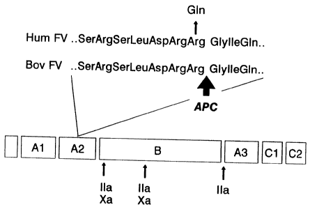
(Arg-506) (Ref. 21 and The Journal of Biological Chemistry, Vol. 262, No.
23, August 15, pp. 11233-11238, 1987, Bruce Odegaard and Kenneth Mann).
respectively.
As a first approach, ectopic transcripts of the factor V gene,
isolated from peripheral blood lymphocytes, were used for first-strand
cDNA synthesis and subsequent amplification of the two regions coding for
the APC binding and cleavage site. Direct sequencing of the PCR-fragments
revealed that two unrelated patients, classified as homozygous deficient
of APC-cofactor II, were both homozygous for a 1691, G ~ A transition
(Fig. 6). This mutation predicts the replacement of Arg-506 (CGA) by Gln
(CAA) (FV(Q506) or FV Leiden). No other sequence abnormalities were
observed in 225 by surrounding 1691 A and in 275 by around the region
coding for the putative APC binding site (Fig. 7).
If cleavage after Arg-506 is instrumental for the inactivation
of human factor Va by APC, one would predict that introduction of a Gln
in position 506 will prevent the inhibitory cleavage. During the
coagulation process plasma factor V is initially activated by factor Xa
(formation of 105/220 kDa heterodimer (ref. 37)) and next further
processed by thrombin (formation of 105/74 kDa heterodimer (Ref. 38))
(Ref. 39). Interestingly, we found that the replacement of Arg-506 by Gln
only prevents the inactivation of the Xa-activated form of factor V by
APC (Fig. 8) but not that of the thrombin-activated form (data not
shown).
The observation that two unrelated APC-resistant patients were
homozygous for the same mutation, suggested that this alteration is
present in the majority of APC-resistant patients. To investigate this
possibility a test was designed for the screening of genomic DNA for the
presence of the 1691 G ~ A transition. Because the mutation is located in
exon 10, 11 nt 51 from the start of intron 10 and only the first 8
nucleotides of intron 10 have been published (Ref. 23), more intron 10
sequence was generated by hemi-nested reverse PCR (Ref. 40) (see also
sequence 14). From this information primers were designed for the
amplification of two overlapping genomic fragments that could be used for
genotyping.
Digestion of the 267 by fragment with Mnl I was used to
WO 95/21938 29 ~ ~ ~ ~ ~ ~ PCT/EP95/00553
demonstrate the presence of a normal (1691 G) or mutated allele, while
hybridisation of the 222 by fragment with oligonucleotides specific for
the normal or mutated allele was used for the positive identification of
1691 A. Using this approach we first studied all the members of the
pedigree from Fig.3. Complete cosegregation of heterozygosity for the
1691, G ~ A transition with APC resistance ( n-APC-SR < 0.84) was
demonstrated as shown in Fig. 10 for a part of the pedigree. In addition,
4 patients (II.6, II.B, II.14, III.22) for whom no n-APC-SR was
available because they were treated with oral anticoagulants, were found
to be heterozygous.
In a previous study of 301 consecutive patients with a first
objectively confirmed episode of deep vein thrombosis and 301 age and sex
matched population controls, 64 APC-resistant thrombosis patients had
been identified (Lancet, December 18, 1993, Vol. 342, pp. 1503-1506, T.
Koster et al). These 64 patients and their 64 controls were screened for
the presence of the G~A transition. From the 128 individuals 70 had a n-
APC-SR < 0.84 (64 patients and 6 controls). Fifty six of these carried
the mutation (53 patients and 3 controls), six of the patients in both
alleles (mean n-APC-SR 0.43; range 0.41-0.44) and the other 50 in one
allele (mean n-APC-SR 0.57; range 0.50-0.67). The remaining 14 APC-
resistant individuals did not carry the mutation, and had only a
marginally reduced n-APC-SR (mean n-APC-SR 0.78; range 0.70-0.83). All 58
non APC-resistant individuals did not carry the mutation (mean n-APC-SR
0.99; range 0.83-1.19). Further none of 100 consecutive thrombosis
patients with a n-APC-SR > 0.84 was carrier of the mutation, while -as
expected- 3 of their 100 matched controls were. These 3 (n-APC-SR of
0.57, 0.58 and 0.59) were the only controls with a n-APC-SR < 0.84.
Our data demonstrate that 80x of the individuals with a n-APC
SR < 0.84 and 100x of those with a n-APC-SR < 0.70 are heterozygotes or
homozygotes for the 1691, G ~ A transition and that vice versa all
carriers of the mutation have a n-APC-SR < 0.7. The relatively high
frequency of the mutated allele in the Dutch population (about 2x)
combined with our previous finding (Lancet, December 18, 1993, Vol. 342,
T. Koster et al) that APC resistance is a common and strong risk factor
for deep vein thrombosis, makes this hereditary factor V defect the most
common hereditary blood coagulation disorder sofar.
Figure 1 and 2.
Measurement of APC-cofactor II levels in plasma
WO 95/21938 PCT/EP95/00553
Fig. 1. Calibration curve for the assay of APC-cofactor II activity in
plasma.
APC-cofactor II refers to the hypothetical new cofactor of APC
(Ref. 2) which is missing or defective in individuals with APC-
5 resistance. n-APC-SRs were measured in dilutions of normal plasma (100x
APC-cofactor II) in plasma of a patient homozygous deficient of APC-
cofactor II {Ox APC-cofactor II). The curve in Fig. 1 is the result of
nine different experiments. The classification homozygous or heterozygous
deficient of APC-cofactor II is based on the results of parentship
10 analysis for 14 probands with APC-resistance (n-APC-SR < 0.84). For 2
probands (n-APC-SR 0.38/0.41) both parents were APC-resistant (mean n-
APC-SR 0.55); for 11 probands (mean n-APC-SR 0.57) one of the parents was
APC resistant (mean n=APC-SR 0.59) while the other was not (mean n-APC-SR
0.96); for one proband (n-APC-SR 0.74) both parents were not affected (n-
15 APC-SR 0.96/0.99). We propose that individuals can be classified as
homozygotes or heterozygotes for APC-cofactor II deficiency on the basis
of their n-APC-SR (homozygotes: mean 0.40, n=2; heterozygotes: mean 0.58,
range 0.51-0.67, n=26).
20 Fig. 2. APC-cofactor II activity ZeveZs in ptasmas deficient (<57) of a
singte coagulation factor.
Plasmas were either from patients with a congenital deficiency
(a, g, f,m,g,r,s,t) or prepared by immunodepletion (b,c,d,e,j,h,i, k,l,p).
Plasmas were deficient of factor II (a), factor VII (b), factor IX (c),
25 factor X (d), factor XI (e), factor XII (j), factor XIII (g), protein C
(1), protein S (i), ~2-glycoprotein (j), antithrombin (k), factor V
(1,m), factor VIII (p,q) or von Willebrand factor (r,s,t). Factor V
deficient plasma (m) was supplemented with two different concentrations -
54x (n) and 90x (o) - of purified human factor V (Serbio, Gennevilliers,
30 France), dialyzed against 20 mM sodium citrate , 150 mM NaCl, 4 mM CaCl2
and tested for APC-cofactor II activity.
The APC-SR was calculated from the results of two APTT
measurements, one in the presence of APC and one in its absence, exactly ,
as previously described. (Lancet, December 18, 1993, Vol. 342, T. Koster
et al) The n-APC-SR was calculated by dividing the APC-SR for the test
sample by the APC-SR for pooled normal plasma. APC-cofactor II activity
was measured by reading the n-APC-SR for two different dilutions (1:1,
WO 95/21938 ~ ~ ~ ~ PCT/EP95100553
31
3:4) of the test plasma in APC-cofactor II deficient plasma on a
calibration curve as shown in Fig. 1.
Figure 3-5. Linkage analysis in a family with APC-resistance
Fig. 3. Pedigree of a family with APC-resistance (or APC-cofactor II
deficiency).
~, ~ , individuals with n-APC-SR< 0.84 (mean 0.65; range 0.59-0.71, n=13);
0, p , individuals with n-APC-SR > 0.84 (mean 1.03; range 0.87-1.29;
n=20); ~, ~lpatients treated with oral anticoagulants (measurement of n-
APC-SR in these patients is not meaningfully 0, C1 , individuals that were
not tested.
Fig. 4. Integrated genetic Ztnkage map of the q21-25 region of chromosome
1.
The relative positions of the loci APOA2, D1S104, D1S61, AT3,
LAMB and F13B were derived from the NIH/CEPH Collaborative Mapping Group
linkage map (Ref. 41). The genetic distance between adjacent loci is
given in cM. The F5 locus was placed on this map within 4 cM of the D1S61
locus by studying the segregation of markers for the F5 and DiS61 loci in
3 CEPH families informative for both markers (in 55 meioses no
recombination between these two loci was observed: Zm~ 16.6 at 0=0.00).
Fig. 5. Patrurtse Zodscores of APC resistance mtth chromosome 1 markers.
All available individuals of the pedigree in Fig. 3 were
analyzed. Oligonucleotide sequences for markers for the loci ApoA2,
D1S104, D1S61, LAMB and F13B are available from the Genome Data Bank. The
primers were obtained from the Dutch primer base. Three different
polymorphic markers for the AT3 locus were not informative in this
family. Two point linkage analysis was performed using the MLINK program
from the LINKAGE package version 5.3, which was obtained from Dr J. Ott.
Sex averaged lodscores are shown.
Methods
~ 35 Microsatellite markers for ApoA2, D1S104, D1S61, LAMB and F13B
were amplified by PCR. Conditions: 50 mM NaCl, 10 mM Tris-HC1 (pH 9.6),
10 mM MgCl2 , 0 . O1 x BSA , 200 uM dGTP , dATP and dTTP , 20 ;,iM dCTP , 0 .
7 PCi
~32P dCTP, 0.43 U Taq polymerase (fetus, Emeryville, CA, USA), 50 ng of
each primer and 30 ng genomic DNA. 27 cycles were run at 94 °C ( 1' ) ,
55
WO 95/21938 PCT/EP95/00553
32
°C (2'), 72 °C (1') with a final elongation step of 10 min. PCR
products
were separated on a 6x denaturing polyacrylamide sequence gel, after
which gels were dried and exposed to X-ray film.
F5 polymorphisms: A 636 by fragment from exon 13 of the factor
V gene (Ref. 23) was amplified by PCR using the primers number 2 (PR-766,
nt 2253-2272 (Ref. 21)) and number 3 (PR-768, nt 2870-2899 (Ref. 21)) of
the Sequence Listing. For PCR conditions see legend Fig. 10 + 11.
Restriction with Hinf I detects a C/T dimorphism at nt 2298 (C: 0.68; T:
0.32) and a rare A/G dimorphism at nt 2411 (A:0.98,; G:0.02). None of
these markers was informative in the pedigree of Fig. 3.
Fig. 6-8. Identification of the factor V gene mutation in a patient
homozygous deficient of APC-cofactor II
Fig. 6. Autoradiogram shoraing the rtucteotide substitution in a patient
classified as homoaygous deficient of APC-cofactor II.
Part of the nucleotide sequence of the non-coding strand of a
cDNA PCR-fragment (coding for aminoacids 417 through 572 in human factor
V (Ref. 21)) is shown for one patient (P) and one non APC-resistant
control (C). Arrows indicate the location of the 1691, G ~ A transition,
which predicts the replacement of Arg 506 by Gln.
Fig. 7. ScYcematic representation of the factor V moZecute.
Human factor V is a 330 kDa glycoprotein which contains several
types of internal repeats.(Ref. 21) Activation by factor Xa results in
the formation of a 105/220 kDa hetero-dimer (AlA2/B'A3C1C2) (Ref. 38),
while activation by thrombin results in the formation of a 105/74 kDa
heterodimer (AlA2/A3ClCz) (Ref. 37) . APC binds to the A3 domain of factor
Va (Ref.35,36) and inbibits bovine factor Va by cleavage in the A2 domain
after Arg-505 (The Journal of Biological Chemistry, Vol. 262, No. 23,
August 15, pp. 11233-11238, 1987, Bruce Odegaard and Kenneth Mann).
The amino acid sequences surrounding the (putative (Ref.43) APC
cleavage site in human (Arg-506) and bovine (Arg-505) factor Va are
shown. In the APC-resistant patient Arg-506 has been replaced by Gln.
Fig. 8. Resistance of factor Xa-activated factor V(Q506) to inactivation
b~ APC.
A1(OH)3-adsorbed and fibrinogen depleted plasma (2 hr 37 °C; 0.3
~J m1'1 Arvin) containing either factor V(R506) or factor V(Q506) was
WO 95/21938 33 ~ ~ ~ ~~ PCT/EP95/00553
treated with factor Xa (2 nM) in the presence of 20 mM CaCl2 and 20 uM
PS/PC (25/75). After 8 min, when factor V activation was complete, 1.9 nM
APC or buffer were added. At different time intervals 10 u1 sample was
diluted 1/100 in stop buffer (50 mM Tris-HC1, pH 7.9, 180 mM NaCl, 0.5 mg
ml-1 OVA, 5 mM CaCl2) and directly assayed for factor Va activity using
the method described by Pieters et al (Ref. 44) The factor Va activity
measured after complete activation of 0.70 U m1'1 FV(R506) (0.64 u1H
thrombin mini) or 0.49 U m1-1 FV(Q506) (0.20 NM thrombin miri 1) is
arbitrary put at 100x;
0, - APC; 1, + APC.
Methods
cDNA synthesis: RNA was isolated (Ref. 45) from the lymphocyte
fraction of 10 ml citrated blood of consenting patients and nonAPC-
resistant controls. 1 ug of RNA was used as template for first strand
cDNA synthesis in the presence of mixed random hexamers using the
superscript kit (BRL, Bethesda, Md, U.S.A.). Amplification of cDNA
fragments. The primers Sequence 4 (PR-764, nt 1421-1440 (Ref. 21)) and
Sequence 5 (PR-856, nt 1867-1891 (Ref. 21)) amplify the region coding for
residues 417 through 572 which contains the putative APC cleavage site;
the primers Sequence 6 (PR-849 nt 5608-5627 (Ref. 21)) and Sequence 7
(PR-848, nt 6040-6063 (Ref. 21) amplify the region coding for amino acid
residues 1812 through 1963, which contains the APC binding region. PCR
conditions were as described in the legend of Fig. 9 + 10 .PCR fragments
were purified on ultra low gelling temperature agarose and directly
sequenced as described before (Ref. 42) using the same primers as in the
PCR reaction. One additional primer was synthesized to aid in sequencing
of the APC-binding region:
Sequence 8(PR-847, nt 5905-5927 (Ref. 21)).
Fig, 9 + 10. Association of APC-resistance with the presence of a 1691 A
allele of factor V
Fig. 9. Cosegregatton of 1691 A with APC resistance.
The upper part gives the position of the individuals in the
pedigree (Fig. 3) and the n-APC-SR, if available (II6 is on oral
anticoagulant treatment). The middle part shows the result of the Mnl I
digestion of the 267 by PCR fragment. The lower part shows the results of
the dot blot hybridisation of the 222 by fragment with the biotinylated
WO 95/21938 PCT/EP95/00553
~ 34
oligonucleotide specific for the 1691A allele (PR 1005).
Ft9. 10. Dot blot hybridisation of the 222 by PCR fragments of 64
thombosis patients with a n-APC-SR < 0.84 and their 64 matched controls
with the biotinylated oligonucleotide specific for the 1691 A allele (PR
1005).
All patients (P) and controls (C) gave their informed consent.
Slashes denote positions of failed PCR reactions in this experiment.
Methods
Amplification of genomic fragments containing 1691 G/A. For
Mnl-I digestion a 267 by fragment was amplified using as 5' primer
Sequence 9(PR-6967;nt 1581-1602 (Ref. 21)) and as 3' primer Sequence
10(PR-990; nt 127 to -146 in intron 10}. For dot blot hybridisation a 222
by fragment was amplified using as 5' primer Sequence 11 (PR-6966, nt
1626-1647 (Ref. 21}) and as 3' primer PR-990 (Sequence 10). Conditions:
125 u1 of a mixture containing 54 mM Tris-HC1 (pH 8.8), 5.4 mM MgCl2, 5.4
uM EDTA, 13.3 mM (NH4)250," 8x DMSO, 8 mM ø-mercaptoethanol, 0.4 mg ml-1
BSA, 0.8 mM of each nucleoside triphosphate, 400 ng of each primer, 200-
500 ng DNA and 2U Taq polymerase (fetus, Emeryville, CA, USA), was
subjected to 36 cycles of 91 °C (40"), 55 °C (40"} and 71
°C (2'). The
267 by fragment (7-10 u1) was digested with 0.4 U Mnl I (Biolabs,
Cambridge, Ma, USA): the 1691 G fragment will give fragments of 67, 37
and 163 bp, while the 1691 A fragment will give fragments of 67 and 200
bp. The 222 by fragment (about 100 ng) was used for dot blot
hybridisation with biotinylated sequence specific oligonucleotides
Sequence 12 (PR 1006; nt 1682-1699 (Ref. 21)) for detection of 1691 G and
Sequence 13 (PR 1005) for detection of 1691 A. Procedures were exactly as
previously described (Ref. 46). After hybridisation stringency washing
With PR-1006 was at 53 °C, and with PR-1005 at 52 °C.
EXAMPLE 2
We investigated the risk of venous thrombosis in individuals
heterozygous and homozygous for a mutation in coagulation factor V .
(factor V Leaden) abnormality. We determined the factor V Leaden genotype
in 471 consecutive patients aged under 70 with a first objectively
confirmed deep-vein thrombosis and in 474 healthy controls. We found 85
heterozygous and 7 homozygous individuals among the cases with
WO 95/21938
PCT/EP95/00553
thrombosis, and 14 heterozygous individuals among the control subjects.
Whereas the relative risk was increased seven-fold for
heterozygous individuals, it was increased 80-fold for homozygous
individuals. These experienced their thrombosis at a much younger age (32
5 versus 44 years). The homozygous individuals were predominantly women,
with mostly blood group A.
Because of the increased risk of thrombosis with age, the
absolute risk difference is most pronounced in older patients, both for
heterozygous and homozygous individuals. For the homozygous individuals,
10 the absolute risk becomes several percent per year. This implies that
most individuals homozygous for factor V Leiden will experience at least
one thrombotic event in their lifetime.
Because of ,the high allele frequency of the mutated factor V
gene, homozygous carriers will not be extremely rare as in other types of
15 hereditary thrombophilia. It was unknown to date whether the homozygous
state confers a higher risk than the heterozygous state. We have
estimated the risk of thrombosis and the clinical features of patients
who were homozygous for Factor V Leiden. These were identified in a large
case-control study on deep-venous thrombosis (The Leiden Thrombophilia
20 Study: LETS) {Koster T, et al. Venous thrombosis due to poor
anticoagulant response to activated protein C: Leiden Thrombophilia
Study. Lancet 1993; 342: 1503-1506).
METHODS
Study design
The details of the design of LETS have been described
previously {Koster T, et al. Venous thrombosis due to poor anticoagulant
responser to activated protein C: Leiden Thrombophilia Study. Lancet
1993; 342: 1503-1506). We included consecutive patients younger than 70
years, who were referred for the out-patient monitoring of anticoagulant
treatment to the Anticogulation Clinics of Leiden, Amsterdam and
Rotterdam, after a first, objectively confirmed episode of deep-vein
thrombosis, in the absence of known malignant disorders. Patients were
seen at least six months (range 6-19 months) after the acute thrombotic
event. 90x of eligible patients were willing to take part in the study.
In addition to 474 thrombosis patients, we included 474 control subjects
who had no history of venous thromboembolism, did not suffer from known
malignancies, and were of the same sex and approximately (plus/minus 5
WO 95/9~~ ~~ PC'f/EP95/00553
years) the same age.
Data collection and laboratory analysis.
All subjects completed a standard questionnaire, which ,
contained questions about the presence of acquired risk situations in the
past, confined to a specific period prior to the index date, i.e. date of ,
the thrombotic event. As acquired risk situations we considered surgery,
hospitalization without surgery or prolonged immobilization at home (z 2
weeks), all in the year preceding the index date, and pregnancy at the
time of the index date.
Blood was collected from the antecubital vein into Sarstedt
Monovett~ tubes, containing 0.106 mmol/L trisodium citrate. High-
molecular-weight DNA 'was isolated from leucocytes and stored at 4'C. The
presence of the mutant factor V-Leiden gene (1691, G~A transition) was
determined as described. By this method we established for each patient
whether he was homozygous normal (GG), heterozygous (AG) for the factor i,'
Leiden mutation, or a homozygous carrier (AA) of this abnormality. The
technicians were at all times blinded to the status of the sample, i.e.
whether it was from a patient or a control subject. Cells for DNA
analysis were available for 471 patients and 474 controls.
Analysis and statistics.
The frequency of heterozygous and homozygous carriers of the
factor V Leiden mutation in cases and controls was compared by simple
cross-tabulation. Since in the analysis of the risk associated with the
heterozygous state sex and age did not appear to be confounding
variables, as they were not expected to be for sutosomal genetic
abnormalities, relative risk estimates for the heterozygous state were
obtained by calculation of unmatched exposure odds ratios. A 95x
confidence interval was constructed according to Woolf (Woolf B. On
estimating the relation between blood group and disease. Ann Hum Genet
1955: 19:251-253).
The risk associated with the homozygous state could not be '
estimated in this standard fashion, since no homozygous individuals were
found among the controls. Therefore, under the assumption of Hardy- '
Weinberg equilibrium (in the controls) the expected number of homozygous
individuals in a control population was calculated, and the odds ratio
was subsequently estimated in the standard fashion. The variance of the
(log) odds ratio for the homozygous state was estimated by a modification
WO 95/21938 ~ ~ ~ PCT/EP95/00553
37
of the method of Woolf. When each cell of the two-by-two table with cell
contents a, b, c and d is considered to be the realisation of a Poisson
distribution, the variance of the log(OR) is 1/a + 1/b +1/c + 1/d (Woolf
B. On estimating the relation between blood group and disease. Ann Hum
Genet 1955; 19:251-253)~ When the number of individuals with GG and AA
genotypes are counted in the cases and calculated from Hardy-Weinberg
equilibrium for the controls, which requires a quadratic transformation,
the variance of the log(OR) becomes 1/AA (cases) + 1/GG (cases) + 4/A
(controls) + 4/A (control) + 4/G (controls), in which AA and GG are the
number of genotypes (individuals), and A and G the number of alleles.
The absolute risk for thrombosis for the various genotypes and
ages was calculated by first partitioning the number of person-years in
the origin population (as derived from information from the municipal
authorities) under the assumption of Hardy-Weinberg equilibrium. Dividing
the cases in each subgroup (genotype, age) by these person-years, leads
to estimates of the absolute risks. Subsequently, these crude incidence
data were modelled after logarithmic transformation in a weighted least
square regression model, with three age classes (0-29: 25 yrs: 30-49: 40
yrs; 50-69: 60 yrs), indicator var~.ables for the heterozygous (0.1) and
the homozygous state (0,1), weighted for the number of cases in each
stratum. This method in which stratum-specific incidence rates are first
estimated and then smoothed ('smooth-last [Greenland S. Multivariate
estimation of exposure-sepcific incidence from case-control studies. J.
Chron. Dis. 1981; 34: 445-453]) by weighted least square regression has
been described by Grizzle et al (Grizzle JE, Starmer CF, Koch GG.
Analysis of categorical data by linear models. Biometrics 1969; 25: 489-
504). Since for a Poisson distribution the variance of the number of
cases equals the number of cases, this is almost identical to fitting a
Poisson regression model. This model will lead to more stable estimates,
especially for the homozygous state, under the assumption that the
incidence rate ratio for the homozygous state (and the heterozygous
state) is constant over the age strata for the log (incidence rate). This
' model can be written as:
Log(I) - a + B1 ~ age + B2 *' AG(0,1) + B3 *' AA(0,1), which can
' 35 subsequently be used to calculate estimates for the absolute risk and for
the relative risk (as the antilogarithm of the coefficients).
RESULTS
WO 95121938 PCT/EP95/00553
3$
Among 471 patients, we found 85x (18x) who were heterozygous
and seven (1,5x) who were homozygous for the defect, whereas the other
379 (80x) did not carry the Factor V Leiden mutation. Among the 474
controls, 14 (2.9x) were heterozygous and all other 460 were normal: ~
there were no homozygous individuals among the controls.
The homozygous individuals experienced thrombosis at a markedly
younger age than the other patients: the median age at thrombosis was 32
years, as compared to 44 years in the heterozygous, and 46 years in the
patients without the mutation (table 2).
The clinical course of the deep vein thrombosis in the
homozygous patients was unremarkable. All suffered from deep venous
thrombosis of the leg. Four were briefly hospitalised for heparinisation,
three were treated as out-patients with cumarin derivatives only. None of
the seven patients had a history of overt arterial disease (myocardial
infarction, stroke or peripheral arterial disease) (table 3).
Six (86x) of the seven homozygous patients were women, as
compared to 46 (54x) of the heterozygous and 217 (57x) of the individuals
without the mutation. Also, six of these seven patients had blood group
A, as compared to 249 (54x) of the other 464 cases. Of the five
homozygous women of 45 years and younger, three used oral contraceptives
at the time of the thrombotic event, which was similar to current use in
all cases. Since non-0 blood group and use of oral contraceptives are in
themselves risk factors for venous thrombosis, these figures indicate an
interaction between these risk factors and homozygous factor V Leiden
which is of a multiplicative nature.
In two (29x) of the seven homozygous patients there had been a
predisposing factor for thrombosis in the year preceding the event (one
had hip surgery 45 days prior to the thrombosis, and one had been
admitted to hospital overnight after giving birth 60 days prior to the
thrombotic event). Among the 85 heterozygous individuals an acquired risk
factor had been present in 25 (29x) patients, and among the normal
patients in 131 (35x) of 379)~
Previous risk situations (operations, pregnancies, hospital
admissions) without thrombotic consequences were less frequent in the
patients homozygous for factor V Leiden than in the other patients. ~
Still, five of the seven homozygous patients had encountered risk
situations in the past without a subsequent thrombosis (two had had
surgery, four had given birth to five children).
The seven patients were followed on average for two years
WO 95/21938 PCT/EP95/00553
39
without long-term oral anticoagulation after the first thrombotic event.
One patient had a recurrent thrombosis (1/13.4 yr: 7.4 percent per year).
Of the 14 parents, three had a history of venous thrombosis, which is
approximately five times higher than expected.
Under Hardy-Weinberg equilibrium, the relative frequency of
normals : heterozygotes : homozygotes is p2 : 2 pg : q2, in which p is the
allele frequency of the normal gene and q of the abnormal gene. Since
p2 : 2pg was 460/474 . 14/474, it follows that the allele frequency of
factor V Leiden (q) is 0Ø15. The allele frequencies of p = 0.985 and q
- 0.015 conform to a distribution among 474 unselected individuals of
459.9 (GG), 14.0 (AG) and 0.107 (AA).
The expected number of homozygous individuals (q2) of 0.107
among 474 controls leads to an odds ration for the homozygous state of
(7/379) / (0.107/460) - 79. So, the risk of thrombosis for homozygous
individuals is almost eighty times increased compared to normal
individuals (CI95x: 22 to 289).
Table 4 shows the odds ratio for the three age groups, when the
allele frequency of 0.015 is used to calculate the expected number of
homozygous controls in each age group. It is evident that the high
relative risk of thrombosis in homozygous individuals diminishes with
advancing age. This is in contrast to the relative risk for heterozygous
individuals, which is more or less constant over age.
Subsequently, we calculated absolute risks (incidence rates)
for the different age groups and genotypes, by using data on the age
distribution in the origin population, and under the assumption of Hardy
Weinberg equilibrium. As is shown in table 4, the incidence increases
from only 0.55 per 10,000 per year in the youngest age group with GG
genotype, to 16.3 per 10,000/year for heterozygous individuals in the
older age groups. It is also clear from the figures in table 4, that at
all age groups the risk for homozygous individuals is much higher than
for heterozygous individuals (78 to 176 per 10,000 per year). However,
since these figures are based on only seven individuals divided over
three age groups, the estimates are unstable, and show an unexpected
lower incidence in the oldest age group. The regression model we used
smooths these estimate, since it assumes a constant relative risk over
the age groups. As figure 12 shows, this model fits excellently for the
normal and heterozygous individuals (coefficients: constant: -10.06,
age: .0293, AG: 1.96, AA: 4.52). The smoothed incidence estimates for
homozygous individuals now increase from 82 per 10,000 person-years in
WO 95/21938 PCTlEP95100553
those aged under 30, to 227 per 10,000 patients-years for those aged 50-
69 (figure 13). These estimates imply that the most homozygous patients
will experience at least one thrombotic event in their lifetime.
5 DISCUSSION
Resistance to APC is a common abnormality with an allele
frequency for the mutant factor V gene of about 1.5 percent. This implies
that three percent of the population is heterozygous, and homozygous
10 individuals can be expected with a prevalence of about two per 10,000
births.
In this study we show that homozygous individuals have a high
risk of thrombosis, which is also considerably higher than the risk of
heterozygous individuals. This conclusion finds support in the young age
15 at which the homozygous individuals experienced their first thrombotic
event.
- It is clear that the risk of thrombosis in homozygous factor V
Leaden is nowhere near the risk of thrombosis in homozygous protein C or
protein S deficiency; these abnormalities lead to neonatal purpura
20 fulminans (Branson HE, et al. Inherited protein C deficiency and
coumarin-responsive chronic relapsing purpura fulminans in a newborn
infant. Lancet 1983; ii: 1165; Mahassndana C, et al. Neonatal purpura
fulminans associated with homozygous protein S deficiency. Lancet 1990;
335: 61-62). All of the individuals with homozygous factor V Leaden lived
25 until adulthood before the first thrombotic event, and one even until
late middle age. Most of the homozygous individuals had experienced risk
situations in the past without thrombosis, for most of which (pregnancy,
puerperium) no anticoagulant prophylaxis will have been prescribed. This
shows that APC-resistance should be seen as a quantitative defect
30 (decreased inactivation rate of factor Va) rather than a qualitative
defect (no protein C activity) as in homozygous protein C deficiency.
A remarkable finding in this study was the predominance of
women among the homozygous patients. Since among these women the use of
oral contraceptives was as prevalent as among the other cases, it is
35 likely that this use played a role by a synergistic effect with APC-
resistance. Since both pill use and APC-resistance are common, further
studies should investigate this association, especially for the
heterozygous carriers (3 percent of all women).
The relative risk for heterozygous individuals appears constant
WO 95/21938 PCT/EP95/00553
4 2~.~~~~'
for the different age groups. This observation has to be seen in the
light of a background incidence that increases with age. This implies, as
we showed in figures 12 and 13 that the absolute risk of thrombosis, or
the absolute risk added by APC-resistance, becomes substantial for older
heterozygous individuals.
It may be noted that our overall estimate for the incidence
rate, at about 2 per 10,000 per year, is lower than the usual estimates
of about 0.5 to 1 per 1000 person-year (Branson HE, et al. Inherited
protein C deficiency and coumarin-responsive chronic relapsing purpura
fulminans in a newborn infant. Lancet 1983; ii: 1165; Koster T, More
objective diagnoses of venous thromboembolism Neth. J. Med. 1991; 38:
246-248). This is most easily explained by the age limits in our study
(<~0 years), by the restriction to confirmed thromboses, by the exclusion
of patients with malignancies and by the restriction to first thrombotic
events.
The homozygous patients had a risk of thrombosis that was
eighty times increased, which leads to an overall incidence of about 1
percent per year. The observed decrease of the rate in the older age
groups, may be explained by a scarcity of individuals of that age in the
population who had not already experienced a first thrombotic event. It
may also have been the result of the small number of homozygous patients
(i.e. only one in the oldest age group). In both instances, the incidence
figures that were recalculated from the weighted regression model seem
the best estimate of the risk, which becomes over two percent per year in
patients aged 50 and older.
We conclude that APC-resistance caused by homozygous factor V
Leiden leads to a high risk of deep venous thrombosis. This thrombosis
appears not to occur before aldulthood, and even does not invariably
become apparent a.n risk situations such as pregnancy and puerperium.
Therefore, although we are convinced that these patients should receive
short-term prophylaxis with anticoagulants in risk situations. Life-long
prophylaxis in individuals homozygous for factor V Leiden may however not
necessarily be required.
WO 95/21938 PCT/EP95/00553
42
Table 2
General characteristics of 471 thrombosis patients by factor V genotype
GG AG AA
n 379 85 7
,
Age
median (yr) 46 44 31
range (yr-yr) 15-69 17-69 22-55
Sex
men (x) 162 (43) 39 (46) 1 (14)
women (x) ~ 217 (57) 46 (54) 6 (86)
GG : homozygous normal factor V
AG : heterozygous for factor V Leiden
AA : homozygous for factor V Leiden
WO 95/21938 4~ ~ ~ ~ ~ ~ PCT/EP95/00553
c
~ a~ o
a~ ~ x x x x x z z c~
c
~ b o
x
.
c~
c ~o
a~
0 0 0 0
a a
N
.u ed c
c
1~ c
' p ~ x x x z x
oao ~ ~ ~ ~ o
r' o " o
a
c s s
x ~ x
Z ~ ~ Z > a ,x, o ~ ~ m
.~ c o .aa .c
~ ~ a~~ ~ M
~i 8 d d d O d d d V 8 .c Gr
y
D ~ O ~ O O
~ .~~ O .~
m y M
U fx M ~ O d M O .,'~-1s0O ~ O hp
-1 ~ a p ~ ~ 0
r1 pV, '' ~ N v-1N N r-1~ Q U 4!
M d ; r1 v-i.-1v-ir-1r1 '~'~ .e X0.1~ O
~ C t0 r
T
m .~ ~.m .~'cv w
M ~ t~.~as
N M N ~ ~ M a
.~ ~ ~ ~ m
' ~
1
d ~
.C y ~ O.
W t 4r4~ 7
~ 4 t. tr4 g' O ,~ O O O j
O
O 0 O G ~
m 0 t~.~. ~,~0'1
pp O 0 tl,~ c C,O
~ c .c
00
C
d
M
r ~ ~ ~ N n ~ ~
O l 1 11
r- r1t
N !~'1a tt1vG
P
WO 95/21938 PCT/EP95/00553
~ 44
a
s~
a
a~
O d .~ O ~ '
N
.-i d ~ N r-~i
.-i
O
O
r~
~ It1 ~ 3
~~-I ~
'
~
.r
t W t
o0 N x
d O .-1
N O
.-1 O~ O
M Ov O r~ 4j
~ l~
to M ~ N ~ 8 ~
w
Ga
O ~ O ~ O O
~ I~ ~-i d0 m
111 III~ N O
b
ri ~ CO ~ O O 6 8
_V O ~ V O .~
~ ~ ~
a If1 O ~ O it
p O N a0 ~ ~ ~ -girl
O vD
L~
L>i ~ G0
O~ O
1~f1 ~ O O ~-1 0
00 N ~ ~ ~ .~ to
w
M ~ ~ ~ ~ b
O .~ D O N
M
~ Q1 ~ '~ O
'b 0 It1
0
~ O
O
N O ~
1 a LOC1 ~ N 11 ~ ~ m
W O O O ~ ~ O ~ O 0
~ N
N
U7 O 6 N C
~ O r1O O
.C .L",
b
d M \ \ ~ 4.0r O U O ~
O \ \ L~ M ~ ~ t~.,G .d
U ~ ~ N .-~~G ~ O ~ V C
O
M O
d ~t r1 N ~o ~ ~:-,'TiO
d N \ W ~ c~ to O '"1
o u~ i~b
it
m
d ~ v v G7. o 's7k O :a
c \ \ vD N ~ TS ~ ~ U R,
O ~D O
~ p
O p~ .. .. .. " ..
6 ~ O
N O 4 C7
t!1W U
O M O O ~
tf1 O
WO 95/21938 ~ ~ °~ PCT/EP95I00553
Figures 12 and 13.
Crude (fig. 12) and smoothed (fig. 13) incidence rates estimates for
factor V Leiden genotypes by age.
The lowest line shows the estimates for the GG genotype, and the upper
5 line for the AG genotype (in the figure). Figure 13 also shows the
estimates for the AA genotype (homozygous factor V Leiden). Crude
incidence estimates are indicated by +, whereas the smoothed rates are
indicated by 0. The smoothed incidence rates per 10,000 person-years
were , for GG : 0 . 9 ( 0-20 yr ) , 1. 4 ( 30-49 yr ) and 2 . 5 ( 50-69 yr ) ;
for AG
10 6.3 (0-29 yr), 9.8 (30-49 yr), 17.6 (50-69 yr); for AA: 81.5 (0-29 yr),
126.5 (30-49 yr) and 227.3 (50-69 yr).
EXAMPLE 3
P~asmids and in vitro RNAS
RNA isolated from PBMCs of a healthy person (homozygous wild-
type) and of two patients, ID90 and ID137 (homozygous mutant, both) was
obtained from the Hemostatis and Thrombosis Research Centre, Leiden, the
Netherlands. Fragments of 297 nt encompassing the mutation at position
amino acid 506 were cloned in the vector pG30 using the restriction
enzymes EcoRI and Csp451. The re8ulting plasmids were named pG30/FVwt and
pG30/FVmut for wild-type and mutant clones respectively.
Cloning of the correct sequence was confirmed by sequence
analysis and subsequently the plasmids were purified by CsCl gradient for
in vitro RNA synthesis. Using plasmid pG30/FVwt as a source a system
control plasmid (pG30/FV E2) was constructed by deletion of the probe
sequence (21 nt) and insertion of the E2 sequence (144 nt). The 3
plasmids described above were used for in vitro RNA transcription using
T'7 RNA polymerase in the standard protocol. The plasmids were linearized
with BamHl which, after transcription with T7 RNAP, would result in RNAs
consisting of 297 nt and 420 nt of respectively the wt, mut and system
control clones followed by 700 nt of vector sequence, so that the overall
length of the in vitro RNA would be approximately 1 kb. After in vitro
transcription the RNA was treated with DNase I, purified using the Tip
100 column (Qiagen) protocol and quantitated spectophotometrically.
Appropriate serial dilutions were made in water and the in vitro RNAs
were stored at -70°C.
primers an probes.
WO 95/21938 ~~ ~ PCT/EP95/00553
46
The sequences of the NASBA amplification primers and detection
probes for ELGA and ECL detection are given in table 5.
Table 5: primers and probes for Factor V NASBA
Name Sequence Length Remarks
(nt)
P1 5'ATT TCTAAT ACGACT TAT AGG
CAC
.,AAA 47
GGT
ACC
AGC
TTT
TGT
TCT
CA
3'
(SEQ. ID.N0. 15)
P2 5'AGT GCTTAA CAAGAC ACT A 22
CAT 3'
(SEQ. ID.N0. 16)
Generic 5' 21 HRP-labelled
TGA
CGT
GGA
CAT
CAT
GAG
AGA
3'
ELGA-probe (SEQ. ID.N0. 1'7)
Generic 5'CAG CAGGCT GTGTTT GTG 3' 21 ECL-labelled
GCT
ECL-probe (SEQ. ID.N0. 18)
WT-probe 5'CTG GACAGG CGAGGA CAG 3' 21 HRP-labelled
ATA
(SEQ. ID.No. 19) or biotin-
labelled
Mut-probe 5'CTG GACAGG CAAGGA CAG 3' 21 HRP-labelled
ATA
(SEQ. ID.N0. 20) or biotin-
labelled
SC-probe 5'GAC ACCAAG GAAGCT GAC3' 21 biotin-
TTA
(SEQ. ID.N0. 21) labelled
a. The underlined part in the P1 sequence is the T~ RNAP promoter sequence.
The P1 is located in exon 10 of the Factor V coding sequence,
while the P2 sequence is located in exon 11. As a result this primer set
can only amplify mRNA sequences from which the intron 10 sequence is
removed by splicing. Due to better performance in either ELGA or ECL
there are two generic probes. However, it should be possible to choose
one generic probe for both ELGA and ECL. The amplification primers were
purified on 20x acryl amide, 7M urea slab gels. After elution and EtOH
precipitation the primers were dissolved in 500 u1 H20 and the
WO 95/21938 ~ , ~; ~ ~, : PCTIEP95/00553
47
concentration determined by spectophotometry (0D260).
Biotin oligos were made on the synthesizer and used after EtOH
precipitation and dissolving in H20. Coi:pling the HRP label to NH2-oligos
was done according to the standard protocol and the probe was used
without further purification (generic ELGA probe) or purified on a slab
gel (wild-type and mutant specific ELGA probes). The ECL oligos were
synthesized and used without further purification.
Nucleic acid isolation.
All nucleic acid isolations were performed using the method
described by Boom et al. (1990, J. Clin. Microbiol 28: 495-503). Nucleic
acid was extracted from 100 u1 whole blood (see clinical samples) and
elution was in 100 u1 H20, typically 5 u1 of eluate was used as input for
NASBA amplification. The remainder of the eluate was stored at -70°C.
NASBA amplifications.
NASBA amplifications were performed as follows . To 5 u1 of RNA
18 u1 of a premix solution was added that consisted of : 10 u1 2. 5 x NRG
buffer (final concentration in 1 x buffer: 40 mM tris, pH = 8.5, 70 mM
KCl, 1 mM each dNTP, 2 mM ATP/CTP/UTP, 1.5 mM GTP, 0.5 mM ITP, 12 mM
MgCl2), 6.25 u1 4 x primer mix (final concentration in 1 x buffer: 15x
v/v DMSO, 0.2 ~xM Pl and 0.2 uM P2) and 1.75 u1 H20. The sample was
incubated at 65°C for 5 minutes and subsequently incubated at
41°C for 5
minutes. Leaving the tubes as much as possible at 41°C 2 u1 enzyme mix
8 units AMV-RT, 40 units T7 RNAP, 0.1 unit E. cola RNase H, 2.6 ug BSA,
1.5 M sorbitol) was added, followed by gentle mixing (i.e. tapping) and
incubation at 41°C for 90 minutes.
ELGA detection.
For ELGA detection 3 different probe solutions were used
containing a generic probe, wild-type probe and mutant probe
respectively. In order to increase the specificity of the wild-type and
mutant HRP labelled probes, these labelled probes were mixed with their
counterpart non-labelled probe (see table 6).
WO 95/21938 PCT/EP95/00553
48
Table 6
Ratios of labelled and non-labelled probes to increase the specificity of
hybridization for ELGA detection
Amplificate to HRP-labelled probe non-labelled probe
detect (molec/hyb) , (molec/hyb)
Generic) ELGA Generic (2x101o) -
Wild-type Wild-type (2x11)b) Mutant (5x10¹³)
Mutant Mutant (2x10¹¹)b) Wild-type (2x10¹³)
a. This includes: wild-type, mutant and system control amplificates
b. Due to the purification process (slab gel) the specific activity of the
wild-type and mutant HRP
probes is lower than normal.
After amplification 1 u1 of amplificate was added to 4 u1 of
appropriate probe mix (final concentration in 5 u1: 1 x SSC, BFB, XCFF,
5x v/v glycerol and the appropriate probes, see table 2), mixed and
incubated for 15 minutes at 45°C. Subsequently 2.5 u1 of the sample was
analyzed on an acrylamide gel (5x acryl/bisacryl, 0.04 dextrane
sulphate, NASBA elfo buffer = 25 mM tris, 25 mM boric acid, 500 11M EDTA,
pH = 8.3) run at 150 V in 0.5 x NASBA elfo buffer. After electrophoresis
the gel was stained using the standard TMB/UP substrate solutions (mixed
at 1:1 ratio) for approximately 6 minutes. Usually the gels were fixed in
50x methanol (0/N) and air dried between 2 sheets of transparent foil.
ECI~ detection.
For ECL also 3 different probe solutions were used for
detection of amplificate (see table 7)~
WO 95/21938 ~ ~ ~ ~ PCTIEP95/00553
49
Table ~
Ratios of labelled and non-labelled probes to increase the specificity of
hybridization for ECL detection.
Amplificate ECL-labelled Biotin capture Non-labelled
to detect probe (molec/hyb)probe (molec/hyb)probe
(molec/hyb)
Wild-type ECL generic Wild-type Mutant
(2X10¹²) (2x10¹²)a) (2X10¹³)
Mutant ECL generic Mutant Wild-type
(2x10¹²) (2X10¹²)) (8x10¹²)
System control ECL generic SC
(2x10¹²) (2X10¹²) -
a. Streptavidin coated beads for 100 hybridisation (200 p1 Dynal beads)
reactions were loaded with
2x10~~ molecules biotin capture probes.
To set up the hybridisation reaction 10 u1 ECL mix (0.1x w/v
BSA, 12.5 x SSC, 2x10¹² molecules ECL generic probe), 10 u1 bead mix (0.1
w/v BSA, lxPBS, 2 u1 appropriate bead solution and the appropriate non-
labelled probe) and 5 yll 21 fold diluted (in water) amplificate were
mixed and incubated for 30 minutes at 45°C under constant shaking
in a
stove. Subsequently 300 111 ECL assay buffer was added and the tubes
placed in the ECL instrument for reading of ECL signals.
RESULTS
Sensitivity.
The primers used for NASBA amplification of the Factor V mRNA
generate an amplificate of 182 nt long for the wild-type and mutant
sequence. When the primers are used for amplification of the system
control (SC) in vitro RNA the result is an amplimer of 305 nt in length.
The sensitivity of the amplification was investigated using serial
dilutions of in vitro generated wild-type, mutant-and SC RNA (Table 8).
WO 95/21938 PCT/EP95/00553
Table 8
Sensitivity of the Factor V mRNA NASBA using ELGA detection with the
generic ELGA probe
5
Input RNA Amount ELGA
(molecules) results
Wild-type 104 +
103 +
102 +
10 ~ 101 -
10 -
Mutant 104 +
103 +
~5 102 +
101 +
10 -
SC 104 +
103 +
102 +
101 t
10 -
20
For analytical
all sensitivity
3 is
input at
RNAs least
the 100
molecules. an
The input
reactions of
with 10
molecules
are
occasionally
positive, that sensitivity
indicating the is
actually
between
10
and
100
molecules.
EXAMPLE 4
The methods used are as described for example 3.
In order to determine the amount of SC RNA that should be
spiked in the nucleic acid isolation without competing with the wild-type
of mutant RNA when present, several amounts of SC RNA were analyzed.
These SC RNA amounts were isolated without addition of sample, with the
WO 95/21938 ~ PCT/EP95/00553
51
addition of 100 u1 whole blood and as a control a dilution series of SC
RNA was directly amplified. The results of ELGA analysis after
amplification are depicted in table 5. Apparently there is some loss of
nucleic acid during nucleic acid isolation (compare lanes 3 with and
without isolation, A and C, respectively).
From the A series (table 9) it can be concluded that the
minimum amount of SC RNA that should be spiked in the lysisbuffer is
1x10⁵ molecules. This amount of SC RNA is not inhibitory for the
amplification of wild-type or mutant RNA isolated from 100 u1 whole
blood. In fact, even when 10 times more SC RNA is used, this is not
inhibitory for wild-type or mutant RNA isolated from 100 u1 whole blood
(table 5, B series). In all further experiments, when appropriate, 105
molecules of SC RNA'were spiked in the lysisbuffer before nucleic acid
isolation.
Table 9. ELGA results of amplification of different amount of System
control RNA spiked before nucleic acid isolation
Input Set-up SC signal WT/Mut
signal
1 A + -
2 A + -
A _ _
4 A - -
1 B - +
2 B - +
3 B _
4 B -
1 c + _
2 C + _
3 C + -
4 C - -
ELGA results of amplification of diffcrent amount of CS RNA spiked before
nucleic acid isolation
A. Nucleic acid isolation without addition of sample
B. Nucleic acid isolation with 100 u1 whole blood
C. control, direct amplification of SC RNA
WO 95/21938 PCT/EP95/00553
52
1. 1x10⁶ molecules SC RNA in lysis buffer (: 5x10⁴ per amplification)
2. 1x10⁴ molecules SC RNA in lysisbuffer (x 5x10³ per amplification)
3. 1x10⁴ molecules SC RNA in lysiabuffer (t 5x101 per amplification)
The probe used for hybridisation was the generic ELGA probe.
EXAMPLE 5
The methods used are as described in example 3
Due to the nature of the mutation in the Factor V mRNA, a G->A
single base mutation, it is expected that the wild-type probe will give
considerable background signal on mutant amplificate and vice versa. This
will be the case for both ELGA and ECL detection. In order to avoid
complicated hybridization protocols the labelled probes are mixed with
their non-labelled counterpart to suppress background hybridization on
the non-homologous amplificate. In table 10 the results of specific
detection of wild-type and mutant amplificates with probe mixtures are
depicted, using ELGA detection.
Table 10.
ELGA detection of wild-type mutant amplificates with specific wild-type
(WT) and mutant (Mut) probes labelled with HRP.
Labelled Non-labelledExcess non- WT-signal Mut signal
probe probe labelled probe
WT Mut 1 ++ ++
5 ++ ++
10 ++ +
loo ++ t
250 ++ -
500 + _
Mut WT 1 + ++
5 + ++
to t ++
100 - ++
250 _ t ,
WT = wild-type HRP-labelled probe ; Mut = mutant HRP-labelled probe
It is apparent that in case of the wild-type HRP labelled probe
a 250 fold excess of non-labelled mutant probe should be added to reduce
WO 95!21938
PCT/EP95/00553
53
the background sufficiently. When the HRP-labelled mutant probe is used,
an excess of 100 fold non-labelled wild-type probe is sufficient to
reduce the background to an acceptable level. A more or less identical
experiment was performed using ECL detection. In the ECL method the non-
labelled probe has to compete with the specific biotinylated capture
probe on the magnetic bead. The result of the ECL detection using
different excess non-labelled probe ratios is depicted in Table 11.
Table 11 ECL detection of wild-type and mutant smplificates with
specific wild-type (WT) and mutant (Mut) probes.
Labelled Non- ~ Excess non- WT-signal Mut signal
probe labelled labelled (x1000) (x1000)
probe probe
WT Mut 0 620 250
1 250 20
2.5 300 1
5 200 1
to loo 0
Mut WT 0 300 600
1 2 200
2.5 3 ioo
5 0 80
to 0 30
Vf = wild-type HRP-labelled probe ; Mvt = mutant HRP-labelled probe
For subsequent ECL detection an excess of 10 fold non-labelled
mutant probe with biotinylated wild-type probe on beads and an excess of
4 fold non-labelled wild-type probe with biotinylated mutant probe on
beads was used. The differences between amount of non-labelled probe that
has to be added for ELGA and ECL detection has to do with the
- hybridization formats. In the ECL format the specific probe is bound to
the magnetic bead and therefore will have slower hybridization kinetics
compared to probes in solution. As a result a relatively small excess of
in solution non-labelled probe has to be added. In the ELGA the
competition takes place between 2 probes in solution, which makes it
WO 95!21938 PCT/EP95/00553
54
necessary to add a relatively high amount of non-labelled probe.
WO 95/21938
PCT/EP95/00553
References
1. Hirsh, J., Hull, R., Raskob, G.E. Epidemiology and pathogenesis of
venous thrombosis. J. Am. CoZZ. CardioZ. 1986; 8:104B-113B
5 2. Dahlbgck, B., Carlsson, M., Svensson, P.J. Proc. Natt. Acad.Sci. 1993;
90:1004-8
3. Clouse, L.H., Comp, P.C. New Engt. J. fled. 1986; 314:1298-1304
10 4. Esmon, C.T., Protein-C: biochemistry, physiology, and clinical
implications. Btood 1993; 62:1155-1158
5. Bakker, H.M., Tans, G., Janssens-Claessen, T., Thomassen, M.C.L.G.D.,
Hemker, H.C., Griffin, J.H., Rosing, J. Eur. J. Biochem. 1992; 208:171
15 1786
6. Koedam, J.A., Meijers, J.C.M., Sixma, J.J., Bouma, B.N. J. Ctin.
Invest. 1988; 82:1236-1243
20 7. Svensson, P.J., Dahlb~ck, B. Thromb. Haemostas. 1993; 69:1252
8. Svensson, P.J., Dahlb~ck, B., Thromb. Haemostas. 1993; 69:999
9. DahlbKck B., Malm J., Thromb. Haemostas. 1993; 69:529
10. Walker, F.J., Sexxton, P.W. & Esmon, C.J. Biochim. Biophys. Acta
1979 571: 333-342
11. Boyer-Neumann, C., Bertina, R.M., Tripodi, A., D'Angelo A., Wolf M.,
Vigano D'Angelo, S., Mannucci, P.M., Meyer, D., Larrieu, M.J. Comparison
of functional assays for protein S; European collaborative study of
outpatients with congenital and acquired deficiency. Thromb. Haemostas.
' 1993 70(6):946-950.
12. Canfield, W., Nesheim, M., Kisiel, W. & Mann, K.G. Circulation. 197$;
58: II-210.
13. Suzuki, K1, Stenflo, J., Dahlbgck, B & Teodorsson, B. J. Biol. Chem.
1983; 258: 1914-1920.
WO 95/21938 PCT/EP95/00553
56
14. Tracy, P.B., Nesheim, M.E., and Mann, K.G. J. Biol. Chem. 1983; 258:
662-669.
15. Miletich, J., Sherman, L., Broze, G., N. Engt. J. Med. 1987; 317:991-
996
16. Miletich, J.P., Prescott, S.M., White R., Majerus, P.W., Bovill,
E.G., CeZt 1993; 72:477-480
17. Allaart, C.F., Poort, S.R., Rosendaal, F.R., Reitsma, P.H., Bertina,
R.M., Brit, E., Lancet 1993; 341:134-138
18. Heijboer, H., Brandjes, D.P.M., Btiller, H.R., Sturk, A., ten Cate,
J.W., N. EngZ. J. Med. 1990; 323:1512-1516
19. Potzsch, B., Kawamura, H., Preissner, K.T., Schmidt, M., Seelig, C.,
Muller-Berghaus, G. Btood 1992; 80:267A (Abstr.)
20. Kane, W.H., Davie, E.W. Proc. NatZ. Acad. Sci. USA 1986; 83:6800
21. Jenny, J.R., Pittman, D.D., Toole, J.J., Kriz, R.W., Aldape, R.A.,
Hewick, R.M., Kaufman, R.J., Mann, K.G. Proc. Natt. Acad. Scf. USA 1987;
84:4846
22. Kane, W.H., Ichinose, A., Hagen, F.S. and Davie, E.W. BiochemistrU
1987; 26:6508-6514
23. Cripe, L.D., Moore, K.D., and Kane, W.H. Biochemistrg 1992; 31:3777-
3785
24. Fass, D.N., Hewick, R.M., Knutson, G.J., Nesheim, M.E., and Mann,
K.G. Proc. NatZ. Acad. Sci. USA 1985; 82:1688-1691
25. Toole, J.J., Knopt, J.L., Wozney, J.M., Sultzman, L.A., Buecker,
J.L., Pittman, D.D., Kaufman, R.J., Brown, E., Shoemaker, C., Orr, E.C., '
Amphlett, G.W., Foster, W. B., Coe, M.L., Knutson, G.J., Fass, D.N., and
Hewick, R.M. Nature 1984; 312:342-347
26. Church, W.R., Jernigan, R.L., Toole, J., Hewick, R.M., Knopf, J.,
WO 95/21938 ~ PCT/EP95/04553
57
Knutson, G.J., Nesheim, M.E., Mann, K.G., and Fass, D.N. Proc. NatZ.
Acaa. sci. vsA 1984; 81:6934-6937
. 27. Esmon, C.T. Arteriosct. and Thromb. 1992; 12:135-145
28. Walker, F.J. & Fay P.J. FASEB. J. 1992; 6:2561-2567
29. Walker, F.J. J. BioZ. Chem. 1980; 255:5521-5524
30. Engesser, L., Broekmans, A.W., Brit, E., Brommer, E.J.P. & Bertina,
R.M. Ann. Int. Med. 1987; 106:677-682
31. Hirsh, J., Piovella, F. & Pini, M. Am. J. Med. 1989; 87:345-385
32. Allaart, C.F. & Brit, E. In: Haemostasis and Thrombosis (Eds. Bloom,
A.L., Forbes, C.D. & Thomas, D.P. & Tuddenham E.G.D.) 1349-1360
(Churchill Livingstone London, 1994).
33~ Wang, H., Riddell, D.C., Quinto, E.R. & MacGillivray, R.T.A. Genomics
1988; 2:324
34. Kalafatis, M. Rand, M.D., Jenny, R.J., Ehrlich, Y.M. & Mann, K.G.,
Blood 1993; 81: 704-719
35. Walker, F.J., Scandella, D. & Fay, P.J. J. Biot. Chem. 1990;
265:1484-1489
36. Walker, F.J. & Fay, P.J., J. BioZ. Chem. 1990; 265:1834-1836
37~ Suzuki, K., Dahlb~ck, B & Stenflo, J. J. Biot. Chem. 1982; 257:6556-
6564
38. Monkovic, D.D. & Tracey, P.B. BiochemistrN 1990; 29:1118-1128 (1990).
' 35 39~ Yang, K.J., Blajchman, M.A., Craven, S., Smith, L.M., Anvari, N. &
Ofosu, F.A. Biochem J. 1990; 272:399-406
40. Triglia, T., Peterson, M.G., & Kemp, D.J. Nuct. Acids Res. 1988;
16:8186
WO 95/21938 PCT/EP95/00553
58
41. NIH/CEPH collaborative mapping group. Sctercce 1992: 258;67-86
42. Reitsma, P.H., Poort, S.R., Allaart, C.F., Brit, E. & Bertina, R.M.
BZood 1991; 78: 890-894
43. Guinto E.R., Esmon C.T., Mann K.G. & MacGillivray R.T.A. J. B~oZ. ,
Chem.. 1992; 267:2971-2978
44. Pieters J. & Lindhout, T. Btood 1988; 72:2048-2052
45. Chromczynski, P. & Sacchi, N. Anal. Btochem. 1987; 162:156-159
46. Verduyn, W., Doxiadis, I.I.N., Anholts J., Drabbels, J.J.M., Naipal,
A., D'Amaro, J., Persyn, G.G., Giphart, M.J. & Schreuder, M.Th. Hum.
Immunot. 1993; 37: 59-67
47. Faioni, E.M., Franchi, F., Asti, D., Sacchi, E., Bernardi, F., &
Mannucci, P.M. Thromb. Haemostas. 1993; 70:1067-1071
48. Kalafatis, M., & Mann, K. J. BtoZ. Chem. 1993; 268:27246-27257
49. Fay, P.J., Smudzin, T.M. & Walker, F.J. J. Biot. Chem. 1991;
266:20139-20145
50. Odegaard, B., Mann, K.G.: Proteolysis of factor Va by factor Xa and
activated protein C. J. Biol. Chem. 262: 11233-11238; 1987
51. Kalafatis, M., Haley, P.E., Mann, K.G.: Membrane-bound human factor
Va is inactivated by activated protein C after cleavage of the heavy
chain at Arg506 and Arg306. Blood 82, suppl. 1, p. 58a; 1993
52. Kalafatis~ M., Rand, M.D., Mann, K.G.: Journ. of biological Chem.
v01. 269, (1994), p31869-31880. '
WO 95/21938 ~ ~ ~' ~~ PCT/EP95/00553
59
SEQUENCE LISTING
(1) GENERAL INFORMATION:
(i) APPLICANT:
(A) NAME: Rijksuniversitieit Leiden
(B) STREET: Stationsweg 46
(C) CITY: Leiden
(E) COUNTRY: The Netherlands
(F) POSTAL CODE (ZIP): 2300 RA
(ii) TITLE OF INVENTION: A method for screening for the presence of a
genetic defect associated with thrombosis and/or poor anticoagulant
response to activated protein C.
(iii) NUMBER OF SEQUENCES: 14
(iv) COMPUTER READABLE FORM:
(A) MEDIUM TYPE: Floppy disk
(B) COMPUTER: IBM PC compatible
(C) OPERATING SYSTEM:PC-DOS/MS-DOS
(D) SOFTWARE: Patentln Release #1.0, Version #1.25 (EPO)
(2) INFORMATION FOR SEQ ID N0: 1:
(i) SEQUENCE CHARACTERISTICS:
(A) LENGTH: 6909 base pairs
(B) TYPE: nucleic acid
(C) STRANDEDNESS: both
(D) TOPOLOGY: unknown
(ii) MOLECULE TYPE: cDNA
(xi) SEQUENCE DESCRIPTION: SEQ ID N0: 1:
WO 95/2193 ~ ~ PCT/EP95/00553
GAATTCCGCA GCCCGGAGTG TGGITAGCAG CTCGGCAAGC GCTGCCCAGG TCCTGGGGTG 60
GTGGCAGCCAGCGGGAGCAGGAAAGGAAGCATGTTCCCAGGCTGCCCACGCCTCTGGGTC 120
5 CTGGTGGTCTTGGGCACCAGCTGGGTAGGCTGGGGGAGCCAAGGGACAGAAGCGGCACAG 180
CTAAGGCAGTTCTACGTGGCTGCTCAGGGCATCAGTTGGAGCTACCGACCTGAGCCCACA 240
AACTCAAGTTTGAATCTTTCTGTAACTTCCTTTAAGAAAATTGTCTACAGAGAGTATGAA 300
10
CCATATTTTAAGAAAGAAAAACCACAATCTACCATTTCAGGACTTCTTGGGCCTACTTTA 360
TATGCTGAAGTCGGAGACATCATAAAAGTTCACTTTAAAAATAAGGCAGATAAGCCCTTG 420
15 AGCATCCATCCTCAAGGAATTAGGTACAGTAAATTATCAGAAGGTGCTTCTTACCTTGAC 480
CACACATTCCCTGCGGAGAAGATGGACGACGCTGTGGCTCCAGGCCGAGAATACACCTAT 540
GAATGGAGTATCAGTGAGGACAGTGGACCCACCCATGATGACCCTCCATGCCTCACACAC 600
20
ATCTATTACTCCCATGAAAATCTGATCGAGGATTTCAACTCGGGGCTGATTGGGCCCCTG 660
CTTATCTGTA AAAAAGGGAC CCTAACTGAG GGTGGGACAC AGAAGACGTT TGACAAGCAA 720
25 ATCGTGCTAC TATTTGCTGT GTTTGATGAA AGCAAGAGCT GGAGCCAGTC ATCATCCCTA 780
ATGTACACAG TCAATGGATA TGTGAATGGG ACAATGCCAG ATATAACAGT TTGTGCCCAT 840
GACCACATCA GCTGGCATCT GCTGGGAATG AGCTCGGGGC CAGAATTATT CTCCATTCAT 900
0
TTCAACGGCC AGGTCCTGGA GCAGAACCAT CATAAGGTCT CAGCCATCAC CCTTGTCAGT 960
GCTACATCCA CTACCGCAAA TATGACTGTG GGCCCAGAGG GAAAGTGGAT CATATCTTCT 1020
35 CTCACCCCAA AACATTTGCA AGCTGGGATG CAGGCTTACA TTGACATTAA AAACTGCCCA 1080 '
AAGAAAACCA GGAATCTTAA GAAAATAACT CGTGAGCAGA GGCGGCACAT GAAGAGGTGG 1140
GAATACTTCA TTGCTGCAGA GGAAGTCATT TGGGACTATG CACCTGTAAT ACCAGCGAAT 1200
WO 95/21938 ~~' ~. ~. PCTIEP95/00553
61
ATGGACAAAA AATACAGGTC TCAGCATTTG GATAATTTCT CAAACCAAAT TGGAAAACAT 1260
TATAAGAAAG TTATGTACAC ACAGTACGAA GATGAGTCCT TCACCAAACA TACAGTGAAT 1320
CCCAATATGA AAGAAGATGG GATTTTGGGT CCTATTATCA GAGCCCAGGT CAGAGACACA 1380
CTCAAAATCG TGTTCAAAAA TATGGCCAGC CGCCCCTATA GCATTTACCC TCATGGAGTG 1440
ACCTTCTCGC CTTATGAAGA TGAAGTCAAC TCTTCTTTCA CCTCAGGCAG GAACAACACC 1500
ATGATCAGAG CAGTTCAACC AGGGGAAACC TATACTTATA AGTGGAACAT CTTAGAGTTT 1560
GATGAACCCA CAGAAAATGA TGCCCAGTGC TTAACAAGAC CATACTACAG TGACGTGGAC 1620
ATCATGAGAG ACATCGCCTC TGGGCTAATA GGACTACTTC TAATCTGTAA GAGCAGATCC 1680
CTGGACAGGC GAGGAATACA GAGGGCAGCA GACATCGAAC AGCAGGCTGT GTT?'GCTGTG 1'740
TTTGATGAGA ACAAAAGCTG GTACCTTGAG GACAACATCA ACAAGTTTTG TGAAAATCCT 1800
ao
GATGAGGTGA AACGTGATGA CCCCAAGTTT TATGAATCAA ACATCATGAG CACTATCAAT 1860
GGCTATGTGC CTGAGAGCATAACTACTCTTGGATTCTGCT TTGATGACACTGTCCAGTGG1920
CACTTCTGTA GTGTGGGGACCCAGAATGAAATTZTGACCA TCCACTTCACTGGGCACTCA1980
TTCATCTATG GAAAGAGGCATGAGGACACCTTGACCCTCT TCCCCATGCGTGGAGAATCT2040
GTGACGGTCA CAATGGATAA TGTtGGAACT TGGATGTTAA CTTCCATGAA TTCTAGTCCA 2100
AGAAGCAAAA AGCTGAGGCT GAAATTCAGG GATGTTAAAT GTATCCCAGA TGATGATGAA 2160
GACTCATATG AGATrITI'GA ACCTCCAGAA TCTACAGTCA TGGCTACACG GAAAATGCAT 2220
GATCGTTTAG AACCTGAAGA TGAAGAGAGT GATGCTGACT ATGATTACCA GAACAGACTG 2280
GCTGCAGCATTAGGAATTAGGTCATTCCGAAACTCATCAT TGAACCAGGAAGAAGAAGAG 2340
TTCAATCTTACTGCCCTAGCTCTGGAGAATGGCACTGAAT TCGTTTCTTCGAACACAGAT 2400
WO 95!21938 ~ ~ ~ PCT/1JP95/00553
62
ATAATTGTTG GTTCAAATTA TTCTTCCCCA AGTAATATTA GTAAGTTCAC TGTCAATAAC 2460
CTTGCAGAACCTCAGAAAGCCCCTTCTCACCAACAAGCCACCACAGCTGG 2520
TTCCCCACTG
AGACACCTCATTGGCAAGAACTCAGTTCTCAATTCTTCCACAGCAGAGCATTCCAGCCCA 2580
TATTCTGAAGACCCTATAGAGGATCCTCTACAGCCAGATGTCACAGGGATACGTCTACTT 2640
TCACTTGGTGCTGGAGAATTCAGAAGTCAAGAACATGCTAAGCGTAAGGGACCCAAGGTA 2700
GAAAGAGATCAAGCAGCAAAGCACAGGTTCTCCTGGATGAAATTACTAGCACATAAAGTT 2760
GGGAGACACCTAAGCCAAGA'CACTGGTTCTCCTTCCGGAATGAGGCCCTGGGAGGACCTT 2820
CCTAGCCAAGACACTGGTTCTCCTTCCAGAATGAGGCCCTGGGAGGACCCTCCTAGTGAT 2880
CTGTTACTCTTAAAACAAAGTAACTCATCTAAGATTTTGGTTGGGAGATGGCATTTGGCT 2940
TCTGAGAAAGGTAGCTATGAAATAATCCAAGATACTGATGAAGACACAGCTGTTAACAAT 3000
TGGCTGATCAGCCCCCAGAATGCCTCACGTGCTTGGGGAGAAAGCACCCCTCTTGCCAAC 3060
AAGCCTGGAAAGCAGAGTGGCCACCCAAAGTTTCCTAGAGTTAGACATAAATCTCTACAA 3120
GTAAGACAGGATGGAGGAAAGAGTAGACTGAAGAAAAGCCAGTTTCTCATTAAGACACGA 3180
AAAAAGAAAA AAGAGAAGCA CACACACCAT GCTCCTTTAT CTCCGAGGAC CTTTCACCCT 3240
CTAAGAAGTG AAGCCTACAA CACATTTTCA GAAAGAAGAC TTAAGCATTC GTTGGTGCTT 3300
CATAAATCCA ATGAAACATC TCTTCCCACA GACCTCAATC AGACATTGCC CTCTATGGAT 3360
TTTGGCTGGA TAGCCTCACT TCCTGACCAT AATCAGAATT CCTCAAATGA CACTGGTCAG 3420
GCAAGCTGTC CTCCAGGTCT TTATCAGACA GTGCCCCCAG AGGAACACTA TCAAACATTC 3480 '
CCCATTCAAG ACCCTGATCA AATGCACTCT ACTTCAGACC CCAGTCACAG ATCCTCTTCT 3540
CCAGAGCTCA GTGAAATGCT TGAGTATGAC CGAAGTCACA AGTCCTTCCC CACAGATATA 3600
WO 95/21938
PCT/EP95/00553
63
AGTCAAATGT CCCCTTCCTC AGAACATGAA GTCTGGCAGA CAGTCATCTC TCCAGACCTC 3660
AGCCAGGTGA CCCTCTCTCC AGAACTCAGC CAGACAAACC TCTCTCCAGA CCTCAGCCAC 3720
ACGACTCTCT CTCCAGAACTCATTCAGAGAAACCTITCCCCAGCCCTCGGTCAGATGCCC3780
ATITCTCCAG ACCTCAGCCATACAACCCTTTCTCCAGACCTCAGCCATACAACCCTITCT3840
TTAGACCTCA GCCAGACAAACCTCTCTCCAGAACTCAGTCAGACAAACCTTTCCCCAGCC3900
CTCGGTCAGA TGCCCCTITCTCCAGACCTCAGCCATACAACCCTITCTCTAGACTTCAGC3960
CAGACAAACC TCTCTCCAGAACTCAGCCATATGACTCTCTCTCCAGAACTCAGTCAGACA4020
AACCTiTCCC CAGCCCTTGG TCAGATGCCC ATTTCTCCAG ACCTCAGCCA TACAACCCTT 4080
TCTCTAGACT TCAGCCAGAC AAACCTCTCT CCAGAACTCA GTCAAACAAA CCTITCCCCA 4140
GCCCTCGGTC AGATGCCCCT TTCTCCAGAC CCCAGCCATA CAACCCTIT~ TCTAGACCTC 4200
AGCCAGACAA ACCTCTCTCC AGAACTCAGT CAGACAAACC TrTCCCCAGACCTCAGTGAG4260
ATGCCCCTCT TTGCAGATCT CAGTCAAATT CCCCTTACCC CAGACCTCGACCAGATGACA4320
CTITCTCCAG ACCTTGGTGAGACAGATCTT TCCCCAAACT TTGGTCAGATGTCCCTTTCC4380
CCAGACCTCA GCCAGGTGAC TCTCTCTCCA GACATCAGTG ACACCACCCTTCTCCCGGAT4440
CTCAGCCAGA TATCACCTCC TCCAGACCTT GATCAGATAT TCTACCCTTC TGAATCTAGT 4500
CAGTCATTGC TTCTTCAAGA ATITAATGAG TCTITTCCTT ATCCAGACCT TGGTCAGATG 4560
CCATCTCCTT CATCTCCTAC TCTCAATGAT ACT1TTCTAT CAAAGGAATT TAATCCACTG 4620
GTTATAGTGG GCCTCAGTAA AGATGGTACA GATTACATTG AGATCATTCC AAAGGAAGAG 4680
GTCCAGAGCA GTGAAGATGA CTATGCTGAA ATTGATTATG TGCCCTATGA TGACCCCTAC 4740
AAAACTGATG TTAGGACAAA CATCAACTCC TCCAGAGATC CTGACAACAT TGCAGCATGG 4800
WO 95/21938 ~' PCT/EP95/00553
64
TACCTCCG~~f" GCAACAATGG AAACAGAAGA AATTATTACA TTGCTGCTGA AGAAATATCC 4860
TGGGATTATTCAGAATTTGTACAAAGGGAA AAGACTCTGATGATATTCCA 4920
ACAGATATTG
GAAGATACCACATATAAGAAAGTAGTTTTTCGAAAGTACCTCGACAGCACTTTTACCAAA 4980
CGTGATCCTCGAGGGGAGTATGAAGAGCATCTCGGAATTCTTGGTCCTATTATCAGAGCT 5040
GAAGTGGATGATGTTATCCAAGTTCGTTTTAAAAATTTAGCATCCAGACCGTATTCTCTA 5100
CATGCCCATGGACTTTCCTATGAAAAATCATCAGAGGGAAAGACTTATGAAGATGACTCT 5160
CCTGAATGGTTTAAGGAAGA,TAATGCTGTTCAGCCAAATAGCAGTTATACCTACGTATGG 5220
CATGCCACTGAGCGATCAGGGCCAGAAAGTCCTGGCTCTGCCTGTCGGGCTTGGGCCTAC 5280
TACTCAGCTGTGAACCCAGAAAAAGATATTCACTCAGGCTTGATAGGTCCCCTCCTAATC 5340
TGCCAAAAAGGAATACTACATAAGGACAGCAACATGCCTGTGGACATGAGAGAATTTGTC 5400
TTACTATTTATGACCTTTGATGAAAAGAAGAGCTGGTACTATGAAAAGAAGTCCCGAAGT 5460
TCTTGGAGACTCACATCCTCAGAAATGAAAAAATCCCATGAGTTTCACGCCATTAATGGG 5520
ATGATCTACAGCTTGCCTGGCCTGAAAATGTATGAGCAAGAGTGGGTGAGGTTACACCTG 5580
CTGAACATAGGCGGCTCCCAAGACATTCACGTGGTTCACTTTCACGGCCAGACCTTGCTG 5640
GAAAATGGCAATAAACAGCACCAGTTAGGGGTCTGGCCCCTTCTGCCTGGTTCATTTAAA 5700
ACTCTTGAAATGAAGGCATCAAAACCTGGCTGGTGGCTCCTAAACACAGAGGTTGGAGAA 5760
AACCAGAGAGCAGGGATGCAAACGCCATTTCTTATCATGGACAGAGACTGTAGGATGCCA 5820
ATGGGACTAAGCACTGGTATCATATCTGATTCACAGATCAAGGCTTCAGAGZ'ITCTGGGT5880
TACTGGGAGCCCAGATTAGCAAGATTAAACAATGGTGGATCTTATAATGCTTGGAGTGTA 5940
GAAAAACTTGCAGCAGAATTTGCCTCTAAACCTTGGATCCAGGTGGACATGCAAAAGGAA 6000
~ ~~Q~
W O 95/21938 PCT/EP95/00553
65
GTCATAATCA CAGGGATCC~~ GACCCAAGGTGCCAAACACTACCTGAAGTCCTGCTATACC 6060
ACAGAGTTCT ATGTAGCTTA CAGTTCCAACCAGATCAACTGGCAGATCTTCAAAGGGAAC 6120
AGCACAAGGA ATGTGATGTA TTTTAATGGCAATTCAGATGCCTCTACAATAAAAGAGAAT 6180
CAGTTTGACC CACCTATTGT GGCTAGATATATTAGGATCTCTCCAACTCGAGCCTATAAC 6240
AGACCTACCC TTCGATTGGA ACTGCAAGGTTGTGAGGTAAATGGATGTTCCACACCCCTG 6300
GGTATGGAAA ATGGAAAGAT AGAAAACAAGCAAATCACAGCTTCTTCGTTTAAGAAATCT 6360
TGGTGGGGAG ATTACTGGGA ACCCTTCCGTGCCCGTCTGAATGCCCAGGGACGTGTGAAT 6420
GCCTGGCAAG CCAAGGCAAA CAACAATAAGCAGTGGCTAGAAATTGATCTACTCAAGATC 6480
AAGAAGATAA CGGCAATTAT AACACAGGGCTGCAAGTCTCTGTCCTCTGAAATGTATGTA 6540
AAGAGCTATA CCATCCACTA CAGTGAGCAGGGAGTGGAATGGAAACCATACAGGCTGAAA 6600
TCCTCCATGG TGGACAAGAT TTTTGAAGGAAATACTAATACCAAAGGACATGTGAAGAAC 6660
TITITCAACC CCCCAATCAT TTCCAGGTTTATCCGTGTCATTCCTAAAACATGGAATCAA 6720
AGTATTACAC TTCGCCTGGA ACTCTTTGGCTGTGATATTTACTAGAATTGAACATTCAAA 6780
AACCCCTGGA AGAGACTCTT TAAGACCTCAAACCATTTAGAATGGGCAATGTATTTTACG 6840
CTGTGTTAAA TGTTAACAGT TTTCCACTATTTCTCTTTCTTTTCTATTAGTGAATAAAAT 6900
TTTATACAA 6909
' (2) INFORMATION FOR SEQ ID
N0: 2:
(i) SEQUENCE CHARACTERISTICS:
(A) LENGTH: 20 base pairs
(B) TYPE: nucleic acid
(C) STRANDEDNESS: both
(D) TOPOLOGY: unknown
WO 95!21938 PCT/EP95/00553
~.
66 a
(ii") MOLECULE TYPE: cDNA
(xi) SEQUENCE DESCRIPTION: SEQ ID N0: 2:
TGCTGACTATGATTACCAGA 20
(2) INFORMATION FOR SEQ ID N0: 3:
(i) SEQUENCE CHARACTERISTICS:
(A) LENGTH: 20 base pairs
(B) TYPE: nucleic acid
(C) STRANDEDNESS: both
(D) TOPOLOGY: unknown
(ii) MOLECULE TYPE: cDNA
(xi) SEQUENCE DESCRIPTION: SEQ ID N0: 3:
GAGTAACAGATCACTAGGAG 20
(2) INFORMATION FOR SEQ ID N0: 4:
(i) SEQUENCE CHARACTERISTICS:
(A) LENGTH: 20 base pairs
(B) TYPE: nucleic acid
(C) STRANDEDNESS: both
(D) TOPOLOGY: unknown
(ii) MOLECULE TYPE: cDNA
'
(xi) SEQUENCE DESCRIPTION: SEQ ID N0: 4:
GCATTTACCCTCATGGAGTG 20
WO 95/21938
PCT/EP95/00553
6~
(2) INFORMATION FOR SEQ ID N0: 5:
(i) SEQUENCE CHARACTERISTICS:
(A) LENGTH: 25 base pairs
{B) TYPE: nucleic acid
(C) STRANDEDNESS: both
(D) TOPOLOGY: unknown
(ii) MOLECULE TYPE: cDNA
(xi) SEQUENCE DESCRIPTION: SEQ ID N0: 5:
CAAGAGTAGTTATGCTCTCAGGCAC 25
(2) INFORMATION FOR SEQ ID N0: 6:
(i) SEQUENCE CHARACTERISTICS:
(A) LENGTH: 20 base pairs
(B) TYPE: nucleic acid
{C) STRANDEDNESS: both
(D) TOPOLOGY: unknown
(ii) MOLECULE TYPE: cDNA
(xi) SEQUENCE DESCRIPTION: SEQ ID N0: 6:
CACGTGGTTCACTTTCACGG 20
(2) INFORMATION FOR SEQ ID N0: 7:
(i) SEQUENCE CHARACTERISTICS:
(A) LENGTH: 24 base pairs
(B) TYPE: nucleic acid
(C) STRANDEDNESS: both
(D) TOPOLOGY: unknown
WO 95/21938 PCT/EP95/00553
68
(ii) MOLECULE TYPE: cDNA
(xi) SEQUENCE DESCRIPTION: SEQ ID N0: 7:
TGTGGTATAGCAGGACTTCAGGTA , 24
(2) INFORMATION FOR SEQ ID N0: 8:
(i) SEQUENCE CHARACTERISTICS:
(A) LENGTH: 18 base pairs
(B) TYPE: nucleic acid
{C) STRANDEDNESS: both
(D) TOPOLOGY: unknown
(ii) MOLECULE TYPE: cDNA
(xi) SEQUENCE DESCRIPTION: SEQ ID N0: 8:
TATAAGATCCACCATTGT 1$
(2) INFORMATION FOR SEQ ID N0: 9:
(i) SEQUENCE CHARACTERISTICS:
{A) LENGTH: 22 base pairs
(B) TYPE: nucleic acid
(C) STRANDEDNESS: both
(D) TOPOLOGY: unknown
(ii) MOLECULE TYPE: cDNA
(xi) SEQUENCE DESCRIPTION: SEQ ID N0: 9:
TGCCCAGTGCTTAACAAGACCA 22
WO 95/21938 ~ ~ PCT/EP95/00553
69
{2) INFORMATION FOR SEQ ID N0: 10:
(i) SEQUENCE CHARACTERISTICS:
(A) LENGTH: 20 base pairs
(B) TYPE: nucleic acid
(C) STRANDEDNESS: both
(D) TOPOLOGY: unknown
(ii) MOLECULE TYPE: DNA
(xi) SEQUENCE DESCRIPTION: SEQ ID N0: 10:
TGTTATCACACTGGTGCTAA 20
(2) INFORMATION FOR SEQ ID N0: 11:
(i) SEQUENCE CHARACTERISTICS:
{A) LENGTH: 22 base pairs
{B) TYPE: nucleic acid
(C) STRANDEDNESS: both
(D) TOPOLOGY: unknown
(ii) MOLECULE TYPE: cDNA
(xi) SEQUENCE DESCRIPTION: SEQ ID N0: 11:
GAGAGACATCGCCTCTGGGCTA 22
(2) INFORMATION FOR SEQ ID N0: 12:
(i) SEQiiENCE CHARACTERISTICS:
(A) LENGTH: 18 base pairs
(B) TYPE: nucleic acid
(C) STRANDEDNESS: both
(D) TOPOLOGY: unknown
WO 95/2~~, ~ PCT/EP95/00553
~0
(ii) MOLECULE TYPE: cDNA
't
(xi) SEQUENCE DESCRIPTION: SEQ ID N0: 12:
TGGACAGGCGAGGAATAC 18
(2) INFORMATION FOR SEQ ID N0: 13:
(i) SEQUENCE CHARACTERISTICS:
(A) LENGTH: 18 base pairs
(B) TYPE: nucleic acid
(C) STRANDEDNESS: both
(D) TOPOLOGY: unknown
(ii) MOLECULE TYPE: cDNA
(xi) SEQUENCE DESCRIPTION: SEQ ID N0: 13:
TGGACAGGCAAGGAATAC 18
(2) INFORMATION FOR SEQ ID N0: 14:
(i) SEQUENCE CHARACTERISTICS:
(A) LENGTH: 149 base pairs
(B) TYPE: nucleic acid
(C) STRANDEDNESS: both
(D) TOPOLOGY: unknown
(ii) MOLECULE TYPE: DNA
(xi) SEQUENCE DESCRIPTION: SEQ ID N0: 14:
GTATTTTGTC CTTGAAGTAA CCTTTCAGAA ATTCTGAGAA TTTCTTCTGG CTAGAACATG 60
WO 95/21938 ~ ~ ~ ~ PCT/EP95/00553
71
TTAGGTCTCC TGGCTAAATA ATGGGGCATT TCCTTCAAGA GAACAGTAAT TGTCAAGTAG 120
TCCTITITAG CACCAGTGTG ATAACATTT 149